User login
AVAHO
div[contains(@class, 'header__large-screen')]
div[contains(@class, 'read-next-article')]
div[contains(@class, 'nav-primary')]
nav[contains(@class, 'nav-primary')]
section[contains(@class, 'footer-nav-section-wrapper')]
footer[@id='footer']
div[contains(@class, 'main-prefix')]
section[contains(@class, 'nav-hidden')]
div[contains(@class, 'ce-card-content')]
nav[contains(@class, 'nav-ce-stack')]
Levothyroxine dose for checkpoint inhibitor toxicity may be too high
CHICAGO – both for patients with preexisting and de novo hypothyroidism.
The real-world data, presented by Megan Kristan, MD, at the annual meeting of the American Thyroid Association, refine recommendations for dosing by body weight for levothyroxine in patients receiving checkpoint inhibitor therapy.
Immune checkpoint inhibitors stand a good chance of turning the tide against melanoma, some lung cancers, and other malignancies that have long been considered lethal. However, as more patients are exposed to the therapies, endocrinologists are seeing a wave of thyroid abnormalities, and must decide when, and at what doses, to treat hypothyroidism, said Dr. Kristan, a diabetes, endocrinology, and nutrition fellow at the University of Maryland, Baltimore.
Six checkpoint inhibitors are currently approved to hit a variety of molecular targets, and the prevalence of thyroid toxicity and hypothyroidism across the drug class ranges from a reported 9% to 40%, said Dr. Kristan.
The acknowledged thyroid toxicity of these drugs led the American Society for Clinical Oncology (ASCO) to issue guidelines advising that oncologists obtain baseline thyroid function tests before initiating checkpoint inhibitors, and that values be rechecked frequently – every 4-6 weeks – during therapy.
The guidelines advise dosing levothyroxine at approximately 1.6 mcg/kg per day, based on ideal patient body weight. The recommendation is limited to patients without risk factors, and approximates full levothyroxine replacement.
However, some patients enter cancer treatment with hypothyroidism, and some develop it de novo after beginning checkpoint inhibitor therapy. It is not known how best to treat each group, said Dr. Kristan.
To help answer that question, she and her collaborators at Georgetown University Hospital, McLean, Va., made use of a database drawn from five hospitals to perform a retrospective chart review. They looked at 822 patients who had received checkpoint inhibitor therapy, and from those patients, they selected 118 who had a diagnosis of hypothyroidism, or who received a prescription for levothyroxine during the 8-year study period.
The investigators assembled all available relevant data for each patient, including thyroid function tests, levothyroxine dosing, type of cancer, and type of therapy. They sorted participants into those who had received a diagnosis of hypothyroidism before or after receiving the first dose of checkpoint inhibitor therapy.
At baseline, 81 patients had preexisting hypothyroidism and were receiving a mean levothyroxine dose of 88.2 mcg. After treatment, the mean dose was 94.3 mcg, a nonsignificant difference. The median dose for this group remained at 88 mcg through treatment.
For the 37 patients who developed hypothyroidism de novo during checkpoint inhibitor therapy, the final observed levothyroxine dose was 71.2 mcg.
The mean age of the patients at baseline was 69 years. About half were women, and 91% were white. Either nivolumab or pembrolizumab was used in 72% of patients, making them the most commonly used checkpoint inhibitors, though 90% of patients received combination therapy. Taken together, melanoma and lung cancer accounted for about two-thirds of the cancers seen.
For both groups, the on-treatment levothyroxine dose was considerably lower than the ASCO-recommended, weight-based dosing, which would have been 122.9 mcg for those with preexisting hypothyroidism and 115.7 mcg for those who developed hypothyroidism on treatment (P less than .001 for both).
Dr. Kristan noted that thyroid stimulating hormone (TSH) values for patients with pretreatment hypothyroidism peaked between weeks 12 and 20, though there was no preemptive adjustment of levothyroxine dosing.
For those who developed on-treatment hypothyroidism, TSH values peaked at a series of times, at about weeks 8, 16, and 32. These waves of TSH elevation, she said, support the 4- to 6-week follow-up interval recommended in the ASCO guidelines.
However, she said, patients with de novo hypothyroidism “should not be started on the 1.6-mcg/kg-a-day weight-based dosing.” The cohort with de novo hypothyroidism in Dr. Kristan’s analysis required a daily dose of about 1 mcg/kg, she said. These real-world results support the idea that many patients on checkpoint inhibitors retain some thyroid reserve.
Dr. Kristan said that based on these findings, she and her collaborators recommend monitoring thyroid function every 4-6 weeks for patients taking immune checkpoint inhibitors. Patients with preexisting thyroid disease should not have an empiric adjustment of levothyroxine dose on checkpoint inhibitor initiation. For patients who develop thyroiditis after starting therapy, initiating a dose at 1 mcg/kg per day of ideal body weight is a good place to start, and treatment response should be monitored.
The study was limited by its retrospective nature and the small sample size, acknowledged Dr. Kristan. In addition, there were confounding variables and different frequencies of testing across institutions, and antibody status was not available and may have affected the results. Testing was performable for all participants.
Dr. Kristan said that the analysis opens up areas for further study, such as which patient populations are at risk for developing thyroid toxicity, what baseline characteristics can help predict which patients develop toxicity, and whether particular checkpoint inhibitors are more likely to cause toxicity. In addition, she said, a subset of patients will develop hyperthyroidism on checkpoint inhibitor therapy, and little is known about how to treat that complication.
Dr. Kristan reported no conflicts of interest. The research she presented was completed during her residency at Georgetown University.
SOURCE: Kristan M et al. ATA 2019, Oral Abstract 25.
REPORTING FROM ATA 2019
Immune checkpoint inhibition in SCLC: Modest outcomes, many questions
BARCELONA – Immune checkpoint inhibitors demonstrate activity in small cell lung cancer (SCLC), but achieving more durable disease control and better survival requires improved understanding of biomarkers and the immune microenvironment.
That was the overarching message from experts speaking at a minisymposium on immunotherapy in SCLC at the World Conference on Lung Cancer.
“None of us are disputing that immunotherapy is clearly active in this space, but I think that we can all agree that the outcomes have been somewhat modest in an unselected population, and there is certainly room to grow,” said Dr. Stephen V. Liu, MD. “Moving forward, while we will look for any advances we can, we also feel strongly that these incremental gains are probably not enough.”
The state of the art
Hints that immunotherapy could be clinically efficacious in SCLC emerged in 2016 when interim findings from the CheckMate 032 study showed that the programmed cell death-1 (PD-1) inhibitor nivolumab, either alone or in combination with the anti-CTLA4 antibody ipilimumab, had efficacy in recurrent SCLC. Efficacy was seen regardless of programmed death-ligand 1 (PD-L1) status, which is “a good thing since PD-L1 is expressed much less frequently in SCLC than in non-SCLC,” Scott J. Antonia, MD, of Duke Cancer Institute, Durham, N.C., who was the first author on that study, said during the symposium.
A particularly encouraging finding was that responders included patients with platinum-refractory SCLC for whom treatments in the relapse setting are lacking, Dr Antonia said.
An exploratory analysis of CheckMate 032 also showed better responses among patients in the highest tumor mutation burden (TMB) tertile, especially in the combination therapy group, leading to the hypothesis-generating finding that TMB may predict response, he said.
Another suggestion of nivolumab’s potential came from the randomized CheckMate 331 study comparing the checkpoint inhibitor with chemotherapy in relapsed SCLC patients. As reported in 2018 at the European Society for Medical Oncology (ESMO), no overall survival (OS) benefit was apparent at 12 months (37% vs. 34%), but a separation of the curves at 36 months suggested a possible OS benefit with nivolumab, Dr. Antonia noted, adding that the difference was “obviously small” and requires “a lot more work related to that.”
Subgroup analyses in that study also were “perhaps revealing” in that patients without liver metastases derived benefit (hazard ratio, 0.75), as did those who were platinum resistant (HR, 0.71), he said.
The phase 1b KEYNOTE-028 study showed that the anti–PD-1 monoclonal antibody pembrolizumab also has activity in PD-L1–positive SCLC patients in the relapsed setting, and pooled data from that study and KEYNOTE-158, which included both PD-L1–positive and –negative patients, showed promising antitumor activity and durable responses with pembrolizumab. The pooled data, as presented at the 2019 annual meeting of the American Association for Cancer Research, showed an objective response rate (ORR) of 19%, including complete and partial response rates of 2% and 17%, respectively.
“And there appears to be, at least preliminarily, some durability to the responses,” he said, noting that 9 of 16 patients experienced at least an 18-month response. “Progression-free survival was 2 months, and overall survival was 7.7 months.”
The IMpower133 study showed significantly longer OS and progression-free survival (PFS) with the addition of atezolizumab to chemotherapy in the first-line treatment of extensive-stage SCLC (HR, 0.70). Some late merging of the survival curves was apparent, but the data haven’t matured.
“Hopefully there will be some evidence of a lifting of the tail of the survival curve with some durability of responsiveness like we see in non–small cell lung cancer,” he said.
When it comes to “making the next leap” toward improved clinical efficacy with immunotherapy for SCLC, “we need to think about three general categories of how it is that tumors evade rejection by the immune system,” he said.
One category involves SCLC patients with an insufficient numbers of T cells generated within the lymphoid compartment; in those patients, an immunotherapeutic approach directed at the tumor microenvironment won’t lead to a response. Another category includes patients who generate enough T cells within the lymphoid compartment but in whom those cells aren’t driven into the tumor parenchyma. The third involves those whose T cells may make it into the tumor parenchyma, but are inhibited in the tumor microenvironment, he explained.
Strategies to increase the number of T cells generated in the lymphoid compartment – such as vaccines, radiation, adoptive cell therapy with chimeric antigen receptors, to name a few – were a focus of research efforts more than a decade ago, but the pendulum swung more toward addressing the tumor microenvironment.
“I think that the pendulum needs to swing back to the middle, and we do need to develop combination immunotherapies paying attention to the lymphoid compartment as well as the tumor microenvironment,” Dr. Antonia said, listing these “guiding principles” for the development of effective SCLC immunotherapy:
- Combination immunotherapy is necessary.
- Mechanisms exploited by SCLC to evade immune-mediated rejection need to be identified.
- Inclusion of strategies for driving tumor-reactive T cells into the tumor microenvironment should be considered.
- PD-1 blockade should continue.
- Biomarkers should be identified for selecting patients for tumor microenvironment–targeted agents.
Clinical and molecular biomarkers
Indeed, there is much work to do with respect to biomarkers, but their use in the selection of SCLC patients for immunotherapy is “finally starting to evolve and evolve more rapidly,” according to Lauren Averett Byers, MD, of the University of Texas MD Anderson Cancer Center, Houston.
Numerous groups are identifying biomarkers for both targeted therapy and immunotherapy, Dr. Byers said, noting that “this has been a really incredible time for those of us who take care of small cell lung cancer patients.”
“It’s been many decades since we’ve had a new option for our patients ... and so I think with the landmark clinical trials ... we really do have a new option in terms of a new standard of care,” she said of immunotherapy. “But I think we also recognize that there is significant room for further improvement.”
Many patients don’t respond or don’t respond as well as hoped, and therefore an “incredible need” exists for personalized biomarker-driven therapy for SCLC and its distinct molecular subsets, she said.
Emerging and potential biomarkers and other factors to guide treatment decisions include TMB, PD-L1, clinical history/duration of response in immunotherapy-naive relapsed patients, gene expression profile–driven SCLC subgroup identification, and DNA damage response (DDR) inhibitors such as Chk1, PARP, and Wee1 inhibitors.
TMB, as described by Dr. Antonia with respect to the CheckMate 032 findings of improved outcomes in those in the highest TMB tertile, is one potentially helpful biomarker for response.
“In thinking about how we apply this, though, we have to think about what we’re deciding between,” Dr. Byers said, explaining that the responses in patients with medium or low TMB – between 0% and 10% in most studies in the relapsed SCLC setting – aren’t that different from those seen with other treatment options.
“Currently we’re not routinely ordering TMB to decide on immunotherapy because there are still patients that can be as likely to benefit from immunotherapy as they are from chemotherapy, and potentially with more durable responses,” she said. “But certainly, it is a way to potentially identify patients where immunotherapy alone may have very high rates of response.”
IMPower133 showed no difference in hazard ratios for death based on TMB detected in the blood in SCLC patients treated with first-line atezolizumab plus chemotherapy, but “this still supports using the immunotherapy/chemotherapy combination broadly, and also emphasizes the need for an improved – and probably expanded – look at other biomarkers that may help predict response,” she said.
PD-L1 appears to have a role as a biomarker in this setting as well, she said, citing the KEYNOTE-028 findings of numerically improved responses in PD-L1–positive SCLC patients treated with pembrolizumab.
“We should be looking at PD-L1 levels, but we need further information to know how we might use this,” she said.
In immunotherapy-naive patients who relapse after front-line chemotherapy, the most important biomarker is clinical history and duration of response to platinum, which helps guide second-line treatment, Dr. Byers said.
“I think there’s consensus among most of us that patients who have platinum-refractory disease and are unlikely to respond to further platinum therapy or other chemotherapy agents are patients who really should get immunotherapy,” she added, explaining that the available data suggest there is no cross-resistance and that there may actually be enhanced benefit with immunotherapy in such patients.
Using molecular data to identify SCLC subtypes based on gene expression profiles is another area of interest, she said.
In fact, new data presented at the WCLC conference by Carl Gay, MD, PhD, a former fellow in her lab and now a junior faculty member at MD Anderson, identified four specific SCLC subgroups; three were driven by activation of the known transcription factors ASCL1, NEUROD1, and POU2F3, but an additional “inflamed” group without expression of those three transcription factors was also identified.
That “triple-negative” group had significantly higher expression of human leukocyte antigen and very high T-cell activation with expression of multiple immune checkpoints representing candidate targets, she said, adding: “We hypothesize that this group may be the group in SCLC that gets relatively greater benefit from immune checkpoint blockade.”
DDR is also garnering attention.
“Since there was this signal that [patients with] DNA damage ... tend to be more sensitive to immunotherapy ... we looked at whether or not targeted agents that prevented repair of DNA damage and induced increased levels of DNA damage ... might activate the innate immune system through the STING pathway and if that could be a potential approach to enhance immunotherapy response,” she said.
The approach showed promise in cell lines in a mouse model and also in an immunocompetent SCLC mouse model.
“It was really interesting to see how these drugs might potentially enhance response to immunotherapy,” Dr. Byers said, noting that the same phenomenon has been seen with PARP inhibitors in breast and colon cancer models and in other solid tumors.
“So I think that there is something there, and fortunately we’re now at a point now where we can start looking at some of these combinations in the clinic across many different cancer types,” she said. “I think we’ll be learning a lot more about what’s happening with these patients.”
At present, however, “there is more that we don’t know about the immune landscape of small cell lung cancer than what we do know, and that’s a real opportunity where, over the next several years, we will gain a deeper understanding ... that will direct where we’re going in terms of translating that back into the clinic.”
The SCLC immune microenvironment
The immune microenvironment will be an integral part of that journey, according to Dr. Liu.
“We consider small cell lung cancer – a carcinogen-associated cancer – to be one that has a high somatic mutation rate, but what we’ve learned over the past few years is that tumor neoantigens are certainly necessary – but not sufficient,” he said, noting that mutational burden represents the potential for immune-mediated antitumor responses, but is not a guarantee.
“As a group, we need to develop strategies to overcome the powerful immunosuppressive microenvironment in small cell lung cancer,” he added.
Lessons learned from studying PD-L1 provided the first insight into the importance of the immune microenvironment: PD-L1 expression, as measured by tumor proportion score (TPS) holds predictive value in non–small cell lung cancer patients treated with PD-1 inhibitors, but the SCLC story is much more complex, he said.
Only 18% of SCLC patients in CheckMate 032 were PD-L1–positive, and “paradoxically, we see responses were better in the PD-L1–negative group,” he explained. The response rates for nivolumab/ipilimumab were 32% in the PD-L1–negative group and 10% in the PD-L1–positive group.
Recent findings regarding the use of the combined positive score (CPS), which unlike the TPS for determining PD-L1 status, includes PD-L1 expression on stromal cells, are also notable. In a phase 2 study of maintenance pembrolizumab in SCLC, for example, 3 of 30 patients were PD-L1 positive by TPS, and 8 of 20 were positive by CPS.
“And that did predict outcomes: We see a higher response rate [38% vs. 8%], better PFS [6.5 vs. 1.3 months], and better overall survival [13 months vs. 8 months] in pretreated small cell lung cancer,” he said.
Similarly, in KEYNOTE-158 when looking at pembrolizumab in previously treated SCLC, the overall response was modest at 18.7%, and median PFS was 2.0 months.
“Again, breaking it down by CPS, we see a different story,” Dr. Liu said. “We see better outcomes in the PD-L1–positive [group] if you’re factoring in expression in the microenvironment.” When assessed by CPS, 39% of patients were PD-L1 positive; those patients, when compared with PD-L1–negative patients, had improved 12-month PFS (28.5% vs. 8.2%, respectively), 12-month OS (53.3% vs. 30.7%), and median OS (14.9 vs. 5.9 months).
Checkpoint expression in tumor-infiltrating lymphocytes (TILs) also has been shown to vary when compared with tumor expression. SCLC tissue microarrays in a study presented at ASCO 2017 (Rivalland et al. Abstract 8569), for example, showed that tumor expression versus TIL expression of PD-L1, TIMS3, and LAG3 was 18% vs. 67%, 0% vs. 59%, and 0% vs. 45%, respectively, and the TIL expression correlated with survival, Dr. Liu said.
“So when we consider things like PD-L1 expression, looking at a narrow scope of just the tumor is not enough. We need to consider the stromal cells, the microenvironment,” he said. “And even larger than that, PD-1/PD-L1 interaction is but a fraction of powerful, dynamic, immunosuppressive factors in small cell lung cancer.
“All of these will need to be accounted for in various patients.”
These findings and others, like those from a recent study showing differentially expressed genes and pathways in the stromal cells of longer- versus shorter-term survivors, raise questions about whether the lymphoid compartment can be manipulated in SCLC to improve immune responses using the strategies discussed by Dr. Antonia and Dr. Byers, he said.
In “cold” tumor phenotypes, one hypothesis has tumor-associated macrophages (TAMs) preventing infiltration of the cytotoxic T lymphocytes, which raises the possibility that TAMs are a therapeutic target, he said.
“At this meeting and others we’ve heard of lurbinectedin as a possible active drug in SCLC,” he said, noting that preclinical data also demonstrate that lurbinectedin targets TAMs. Perhaps the agent’s future role will be that of an immune modulator rather than a cytotoxic agent, he suggested.
Regulatory T cells (Tregs) are another potential immunomodulatory target, but the problem is their redundancy and the lack of good models to identify which ones are active, he said.
“Myeloid-derived suppressor cells [MDSC] are another important part of the microenvironment and could be potential targets to restore immune responses,” he added.
But many questions remain, he said.
For example: How can we overcome an immunosuppressive tumor microenvironment? Can we inhibit arginine or adenosine? Can we restore interleukin-2? Can we target things like LAG3? Can we eliminate the Treg and MDSC population? Which strategies are appropriate? Are they the same in immunotherapy-naive vs. immunotherapy-experienced patients – is intrinsic resistance the same as acquired resistance? Are they the same in each patient, or even throughout each tumor?
And importantly, “how will we choose between these various molecules we have?” he asked.
“At this point we’ve learned that empiric strategies are unlikely to yield meaningful results. We’ve been through empiric strategies in SCLC for years, and it doesn’t work because of that heterogeneity – unless there’s a universal underlying mechanism,” he said. “I think more than likely the studies have to be enriched for the right patients; we need to apply everything we’ve learned from non–small cell lung cancer and apply the principles of targeted therapy to immunotherapy – and that requires the identification of predictive biomarkers.”
It’s a challenging task in SCLC, but “it still needs to be done,” he said, noting that the lack of “perfect models” means relying on cell lines in surgical specimens.
However, while surgical tissue banks are an important resource, there is doubt about whether the specimens are representative of patients in the clinic, he noted.
“At best need to confirm what we know; at worst we may need to rework a lot of the underlying maps,” he said.
Therefore, future SCLC studies “are simply going to need more biopsies,” and that is yet another challenge, he added, explaining that the largely central tumors and fairly aggressive, rapid course of disease in SCLC make it difficult to obtain meaningful biopsies.
“But it’s the only way to move forward,” he said. “As a community we have to stand up and obtain more biopsies and tissue for in-depth analysis.”
As much as that will advance the field, the greatest impact for SCLC will be through prevention, including by smoking cessation, he added.
“Our overarching goal for small cell lung cancer remains achieving durable disease control and long-term survival for our patients,” Dr. Liu said. “That certainly is a lofty goal, but those are probably the only goals worth having.”
Dr. Liu, Dr. Byers, and Dr. Antonia reported relationships with numerous pharmaceutical companies.
EXPERT ANALYSIS FROM WCLC 2019
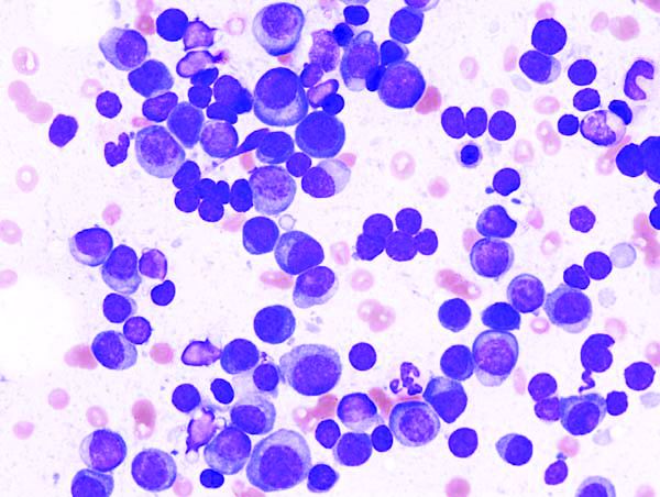
Levofloxacin prophylaxis improves survival in newly diagnosed myeloma
Adding levofloxacin to antimyeloma therapy improved survival and reduced infections in patients with newly diagnosed myeloma, findings from a phase 3 trial suggest.
The advantages of levofloxacin prophylaxis appear to offset the potential risks in patients with newly diagnosed disease, explained Mark T. Drayson, MBChB, PhD, of the University of Birmingham (England) and colleagues. The study was published in the Lancet Oncology.
The randomized, placebo-controlled, phase 3 TEAMM study enrolled 977 patients with newly diagnosed myeloma. The effects of antimicrobial prophylaxis on infection risk and infection-related mortality were evaluated across 93 hospitals throughout the United Kingdom.
Study patients were randomly assigned to receive 500 mg of oral levofloxacin once daily or placebo for a total of 12 weeks. If applicable, dose adjustments were made based on estimated glomerular filtration rate.
At baseline, the team collected stool samples and nasal swabs, and follow-up assessment occurred every 4 weeks for up to 1 year. The primary endpoint was time to death (all causes) or first febrile event from the start of prophylactic therapy to 12 weeks.
After a median follow-up of 12 months, first febrile episodes or deaths were significantly lower for patients in the levofloxacin arm (19%), compared with the placebo arm (27%) for a hazard ratio for time to first event of 0.66 (95% confidence interval, 0.51-0.86; P = .0018).
With respect to safety, the rates of serious adverse events were similar between the study arms, with the exception of tendinitis in the levofloxacin group (1%). Among all patients, a total of 597 serious toxicities were observed from baseline to 16 weeks (52% in the levofloxacin arm vs. 48% in the placebo arm).
“To our knowledge, this is the first time that the use of prophylactic antibiotics has shown a survival benefit in patients with newly diagnosed myeloma,” the researchers reported.
One key limitation of the study was the younger patient population relative to the general population. As a result, differences in survival estimates could exist between the trial and real-world populations, they noted.
“Patients with newly diagnosed myeloma could benefit from levofloxacin prophylaxis, although local antibiotic resistance proportions must be considered,” the researchers cautioned.
The study was funded by the National Institute for Health Research in the United Kingdom. The authors reported financial affiliations with Actelion, Astellas, Celgene, Gilead, Janssen, Pfizer, Takeda, and other companies.
SOURCE: Drayson MT et al. Lancet Oncol. 2019 Oct 23. doi: 10.1016/S1470-2045(19)30506-6.
FROM LANCET ONCOLOGY
New data further define role of PD-L1 status, immunotherapy in metastatic breast cancer
BARCELONA – Programmed death-ligand 1 (PD-L1) status in patients with advanced triple negative or HER2-positive breast cancer appears to identify distinct disease entities with varying likelihood of benefit from immune checkpoint inhibition, according to Giampaolo Bianchini, MD.
This observation, which contrasts with findings in other solid tumors and expands the road map to improved outcomes with immunotherapy for metastatic breast cancer, is based in part on new findings presented at the European Society for Medical Oncology Congress.
Among additional lessons from those findings: PD-L1 assays are not easily interchangeable, and studies with a “one size fits all” approach should be avoided, Dr. Bianchini, head of the Breast Cancer Group – Medical Oncology and clinical translational and immunotherapy research at Ospedale San Raffaele, Milan, said at the congress.
IMPassion130 and PD-L1 assays
In the phase 3 IMpassion130 trial assessing nanoparticle, albumin-bound (nab)-paclitaxel chemotherapy + either the anti-PD-L1 monoclonal antibody atezolizumab or placebo for the first-line treatment of metastatic triple-negative breast cancer (mTNBC), investigators used, and validated, the VENTANA PD-L1 SP142 assay to evaluate PD-L1 expression in immune cells (IC). PD-L1 positivity was defined using a 1% cutoff, meaning that PD-L1-stained IC encompassed at least 1% of the tumor area.
The trial demonstrated significantly improved progression-free survival (PFS) in the atezolizumab arm, both in the intention-to-treat (ITT) analysis (7.2 vs. 5.5 months in the placebo arm; hazard ratio, 0.80), and the PD-L1-positive subgroup (7.5 vs. 5.0 months; HR, 0.62), and the results were published in November 2018 (N Engl J Med. 2018; 379:2108-21).
“IMpassion130 is the first phase 3 trial demonstrating clinical benefit of cancer immunotherapy in patients with PD-L1-positive, metastatic triple-negative breast cancer,” Hope S. Rugo, MD, said at the congress. “The combination of atezolizumab and nab-paclitaxel is now approved in the United States and Europe for this indication.”

In addition, the SP142 antibody (which binds to PD-L1), at the 1% cutoff, predicted PFS and overall survival (OS) with atezolizumab + nab-paclitaxel, compared with nab-paclitaxel + placebo; the absolute improvement in OS in the PD-L1-positive population was 7 months (HR, 0.71), whereas no impact was seen in PFS or OS in patients who were PD-L1-negative using the SP142 assay, said Dr. Rugo, professor of hematology/oncology, and director of breast oncology and clinical trials education at the University of California, San Francisco.
Based on the IMPassion130 findings, the Food and Drug Administration approved the SP142 assay, using the 1% cutoff, as a “companion diagnostic device for selecting TNBC patients for atezolizumab.”
However, questions remain about how to best identify patients who could benefit from the atezolizumab + nab-paclitaxel combination, Dr. Rugo said.
Therefore, she and her colleagues performed a retrospective post hoc subgroup analysis of data from the trial to assess the performance and analytical concordance of the SP142 assay and two other commonly used PD-L1 immunohistochemistry (IHC) assays: the VENTANA SP263 IHC assay typically used as a companion diagnostic with durvalumab, and the Dako PD-L1 IHC 22C3 assay typically used with pembrolizumab.
In addition, the investigators assessed PD-L1 prevalence and clinical activity.
“We also included an evaluation of important factors related to PD-L1 testing and ... relationship to clinical outcome,” Dr. Rugo said.
In 614 biomarker-evaluable patients, representing 68% of the IMPassion130 ITT population, PD-L1-positive prevalence was 46% with the SP142 assay, 75% with the SP263 assay (also based on a 1% IC cutoff), and 81% with the 22C3 assay (with positivity defined as a combined positive score [CPS] of 1 or more based on an algorithm including both tumor and IC counts).
“Almost all SP142-positive cases are captured by either 22C3 or SP263. However, about a third of patients’ tumors were positive for PD-L1 using only one of the other two assays,” she noted, explaining that “this leads to suboptimal analytical concordance.”
The overall percentage agreement between SP142 and the other assays was only 64%-68%, she said.
Positive percentage agreement rates of 98% for both SP263 and 22C3 suggest that the patients identified as PD-L1 positive using the SP142 assay are captured by the other two assays. However, negative percentage agreement rates were less than 45%.
The HRs for PFS were 0.60 in SP142-positive patients, 0.64 in SP263-positive patients, and 0.68 in 22C3-positive patients, and the HRs for OS were 0.74, 0.75, and 0.78, respectively.
Subgroup analyses indicated that PFS and OS benefit with atezolizumab + nab-paclitaxel vs. nab-paclitaxel alone was greater in double-positive patients (those with SP142 positivity and either SP263 or 22C3 positivity) than in patients who were SP263-positive/SP142-negative or 22C3-positive/SP142-negative.
Dr. Rugo and her colleagues also found that the benefits with atezolizumab + nab-paclitaxel in PD-L1-positive patients were apparent regardless of the source of tissue for testing (breast or distant metastases).
They concluded that the findings of the assays are not equivalent; 22C3 and SP263 identified more patients as PD-L1 positive, and SP142-positivity was encompassed in positive tests for both.
“The clinical benefit in the 22C3-positive and the SP263-positive subgroups appear to be driven by the SP142-positive subgroup, and [SP142] identifies patients with the longest median progression-free and overall survival from the addition of atezolizumab to nab-paclitaxel,” she said “The SP142 assay with an IC cutoff of 1% or greater is the approved diagnostic test used to identify patients with metastatic triple-negative breast cancer who are most likely to benefit from the addition of the checkpoint inhibitor atezolizumab to nab-paclitaxel.”
As for whether the SP142 should be the assay of choice in other settings in which it hasn’t been validated, Dr. Rugo said it is advisable to use the assay that has been validated in a positive trial.
“That’s what we would generally do ... however, recognizing that some countries are not using SP142, and some sites may not have access, certainly you encompass that population in the patients whose tumors are positive by both other assays,” she said. “The risk is that you might overtreat, and the cost of treatment is greater.”
Excess toxicity is also a concern in that situation, she said, adding that “hopefully in the future we’ll be able to figure out ways to have even more patients benefit from the addition of immunotherapy so that won’t be an issue.”
“What this data shows is that you can feel secure that you are encompassing the patient population identified by the parent trial to benefit from the addition of atezolizumab by using either of the other two assays; you’re only missing 1% – so that’s very reasonable,” she said. “The risk is that you’re overtreating; it’s quite likely that there’s a population there that isn’t benefiting as much, but that’s a balance.”
The findings from IMPassion130 with regard to OS in the unselected population that included PD-L1-negative patients (18.7 vs. 21.0 months with vs. without atezolizumab; HR, 0.86) underscore the fact that “one size does not fit all” when it comes to immunotherapy benefit, Dr. Bianchini said.
This is further demonstrated by the post hoc analysis comparing IHC assays, he said, explaining that 63% of IMPassion130 patients who were considered PD-L1-negative based on the SP142 “actually scored as PD-L1-positive by the other tests.
“So the very clinically important question is if there is any evidence from the data that [the PD-L1-negative group] benefits in a significant way from the addition of atezolizumab,” he said. “I don’t see evidence for a clinical benefit, I see evidence to look for new biomarkers to identify a potential population who will benefit.”
The “absence of evidence is not evidence of absence,” he stressed, noting that it may be possible – with the right biomarkers – to identify PD-L1-negative patients who would benefit.
What the findings do show, however, is support for the FDA decision to approve the SP142 assay with an IC cutoff of 1% as a companion diagnostic tool, and that PD-L1 is ideally assessed using samples from both the primary and metastatic site, as the IMPassion130 data “do not inform whether PD-L1 assessment in primary and metastatic sites is equally informative,” he said.
In addition, Dr. Bianchini said the findings suggest that more information is needed about using different cutoffs for SP263 and 22C3, and he cautioned against “directly translating these finding to other disease settings or immune combinations.
“Defining new biomarkers to identify who within the PD-L1-negative group might benefit from this combination remains an unmet need,” he said. “For sure, I don’t see a space for the other tests to define this population,” he added.
KEYNOTE-119, KATE2, and future directions
Both the randomized, open-label, phase 3 KEYNOTE-119 study of the checkpoint inhibitor pembrolizumab vs. single-agent chemotherapy for mTNBC, and the phase 2 KATE2 trial of the antibody-drug conjugate trastuzumab emtansine (T-DM1) + either atezolizumab or placebo in previously treated HER2-positive breast cancer patients, failed to meet their respective primary study endpoints.
But the news isn’t all bad, Dr. Bianchini said.
For example, in KEYNOTE-119, second- or third-line pembrolizumab monotherapy did not significantly improve OS vs. chemotherapy for mTNBC, but the pembrolizumab treatment effect increased as PD-L1 enrichment increased, he explained.
Pembrolizumab showed promising antitumor activity and manageable safety in mTNBC in prior trials, and was therefore further assessed in the KEYNOTE-119 study of 601 patients with centrally confirmed TNBC, 1-2 prior systemic treatments for mTNBC, progression on the latest therapy, and a prior anthracycline or taxane, Javier Cortés, MD, PhD, of Instituto Oncológico, Madrid, reported at the congress.
Pembrolizumab was given at a dose of 200 mg every 3 weeks, and chemotherapy was physician’s choice of capecitabine, eribulin, gemcitabine, or vinorelbine.
At a median follow-up of 9.9 months in the pembrolizumab group and 10.9 months in the chemotherapy group, OS did not differ significantly between the groups; this was true overall, in patients with a CPS of 10 or greater, and in those with a CPS of 1 or greater.
In all-comers, the HR for OS was 0.97, compared with 0.78 in patients with CPS of 10 or greater, and 0.86 in those with CPS of 1 or greater, Dr. Cortés said.
“One of the most interesting exploratory analyses was OS in those patients with CPS of 20 or higher,” he said, noting that median OS in that group was 14.9 vs.12.5 months with pembrolizumab vs. with chemotherapy (HR, 0.58).
Pembrolizumab did not improve overall PFS, but again, the rates improved with higher CPS. Duration of response, however, was longer with pembrolizumab vs. chemotherapy (12.2 vs. 8.3 months overall; 12.2 vs. 6.5 months for CPS of 1 or greater; and not reached vs. 7.1 months for CPS of 10 or greater).
Grade 3-5 AEs occurred in 35% vs. 49% of patients in the pembrolizumab vs. chemotherapy groups, with nine deaths occurring in each, Dr. Cortés said, adding that treatment-related AEs occurred in 14% (with one death) and 36% (with two deaths), respectively, and grade 3-4 immune-mediated AEs and infusion reactions occurred in 3.2% vs. 1.0% (no deaths), respectively.
In the double-blind, signal-seeking KATE2 trial, as reported in 2018 at the San Antonio Breast Cancer Symposium, no overall PFS improvement was seen with atezolizumab + T-DM1 (median of 8.2 vs. 6.8 months; HR, 0.82; 12-month PFS 38% vs. 34%), but again, a possible benefit was seen in PD-L1-positive patients (8.5 vs. 4.1 months; HR, 0.60).
KATE2 included 202 patients with advanced HER2-positive breast cancer that progressed after treatment with T-DM1 and a taxane. They were randomized 2:1 to receive intravenous T-DM1 at a dose of 3.6 mg/kg plus atezolizumab (1,200 mg) or placebo every 3 weeks until loss of clinical benefit or intolerable toxicity.
The “overall survival and final safety results” show that at a median follow-up of 19.0 months in the atezolizumab arm and 18.2 months in the placebo arm, with 52 OS events reported, median OS was not reached in either arm and 1-year survival was similar in the two groups (89.1% and 89.0%), Leisha A. Emens, MD, PhD, professor of medicine in hematology/oncology and co-leader of the Hillman Cancer Immunology and Immunotherapy Program at Hillman Cancer Center, University of Pittsburgh Medical Center (UPMC) reported at the congress.
The 1-year OS rate in the PD-L1-positive subgroup, however, was numerically higher with vs. without atezolizumab (94.2% vs. 87.9%), said Dr. Emens, director of translational immunotherapy for the Women’s Cancer Research Center at UPMC.
Of note, all additional biomarkers of T-cell activation and quantity analyzed, including PD-L1 gene expression, CD8 gene expression, T effector signature gene expression, and stromal tumor infiltrating lymphocytes (TILs), were enriched in the PD-L1-positive subgroup vs. the PD-L1-negative patients.
Further, OS rates in other immune biomarker subgroups (those with PD-L1 RNA expression, CD8 RNA expression, and T effector signature at or below vs. above the median, and those with TILs less than 5% vs. 5% or greater) were consistent with those in the PD-L1 IC-positive subgroup, and the biggest difference between the atezolizumab and placebo arms related to stromal TILs, she said.
The safety profile in this final analysis was consistent with the known safety profile of each drug, she added, noting that grade 3 or greater AEs occurred in 52.6% vs. 44.8% of patients in the atezolizumab vs. placebo arms, and serious AEs – primarily pyrexia – occurred in 36.1% vs 20.9%, respectively.
The rate of grade 5 AEs was similar in the groups.
T-DM1 is indicated for the treatment of HER2-positive metastatic breast cancer previously treated with trastuzumab and a taxane, either separately or in combination, Dr. Emens said.
“In addition to its cytotoxic activity, T-DM1 may potentiate tumor immunity,” she explained, adding that KATE2 was designed to assess whether combining T-DM1 with atezolizumab, an anti-PD-L1 antibody that restores anti-tumor immunity, would result in greater clinical activity than either drug alone.
Although the number of OS events was small, the data suggest an OS benefit with the addition of atezolizumab to T-DM1, specifically in the PD-L1 IC-positive patients, but follow-up was short and the study lacked statistical power, therefore additional study of HER2-targeted agents with atezolizumab in previously treated HER2-positive, PD-L1 IC-positive advanced breast cancer is warranted, Dr. Emens concluded.
Indeed, the finding of improved OS in the PD-L1-positive subgroups of both KEYNOTE-119 and KATE2, is of interest, Dr. Bianchini said.
Both trials failed to meet their primary endpoints, but a closer look into KEYNOTE-119 shows that PD-L1 as a continuous biomarker (using CPS, 22C3) was associated with a “continuous and strong trend” toward improved ORR with the addition of pembrolizumab.
The ORR was 9.6% vs. 10.6% in unselected patients, compared with 26.3% vs. 11.5% in those with CPS of 20 or greater.
“And when you look at duration of response, you see an increase not just in the number ... but the quality of the response,” he said, noting that for PFS, as well, a trend toward superiority is seen “that is consistent with all the other endpoints.”
“So overall, the application of incrementally restrictive cut-off of CPS lends weight to the exploratory analysis showing better survival from pembrolizumab in tumors with CPS more than 20,” Dr. Bianchini said, noting that the “real question,” however, is whether the finding “is worth clinical implementation.
“We know a lot about the primary tumor and immune infiltration. We’ve learned ... that if you wait and look ahead at immune infiltration in the advanced stage, you find that the tumor becomes smart,” he said, explaining that tumor/immune co-evolution leads to increased immuno-editing and immune subversion and it becomes “much harder to just hit the tumor with PD-L1, because this is not the only mechanism of immune escape.”
A review of several studies shows that in similar populations defined by biomarkers, response rates in patients treated with checkpoint inhibitors decrease in the second- and third-line setting vs. the first-line setting, he said.
For example, pembrolizumab response rates in the first-line and second-line or greater setting in cohort B of the KEYNOTE-086 study were 21.4% and 5.7%, respectively, compared with 12.3% in the second- to third-line setting in KEYNOTE-119, he said.
Another consideration is whether monotherapy or combination therapy is preferable, and the data suggest that regardless of how PD-L1-positivity is defined (by CPS cutoff of 1 vs. 20, for example), most patients treated with monotherapy progress within the first 3 months, he said.
“I don’t see that this is a safe approach for the majority of these patients. So without better biomarkers, combinations should always be preferred, at least to avoid early progression,” Dr. Bianchini said, adding that the open question, then, is: “If we set the new standard in the first-line as the combination of nab-paclitaxel and atezolizumab for PD-L1-positive patients defined by the VENTANA [SP142 assay], should we continue with immune checkpoint [inhibition] using different combinations?”
“Of course, at the time the trial was designed, the results of IMpassion were not available, but it’s very important, because [the findings] add to the evidence that immunotherapy is extremely relevant for some patients,” he said.
KATE2 further demonstrated the importance of PD-L1 status, he said, adding that due to its limitations, including small sample size and short follow-up, longer follow-up is needed to better evaluate duration of response and PFS.
“Despite the trial limitations, the qualitative effect seen in all clinical endpoints – overall response rate, progression-free survival, overall survival – in PD-L1-positive tumors defined by SP142 ... provided strong and robust signals supporting the investigation of immune checkpoint inhibitors in HER-positive breast cancer,” he said, noting that “many trials are ongoing in the early setting and the advanced setting.”
In addition to the lessons of these trials with respect to the interchangeability of PD-L1 IHC assays and the value of PD-L1 assessment for identifying the likelihood of benefit from immune checkpoint inhibitors, the findings highlight the possibility that PD-L1-negative tumors require different immunotherapy approaches or alternative therapeutic strategies, and underscore that the benefit of immunotherapy in PD-L1-positive patients is still restricted to a minority.
“So new studies and approaches with immuno-oncology are needed, and we need more effective biomarkers, because we need to have precision oncology applied – we need to go in that direction,” he concluded.
Dr. Bianchini reported consultancy/honorarium and or advisory board activity associated with Roche, MSD, AstraZeneca, Pfizer, Chugai, EISAI, Lilly, Novartis, Amgen, Sanofi, Neopharm, and Genomic Health. The IMPassion30 trial was funded by F. Hoffmann-La Roche Ltd.; Dr. Rugo reported research grants, other funding, and or travel/accommodation/expenses from Pfizer, Novartis, Eli Lilly, Merck, OBI, EISAI, Plexxikon, Genentech/Roche, MacroGenics, PUMA, Mylan, Immunomedics, Daiichi Sankyo, and Celltrion. KEYNOTE-119 was funded by Merck Sharp & Dohme Corp.; Dr. Cortés and Dr. Emens reported numerous funding relationships but none with F. Hoffman-La Roche. KATE2 was funded by F. Hoffmann-La Roche.
Sources: IMPassion130; ESMO Abstract LBA20; KEYNOTE-119: ESMO Abstract LBA21; KATE2: ESMO Abstract 305O.
BARCELONA – Programmed death-ligand 1 (PD-L1) status in patients with advanced triple negative or HER2-positive breast cancer appears to identify distinct disease entities with varying likelihood of benefit from immune checkpoint inhibition, according to Giampaolo Bianchini, MD.
This observation, which contrasts with findings in other solid tumors and expands the road map to improved outcomes with immunotherapy for metastatic breast cancer, is based in part on new findings presented at the European Society for Medical Oncology Congress.
Among additional lessons from those findings: PD-L1 assays are not easily interchangeable, and studies with a “one size fits all” approach should be avoided, Dr. Bianchini, head of the Breast Cancer Group – Medical Oncology and clinical translational and immunotherapy research at Ospedale San Raffaele, Milan, said at the congress.
IMPassion130 and PD-L1 assays
In the phase 3 IMpassion130 trial assessing nanoparticle, albumin-bound (nab)-paclitaxel chemotherapy + either the anti-PD-L1 monoclonal antibody atezolizumab or placebo for the first-line treatment of metastatic triple-negative breast cancer (mTNBC), investigators used, and validated, the VENTANA PD-L1 SP142 assay to evaluate PD-L1 expression in immune cells (IC). PD-L1 positivity was defined using a 1% cutoff, meaning that PD-L1-stained IC encompassed at least 1% of the tumor area.
The trial demonstrated significantly improved progression-free survival (PFS) in the atezolizumab arm, both in the intention-to-treat (ITT) analysis (7.2 vs. 5.5 months in the placebo arm; hazard ratio, 0.80), and the PD-L1-positive subgroup (7.5 vs. 5.0 months; HR, 0.62), and the results were published in November 2018 (N Engl J Med. 2018; 379:2108-21).
“IMpassion130 is the first phase 3 trial demonstrating clinical benefit of cancer immunotherapy in patients with PD-L1-positive, metastatic triple-negative breast cancer,” Hope S. Rugo, MD, said at the congress. “The combination of atezolizumab and nab-paclitaxel is now approved in the United States and Europe for this indication.”
In addition, the SP142 antibody (which binds to PD-L1), at the 1% cutoff, predicted PFS and overall survival (OS) with atezolizumab + nab-paclitaxel, compared with nab-paclitaxel + placebo; the absolute improvement in OS in the PD-L1-positive population was 7 months (HR, 0.71), whereas no impact was seen in PFS or OS in patients who were PD-L1-negative using the SP142 assay, said Dr. Rugo, professor of hematology/oncology, and director of breast oncology and clinical trials education at the University of California, San Francisco.
Based on the IMPassion130 findings, the Food and Drug Administration approved the SP142 assay, using the 1% cutoff, as a “companion diagnostic device for selecting TNBC patients for atezolizumab.”
However, questions remain about how to best identify patients who could benefit from the atezolizumab + nab-paclitaxel combination, Dr. Rugo said.
Therefore, she and her colleagues performed a retrospective post hoc subgroup analysis of data from the trial to assess the performance and analytical concordance of the SP142 assay and two other commonly used PD-L1 immunohistochemistry (IHC) assays: the VENTANA SP263 IHC assay typically used as a companion diagnostic with durvalumab, and the Dako PD-L1 IHC 22C3 assay typically used with pembrolizumab.
In addition, the investigators assessed PD-L1 prevalence and clinical activity.
“We also included an evaluation of important factors related to PD-L1 testing and ... relationship to clinical outcome,” Dr. Rugo said.
In 614 biomarker-evaluable patients, representing 68% of the IMPassion130 ITT population, PD-L1-positive prevalence was 46% with the SP142 assay, 75% with the SP263 assay (also based on a 1% IC cutoff), and 81% with the 22C3 assay (with positivity defined as a combined positive score [CPS] of 1 or more based on an algorithm including both tumor and IC counts).
“Almost all SP142-positive cases are captured by either 22C3 or SP263. However, about a third of patients’ tumors were positive for PD-L1 using only one of the other two assays,” she noted, explaining that “this leads to suboptimal analytical concordance.”
The overall percentage agreement between SP142 and the other assays was only 64%-68%, she said.
Positive percentage agreement rates of 98% for both SP263 and 22C3 suggest that the patients identified as PD-L1 positive using the SP142 assay are captured by the other two assays. However, negative percentage agreement rates were less than 45%.
The HRs for PFS were 0.60 in SP142-positive patients, 0.64 in SP263-positive patients, and 0.68 in 22C3-positive patients, and the HRs for OS were 0.74, 0.75, and 0.78, respectively.
Subgroup analyses indicated that PFS and OS benefit with atezolizumab + nab-paclitaxel vs. nab-paclitaxel alone was greater in double-positive patients (those with SP142 positivity and either SP263 or 22C3 positivity) than in patients who were SP263-positive/SP142-negative or 22C3-positive/SP142-negative.
Dr. Rugo and her colleagues also found that the benefits with atezolizumab + nab-paclitaxel in PD-L1-positive patients were apparent regardless of the source of tissue for testing (breast or distant metastases).
They concluded that the findings of the assays are not equivalent; 22C3 and SP263 identified more patients as PD-L1 positive, and SP142-positivity was encompassed in positive tests for both.
“The clinical benefit in the 22C3-positive and the SP263-positive subgroups appear to be driven by the SP142-positive subgroup, and [SP142] identifies patients with the longest median progression-free and overall survival from the addition of atezolizumab to nab-paclitaxel,” she said “The SP142 assay with an IC cutoff of 1% or greater is the approved diagnostic test used to identify patients with metastatic triple-negative breast cancer who are most likely to benefit from the addition of the checkpoint inhibitor atezolizumab to nab-paclitaxel.”
As for whether the SP142 should be the assay of choice in other settings in which it hasn’t been validated, Dr. Rugo said it is advisable to use the assay that has been validated in a positive trial.
“That’s what we would generally do ... however, recognizing that some countries are not using SP142, and some sites may not have access, certainly you encompass that population in the patients whose tumors are positive by both other assays,” she said. “The risk is that you might overtreat, and the cost of treatment is greater.”
Excess toxicity is also a concern in that situation, she said, adding that “hopefully in the future we’ll be able to figure out ways to have even more patients benefit from the addition of immunotherapy so that won’t be an issue.”
“What this data shows is that you can feel secure that you are encompassing the patient population identified by the parent trial to benefit from the addition of atezolizumab by using either of the other two assays; you’re only missing 1% – so that’s very reasonable,” she said. “The risk is that you’re overtreating; it’s quite likely that there’s a population there that isn’t benefiting as much, but that’s a balance.”
The findings from IMPassion130 with regard to OS in the unselected population that included PD-L1-negative patients (18.7 vs. 21.0 months with vs. without atezolizumab; HR, 0.86) underscore the fact that “one size does not fit all” when it comes to immunotherapy benefit, Dr. Bianchini said.
This is further demonstrated by the post hoc analysis comparing IHC assays, he said, explaining that 63% of IMPassion130 patients who were considered PD-L1-negative based on the SP142 “actually scored as PD-L1-positive by the other tests.
“So the very clinically important question is if there is any evidence from the data that [the PD-L1-negative group] benefits in a significant way from the addition of atezolizumab,” he said. “I don’t see evidence for a clinical benefit, I see evidence to look for new biomarkers to identify a potential population who will benefit.”
The “absence of evidence is not evidence of absence,” he stressed, noting that it may be possible – with the right biomarkers – to identify PD-L1-negative patients who would benefit.
What the findings do show, however, is support for the FDA decision to approve the SP142 assay with an IC cutoff of 1% as a companion diagnostic tool, and that PD-L1 is ideally assessed using samples from both the primary and metastatic site, as the IMPassion130 data “do not inform whether PD-L1 assessment in primary and metastatic sites is equally informative,” he said.
In addition, Dr. Bianchini said the findings suggest that more information is needed about using different cutoffs for SP263 and 22C3, and he cautioned against “directly translating these finding to other disease settings or immune combinations.
“Defining new biomarkers to identify who within the PD-L1-negative group might benefit from this combination remains an unmet need,” he said. “For sure, I don’t see a space for the other tests to define this population,” he added.
KEYNOTE-119, KATE2, and future directions
Both the randomized, open-label, phase 3 KEYNOTE-119 study of the checkpoint inhibitor pembrolizumab vs. single-agent chemotherapy for mTNBC, and the phase 2 KATE2 trial of the antibody-drug conjugate trastuzumab emtansine (T-DM1) + either atezolizumab or placebo in previously treated HER2-positive breast cancer patients, failed to meet their respective primary study endpoints.
But the news isn’t all bad, Dr. Bianchini said.
For example, in KEYNOTE-119, second- or third-line pembrolizumab monotherapy did not significantly improve OS vs. chemotherapy for mTNBC, but the pembrolizumab treatment effect increased as PD-L1 enrichment increased, he explained.
Pembrolizumab showed promising antitumor activity and manageable safety in mTNBC in prior trials, and was therefore further assessed in the KEYNOTE-119 study of 601 patients with centrally confirmed TNBC, 1-2 prior systemic treatments for mTNBC, progression on the latest therapy, and a prior anthracycline or taxane, Javier Cortés, MD, PhD, of Instituto Oncológico, Madrid, reported at the congress.
Pembrolizumab was given at a dose of 200 mg every 3 weeks, and chemotherapy was physician’s choice of capecitabine, eribulin, gemcitabine, or vinorelbine.
At a median follow-up of 9.9 months in the pembrolizumab group and 10.9 months in the chemotherapy group, OS did not differ significantly between the groups; this was true overall, in patients with a CPS of 10 or greater, and in those with a CPS of 1 or greater.
In all-comers, the HR for OS was 0.97, compared with 0.78 in patients with CPS of 10 or greater, and 0.86 in those with CPS of 1 or greater, Dr. Cortés said.
“One of the most interesting exploratory analyses was OS in those patients with CPS of 20 or higher,” he said, noting that median OS in that group was 14.9 vs.12.5 months with pembrolizumab vs. with chemotherapy (HR, 0.58).
Pembrolizumab did not improve overall PFS, but again, the rates improved with higher CPS. Duration of response, however, was longer with pembrolizumab vs. chemotherapy (12.2 vs. 8.3 months overall; 12.2 vs. 6.5 months for CPS of 1 or greater; and not reached vs. 7.1 months for CPS of 10 or greater).
Grade 3-5 AEs occurred in 35% vs. 49% of patients in the pembrolizumab vs. chemotherapy groups, with nine deaths occurring in each, Dr. Cortés said, adding that treatment-related AEs occurred in 14% (with one death) and 36% (with two deaths), respectively, and grade 3-4 immune-mediated AEs and infusion reactions occurred in 3.2% vs. 1.0% (no deaths), respectively.
In the double-blind, signal-seeking KATE2 trial, as reported in 2018 at the San Antonio Breast Cancer Symposium, no overall PFS improvement was seen with atezolizumab + T-DM1 (median of 8.2 vs. 6.8 months; HR, 0.82; 12-month PFS 38% vs. 34%), but again, a possible benefit was seen in PD-L1-positive patients (8.5 vs. 4.1 months; HR, 0.60).
KATE2 included 202 patients with advanced HER2-positive breast cancer that progressed after treatment with T-DM1 and a taxane. They were randomized 2:1 to receive intravenous T-DM1 at a dose of 3.6 mg/kg plus atezolizumab (1,200 mg) or placebo every 3 weeks until loss of clinical benefit or intolerable toxicity.
The “overall survival and final safety results” show that at a median follow-up of 19.0 months in the atezolizumab arm and 18.2 months in the placebo arm, with 52 OS events reported, median OS was not reached in either arm and 1-year survival was similar in the two groups (89.1% and 89.0%), Leisha A. Emens, MD, PhD, professor of medicine in hematology/oncology and co-leader of the Hillman Cancer Immunology and Immunotherapy Program at Hillman Cancer Center, University of Pittsburgh Medical Center (UPMC) reported at the congress.
The 1-year OS rate in the PD-L1-positive subgroup, however, was numerically higher with vs. without atezolizumab (94.2% vs. 87.9%), said Dr. Emens, director of translational immunotherapy for the Women’s Cancer Research Center at UPMC.
Of note, all additional biomarkers of T-cell activation and quantity analyzed, including PD-L1 gene expression, CD8 gene expression, T effector signature gene expression, and stromal tumor infiltrating lymphocytes (TILs), were enriched in the PD-L1-positive subgroup vs. the PD-L1-negative patients.
Further, OS rates in other immune biomarker subgroups (those with PD-L1 RNA expression, CD8 RNA expression, and T effector signature at or below vs. above the median, and those with TILs less than 5% vs. 5% or greater) were consistent with those in the PD-L1 IC-positive subgroup, and the biggest difference between the atezolizumab and placebo arms related to stromal TILs, she said.
The safety profile in this final analysis was consistent with the known safety profile of each drug, she added, noting that grade 3 or greater AEs occurred in 52.6% vs. 44.8% of patients in the atezolizumab vs. placebo arms, and serious AEs – primarily pyrexia – occurred in 36.1% vs 20.9%, respectively.
The rate of grade 5 AEs was similar in the groups.
T-DM1 is indicated for the treatment of HER2-positive metastatic breast cancer previously treated with trastuzumab and a taxane, either separately or in combination, Dr. Emens said.
“In addition to its cytotoxic activity, T-DM1 may potentiate tumor immunity,” she explained, adding that KATE2 was designed to assess whether combining T-DM1 with atezolizumab, an anti-PD-L1 antibody that restores anti-tumor immunity, would result in greater clinical activity than either drug alone.
Although the number of OS events was small, the data suggest an OS benefit with the addition of atezolizumab to T-DM1, specifically in the PD-L1 IC-positive patients, but follow-up was short and the study lacked statistical power, therefore additional study of HER2-targeted agents with atezolizumab in previously treated HER2-positive, PD-L1 IC-positive advanced breast cancer is warranted, Dr. Emens concluded.
Indeed, the finding of improved OS in the PD-L1-positive subgroups of both KEYNOTE-119 and KATE2, is of interest, Dr. Bianchini said.
Both trials failed to meet their primary endpoints, but a closer look into KEYNOTE-119 shows that PD-L1 as a continuous biomarker (using CPS, 22C3) was associated with a “continuous and strong trend” toward improved ORR with the addition of pembrolizumab.
The ORR was 9.6% vs. 10.6% in unselected patients, compared with 26.3% vs. 11.5% in those with CPS of 20 or greater.
“And when you look at duration of response, you see an increase not just in the number ... but the quality of the response,” he said, noting that for PFS, as well, a trend toward superiority is seen “that is consistent with all the other endpoints.”
“So overall, the application of incrementally restrictive cut-off of CPS lends weight to the exploratory analysis showing better survival from pembrolizumab in tumors with CPS more than 20,” Dr. Bianchini said, noting that the “real question,” however, is whether the finding “is worth clinical implementation.
“We know a lot about the primary tumor and immune infiltration. We’ve learned ... that if you wait and look ahead at immune infiltration in the advanced stage, you find that the tumor becomes smart,” he said, explaining that tumor/immune co-evolution leads to increased immuno-editing and immune subversion and it becomes “much harder to just hit the tumor with PD-L1, because this is not the only mechanism of immune escape.”
A review of several studies shows that in similar populations defined by biomarkers, response rates in patients treated with checkpoint inhibitors decrease in the second- and third-line setting vs. the first-line setting, he said.
For example, pembrolizumab response rates in the first-line and second-line or greater setting in cohort B of the KEYNOTE-086 study were 21.4% and 5.7%, respectively, compared with 12.3% in the second- to third-line setting in KEYNOTE-119, he said.
Another consideration is whether monotherapy or combination therapy is preferable, and the data suggest that regardless of how PD-L1-positivity is defined (by CPS cutoff of 1 vs. 20, for example), most patients treated with monotherapy progress within the first 3 months, he said.
“I don’t see that this is a safe approach for the majority of these patients. So without better biomarkers, combinations should always be preferred, at least to avoid early progression,” Dr. Bianchini said, adding that the open question, then, is: “If we set the new standard in the first-line as the combination of nab-paclitaxel and atezolizumab for PD-L1-positive patients defined by the VENTANA [SP142 assay], should we continue with immune checkpoint [inhibition] using different combinations?”
“Of course, at the time the trial was designed, the results of IMpassion were not available, but it’s very important, because [the findings] add to the evidence that immunotherapy is extremely relevant for some patients,” he said.
KATE2 further demonstrated the importance of PD-L1 status, he said, adding that due to its limitations, including small sample size and short follow-up, longer follow-up is needed to better evaluate duration of response and PFS.
“Despite the trial limitations, the qualitative effect seen in all clinical endpoints – overall response rate, progression-free survival, overall survival – in PD-L1-positive tumors defined by SP142 ... provided strong and robust signals supporting the investigation of immune checkpoint inhibitors in HER-positive breast cancer,” he said, noting that “many trials are ongoing in the early setting and the advanced setting.”
In addition to the lessons of these trials with respect to the interchangeability of PD-L1 IHC assays and the value of PD-L1 assessment for identifying the likelihood of benefit from immune checkpoint inhibitors, the findings highlight the possibility that PD-L1-negative tumors require different immunotherapy approaches or alternative therapeutic strategies, and underscore that the benefit of immunotherapy in PD-L1-positive patients is still restricted to a minority.
“So new studies and approaches with immuno-oncology are needed, and we need more effective biomarkers, because we need to have precision oncology applied – we need to go in that direction,” he concluded.
Dr. Bianchini reported consultancy/honorarium and or advisory board activity associated with Roche, MSD, AstraZeneca, Pfizer, Chugai, EISAI, Lilly, Novartis, Amgen, Sanofi, Neopharm, and Genomic Health. The IMPassion30 trial was funded by F. Hoffmann-La Roche Ltd.; Dr. Rugo reported research grants, other funding, and or travel/accommodation/expenses from Pfizer, Novartis, Eli Lilly, Merck, OBI, EISAI, Plexxikon, Genentech/Roche, MacroGenics, PUMA, Mylan, Immunomedics, Daiichi Sankyo, and Celltrion. KEYNOTE-119 was funded by Merck Sharp & Dohme Corp.; Dr. Cortés and Dr. Emens reported numerous funding relationships but none with F. Hoffman-La Roche. KATE2 was funded by F. Hoffmann-La Roche.
Sources: IMPassion130; ESMO Abstract LBA20; KEYNOTE-119: ESMO Abstract LBA21; KATE2: ESMO Abstract 305O.
REPORTING FROM ESMO 2019
KRAS-mutation colon, rectal cancers have distinct survival profiles
SAN FRANCISCO – When it comes to KRAS mutational status, liver metastases originating from left-sided colon tumors have different clinical and survival characteristics from those originating from primary rectal tumors. There was no significant difference in survival between bearers of mutated versus wild-type (WT) KRAS in rectal tumor cases, while there was a significant difference in survival from left-sided colon cases, according to a first-time analysis of the effect of KRAS status in this specific population.
The work was presented at the annual clinical congress of the American College of Surgeons by Neda Amini, MD. “The liver metastasis originating from a rectal tumor might have a different biology than from a primary colon tumor, and we should have stratification according to the primary tumor location in clinical trials testing chemotherapy and targeted agents,” said Dr. Amini, who is a surgical resident at Sinai Hospital of Baltimore, during her presentation of the research.
“I thought that was interesting because most of the studies that have been done look at KRAS mutations in colorectal cancers, and [colon and rectal cancers] are two completely different entities,” said session comoderator Valentine Nfonsam, MD, associate professor of surgery at the University of Arizona, Tucson, in an interview. The findings could also impact clinical practice. “If a patient has rectal cancer, if they have a KRAS mutation, whether you treat them with cetuximab or not, the overall survival doesn’t really change. Whereas for colon cancer patients you really want to make that distinction. You want to truly personalize their therapy, because of the difference in survival in a patient with the KRAS mutation in colon cancer” Dr. Nfonsam said.
“It gets to the heart that there might be different biology between colon cancers and rectal cancers. It’s important to understand the differences in the basic biology, which affects the treatment and the surgery,” said the other comoderator, Jonathan Mitchem, MD, in an interview. Dr. Mitchem is an assistant professor at the University of Missouri–Columbia.
KRAS is common in colorectal cancer, occurring in 30% of cases, and multiple trials have shown it is associated with nonresponse to the epidermal growth factor receptor inhibitors cetuximab or panitumumab. All colorectal cancer patients with liver metastases should be screened for KRAS mutations, according to National Comprehensive Cancer Network guidelines.
The researchers conducted a retrospective analysis of 1,304 patients who underwent curative-intent surgery for colorectal liver metastases at nine institutions between 2000 and 2016. The KRAS mutation rate was similar in the primary colon and rectal tumors (34.2% vs. 30.9%; P = .24). The frequency was highest in right-sided colon tumors (39.4%). There was a statistically significant difference in the frequency of KRAS mutation between primary rectal tumors (30.9%), and left-sided colon tumors (21.1%; P = .001).
There were several differences in clinical characteristics between left-sided colon cancers and rectal cancers. Rectal cancer patients were more likely to be male (73.4% vs. 62.4%; P = .001); more likely to be stage T1-T2 (16.6% vs. 10.6%; P = .012); less likely to have serum carcinoembryonic antigen greater than 100 ng/mL (8.4% vs. 14.1%; P = .018); and less likely to have a liver metastasis under 3 cm (36.1% vs. 49.3%).
There were significant differences between KRAS mutant and KRAS wild-type patients with a colon primary tumor, including greater likelihood of lymph node metastasis in WT (65.2% vs. 55.37%; P = .004), greater likelihood of liver metastasis greater than 3 cm in WT (48.8% versus 39.3%; P = .01), greater likelihood of extrahepatic disease in mutant KRAS (16.3% vs. 10.4%; P = .01), greater likelihood of prehepatic resection chemotherapy in WT (65.5% vs. 56.0%; P = .005), greater likelihood of posthepatic resection chemotherapy in mutant KRAS (64.5% vs. 55.5%; P = .01), and greater likelihood of receiving anti–epidermal growth factor therapy in WT (5.7% vs. 0.3%; P less than .001). The only difference seen in patients with rectal primary tumors was the odds of receiving post-hepatic surgery chemotherapy, which was higher among patients with mutated KRAS (70.8% vs. 59.0%; P = .03).
After a median follow-up of 26.4 months, the 1-, 3-, and 5-year overall survival rates were 88.9%, 62.5%, and 44.5%. Among patients with primary colon cancer, there was a statistically significant lower survival curve in patients with a KRAS mutation overall and in those with left-sided colon tumors (log rank P less than .001 for both), but there was no significant survival difference between mutation bearers and wild-type patients with a primary rectal tumor (log rank P = .53). A multivariate analysis showed an 82% risk of death from KRAS mutation in primary colon cancer (hazard ratio, 1.82; P less than .001), but a univariate analysis showed no significant mortality association in rectal primary tumors (hazard ratio, 1.13; P = .46).
The funding source was not disclosed. The authors had no relevant financial disclosures.
SOURCE: Amini N et al. J Am Coll Surg. 2019 Oct;229(4):Suppl 1, S69-70.
REPORTING FROM CLINICAL CONGRESS 2019
Behind the scenes of a radical new cancer cure
An unexpected early morning phone call from the hospital is never good news. When Joy Johnson answered, her first thought was that Sharon Birzer, her partner of 15 years, was dead. Her fears were amplified by the voice on the other end refusing to confirm or deny it. Just “come in and talk to one of the doctors,” she remembers the voice saying.
Johnson knew this was a real possibility. A few weeks earlier, she and Birzer sat in the exam room of a lymphoma specialist at Stanford University. Birzer’s cancer had grown, and fast — first during one type of chemotherapy, then through a second. Out of standard options, Birzer’s local oncologist had referred her for a novel treatment called chimeric antigen receptor T-cell therapy — or CAR-T. Birzer and Johnson knew the treatment was risky. They were warned there was a chance of death. There was also a chance of serious complications such as multi-organ failure and neurological impairment. But it was like warning a drowning person that her lifeboat could have problems. Without treatment, the chance of Birzer’s death was all but certain. She signed the consent form.
Johnson hung up the phone that early morning and sped to the hospital. She met with a doctor and two chaplains in a windowless room in the cancer ward, where happy photos of cancer “alumni” smiled down from the walls. This is getting worse and worse, Johnson thought. As she remembers it, the doctor went through the timeline of what happened for 10 minutes, explaining how Birzer became sicker and sicker, before Johnson interrupted with the thought splitting her world in two: “I need you to tell me whether she’s alive or dead.”
Birzer wasn’t dead. But she was far from okay. The ordeal began with Birzer speaking gibberish. Then came seizures so severe there was concern she wouldn’t be able to breathe on her own. When it took a few different medications to stop Birzer from seizing, her doctors sedated her, put a breathing tube down her throat, and connected her to a ventilator. Now, she was unconscious and in the intensive care unit (ICU).
Birzer was one of the early patients to receive CAR-T, a radical new therapy to treat cancer. It involved removing Birzer’s own blood, filtering for immune cells called T-cells, and genetically engineering those cells to recognize and attack her lymphoma. CAR-T made history in 2017 as the first FDA-approved gene therapy to treat any disease. After three to six months of follow-up, the trials that led to approval showed response rates of 80 percent and above in aggressive leukemias and lymphomas that had resisted chemotherapy. Patients on the brink of death were coming back to life.
This is something I often dream of seeing but rarely do. As a doctor who treats cancer, I think a lot about how to frame new treatments to my patients. I never want to give false hope. But the uncertainty inherent to my field also cautions me against closing the door on optimism prematurely. We take it as a point of pride that no field of medicine evolves as rapidly as cancer — the FDA approves dozens of new treatments a year. One of my biggest challenges is staying up to date on every development and teasing apart what should — and shouldn’t — change my practice. I am often a mediator for my patients, tempering theoretical promises with everyday realism. To accept a research finding into medical practice, I prefer slow steps showing me proof of concept, safety, and efficacy.
CAR-T, nearly three decades in the making, systemically cleared these hurdles. Not only did the product work, its approach was also unique among cancer treatments. Unlike our usual advances, this wasn’t a matter of prescribing an old drug for a new disease or remixing known medications. CAR-T isn’t even a drug. This is a one-time infusion giving a person a better version of her own immune system. When the FDA approved its use, it wasn’t a question of whether my hospital would be involved, but how we could stay ahead. We weren’t alone.
Today, two FDA-approved CAR-T products called Kymriah and Yescarta are available in more than 100 hospitals collectively across the U.S. Hundreds of clinical trials are tinkering with dosages, patient populations, and types of cancer. Some medical centers are manufacturing the cells on-site.
The FDA approved CAR-T with a drug safety program called a Risk Evaluation and Mitigation Strategy (REMS). As I cared for these patients, I quickly realized the FDA’s concerns. Of the 10 or so patients I’ve treated, more than half developed strange neurologic side effects ranging from headaches to difficulty speaking to seizures to falling unconscious. We scrambled to learn how to manage the side effects in real time.
Johnson and Birzer, who I didn’t treat personally but spoke to at length for this essay, understood this better than most. Both had worked in quality control for a blood bank and were medically savvier than the average patient. They accepted a medical system with a learning curve. They were fine with hearing “I don’t know.” Signing up for a trailblazing treatment meant going along for the ride. Twists and bumps were par for the course.
Cancer, by definition, means something has gone very wrong within — a cell has malfunctioned and multiplied. The philosophy for fighting cancer has been, for the most part, creating and bringing in treatments from outside the body. That’s how we got to the most common modern approaches: Chemotherapy (administering drugs to kill cancer), radiation (using high energy beams to kill cancer), and surgery (cutting cancer out with a scalpel and other tools). Next came the genetics revolution, with a focus on creating drugs that target a precise genetic mutation separating a cancer cell from a normal one. But cancers are genetically complex, with legions of mutations and the talent to develop new ones. It’s rare to have that one magic bullet.
Over the last decade or so, our approach shifted. Instead of fighting cancer from the outside, we are increasingly turning in. The human body is already marvelously equipped to recognize and attack invaders, from the common cold to food poisoning, even if the invaders are ones the body has never seen before. Cancer doesn’t belong either. But since cancer cells come from normal ones, they’ve developed clever camouflages to trick and evade the immune system. The 2018 Nobel Prize in Physiology or Medicine was jointly awarded to two researchers for their work in immunotherapy, a class of medications devoted to wiping out the camouflages and restoring the immune system’s upper hand. As I once watched a fellow oncologist describe it to a patient: “I’m not treating you. You are treating you.”
What if we could go one step further? What if we could genetically engineer a patient’s own immune cells to spot and fight cancer, as a sort of “best hits” of genetic therapy and immunotherapy?
Enter CAR-T. The technology uses T-cells, which are like the bouncers of the immune system. T-cells survey the body and make sure everything belongs. CAR-T involves removing a person’s T-cells from her blood and using a disarmed virus to deliver new genetic material to the cells. The new genes given to the T-cells help them make two types of proteins. The first — giving the technology its name — is a CAR, which sits on the T-cell’s surface and binds to a protein on the tumor cell’s surface, like a lock and key. The second serves as the T-cell’s caffeine jolt, rousing it to activate. Once the genetic engineering part is done, the T-cells are prodded to multiply by being placed on a rocking device that feeds them nutrients while filtering their wastes. When the cells reach a high enough number — a typical “dose” ranges from hundreds of thousands to hundreds of millions — they are formidable enough to go back into the patient. Once inside, the cancer provokes the new cells to replicate even more. After one week, a typical expansion means multiplying by about another 1,000-fold.
Practically, it looks like this: A person comes in for an appointment. She has a catheter placed in a vein, perhaps in her arm or her chest, that connects to a large, whirring machine which pulls in her blood and separates it into its components. The medical team set the T-cells aside to freeze while the rest of the blood circulates back into the patient in a closed loop. Then, the hospital ships the cells frozen to the relevant pharmaceutical company’s headquarters or transports them to a lab on-site, where thawing and manufacturing takes from a few days to a few weeks. When the cells are ready, the patient undergoes about three days of chemotherapy to kill both cancer and normal cells, making room for the millions of new cells and eradicating normal immune players that could jeopardize their existence. She then gets a day or two to rest. When the new cells are infused back into her blood, we call that Day 0.
I remember the first time I watched a patient get his Day 0 infusion. It felt anti-climactic. The entire process took about 15 minutes. The CAR-T cells are invisible to the naked eye, housed in a small plastic bag containing clear liquid.
“That’s it?” my patient asked when the nurse said it was over. The infusion part is easy. The hard part is everything that comes next.
Once the cells are in, they can’t turn off. That this may cause collateral damage was evident from the start. In 2009 — working in parallel with other researchers at Memorial Sloan Kettering Cancer Center in New York and the National Cancer Institute in Maryland — oncologists at the University of Pennsylvania opened a clinical trial for CAR-T in human leukemia patients. (Carl June, who led the CAR-T development, did not respond to Undark’s interview request.) Of the first three patients who got CAR-T infusions, two achieved complete remission — but nearly died in the process. The first was a retired corrections officer named Bill Ludwig, who developed extremely high fevers and went into multi-organ failure requiring time in the ICU. At the time, the medical teams had no idea why it was happening or how to stop it. But time passed. Ludwig got better. Then came the truly incredible part: His cancer was gone.
With only philanthropic support, the trial ran out of funding. Of the eligible patients they intended to treat, the Penn doctors only treated three. So they published the results of one patient in the New England Journal of Medicine and presented the outcomes of all three patients, including Ludwig, at a cancer conference anyway. From there, the money poured in. Based on the results, the Swiss pharmaceutical company Novartis licensed the rights of the therapy.
The next year, six-year-old Emily Whitehead was on the brink of death when she became the first child to receive CAR-T. She also became extremely ill in the ICU, and her cancer was also eventually cured. Her media savvy parents helped bring her story public, making her the poster child for CAR-T. In 2014, the FDA granted CAR-T a breakthrough therapy designation to expedite the development of extremely promising therapies. By 2017, a larger trial gave the treatment to 75 children and young adults with a type of leukemia — B-cell acute lymphoblastic leukemia — that failed to respond to chemotherapy. Eighty-one percent had no sign of cancer after three months.
In August 2017, the FDA approved a CAR-T treatment as the first gene therapy in the U.S. The decision was unanimous. The Oncologic Drugs Advisory Committee, a branch of the FDA that reviews new cancer products, voted 10 to zero in favor of Kymriah. Committee members called the responses “remarkable” and “potentially paradigm changing.” When the announcement broke, a crowd formed in the medical education center of Penn Medicine, made up of ecstatic faculty and staff. There were banners and T-shirts. “A remarkable thing happened” was the tagline, above a cartoon image of a heroic T-cell. Two months later, in October 2017, the FDA approved a second CAR-T formulation called Yescarta from Kite Pharma, a subsidiary of Gilead Sciences, to treat an aggressive blood cancer in adults called diffuse large B-cell lymphoma, the trial of which had shown a 54 percent complete response rate, meaning all signs of cancer had disappeared. In May 2018, Kymriah was approved to treat adults with non-Hodgkin lymphoma.
That year, the American Society of Clinical Oncology named CAR-T the Advance of the Year, beating out immunotherapy, which had won two years in a row. When I attended the last American Society of Hematology meeting in December 2018, CAR-T stole the show. Trying to get into CAR-T talks felt like trying to get a photo with a celebrity. Running five minutes late to one session meant facing closed doors. Others were standing room only. With every slide, it became difficult to see over a sea of smartphones snapping photos. At one session I found a seat next to the oncologist from my hospital who treated Birzer. “Look,” she nudged me. “Do you see all these ‘non-member’ badges?” I turned. Members were doctors like us who treated blood cancers. I couldn’t imagine who else would want to be here. “Who are they?” I asked. “Investors,” she said. It felt obvious the moment she said it.
For patients, the dreaded “c” word is cancer. For oncologists, it’s cure. When patients ask, I’ve noticed how we gently steer the conversation toward safer lingo. We talk about keeping the cancer in check. Cure is a dangerous word, used only when so much time has passed from her cancer diagnosis we can be reasonably certain it’s gone. But that line is arbitrary. We celebrate therapies that add weeks or months because the diseases are pugnacious, the biology diverse, and the threat of relapse looming. Oncologists are a tempered group, or so I’ve learned, finding inspiration in slow, incremental change.
This was completely different. These were patients who would have otherwise died, and the trials were boasting that 54 to 81 percent were cancer-free upon initial follow-up. PET scans showed tumors that had speckled an entire body melt away. Bone marrow biopsies were clear, with even the most sensitive testing unable to detect disease.
The dreaded word was being tossed around — could this be the cure we’ve always wanted?
When a new drug gets FDA approval, it makes its way into clinical practice, swiftly and often with little fanfare. Under the drug safety program REMS, hospitals offering CAR-T were obligated to undergo special training to monitor and manage side effects. As hospitals worked to create CAR-T programs, oncologists like me made the all too familiar transition from first-time user to expert.
It was May 2018 when I rotated through my hospital’s unit and cared for my first patients on CAR-T. As I covered 24-hour shifts, I quickly learned that whether I would sleep that night depended on how many CAR-T patients I was covering. With each treatment, it felt like we were pouring gasoline on the fire of patients’ immune systems. Some developed high fevers and their blood pressures plummeted, mimicking a serious infection. But there was no infection to be found. When resuscitating with fluids couldn’t maintain my patients’ blood pressures, I sent them to the ICU where they required intensive support to supply blood to their critical organs.
We now have a name for this effect — cytokine release syndrome — that occurs in more than half of patients who receive CAR-T, starting with Ludwig and Whitehead. The syndrome is the collateral damage of an immune system on the highest possible alert. This was first seen with other types of immunotherapy, but CAR-T took its severity to a new level. Usually starting the week after CAR-T, cytokine release syndrome can range from simple fevers to multi-organ failure affecting the liver, kidneys, heart, and more. The activated T-cells make and recruit other immune players called cytokines to join in the fight. Cytokines then recruit more immune cells. Unlike in the early trials at Penn, we now have two medicines to dampen the effect. Steroids calm the immune system in general, while a medication called tocilizumab, used to treat autoimmune disorders such as rheumatoid arthritis, blocks cytokines specifically.
Fortuity was behind the idea of tocilizumab: When Emily Whitehead, the first child to receive CAR-T, developed cytokine release syndrome, her medical team noted that her blood contained high levels of a cytokine called interleukin 6. Carl June thought of his own daughter, who had juvenile rheumatoid arthritis and was on a new FDA-approved medication that suppressed the same cytokine. The team tried the drug, tocilizumab, in Whitehead. It worked.
Still, we were cautious in our early treatments. The symptoms of cytokine release syndrome mimic the symptoms of severe infection. If this were infection, medicines that dampen a patient’s immune system would be the opposite of what you’d want to give. There was another concern: Would these medications dampen the anti-cancer activity too? We didn’t know. Whenever a CAR-T patient spiked a fever, I struggled with the question — is it cytokine release syndrome, or is it infection? I often played it safe and covered all bases, starting antibiotics and steroids at the same time. It was counterintuitive, like pressing both heat and ice on a strain, or treating a patient simultaneously with fluids and diuretics.
The second side effect was even scarier: Patients stopped talking. Some, like Sharon Birzer spoke gibberish or had violent seizures. Some couldn’t interact at all, unable to follow simple commands like “squeeze my fingers.” How? Why? At hospitals across the nation, perfectly cognitively intact people who had signed up to treat their cancer were unable to ask what was happening.
Our nurses learned to ask a standardized list of questions to catch the effect, which we called neurotoxicity: Where are we? Who is the president? What is 100 minus 10? When the patients scored too low on these quizzes, they called me to the bedside.
In turn, I relied heavily on a laminated booklet, made by other doctors who were using CAR-T, which we tacked to a bulletin board in our doctors’ workroom. It contained a short chart noting how to score severity and what to do next. I flipped through the brightly color-coded pages telling me when to order a head CT-scan to look for brain swelling and when to place scalp electrodes looking for seizures. Meanwhile, we formed new channels of communication. As I routinely called a handful of CAR-T specialists at my hospital in the middle of the night, national consortiums formed where specialists around the country shared their experiences. As we tweaked the instructions, we scribbled updates to the booklet in pen.
I wanted to know whether my experience was representative. I came across an abstract and conference talk that explored what happened to 277 patients who received CAR-T in the real world, so I emailed the lead author, Loretta Nastoupil, director of the Department of Lymphoma and Myeloma at the University of Texas MD Anderson Cancer Center in Houston. Fortuitously, she was planning a trip to my university to give a talk that month. We met at a café and I asked what her research found. Compared to the earlier trials, the patients were much sicker, she said. Of the 277 patients, more than 40 percent wouldn’t have been eligible for the very trials that got CAR-T approved. Was her team calling other centers for advice? “They were calling us,” she said.
Patients included in clinical trials are carefully selected. They tend not to have other major medical problems, as we want them to survive whatever rigorous new therapy we put them through. Nastoupil admits some of it is arbitrary. Many criteria in the CAR-T trials were based on criteria that had been used in chemotherapy trials. “These become standard languages that apply to all studies,” she said, listing benchmarks like a patient’s age, kidney function, and platelet count. “But we have no idea whether criteria for chemotherapy would apply to cellular therapy.”
Now, with a blanket FDA approval comes clinical judgment. Patients want a chance. Oncologists want to give their patients a chance. Young, old, prior cancer, heart disease, or liver disease — without strict trial criteria, anyone is fair game.
When I was making rounds at my hospital, I never wandered too far from these patients’ rooms, medically prepared for them to crash at any moment. At the same time, early side effects made me optimistic. A bizarre truism in cancer is that side effects may bode well. They could mean the treatment is working. Cancer is usually a waiting game, requiring months to learn an answer. Patients and doctors alike seek clues, but the only real way to know is waiting: Will the next PET scan show anything? What are the biopsy results?
CAR-T was fundamentally different from other cancer treatments in that it worked fast. Birzer’s first clue came just a few hours after her infusion. She developed pain in her lower back. She described it as feeling like she had menstrual cramps. A heavy burden of lymphoma lay in her uterus. Could the pain mean that the CAR-T cells had migrated to the right spot and started to work? Her medical team didn’t know, but the lead doctor’s instinct was that it was a good sign.
Two days later, her temperature shot up to 102. Her blood pressure dropped. The medical team diagnosed cytokine release syndrome, as though right on schedule, and gave her tocilizumab.
Every day, the nurses would ask her questions and have her write simple sentences on a slip of paper to monitor for neurotoxicity. By the fifth day, her answers changed. “She started saying things that were crazy,” Johnson explained.
One of Birzer's sentences was “guinea pigs eat greens like hay and pizza.” Birzer and Johnson owned two guinea pigs, so their diet would be something Birzer normally knew well. So Johnson tried to reason with her: “They don’t eat pizza.” And Birzer replied, “They do eat pizza, but only gluten-free.”
Johnson remembers being struck by the certainty in her partner’s delirium. Not only was Birzer confused, she was confident she was not. “She was doubling down on everything,” Johnson described. “She was absolutely sure she was right.”
Johnson vividly remembers the evening before the frightening early-morning phone call that brought her rushing back to the hospital. Birzer had said there was no point in Johnson staying overnight; she would only watch her be in pain. So Johnson went home. After she did, the doctor came by multiple times to evaluate Birzer. She was deteriorating — and fast. Her speech became more and more garbled. Soon she couldn’t name simple objects and didn’t know where she was. At 3 a.m., the doctor ordered a head CT to make sure Birzer wasn’t bleeding into her brain.
Fortunately, she wasn’t. But by 7 a.m. Birzer stopped speaking altogether. Then she seized. Birzer’s nurse was about to step out of the room when she noticed Birzer’s arms and legs shaking. Her eyes stared vacantly and she wet the bed. The nurse called a code blue, and a team of more doctors and nurses ran over. Birzer was loaded with high-dose anti-seizure medications through her IV. But she continued to seize. As nurses infused more medications into her IV, a doctor placed a breathing tube down her throat.
Birzer’s saga poses the big question: Why does CAR-T cause seizures and other neurologic problems? No one seemed to know. My search of the published scientific literature was thin, but one name kept cropping up. So I called her. Juliane Gust, a pediatric neurologist and scientist at Seattle Children’s Hospital, told me her investigations of how CAR-T affects the brain were motivated by her own experiences. When the early CAR-T trials opened at her hospital in 2014, she and her colleagues began getting calls from oncologists about brain toxicities they knew nothing about. “Where are the papers?” she remembered thinking. “There was nothing.”
Typically, the brain is protected by a collection of cells aptly named the blood-brain-barrier. But with severe CAR-T neurotoxicity, research suggests, this defense breaks down. Gust explained that spinal taps on these patients show high levels of cytokines floating in the fluid surrounding the spine and brain. Some CAR-T cells circulate in the fluid too, she said, but these numbers do not correlate with sicker patients. CAR-T cells are even seen in the spinal fluid of patients without any symptoms.
What does this mean? Gust interprets it as a patient’s symptoms having more to do with cytokines than the CAR-T cells. “Cytokine release syndrome is the number one risk factor” for developing neurotoxicity over the next few days, she said. The mainstay for neurotoxicity is starting steroids as soon as possible. “In the beginning we didn’t manage as aggressively. We were worried about impairing the function of the CAR-T,” she added. “Now we give steroids right away.”
But the steroids don’t always work. Several doses of steroids didn’t prevent Birzer from seizing. The morning after Johnson’s alarming phone call, after the meeting at the hospital when she learned what had happened, a chaplain walked her from the conference room to the ICU. The first day, Johnson sat by her partner’s bedside while Birzer remained unconscious. By the next evening, she woke up enough to breathe on her own. The doctors removed her breathing tube, and Birzer looked around. She had no idea who she was or where she was.
Birzer was like a newborn baby, confused and sometimes frightened by her surroundings. She frequently looked like she was about to say something, but she couldn’t find the words despite the nurses and Johnson’s encouragement. One day she spoke a few words. Eventually she learned her name. A few days later she recognized Johnson. Her life was coming back to her, though she was still suspicious of her reality. She accused the nurses of tricking her, for instance, when they told her Donald Trump was president.
She took cues from the adults around her on whether her actions were appropriate. The best example of this was her “I love you” phase. One day, she said it to Johnson in the hospital. A few nurses overheard it and commented on how sweet it was. Birzer was pleased with the reaction. So she turned to the nurse: “I love you!” And the person emptying the trash: “I love you!” Months later, she was having lunch with a friend who asked, “Do you remember when you told me you loved me?” Birzer said, “Well, I stand by that one.”
When she got home, she needed a walker to help with her shakiness on her feet. When recounting her everyday interactions, she would swap in the wrong people, substituting a friend for someone else. She saw bugs that didn’t exist. She couldn’t hold a spoon or a cup steady. Johnson would try to slow her down, but Birzer was adamant she could eat and drink without help. “Then peas would fly in my face,” Johnson said.
Patients who experience neurotoxicity fall into one of three categories. The majority are impaired but then return to normal without long-term damage. A devastating handful, less than 1 percent, develop severe brain swelling and die. The rest fall into a minority that have lingering problems even months out. These are usually struggles to think up the right word, trouble concentrating, and weakness, often requiring long courses of rehabilitation and extra help at home.
As Birzer told me about her months of rehab, I thought how she did seem to fall somewhere in the middle among the patients I’ve treated. On one end of the spectrum was the rancher who remained profoundly weak a year after his infusion. Before CAR-T, he walked across his ranch without issue; six months later, he needed a walker. Even with it, he fell on a near weekly basis. On the other end was the retired teacher who couldn’t speak for a week – she would look around her ICU room and move her mouth as though trying her hardest — and then woke up as though nothing happened. She left the hospital and instantly resumed her life, which included a recent trip across the country. In hindsight, I remember how we worried more about giving the therapy to the teacher than the rancher, as she seemed frailer. Outcomes like theirs leave me with a familiar humility I keep learning in new ways as a doctor: We often can’t predict how a patient will do. Our instincts can be just plain wrong.
I asked Gust if we have data to predict who will land in which group. While we can point to some risk factors — higher burdens of cancer, baseline cognitive problems before therapy — “the individual patient tells you nothing,” she confirmed.
So we wait.
Doctors like me who specialize in cancer regularly field heart-wrenching questions from patients. They have read about CAR-T in the news, and now they want to know: What about me? What about my cancer?
So, who gets CAR-T? That leads to the tougher question — who doesn’t? That depends on the type of cancer and whether their insurance can pay.
CAR-T is approved to treat certain leukemias and lymphomas that come from the blood and bone marrow. Since the initial approval, researchers have also set up new CAR-T trials for all sorts of solid tumors from lung cancer to kidney cancer to sarcoma. But progress has been slow. While some promising findings are coming from the lab and in small numbers of patients on early phase trials, nothing is yet approved in humans. The remarkable responses occurring in blood cancers just weren’t happening in solid tumors.
Cancer is one word, but it’s not one disease. “It’s easier to prove why something works when it works than show why it doesn’t work when it doesn’t work,” said Saar Gill, a hematologist and scientist at the University of Pennsylvania who co-founded a company called Carisma Therapeutics using CAR-T technology against solid tumors. That was his short answer, at least. The longer answer to why CAR-T hasn’t worked in solid cancers involves what Gill believes are two main barriers. First, it’s a trafficking problem. Leukemia cells tend to be easier targets; they bob through the bloodstream like buoys in an ocean. Solid tumors are more like trash islands. The cancer cells stick together and grow an assortment of supporting structures to hold the mound together. The first problem for CAR-T is that the T-cells may not be able to penetrate the islands. Then, even if the T-cells make it in, they’re faced with a hostile environment and will likely die before they can work.
At Carisma, Gill and his colleagues look to get around these obstacles though a different immune cell called the macrophage. T-cells are not the only players of the immune system, after all. Macrophages are gluttonous cells that recognize invaders and engulf them for destruction. But studies have shown they cluster in solid tumors in a way T-cells don’t. Gill hopes genetically engineered macrophages can be the stowaways that sneak into solid tumor and attack from the inside out.
Another big challenge, even for leukemias and lymphomas, is resistance, where the cancers learn to survive the CAR-T infusion. While many patients in the trials achieved remission after a month, we now have two years’ worth of data and the outlook isn’t as rosy. For lymphoma, that number is closer to 40 percent. Patients celebrating cures initially are relapsing later. Why?
The CAR-T cells we use target a specific protein on cancer cells. But if the cancer no longer expresses that protein, that can be a big problem, and we’re finding that’s exactly what’s happening. Through blood testing, we see that many patients who relapse lose the target.
Researchers are trying to regain the upper hand by designing CAR-Ts to target more than one receptor. It’s an old idea in a new frame: An arms race between our medicines and the illnesses that can evolve to evade them. Too much medical precision in these cases is actually not what we want, as it makes it easier for cancer to pinpoint what’s after it and develop an escape route. So, the reasoning goes, target multiple pieces at once. Confuse the cancer.
Then there’s the other dreaded “c” word: Cost. Novartis’ Kymriah runs up to $475,000 while Kite Pharma’s Yescarta is $373,000. That covers manufacturing and infusion. Not included is the minimum one-week hospital stay or any complications.
They are daunting numbers. Some limitations on health care we accept — maybe the patients are too sick; maybe they have the wrong disease. The wrong cost is not one we as a society look kindly upon. And drug companies shy away from that kind of attention.
Cost origins in medicine are notoriously murky. Novartis, confident in its technology, made an offer to offset the scrutiny in CAR-T. If the treatment didn’t work after one month, the company said it wouldn’t send a bill.
Not everyone agrees that cost is an issue. Gill, for example, believes the concern is over-hyped. It’s not “a major issue,” he told me over the phone. “Look, of course — [with] health care in this country, if you don’t have insurance, then you’re screwed. That is no different when it comes to CAR-T as it is for anything else,” he said. The cost conversation must also put CAR-T in context. Gill went on to list what these patients would be doing otherwise — months of chemotherapy, bone marrow transplants, hospital stays for cancer-associated complications and the associated loss of income as patients and caregivers miss work. These could add up to far more than a one-time CAR-T infusion. A bone marrow transplant, for example, can cost from $100,000 to more than $300,000. The cancer-fighting drug blinatumomab, also used to treat relapsed leukemia, costs $178,000 a year. “Any discussion of cost is completely irresponsible without weighing the other side of the equation,” Gill said.
How the system will get on board is another question. Logistics will be an issue, Gill conceded. The first national Medicare policy for covering CAR-T was announced in August 2019, two years after the first product was approved. The Centers for Medicare and Medicaid Services has offered to reimburse a set rate for CAR T-cell infusion, and while this figure was recently raised, it remains less than the total cost. Despite the expansion of medical uses, at some centers referrals for CAR-T are dropping as hospitals worry it’s a net loss. And while most commercial insurers are covering CAR-T therapies, companies less accustomed to handling complex therapies can postpone approval. Ironically, the patients considering CAR-T are the ones for whom the window for treatment is narrowest. A delay of even a few weeks can mean the difference between a cure and hospice.
This, of course, poses a big problem. A breakthrough technology is only as good as its access. A major selling point of CAR-T — besides the efficacy — is its ease. It’s a one-and-done treatment. Engineered T-cells are intended to live indefinitely, constantly on the alert if cancer tries to come back. Compare that to chemotherapy or immunotherapy, which is months of infusions or a pill taken indefinitely. CAR-T is more akin to surgery: Cut it out, pay the entire cost upfront, and you’re done.
Birzer was lucky in this respect. I asked her and Johnson if cost had factored into their decision to try CAR-T. They looked at each other. “It wasn’t an issue,” said Johnson. They remembered getting a statement in the mail for a large sum when they got home. But Birzer had good insurance. She didn’t pay a cent.
One year after Birzer’s infusion, I met her and Johnson at a coffee shop near their home in San Francisco. They had saved a table. Johnson had a newspaper open. Birzer already had her coffee, and I noticed her hand trembling as she brought it to her mouth. She described how she still struggles to find exactly the right words. She sometimes flings peas. But she’s mostly back to normal, living her everyday life. She has even returned to her passion, performing stand-up comedy, though she admitted that at least for general audiences: “My jokes about cancer didn’t kill.”
People handed a devastating diagnosis don’t spend most of their time dying. They are living, but with a heightened awareness for a timeline the rest of us take for granted. They sip coffee, enjoy their hobbies, and read the news while also getting their affairs in order and staying on the lookout, constantly, for the next treatment that could save them.
Hoping for a miracle while preparing to die are mutually compatible ideas. Many of my patients have become accustomed to living somewhere in that limbo. It is humbling to witness. They hold out hope for a plan A, however unlikely it may be, while also adjusting to the reality of a plan B. They live their lives; and they live in uncertainty.
I see patients in various stages of this limbo. In clinic, I met a man with multiple myeloma six months after a CAR-T trial that supposedly cured him. He came in with a big smile but then quietly began praying when it was time to view PET results. He asked how the other patients on the trial were doing, and I shared the stats. While percentages don’t say anything about an individual experience, they’re also all patients have to go on. When someone on the same treatment dies, it’s shattering for everyone. Was one person the exception, or a harbinger another’s fate? Who is the outlier?
I look at these patients and think a sober truth: Before CAR-T, all would likely die within six months. Now, imagine taking 40 percent and curing them. Sure, a naysayer might point out, it’s only 40 percent. What’s the hype if most still succumb to their cancer? But there was nothing close to that before CAR-T. I agree with how Gill described it: “I think CAR-T cells are like chemotherapy in the 1950s. They’re not better than chemotherapy — they’re just different.” For an adversary as tough as cancer, we’ll take any tool we can get.
There remain many questions. Can we use CAR-T earlier in a cancer’s course? Lessen the side effects? Overcome resistance? Streamline manufacturing and reimbursement? Will it work in other cancers? Patients will sign up to answer.
For now, Birzer seems to be in the lucky 40 percent. Her one-year PET scan showed no cancer. I thought of our last coffee meeting, where I had asked if she ever worried she wouldn’t return to normal. She didn’t even pause. “If you’re not dead,” she said, “you’re winning.”
Ilana Yurkiewicz, M.D., is a physician at Stanford University and a medical journalist. She is a former Scientific American Blog Network columnist and AAAS Mass Media Fellow. Her writing has also appeared in Aeon Magazine, Health Affairs, and STAT News, and has been featured in "The Best American Science and Nature Writing."
This article was originally published on Undark. Read the original article.
MMS linked with better survival in early-stage melanoma
according to a retrospective cohort study.
In the study, which was published in JAMA Dermatology, patients who underwent MMS had a “modest survival advantage” when compared with those who were treated with WME, the approach recommended for treatment of invasive melanoma without nodal or extralymphatic metastases in national guidelines, reported the investigators.
“We sought herein to investigate the association of the type of surgical excision – WME or MMS – with overall survival for cases of American Joint Committee on Cancer Cancer Staging Manual 8th edition (AJCC-8) stage I invasive melanoma,” wrote Shayan Cheraghlou, of Yale University, New Haven, Conn., and colleagues.
The researchers identified a total of 70,319 patients diagnosed with stage I invasive melanoma between Jan. 1, 2004, and Dec. 31, 2014. Data were collected from the National Cancer Database, including 3,234 (4.6%) and 67,085 (95.4%) patients who underwent MMS and WME, respectively. The median age of patients in the cohort was 57 years; 47.7% were female, and almost 97% were white.
In the survival analysis, the team adjusted for clinical and tumor-specific variables and conducted a matched analysis using propensity scores. The primary outcome measured was overall survival.
After analysis, the researchers found that MMS was associated with modestly better overall survival when compared with WME after adjustments (hazard ratio, 0.86; 95% confidence interval, 0.76-0.97). In the propensity score–matched analysis, a similar modest survival advantage was seen for patients who underwent MMS (hazard ratio, 0.82; 95% CI, 0.68-0.98).
“Significant differences in treatment practices based on the treatment facility were noted, with academic facilities more than twice as likely as nonacademic facilities to use MMS,” they wrote.
The researchers acknowledged a key limitation of the study was the use of a convenience sample, as opposed to a population-based sample. As a result, the generalizability of the findings may be limited to certain treatment facilities.
“These data suggest that MMS is an effective approach compared with WME for AJCC-8 stage I invasive melanoma,” they concluded.
No funding sources were reported. The authors reported having no conflicts of interest.
SOURCE: Cheraghlou S et al. JAMA Dermatol. 2019 Sep 25. doi: 10.1001/jamadermatol.2019.2890.
While controversial historically, evidence showing benefit for Mohs micrographic surgery (MMS) in patients with melanoma has been reported. The findings from the current study add to the body of retrospective data suggesting improved survival for those with early-stage disease.
The survival benefit found by Cheraghlou et al., “although relatively novel,” is not surprising. Previous population-based and database studies have demonstrated a nonsignificant trend toward a survival advantage in patients with early-stage melanoma. In addition, no survival disadvantages have been reported in any other stage of malignancy.
The primary advantage of MMS is the ability of the surgery to allow for full tumor resection. Reducing the likelihood of recurrence and ensuring local control is maximized remain key strategies to ensuring survival in patients with melanoma.
Database studies have limitations, and care should be taken not to overinterpret the results of a study with two groups of patients that are disproportionate in size. As the authors of the study note, their results support the need for prospective studies to compare surgical melanoma treatments. And until those studies can be done, “the weight of existing evidence suggests that MMS is a safe and effective treatment for melanoma.”
These comments are adapted from an accompanying editorial (JAMA Dermatol. 2019 Sep 25. doi: 10.1001/jamadermatol.2019.2622) by Ian Maher, MD, professor and director of dermatologic surgery at the University of Minnesota, Minneapolis. He reported having no conflicts of interest.
according to a retrospective cohort study.
In the study, which was published in JAMA Dermatology, patients who underwent MMS had a “modest survival advantage” when compared with those who were treated with WME, the approach recommended for treatment of invasive melanoma without nodal or extralymphatic metastases in national guidelines, reported the investigators.
“We sought herein to investigate the association of the type of surgical excision – WME or MMS – with overall survival for cases of American Joint Committee on Cancer Cancer Staging Manual 8th edition (AJCC-8) stage I invasive melanoma,” wrote Shayan Cheraghlou, of Yale University, New Haven, Conn., and colleagues.
The researchers identified a total of 70,319 patients diagnosed with stage I invasive melanoma between Jan. 1, 2004, and Dec. 31, 2014. Data were collected from the National Cancer Database, including 3,234 (4.6%) and 67,085 (95.4%) patients who underwent MMS and WME, respectively. The median age of patients in the cohort was 57 years; 47.7% were female, and almost 97% were white.
In the survival analysis, the team adjusted for clinical and tumor-specific variables and conducted a matched analysis using propensity scores. The primary outcome measured was overall survival.
After analysis, the researchers found that MMS was associated with modestly better overall survival when compared with WME after adjustments (hazard ratio, 0.86; 95% confidence interval, 0.76-0.97). In the propensity score–matched analysis, a similar modest survival advantage was seen for patients who underwent MMS (hazard ratio, 0.82; 95% CI, 0.68-0.98).
“Significant differences in treatment practices based on the treatment facility were noted, with academic facilities more than twice as likely as nonacademic facilities to use MMS,” they wrote.
The researchers acknowledged a key limitation of the study was the use of a convenience sample, as opposed to a population-based sample. As a result, the generalizability of the findings may be limited to certain treatment facilities.
“These data suggest that MMS is an effective approach compared with WME for AJCC-8 stage I invasive melanoma,” they concluded.
No funding sources were reported. The authors reported having no conflicts of interest.
SOURCE: Cheraghlou S et al. JAMA Dermatol. 2019 Sep 25. doi: 10.1001/jamadermatol.2019.2890.
FROM JAMA DERMATOLOGY
USPSTF recommendations on risk assessment, genetic counseling, and genetic testing for BRCA-related cancer
Breast cancer screening recommendations have evolved over the past decade. BRCA1/2 genes are tumor-suppressor genes. Mutations in these genes place women at an increased risk for developing breast, ovarian, fallopian tube, and peritoneal cancer. Detection of BRCA1/2 mutations through genetic screening can provide patients with more information about their cancer risk and can lead to discussion of prophylactic therapies. This includes increased screening frequency, medical therapy, and surgical interventions.
New USPSTF recommendations address who is at an increased risk for BRCA1/2 mutations. They recommend using screening tools focusing on family history that primary care physicians can utilize to determine who should be referred for genetic counseling to discuss the risks and benefits of genetic screening. The following are the task force’s two primary recommendations:
The USPSTF recommends that primary care clinicians assess women with a personal or family history of breast, ovarian, tubal, or peritoneal cancer or who have an ancestry associated with BRCA1/2 gene mutations with an appropriate brief familial risk assessment tool. Women with a positive result on the risk assessment tool should receive genetic counseling and, if indicated after counseling, genetic testing. (B recommendation)
The USPSTF recommends against routine risk assessment, genetic counseling, or genetic testing for women whose personal or family history or ancestry is not associated with potentially harmful BRCA1/2 gene mutations. (D recommendation)
Breast cancer is the second leading cause of cancer and cancer death for women in the United States. Ovarian cancer ranks fifth in cancer deaths for women in the U.S. By age 70, women with BRCA1/2 mutations have a 45%-65% cumulative lifetime risk of developing breast cancer.
Mutations in BRCA1, specifically, are associated with a 39% lifetime risk for ovarian, fallopian tube, and peritoneal cancer. In contrast, mutations in BRCA2 are associated with a 10%-17% lifetime risk.
The USPSTF also underscores the increased prevalence of BRCA1/2 mutations in the Ashkenazi Jewish population. Three out of seven familial risk assessment tools inquire about Jewish ancestry. This is because the Ashkenazi Jewish population have a higher prevalence of three founder mutations in the BRCA1/2 gene. A member of this population has a 1 in 40 chance of carrying one of these three mutations, whereas the general population has a 1 in 300 chance.
The USPSTF recommends a multistep process of screening. The first step is taking a family history of cancer. For women who have a family history of breast, ovarian, tubal, or peritoneal cancer or a personal history of these cancers, a brief familial risk assessment tool should be used to determine the need for referral for in-depth genetic counseling to determine the need for genetic testing.
It is important to recognize that the validated tools recommended by the USPSTF are specific for genetic risk assessment. General breast cancer risk assessment tools, including the National Cancer Institute Breast Cancer Risk Assessment Tool, which is based on the Gail model, are not recommended.
The sensitivity of the tools recommended by the USPSTF range between 77% and 100%. A number of tools are given as an option with no one tool being better than the other. Perhaps the easiest to implement of the validated tools recommended is the Pedigree Assessment Tool. For this tool, points are assigned for every family member with breast or ovarian cancer as indicated in the table below.
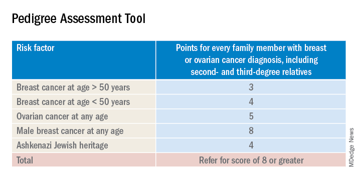
A positive result on a screening tool will lead primary care physicians to appropriately refer patients for genetic counseling. Genetic testing for BRCA1/2 mutations should be limited to those individuals whose personal and/or family history reflects an increased risk for gene mutations after detailed genetic assessment and counseling. The results of the genetic screening should assist a patient in their decision making.
Prophylactic treatment for BRCA1/2 mutation carriers are outside the scope of this recommendation. However the guidelines briefly review risk-reducing therapies including screening, medical, and surgical options. Medical therapy for patients who have BRCA1/2 mutations include the use of tamoxifen, raloxifene, and aromatase inhibitors. Surgical interventions include bilateral mastectomy and salpingo-oopherectomy.
Screening options include earlier and more frequent mammograms and breast MRIs. Screening is largely based on family history and the USPSTF acknowledges their uncertainty when screening women with an unknown family history. Male breast cancer, pancreatic cancer, prostate cancer, and melanoma are also associated with BRCA1/2 mutations. They are not included in this recommendation.
The bottom line
USPSTF recommended that primary care physicians should use familial risk assessment tools to screen women for BRCA1/2 mutations. This includes women with a personal and/or family history of breast, ovarian, tubal, or peritoneal cancer or women with a family history of BRCA1/2 gene mutations. Patients who test positive through one of the suggested screening tools should be referred for genetic counseling. This could lead to genetic testing and subsequent prophylactic therapies and/or increased screenings if the patient so desires. It is of importance to note the USPSTF recommends against routine screening of BRCA1/2 gene mutations for women who do not meet the above requirements.
Reference
USPSTF Recommendation: Assessment, counseling, and testing for BRCA-related cancer. JAMA. 2019;322(7):652-65. doi: 10.1001/jama.2019.10987.
Dr. Style is a second-year resident in the Family Medicine Residency Program at Abington (Pa.) Jefferson Health. Dr. Skolnik is professor of family and community medicine at Jefferson Medical College, Philadelphia, and an associate director of the family medicine residency program at Abington Jefferson Health.
Breast cancer screening recommendations have evolved over the past decade. BRCA1/2 genes are tumor-suppressor genes. Mutations in these genes place women at an increased risk for developing breast, ovarian, fallopian tube, and peritoneal cancer. Detection of BRCA1/2 mutations through genetic screening can provide patients with more information about their cancer risk and can lead to discussion of prophylactic therapies. This includes increased screening frequency, medical therapy, and surgical interventions.
New USPSTF recommendations address who is at an increased risk for BRCA1/2 mutations. They recommend using screening tools focusing on family history that primary care physicians can utilize to determine who should be referred for genetic counseling to discuss the risks and benefits of genetic screening. The following are the task force’s two primary recommendations:
The USPSTF recommends that primary care clinicians assess women with a personal or family history of breast, ovarian, tubal, or peritoneal cancer or who have an ancestry associated with BRCA1/2 gene mutations with an appropriate brief familial risk assessment tool. Women with a positive result on the risk assessment tool should receive genetic counseling and, if indicated after counseling, genetic testing. (B recommendation)
The USPSTF recommends against routine risk assessment, genetic counseling, or genetic testing for women whose personal or family history or ancestry is not associated with potentially harmful BRCA1/2 gene mutations. (D recommendation)
Breast cancer is the second leading cause of cancer and cancer death for women in the United States. Ovarian cancer ranks fifth in cancer deaths for women in the U.S. By age 70, women with BRCA1/2 mutations have a 45%-65% cumulative lifetime risk of developing breast cancer.
Mutations in BRCA1, specifically, are associated with a 39% lifetime risk for ovarian, fallopian tube, and peritoneal cancer. In contrast, mutations in BRCA2 are associated with a 10%-17% lifetime risk.
The USPSTF also underscores the increased prevalence of BRCA1/2 mutations in the Ashkenazi Jewish population. Three out of seven familial risk assessment tools inquire about Jewish ancestry. This is because the Ashkenazi Jewish population have a higher prevalence of three founder mutations in the BRCA1/2 gene. A member of this population has a 1 in 40 chance of carrying one of these three mutations, whereas the general population has a 1 in 300 chance.
The USPSTF recommends a multistep process of screening. The first step is taking a family history of cancer. For women who have a family history of breast, ovarian, tubal, or peritoneal cancer or a personal history of these cancers, a brief familial risk assessment tool should be used to determine the need for referral for in-depth genetic counseling to determine the need for genetic testing.
It is important to recognize that the validated tools recommended by the USPSTF are specific for genetic risk assessment. General breast cancer risk assessment tools, including the National Cancer Institute Breast Cancer Risk Assessment Tool, which is based on the Gail model, are not recommended.
The sensitivity of the tools recommended by the USPSTF range between 77% and 100%. A number of tools are given as an option with no one tool being better than the other. Perhaps the easiest to implement of the validated tools recommended is the Pedigree Assessment Tool. For this tool, points are assigned for every family member with breast or ovarian cancer as indicated in the table below.
A positive result on a screening tool will lead primary care physicians to appropriately refer patients for genetic counseling. Genetic testing for BRCA1/2 mutations should be limited to those individuals whose personal and/or family history reflects an increased risk for gene mutations after detailed genetic assessment and counseling. The results of the genetic screening should assist a patient in their decision making.
Prophylactic treatment for BRCA1/2 mutation carriers are outside the scope of this recommendation. However the guidelines briefly review risk-reducing therapies including screening, medical, and surgical options. Medical therapy for patients who have BRCA1/2 mutations include the use of tamoxifen, raloxifene, and aromatase inhibitors. Surgical interventions include bilateral mastectomy and salpingo-oopherectomy.
Screening options include earlier and more frequent mammograms and breast MRIs. Screening is largely based on family history and the USPSTF acknowledges their uncertainty when screening women with an unknown family history. Male breast cancer, pancreatic cancer, prostate cancer, and melanoma are also associated with BRCA1/2 mutations. They are not included in this recommendation.
The bottom line
USPSTF recommended that primary care physicians should use familial risk assessment tools to screen women for BRCA1/2 mutations. This includes women with a personal and/or family history of breast, ovarian, tubal, or peritoneal cancer or women with a family history of BRCA1/2 gene mutations. Patients who test positive through one of the suggested screening tools should be referred for genetic counseling. This could lead to genetic testing and subsequent prophylactic therapies and/or increased screenings if the patient so desires. It is of importance to note the USPSTF recommends against routine screening of BRCA1/2 gene mutations for women who do not meet the above requirements.
Reference
USPSTF Recommendation: Assessment, counseling, and testing for BRCA-related cancer. JAMA. 2019;322(7):652-65. doi: 10.1001/jama.2019.10987.
Dr. Style is a second-year resident in the Family Medicine Residency Program at Abington (Pa.) Jefferson Health. Dr. Skolnik is professor of family and community medicine at Jefferson Medical College, Philadelphia, and an associate director of the family medicine residency program at Abington Jefferson Health.
Patients have good recall on lung cancer screening scan benefits, not risks
NEW ORLEANS – in a recent survey, underscoring the need for ongoing patient education beyond the initial shared decision-making encounter, a researcher said.
While about 9 in 10 patients recalled key information on the benefits, only about 4 in 10 recalled information on risks, according to Erin Hirsch, MSPH, of the Colorado School of Public Health in Aurora.
“This may mean we need ongoing clinician involvement or continued education about this important information, especially if the patients aren’t screening annually,” Ms. Hirsch said in a podium presentation at the annual meeting of the American College of Chest Physicians.
Even fewer patients could correctly recall eligibility criteria for lung cancer screening, which suggests “ongoing clinician involvement” is needed to identify appropriate patients, Hirsch added in her presentation.
Shared decision making about lung cancer screening, which is supposed to entail a balanced patient-provider conversation about eligibility, risks, and benefits, is required by the Centers for Medicare & Medicaid Services to cover the cost of lung cancer screening as a preventative service, Hirsch noted in her presentation.
However, it’s largely unknown to what extent patients due for an annual screening recall information that should have been imparted in that initial discussion.
“The gap in knowledge centers around the fact that shared decision making is only required for baseline scan, but screening is recommended on an annual basis,” she said.
To test patient recall, Hirsch and colleagues developed a knowledge survey including 34 questions about lung cancer screening eligibility, risks, and benefits. The surveys went out by mail or email to 228 patients who had a baseline screening CT scan 6-12 months earlier; a total of 53 complete responses were included in the analysis, which focused on seven key questions about benefit, risk, and eligibility.
Recall was “excellent” for the benefit questions, Ms. Hirsch said, with 91% of patients able to recall that a computed tomography (CT) scan is better at detecting a possible lung cancer than a chest x-ray, while 87% recalled that without screening lung cancer is often found at a later stage when cure is less likely.
By contrast, a “moderate” amount (40%) remembered that a CT scan can suggest the patient has lung cancer when in fact they do not, she said, and only 38% recalled that radiation exposure was one of the harms of lung cancer screening, she added.
Eligibility recall was “poor,” she added, with only 21% affirming that not all current and former smokers need to be screened for lung cancer. Just 8% recalled that 55 years is the age at which beginning lung cancer screening is recommended, and 4% knew that 30 is the minimum number of pack-years required to be eligible for screening.
While these results may have clinical implications, Ms. Hirsch acknowledged a number of limitations to this pilot study. Among those was the fact that the content of the initial shared decision-making conversation could not be assessed: “We assumed that the patients were exposed to the information asked about in the survey ahead of time, but we can’t say for sure if that was true,” she explained.
Ms. Hirsch and coauthors disclosed no relationships relevant to their study.
SOURCE: Hirsch E et al. CHEST 2019. Abstract, doi: 10.1016/j.chest.2019.08.107.
REPORTING FROM CHEST 2019
How to overcome barriers to exercise for cancer patients
There is increasing evidence that exercise lowers the risk of developing cancer, improves survival after a cancer diagnosis, and helps ease related health outcomes. However, relatively few cancer patients meet current physical activity guidelines – often because it wasn’t recommended by their oncologist.

“Observed barriers to clinicians referring patients to exercise programming include lack of awareness of the potential value of exercise in cancer populations, uncertainty regarding the safety or suitability of exercise for a particular patient, lack of awareness regarding available programs to help facilitate exercise in cancer populations, need for education and skills development for making referrals, and a belief that referrals to exercise programming are not within the scope of practice for oncology clinicians,” Kathryn H. Schmitz, PhD, from Penn State University in Hershey and coauthors from the American College of Sports Medicine International Multidisciplinary Roundtable wrote in CA: A Cancer Journal for Clinicians.
Dr. Schmitz and colleagues proposed using the American College of Sports Medicine’s Exercise Is Medicine initiative to address this gap, with a focus on asking physicians to take an assess, revise, and refer approach to recommending exercise.
Noting that there is clear evidence that patients are more likely to exercise if their oncologist recommends that they do so, the authors said a clinician’s silence on the subject of exercise could be interpreted as “tacit approval to maintain inactivity.”
To assess a patient’s level of physical activity, the authors suggested asking patients how many days during the past week they had undertaken physical activity during which their heart beat faster or they breathed harder than normal for more than 30 minutes or how often they had undertaken activity to increase muscle strength.
If the patient can safely exercise without medical supervision, the authors recommend that clinicians advise patients to increase their physical activity to achieve current recommended levels. Clinicians can refer patients to community programs to help ramp up their activity.
However, for patients who may not be able to exercise safely on their own, the authors recommend referring them to an outpatient rehabiliation professional.
“Referral to appropriate and effective programs and follow-up with an assessment of progress (or lack thereof) at subsequent visits can serve as key transition points to change a patient’s behavior and affect their tolerance of or recovery from treatment,” they wrote.
The authors stressed that oncology clinicians were not expected to prescribe specific levels of exercise or to perform extensive screening and triage of patients based on their fitness.
“Oncology clinicians, however, play a vital role in telling the patient that it is important to exercise and pointing patients in the right direction to make that happen,” they wrote.
The American College of Sports Medicine International Multidisciplinary Roundtable was funded by the American College of Sports Medicine, the American Cancer Society, and other groups. Four authors declared grant support for participating in the study. One declared private industry funding unrelated to the study. No other conflicts of interest were declared.
SOURCE: Schmitz K et al. CA Cancer J Clin. 2019 Oct 16. doi: 10.3322/caac.21579.
There is increasing evidence that exercise lowers the risk of developing cancer, improves survival after a cancer diagnosis, and helps ease related health outcomes. However, relatively few cancer patients meet current physical activity guidelines – often because it wasn’t recommended by their oncologist.
“Observed barriers to clinicians referring patients to exercise programming include lack of awareness of the potential value of exercise in cancer populations, uncertainty regarding the safety or suitability of exercise for a particular patient, lack of awareness regarding available programs to help facilitate exercise in cancer populations, need for education and skills development for making referrals, and a belief that referrals to exercise programming are not within the scope of practice for oncology clinicians,” Kathryn H. Schmitz, PhD, from Penn State University in Hershey and coauthors from the American College of Sports Medicine International Multidisciplinary Roundtable wrote in CA: A Cancer Journal for Clinicians.
Dr. Schmitz and colleagues proposed using the American College of Sports Medicine’s Exercise Is Medicine initiative to address this gap, with a focus on asking physicians to take an assess, revise, and refer approach to recommending exercise.
Noting that there is clear evidence that patients are more likely to exercise if their oncologist recommends that they do so, the authors said a clinician’s silence on the subject of exercise could be interpreted as “tacit approval to maintain inactivity.”
To assess a patient’s level of physical activity, the authors suggested asking patients how many days during the past week they had undertaken physical activity during which their heart beat faster or they breathed harder than normal for more than 30 minutes or how often they had undertaken activity to increase muscle strength.
If the patient can safely exercise without medical supervision, the authors recommend that clinicians advise patients to increase their physical activity to achieve current recommended levels. Clinicians can refer patients to community programs to help ramp up their activity.
However, for patients who may not be able to exercise safely on their own, the authors recommend referring them to an outpatient rehabiliation professional.
“Referral to appropriate and effective programs and follow-up with an assessment of progress (or lack thereof) at subsequent visits can serve as key transition points to change a patient’s behavior and affect their tolerance of or recovery from treatment,” they wrote.
The authors stressed that oncology clinicians were not expected to prescribe specific levels of exercise or to perform extensive screening and triage of patients based on their fitness.
“Oncology clinicians, however, play a vital role in telling the patient that it is important to exercise and pointing patients in the right direction to make that happen,” they wrote.
The American College of Sports Medicine International Multidisciplinary Roundtable was funded by the American College of Sports Medicine, the American Cancer Society, and other groups. Four authors declared grant support for participating in the study. One declared private industry funding unrelated to the study. No other conflicts of interest were declared.
SOURCE: Schmitz K et al. CA Cancer J Clin. 2019 Oct 16. doi: 10.3322/caac.21579.
FROM CA: A CANCER JOURNAL FOR CLINICIANS