User login
Bringing you the latest news, research and reviews, exclusive interviews, podcasts, quizzes, and more.
div[contains(@class, 'header__large-screen')]
div[contains(@class, 'read-next-article')]
div[contains(@class, 'nav-primary')]
nav[contains(@class, 'nav-primary')]
section[contains(@class, 'footer-nav-section-wrapper')]
footer[@id='footer']
div[contains(@class, 'main-prefix')]
section[contains(@class, 'nav-hidden')]
div[contains(@class, 'ce-card-content')]
nav[contains(@class, 'nav-ce-stack')]
VTEs tied to immune checkpoint inhibitor cancer treatment
Cancer patients who receive an immune checkpoint inhibitor have more than a doubled rate of venous thromboembolism during the subsequent 2 years, compared with their rate during the 2 years before treatment, according to a retrospective analysis of more than 2,800 patients treated at a single U.S. center.
The study focused on cancer patients treated with an immune checkpoint inhibitor (ICI) at Massachusetts General Hospital in Boston. It showed that during the 2 years prior to treatment with any type of ICI, the incidence of venous thromboembolic events (VTE) was 4.85/100 patient-years that then jumped to 11.75/100 patient-years during the 2 years following treatment. This translated into an incidence rate ratio of 2.43 during posttreatment follow-up, compared with pretreatment, Jingyi Gong, MD, said at the virtual American Heart Association scientific sessions.
The increased VTE rate resulted from rises in both the rate of deep vein thrombosis, which had an IRR of 3.23 during the posttreatment period, and for pulmonary embolism, which showed an IRR of 2.24, said Dr. Gong, a physician at Brigham and Women’s Hospital in Boston. She hypothesized that this effect may result from a procoagulant effect of the immune activation and inflammation triggered by ICIs.
Hypothesis-generating results
Cardiologists cautioned that these findings should only be considered hypothesis generating, but raise an important alert for clinicians to have heightened awareness of the potential for VTE following ICI treatment.
“A clear message is to be aware that there is this signal, and be vigilant for patients who might present with VTE following ICI treatment,” commented Richard J. Kovacs, MD, a cardiologist and professor at Indiana University, Indianapolis. The data that Dr. Gong reported are “moderately convincing,” he added in an interview.
“Awareness that patients who receive ICI may be at increased VTE risk is very important,” agreed Umberto Campia, MD, a cardiologist, vascular specialist, and member of the cardio-oncology group at Brigham and Women’s Hospital, who was not involved in the new study.
The potential impact of ICI treatment on VTE risk is slowly emerging, added Dr. Campia. Until recently, the literature primarily was case reports, but recently another retrospective, single-center study came out that reported a 13% incidence of VTE in cancer patients following ICI treatment. On the other hand, a recently published meta-analysis of more than 20,000 patients from 68 ICI studies failed to find a suggestion of increased VTE incidence following ICI interventions.
Attempting to assess the impact of treatment on VTE risk in cancer patients is challenging because cancer itself boosts the risk. Recommendations on the use of VTE prophylaxis in cancer patients most recently came out in 2014 from the American Society of Clinical Oncology, which said that VTE prophylaxis for ambulatory cancer patients “may be considered for highly select high-risk patients.” The impact of cancer therapy on VTE risk and the need for prophylaxis is usually assessed by applying the Khorana score, Dr. Campia said in an interview.
VTE spikes acutely after ICI treatment
Dr. Gong analyzed VTE incidence rates by time during the total 4-year period studied, and found that the rate gradually and steadily rose with time throughout the 2 years preceding treatment, spiked immediately following ICI treatment, and then gradually and steadily fell back to roughly the rate seen just before treatment, reaching that level about a year after treatment. She ran a sensitivity analysis that excluded patients who died during the first year following their ICI treatment, and in this calculation an acute spike in VTE following ICI treatment still occurred but with reduced magnitude.
She also reported the results of several subgroup analyses. The IRRs remained consistent among women and men, among patients who were aged over or under 65 years, and regardless of cancer type or treatment with corticosteroids. But the subgroup analyses identified two parameters that seemed to clearly split VTE rates.
Among patients on treatment with an anticoagulant agent at the time of their ICI treatment, roughly 10% of the patients, the IRR was 0.56, compared with a ratio of 3.86 among the other patients, suggesting possible protection. A second factor that seemed linked with VTE incidence was the number of ICI treatment cycles a patient received. Those who received more than five cycles had a risk ratio of 3.95, while those who received five or fewer cycles had a RR of 1.66.
Her analysis included 2,842 cancer patients who received treatment with an ICI at Massachusetts General Hospital. Patients averaged 64 years of age, slightly more than half were men, and 13% had a prior history of VTE. Patients received an average of 5 ICI treatment cycles, but a quarter of the patients received more than 10 cycles.
During the 2-year follow-up, 244 patients (9%) developed VTE. The patients who developed VTE were significantly younger than those who did not, with an average age of 63 years, compared with 65. And the patients who eventually developed VTE had a significantly higher prevalence of prior VTE at 18%, compared with 12% among the patients who stayed VTE free.
The cancer types patients had were non–small cell lung, 29%; melanoma, 28%; head and neck, 12%; renal genitourinary, 6%; and other, 25%. ICIs have been available for routine U.S. practice since 2011. The class includes agents such as pembrolizumab (Keytruda) and durvalumab (Imfinzi).
Researchers would need to perform a prospective, randomized study to determine whether anticoagulant prophylaxis is clearly beneficial for patients receiving ICI treatment, Dr. Gong said. But both Dr. Kovacs and Dr. Campia said that more data on this topic are first needed.
“We need to confirm that treatment with ICI is associated with VTEs. Retrospective data are not definitive,” said Dr. Campia. “We would need to prospectively assess the impact of ICI,” which will not be easy, as it’s quickly become a cornerstone for treating many cancers. “We need to become more familiar with the adverse effects of these drugs. We are still learning about their toxicities.”
The study had no commercial funding. Dr. Gong, Dr. Kovacs, and Dr. Campia had no disclosures.
FROM AHA 2020
SAMSON pins most muscle pain experienced with statins on the nocebo effect
A novel randomized trial taking on a vexing issue around one of the world’s most commonly prescribed medications has concluded that frequently intolerable statin side effects, such as muscle weakness or pain, are almost entirely a nocebo effect, the placebo effect’s darker cousin.
The many patients who report such symptoms while taking statins are indeed probably feeling them, but they are a result of taking the pills rather than any pharmacologic effects, concluded researchers based on their 60-patient study, Self-Assessment Method for Statin Side-effects or Nocebo (SAMSON).
“SAMSON leaves no doubt that patients really do get side effects from statin tablets, but what it shows us is that 90% of this symptomatic burden is elicited by placebo tablets too,” said James P. Howard, MB, PhD, Imperial College London, when presenting the results Nov. 15 at the American Heart Association scientific sessions. They were published simultaneously in the New England Journal of Medicine.
Studies have shown that in practice “more than half of patients abandon statins completely within 2 years. And yet, in placebo-controlled trials, no more people stop statins than placebo,” Dr. Howard said.
“The most important message from SAMSON is that side effects from statin tablets are very real, but they are mainly caused by the act of taking the tablets, not by the statin that is contained within them.”
Patients in the trial, all of whom had a history of dropping statins because of side effects, each took atorvastatin 20 mg/day, a placebo, or neither pill for 1 month, alternating the regimens in randomized order over 1 year so that each was followed a total of 4 months. They used a smartphone app to record the severity of any side effects, not necessarily just pain, on a scale of 0-100.
Symptom intensity scores averaged 16.3 for atorvastatin and 15.4 for placebo, for a nonsignificant difference, but only 8.0 for no-pill months (P < .001 compared with the statin or placebo).
Because such symptoms seem to be based on patient expectations from statin therapy, positive communication about what the drugs can achieve and how the next treatment steps are described can play a big role in their continued use.
For example, “changing them to another statin is a very reasonable thing to do, but as soon as you start trying people on lower doses and working up, you’re sort of telling them that you’re expecting at some dose that they are going to get side effects,” cautioned Dr. Howard at a media briefing on SAMSON.
“The most important thing is to explain the evidence, and what our expectations are, maybe be a bit more optimistic about statins, and tell them they’re very unlikely to suffer from side effects,” he explained, “because the nocebo effect can only really rear its head if the patients are expecting to feel worse – just like the placebo effect will only work if people are expecting to feel better.”
Amit Khera, MD, who moderated the media briefing, said he always tells such patients: “Yes, 1 in 10 patients report having muscle ache. But first and foremost, 9 in 10 don’t. The vast majority of patients don’t get muscle aches. I think that’s really an important part of the communication.”
Now, after SAMSON, “I have an additional point that I’m going to tell them: out of the patients that get muscle aches, probably 90% of that is the anticipation of getting the statin, the nocebo effect,” said Dr. Khera, who directs the preventive cardiology program at the University of Texas Southwestern Medical Center, Dallas.
In practice, however, many patients who report adverse statin effects do so later than 2 weeks after starting therapy, “so these findings cannot be generalized to them,” proposed Francine K. Welty, MD, PhD, Beth Israel Deaconess Medical Center, Boston, as the invited discussant after Dr. Howard’s presentation.
All 60 patients recruited for SAMSON had previously stopped taking a statin because of side effects that arose within 2 weeks of their first dose. That requirement was intended to boost chances that any further symptoms during the trial would arise within a month of starting each new round of pills, Dr. Howard said.
So the trial’s results, Dr. Welty said, “are limited to those subjects who develop symptoms within 2 weeks of starting a statin.”
Including only such patients may have created bias toward a nocebo effect, she said, because “non–drug-related side effects of medications are often greatest during the initial weeks of treatment and tend to abate over time.” For example, “metformin causes diarrhea and beta-blockers cause fatigue, but subjects do adapt and generally tolerate them very well.”
The patients, 25 women and 35 men, 90% of whom were white, received four pill bottles, each with a month’s supply of atorvastatin, four bottles each with 1 month of placebo, and four empty bottles each, to be used double blind for a month in randomized order.
Patients used the smartphone app to document their symptom scores, which ranged from 0 for no symptoms to 100 for symptoms that were the “worst imaginable,” the published report noted. Patients who experienced symptoms so severe as to be intolerable could stop the 1-month regimen they were then following, with instructions to resume the regimens in order starting the next month.
Eleven patients were unable to complete all 12 1-month segments of the trial.
The study’s overall “nocebo ratio” of 0.90 was calculated as the difference between symptom intensity scores on placebo and on no treatment divided by the difference between symptom intensity on the statin and on no treatment. The interpretation: 90% of the symptom burden felt by patients receiving atorvastatin was also felt during placebo use.
A total of 30 patients, contacted 6 months after the trial concluded, had resumed taking a statin, while “4 planned to do so and one could not be contacted,” the report noted. The 25 other patients weren’t receiving a statin and had no plans to take one.
In an important part of the trial, Dr. Howard said, at its conclusion the patients were shown their pattern of symptoms in relation to whether they were taking the statin, placebo, or neither. “Participants could see as clearly as we could the surprisingly powerful magnitude of the nocebo effect. And this led to half of our patients happily restarting statins.”
The implications of SAMSON, Dr. Welty said, “are very important, in that those developing symptoms within 2 weeks of starting a statin should be reassured that approximately half will be able to successful restart the statin.”
SAMSON was funded by the British Heart Foundation. Howard had no disclosures. Dr. Welty disclosed chairing the data safety monitoring committee for Empagliflozin International Clinical Trials, supported by Boehringer Ingelheim.
A version of this article originally appeared on Medscape.com.
Chronic inflammatory diseases vary widely in CHD risk
Not all chronic systemic inflammatory diseases are equal enhancers of atherosclerotic cardiovascular disease risk, according to a large case-control study.

Current AHA/American College of Cardiology guidelines cite three chronic inflammatory diseases as atherosclerotic cardiovascular disease risk enhancers: rheumatoid arthritis, psoriasis, and HIV infection. But this study of those three diseases, along with three others marked by elevated high sensitivity C-reactive protein (systemic sclerosis, inflammatory bowel disease, and systemic lupus erythematosus [SLE]), showed that chronic inflammatory diseases are not monolithic in terms of their associated risk of incident coronary heart disease (CHD).
Indeed, two of the six inflammatory diseases – psoriasis and inflammatory bowel disease – turned out to be not at all associated with increased cardiovascular risk in the 37,117-patient study. The highest-risk disease was SLE, not specifically mentioned in the guidelines, Arjun Sinha, MD, a cardiology fellow at Northwestern University, Chicago, noted in his presentation at the virtual American Heart Association scientific sessions.
The study included 18,129 patients with one of the six chronic inflammatory diseases and 18,988 matched controls, none with CHD at baseline. All regularly received outpatient care at Northwestern during 2000-2019. There were 1,011 incident CHD events during a median of 3.5 years of follow-up.
In a Cox proportional hazards analysis adjusted for demographics, insurance status, hypertension, diabetes, current smoking, total cholesterol, and estimated glomerular filtration rate, here’s how the chronic inflammatory diseases stacked up in terms of incident CHD and MI risks:
- SLE: hazard ratio for CHD, 2.85; for MI, 4.76.
- Systemic sclerosis: HR for CHD, 2.14; for MI, 3.19.
- HIV: HR for CHD, 1.38; for MI, 1.69.
- Rheumatoid arthritis: HR for CHD, 1.22; for MI, 1.45.
- Psoriasis: no significant increase.
- Inflammatory bowel disease: no significant increase.
In an exploratory analysis, Dr. Sinha and coinvestigators evaluated the risk of incident CHD stratified by disease severity. For lack of standardized disease severity scales, the investigators relied upon tertiles of CD4 T cell count in the HIV group and CRP in the others. The HR for new-onset CHD in the more than 5,000 patients with psoriasis didn’t vary by CRP tertile. However, there was a nonsignificant trend for greater disease severity, as reflected by CRP tertile, to be associated with increased incident CHD risk in the HIV and inflammatory bowel disease groups.
In contrast, patients with rheumatoid arthritis or systemic sclerosis who were in the top CRP tertile had a significantly greater risk of developing CHD than that of controls, with HRs of 2.11 in the rheumatoid arthritis group and 4.59 with systemic sclerosis, although patients in the other two tertiles weren’t at significantly increased risk. But all three tertiles of CRP in patients with SLE were associated with significantly increased CHD risk: 3.17-fold in the lowest tertile of lupus severity, 5.38-fold in the middle tertile, and 4.04-fold in the top tertile for inflammation.
These findings could be used in clinical practice to fine-tune atherosclerotic cardiovascular disease risk assessment based upon chronic inflammatory disease type and severity. That’s information which in turn can help guide the timing and intensity of preventive therapy for patients with each disease type.
But studying the association between chronic systemic inflammatory diseases and CHD risk can be useful in additional ways, according to Dr. Sinha. These inflammatory diseases can serve as models of atherosclerosis that shed light on the non–lipid-related mechanisms involved in cardiovascular disease.
“The gradient in risk may be hypothesis-generating with respect to which specific inflammatory pathways may contribute to CHD,” he explained.
Each of these six chronic inflammatory diseases is characterized by a different form of major immune dysfunction, Dr. Sinha continued. A case in point is SLE, the inflammatory disease associated with the highest risk of CHD and MI. Lupus is characterized by a form of neutrophil dysfunction marked by increased formation and reduced degradation of neutrophil extracellular traps, or NETs, as well as by an increase in autoreactive B cells and dysfunctional CD4+ T helper cells. The increase in NETs of of particular interest because NETs have also been shown to contribute to the development of atherosclerosis, endothelial dysfunction, plaque erosion, and thrombosis.
In another exploratory analysis, Dr. Sinha and coworkers found that SLE patients with a neutrophil count above the median level were twice as likely to develop CHD than were those with a neutrophil count below the median.
A better understanding of the upstream pathways linking NET formation in SLE and atherosclerosis could lead to development of new or repurposed medications that target immune dysfunction in order to curb atherosclerosis, said Dr. Sinha, whose study won the AHA’s Samuel A. Levine Early Career Clinical Investigator Award.
He reported having no financial conflicts regarding his study.
Not all chronic systemic inflammatory diseases are equal enhancers of atherosclerotic cardiovascular disease risk, according to a large case-control study.
Current AHA/American College of Cardiology guidelines cite three chronic inflammatory diseases as atherosclerotic cardiovascular disease risk enhancers: rheumatoid arthritis, psoriasis, and HIV infection. But this study of those three diseases, along with three others marked by elevated high sensitivity C-reactive protein (systemic sclerosis, inflammatory bowel disease, and systemic lupus erythematosus [SLE]), showed that chronic inflammatory diseases are not monolithic in terms of their associated risk of incident coronary heart disease (CHD).
Indeed, two of the six inflammatory diseases – psoriasis and inflammatory bowel disease – turned out to be not at all associated with increased cardiovascular risk in the 37,117-patient study. The highest-risk disease was SLE, not specifically mentioned in the guidelines, Arjun Sinha, MD, a cardiology fellow at Northwestern University, Chicago, noted in his presentation at the virtual American Heart Association scientific sessions.
The study included 18,129 patients with one of the six chronic inflammatory diseases and 18,988 matched controls, none with CHD at baseline. All regularly received outpatient care at Northwestern during 2000-2019. There were 1,011 incident CHD events during a median of 3.5 years of follow-up.
In a Cox proportional hazards analysis adjusted for demographics, insurance status, hypertension, diabetes, current smoking, total cholesterol, and estimated glomerular filtration rate, here’s how the chronic inflammatory diseases stacked up in terms of incident CHD and MI risks:
- SLE: hazard ratio for CHD, 2.85; for MI, 4.76.
- Systemic sclerosis: HR for CHD, 2.14; for MI, 3.19.
- HIV: HR for CHD, 1.38; for MI, 1.69.
- Rheumatoid arthritis: HR for CHD, 1.22; for MI, 1.45.
- Psoriasis: no significant increase.
- Inflammatory bowel disease: no significant increase.
In an exploratory analysis, Dr. Sinha and coinvestigators evaluated the risk of incident CHD stratified by disease severity. For lack of standardized disease severity scales, the investigators relied upon tertiles of CD4 T cell count in the HIV group and CRP in the others. The HR for new-onset CHD in the more than 5,000 patients with psoriasis didn’t vary by CRP tertile. However, there was a nonsignificant trend for greater disease severity, as reflected by CRP tertile, to be associated with increased incident CHD risk in the HIV and inflammatory bowel disease groups.
In contrast, patients with rheumatoid arthritis or systemic sclerosis who were in the top CRP tertile had a significantly greater risk of developing CHD than that of controls, with HRs of 2.11 in the rheumatoid arthritis group and 4.59 with systemic sclerosis, although patients in the other two tertiles weren’t at significantly increased risk. But all three tertiles of CRP in patients with SLE were associated with significantly increased CHD risk: 3.17-fold in the lowest tertile of lupus severity, 5.38-fold in the middle tertile, and 4.04-fold in the top tertile for inflammation.
These findings could be used in clinical practice to fine-tune atherosclerotic cardiovascular disease risk assessment based upon chronic inflammatory disease type and severity. That’s information which in turn can help guide the timing and intensity of preventive therapy for patients with each disease type.
But studying the association between chronic systemic inflammatory diseases and CHD risk can be useful in additional ways, according to Dr. Sinha. These inflammatory diseases can serve as models of atherosclerosis that shed light on the non–lipid-related mechanisms involved in cardiovascular disease.
“The gradient in risk may be hypothesis-generating with respect to which specific inflammatory pathways may contribute to CHD,” he explained.
Each of these six chronic inflammatory diseases is characterized by a different form of major immune dysfunction, Dr. Sinha continued. A case in point is SLE, the inflammatory disease associated with the highest risk of CHD and MI. Lupus is characterized by a form of neutrophil dysfunction marked by increased formation and reduced degradation of neutrophil extracellular traps, or NETs, as well as by an increase in autoreactive B cells and dysfunctional CD4+ T helper cells. The increase in NETs of of particular interest because NETs have also been shown to contribute to the development of atherosclerosis, endothelial dysfunction, plaque erosion, and thrombosis.
In another exploratory analysis, Dr. Sinha and coworkers found that SLE patients with a neutrophil count above the median level were twice as likely to develop CHD than were those with a neutrophil count below the median.
A better understanding of the upstream pathways linking NET formation in SLE and atherosclerosis could lead to development of new or repurposed medications that target immune dysfunction in order to curb atherosclerosis, said Dr. Sinha, whose study won the AHA’s Samuel A. Levine Early Career Clinical Investigator Award.
He reported having no financial conflicts regarding his study.
FROM AHA 2020
Combined OCT, cardiac MRI unravels root cause in most MINOCA
Optical CT (OCT) plus cardiac MRI (CMR) provides a more specific diagnosis in the majority of women presenting with myocardial infarction with nonobstructive coronary arteries (MINOCA).
The multimodal imaging strategy identified the underlying cause of MINOCA in 85% of women in the HARP-MINOCA study. Overall, 64% of women had a true MI and 21% had an alternate nonischemic diagnosis, most commonly myocarditis.
“OCTCMR findings correlated well with OCT culprit lesions, demonstrating that nonobstructive culprit lesions frequently cause MINOCA,” said study author Harmony Reynolds, MD, director of New York University Langone’s Sarah Ross Soter Center for Women’s Cardiovascular Research.
The results were presented at the virtual American Heart Association (AHA) Scientific Sessions 2020 and published simultaneously in Circulation.
MINOCA occurs in up to 15% of patients with MI and is defined as MI meeting the universal definition but with less than 50% stenosis in all major epicardial arteries on angiography and no specific alternate diagnosis to explain the presentation.
It is three times more common in women than in men and also disproportionately affects Black, Hispanic, Maori, and Pacific persons. MINOCA has several causes, leading to uncertainty in diagnostic testing and treatment.
“Different doctors tell patients different messages about MINOCA and may incorrectly say the event wasn’t a heart attack,” Dr. Reynolds said in an earlier press briefing. “I had a patient who was told ‘your arteries are open,’ and they gave her Xanax.”
As part of the Women’s Heart Attack Research Program (HARP), researchers enrolled 301 women with a clinical diagnosis of MI, of whom 170 were diagnosed with MINOCA during angiography and underwent OCT at that time, followed by CMR within 1 week of the acute presentation.
All images were interpreted by an independent core laboratory blinded to results of the other tests and clinical information. The final cohort included 145 women with interpretable OCT images.
Their median age was 60 years, 49.7% were white non-Hispanic, and 97% presented with a provisional diagnosis of non–ST-segment MI. Their median peak troponin level was 0.94 ng/mL.
OCT identified a definite or probable culprit lesion in 46% of women, most commonly atherosclerosis or thrombosis. On multivariable analysis, having a culprit lesion was associated with older age, abnormal angiography findings at the site, and diabetes, but not peak troponin level or severity of angiographic stenosis.
CMR available in 116 women showed evidence of infarction or regional injury in 69%. Multivariate predictors of an abnormal CMR were higher peak troponin and diastolic blood pressure but not an OCT culprit lesion or angiographic stenosis severity.
When the OCT and CMR results were combined, a cause of MINOCA was identified in 84.5% of women. Three-fourths of the causes were ischemic (64% MI) and one-quarter were nonischemic (15% myocarditis, 3% Takotsubo syndrome, and 3% nonischemic cardiomyopathy). In the remaining 15%, no cause of MINOCA was identified.
To emphasize the effect multimodal imaging can have on treatment, Dr. Reynolds highlighted a 44-year-old woman with no risk factors for coronary artery disease who had chest pain in the context of heavy menstrual bleeding, a low hemoglobin level, and peak troponin level of 3.25 ng/mL.
Unexpectedly, imaging revealed a left anterior descending (LAD) plaque rupture in a thin-cap fibroatheroma, causing a small transmural infarction at the terminus of the LAD.
“Without this diagnosis, it’s unlikely she would have received antiplatelet therapy or statins and might have been given a diagnosis of supply/demand mismatch, when the real diagnosis was MI,” Dr. Reynolds observed.
“Finally we can say this is not just crazy women. There is really something going on,” said panelist Roxana Mehran, MD, of the Icahn School of Medicine at Mount Sinai in New York. “You have now told us this is most likely atherosclerosis for pretty much 85% of the cases. So make the diagnosis and, of course, make sure you treat these patients accordingly for risk factor modification, really thinking about a ruptured plaque.”
Combining OCT and MRI may result in a more specific diagnosis and better treatment but also raises costs and logistical considerations.
“Implementation challenges are that not every form of testing is available in every medical center,” Dr. Reynolds said in an interview. “Many centers have cardiac MRI,” whereas “OCT is not currently available at most medical centers where heart attack patients are treated but is available at specialized centers.”
Asked during the session about the use of CT angiography, invited discussant Martha Gulati, MD, president-elect of the American Society for Preventive Cardiology, said, “For me, CT is helpful when I’m not sure if there’s any plaque because the angiogram looked really normal and there was no opportunity to do intracoronary imaging. And sometimes that will help me, in particular, if a patient doesn’t want to take a statin.”
Dr. Gulati pointed out that the European Society of Cardiology MINOCA guidelines recommend OCT and CMR, whereas the 2019 AHA statement on MINOCA, which she coauthored, also recommends OCT and CMR, but almost as one or the other.
“We already said that you should do cardiac MR to try to make a diagnosis, but I think the combination of the two needs to be emphasized when we next draft these guidelines. It really will help,” Dr. Gulati said in an interview.
“But using OCT, particularly, needs to be in the setting of the MI. I don’t think you want to do a procedure again,” she said. “So we really need it to become more widely available because at the time of an MI, you won’t necessarily know that you’re not going to find an obstructive lesion.”
Dr. Gulati pointed out several unanswered questions, including whether the diagnosis was missed in some patients, because OCT of all three vessels was available in only 59%, and how the use of high-sensitivity troponin, which was left up to the individual institution, might affect the usefulness of OCT and CMR.
It’s also unknown whether the mechanism is different for ST-segment elevation MI, as the trial included very few cases, although MINOCA often occurs in this setting. Future OCT/CMR studies will also need to enroll men to determine potential sex differences, if any.
Commenting on the study, B. Hadley Wilson, MD, Sanger Heart & Vascular Institute in Charlotte, N.C., said, “There would need to be further justification of this invasive interventional procedure to be sure that the benefit outweighed the risk of putting a wire and an OCT catheter down patients without any significant angiographic blockage and to assure interventional cardiologists of its value here.”
He pointed out that noninvasive CMR appears helpful in the diagnosis of nearly three-quarters of these patients and perhaps could be done first to direct which of those with an ischemic cause might benefit from invasive OCT at catheterization. This seems most pertinent in patients with a high suspicion of coronary artery disease or recurrent MINOCA.
“Overall, we need to consider the expense, logistics, and small risk of these combined modalities, particularly in everyday practice, before making recommendations,” Dr. Wilson said. “ Since OCT is much less available than intravascular ultrasound, it would require a challenging marketplace paradigm shift to implement this multimodality imaging strategy regionally and locally in the U.S., including the added costs. However, further study to direct the more judicious use of either CMR and/or combined with OCT is warranted in these patients.”
The study was funded by the AHA through a grant from the Go Red for Women Strategically Focused Research Network. Dr. Reynolds reported in-kind donations from Abbott Vascular and Siemens related to the study and nonfinancial support from BioTelemetry outside the study. Dr. Gulati and Dr. Wilson reported having no relevant disclosures.
A version of this article originally appeared on Medscape.com.

Empagliflozin favorably reshaped left ventricles in HFrEF patients
Treatment with the SGLT2 inhibitor empagliflozin led to significant reductions in both left ventricular end systolic and diastolic volumes in two independent randomized studies of patients with heart failure with reduced ejection fraction.
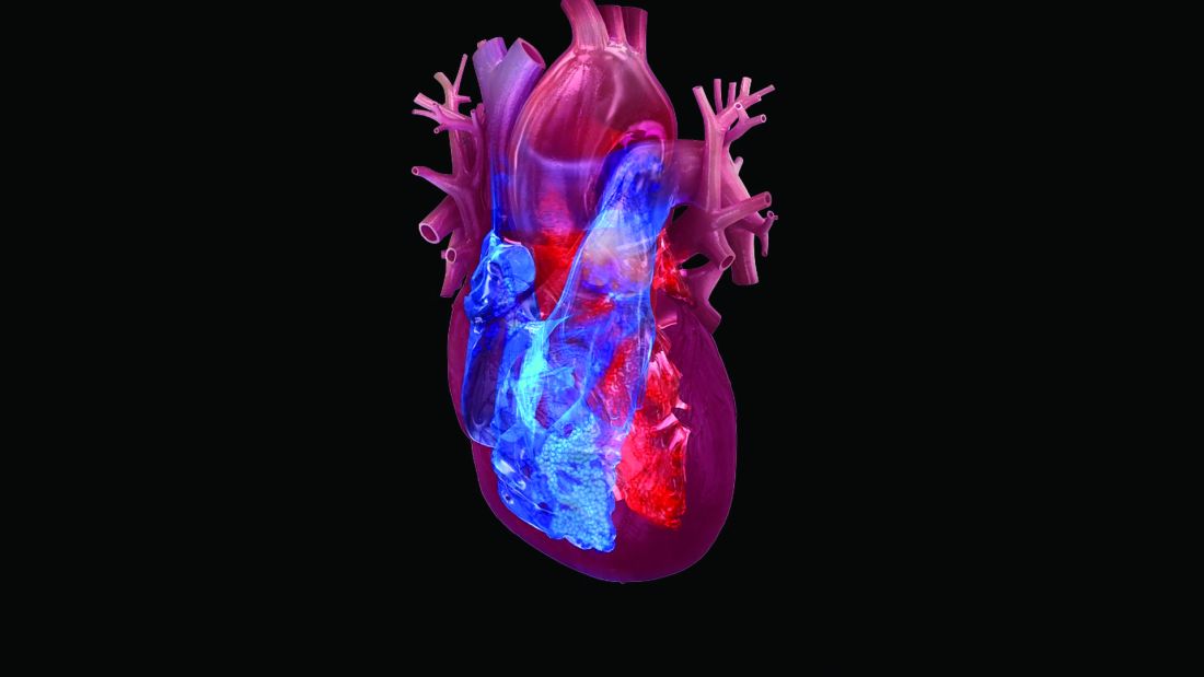
These results provide important new evidence that one way a drug from this class exerts its beneficial effects on cardiovascular outcomes in these patients is by producing favorable left-ventricular remodeling.
One of the two studies involved only patients with heart failure with reduced ejection fraction (HFrEF) with diabetes and examined treatment impact after 36 weeks. The second study focused exclusively on HFrEF patients without diabetes and followed patients for 6 months. Both studies also generated additional significant evidence of favorable left-ventricular effects.
“The results of these two new trials are incredibly important, as they tell cardiologists one of the mechanisms by which SGLT2 [sodium glucose co-transporter 2] inhibitors reduce heart failure hospitalizations and cardiovascular death,” said Mark C. Petrie, MBChB, professor at the Institute of Cardiovascular & Medical Sciences at the University of Glasgow, and principal investigator for one of the two studies.
“Many cardiologists want to know mechanisms as well as clinical benefit. These remodeling data showing that these drugs reduce the size of abnormally large hearts [and] are also very important for patients,” Dr. Petrie said in an interview. “There have been more than 50 publications on potential mechanisms of benefit of SGLT2 inhibitors in HFrEF, but these are the first randomized, mechanistic data.”
Mechanistic clues follow large cardiovascular outcome trials
Results from a large randomized trial, EMPEROR-Reduced, recently showed that treatment with empagliflozin (Jardiance) on top of standard HFrEF treatment led to significant benefits in patients with or without type 2 diabetes (T2D), compared with placebo, for major cardiovascular and renal endpoints, including the combination of cardiovascular death or hospitalization for heart failure. And results from a second large randomized trial, DAPA-HF, showed similar results with a different drug from the same class, dapagliflozin (Farxiga), in an earlier report.
But while these reports led to quick uptake of these two drugs for the treatment of patients with HFrEF, the means by which these agents exert their HFrEF benefits have been unclear.
“Our study identifies why this drug [empagliflozin] is effective – because it improves heart function, something that has not been understood until now,” Carlos G. Santos-Gallego, MD, lead investigator for the second new report, said in a written statement. “Many doctors are afraid of prescribing a drug they do not understand, and our findings will help clinicians feel more comfortable giving this to patients once approved.”
On the strength of the DAPA-HF results, dapagliflozin received a revised U.S. label in May 2020 that added the indication for treating patients with HFrEF regardless of the whether patients also have T2D, the original indication for prescribing the drug. Many experts anticipate that a similar addition to the label for empagliflozin will soon occur.
EMPA-TROPISM examines patients with no T2D
The single-center study reported by Dr. Santos-Gallego randomized 84 patients with HFrEF and no diabetes to standard treatment with empagliflozin or placebo and measured several parameters in 80 patients who completed the planned 6 months of treatment. The primary endpoints were the changes in both left ventricular end systolic and diastolic volume from baseline in the empagliflozin-treated patients compared with patients on placebo, measured by cardiac MR.
The results showed an average reduction of end systolic volume of 26.6 mL from baseline compared with a small rise in the placebo patients, and an average drop in end diastolic volume of 25.1 mL from baseline compared again with a small increase in the controls. Both differences were statistically significant, reported the senior author of the study, Juan J. Badimon, PhD, in a talk at the virtual scientific sessions of the American Heart Association. Concurrently, the results were published online in the Journal of the American College of Cardiology.
Results from the EMPA-TROPISM study also showed several other significant benefits from empagliflozin treatment, both to left ventricular shape and function as well as to other measures of patient well being. The drug regimen led to an increase in left ventricular ejection fraction, a decrease in left ventricular mass, reduced myocardial fibrosis and aortic stiffness, increased peak oxygen consumption, an increased distance traveled in a 6-minute walk test, and improved quality of life, said Dr. Badimon, professor of medicine and director of the Atherothrombosis Research Unit at the Cardiovascular Institute at the Icahn School of Medicine at Mount Sinai in New York.
SUGAR-DM-HF enrolled only T2D patients
The second study, SUGAR-DM-HF, randomized 105 patients with HFrEF and T2D to treatment with empagliflozin or placebo at any of 15 centers in Scotland, with 92 patients completing the full 36 weeks on treatment. One of the study’s two primary endpoints was the change in left ventricular end systolic volume index, which dropped by an average of 7.9 mL/m2 in patients who received empagliflozin and by 1.5 mL/m2 in the controls, a significant average between-group difference of 6.0 mL/m2, reported Matthew M.Y. Lee, MBChB, at the same meeting.
However, the second primary endpoint, change in left ventricular global longitudinal strain, showed no significant difference in effect on empagliflozin compared with placebo, said Dr. Lee, a cardiologist at the University of Glasgow. Concurrently with his report the results appeared in an article published online in Circulation.
The results also showed a significant drop in left ventricular end diastolic volume index from baseline compared with the control patients, with an average between-group difference in the reduction from baseline of 8.2 mL/m2.
“Reverse cardiac remodeling is a mechanism by which SGLT2 inhibitors reduce heart failure hospitalizations and cardiovascular mortality,” Dr. Lee concluded during his presentation at the meeting.
Although the findings from both studies together provide strong evidence for an effect by empagliflozin on left ventricular shape and function, neither study provides much insight into how this drug exerts these effects. The authors of both studies agreed on several potential explanations, including reductions in cardiac preload and afterload that could reduce left ventricular stretch and volume; a change triggered in myocardial energetics that switches from a metabolism mostly dependent on glucose to one more geared to using fatty acids, ketone bodies, and branched chain amino acids; and a possible drug-induced reduction in oxidative stress and inflammation.
SUGAR-DM-HF was sponsored by a grant from Boehringer Ingelheim, the company that along with Eli Lilly markets empagliflozin (Jardiance). Dr. Lee had no disclosures. Dr. Petrie has been a consultant to Boehringer Ingelheim and Eli Lilly and to several other companies. EMPA-TROPISM was sponsored by a grant from Boehringer Ingelheim. Dr. Badimon and Dr. Santos-Gallego had no disclosures.
Treatment with the SGLT2 inhibitor empagliflozin led to significant reductions in both left ventricular end systolic and diastolic volumes in two independent randomized studies of patients with heart failure with reduced ejection fraction.
These results provide important new evidence that one way a drug from this class exerts its beneficial effects on cardiovascular outcomes in these patients is by producing favorable left-ventricular remodeling.
One of the two studies involved only patients with heart failure with reduced ejection fraction (HFrEF) with diabetes and examined treatment impact after 36 weeks. The second study focused exclusively on HFrEF patients without diabetes and followed patients for 6 months. Both studies also generated additional significant evidence of favorable left-ventricular effects.
“The results of these two new trials are incredibly important, as they tell cardiologists one of the mechanisms by which SGLT2 [sodium glucose co-transporter 2] inhibitors reduce heart failure hospitalizations and cardiovascular death,” said Mark C. Petrie, MBChB, professor at the Institute of Cardiovascular & Medical Sciences at the University of Glasgow, and principal investigator for one of the two studies.
“Many cardiologists want to know mechanisms as well as clinical benefit. These remodeling data showing that these drugs reduce the size of abnormally large hearts [and] are also very important for patients,” Dr. Petrie said in an interview. “There have been more than 50 publications on potential mechanisms of benefit of SGLT2 inhibitors in HFrEF, but these are the first randomized, mechanistic data.”
Mechanistic clues follow large cardiovascular outcome trials
Results from a large randomized trial, EMPEROR-Reduced, recently showed that treatment with empagliflozin (Jardiance) on top of standard HFrEF treatment led to significant benefits in patients with or without type 2 diabetes (T2D), compared with placebo, for major cardiovascular and renal endpoints, including the combination of cardiovascular death or hospitalization for heart failure. And results from a second large randomized trial, DAPA-HF, showed similar results with a different drug from the same class, dapagliflozin (Farxiga), in an earlier report.
But while these reports led to quick uptake of these two drugs for the treatment of patients with HFrEF, the means by which these agents exert their HFrEF benefits have been unclear.
“Our study identifies why this drug [empagliflozin] is effective – because it improves heart function, something that has not been understood until now,” Carlos G. Santos-Gallego, MD, lead investigator for the second new report, said in a written statement. “Many doctors are afraid of prescribing a drug they do not understand, and our findings will help clinicians feel more comfortable giving this to patients once approved.”
On the strength of the DAPA-HF results, dapagliflozin received a revised U.S. label in May 2020 that added the indication for treating patients with HFrEF regardless of the whether patients also have T2D, the original indication for prescribing the drug. Many experts anticipate that a similar addition to the label for empagliflozin will soon occur.
EMPA-TROPISM examines patients with no T2D
The single-center study reported by Dr. Santos-Gallego randomized 84 patients with HFrEF and no diabetes to standard treatment with empagliflozin or placebo and measured several parameters in 80 patients who completed the planned 6 months of treatment. The primary endpoints were the changes in both left ventricular end systolic and diastolic volume from baseline in the empagliflozin-treated patients compared with patients on placebo, measured by cardiac MR.
The results showed an average reduction of end systolic volume of 26.6 mL from baseline compared with a small rise in the placebo patients, and an average drop in end diastolic volume of 25.1 mL from baseline compared again with a small increase in the controls. Both differences were statistically significant, reported the senior author of the study, Juan J. Badimon, PhD, in a talk at the virtual scientific sessions of the American Heart Association. Concurrently, the results were published online in the Journal of the American College of Cardiology.
Results from the EMPA-TROPISM study also showed several other significant benefits from empagliflozin treatment, both to left ventricular shape and function as well as to other measures of patient well being. The drug regimen led to an increase in left ventricular ejection fraction, a decrease in left ventricular mass, reduced myocardial fibrosis and aortic stiffness, increased peak oxygen consumption, an increased distance traveled in a 6-minute walk test, and improved quality of life, said Dr. Badimon, professor of medicine and director of the Atherothrombosis Research Unit at the Cardiovascular Institute at the Icahn School of Medicine at Mount Sinai in New York.
SUGAR-DM-HF enrolled only T2D patients
The second study, SUGAR-DM-HF, randomized 105 patients with HFrEF and T2D to treatment with empagliflozin or placebo at any of 15 centers in Scotland, with 92 patients completing the full 36 weeks on treatment. One of the study’s two primary endpoints was the change in left ventricular end systolic volume index, which dropped by an average of 7.9 mL/m2 in patients who received empagliflozin and by 1.5 mL/m2 in the controls, a significant average between-group difference of 6.0 mL/m2, reported Matthew M.Y. Lee, MBChB, at the same meeting.
However, the second primary endpoint, change in left ventricular global longitudinal strain, showed no significant difference in effect on empagliflozin compared with placebo, said Dr. Lee, a cardiologist at the University of Glasgow. Concurrently with his report the results appeared in an article published online in Circulation.
The results also showed a significant drop in left ventricular end diastolic volume index from baseline compared with the control patients, with an average between-group difference in the reduction from baseline of 8.2 mL/m2.
“Reverse cardiac remodeling is a mechanism by which SGLT2 inhibitors reduce heart failure hospitalizations and cardiovascular mortality,” Dr. Lee concluded during his presentation at the meeting.
Although the findings from both studies together provide strong evidence for an effect by empagliflozin on left ventricular shape and function, neither study provides much insight into how this drug exerts these effects. The authors of both studies agreed on several potential explanations, including reductions in cardiac preload and afterload that could reduce left ventricular stretch and volume; a change triggered in myocardial energetics that switches from a metabolism mostly dependent on glucose to one more geared to using fatty acids, ketone bodies, and branched chain amino acids; and a possible drug-induced reduction in oxidative stress and inflammation.
SUGAR-DM-HF was sponsored by a grant from Boehringer Ingelheim, the company that along with Eli Lilly markets empagliflozin (Jardiance). Dr. Lee had no disclosures. Dr. Petrie has been a consultant to Boehringer Ingelheim and Eli Lilly and to several other companies. EMPA-TROPISM was sponsored by a grant from Boehringer Ingelheim. Dr. Badimon and Dr. Santos-Gallego had no disclosures.
FROM AHA 2020
Clopidogrel bests ticagrelor head-to-head for elective PCI in ALPHEUS
Ticagrelor failed to unseat clopidogrel as the guideline-recommended P2Y12 inhibitor of choice in patients undergoing elective percutaneous coronary intervention for stable CAD in the randomized ALPHEUS trial.
“The higher level of platelet inhibition obtained with ticagrelor does not translate into a reduction of periprocedural MI or myocardial injury within 48 hours of high-risk PCI performed in stable coronary patients,” reported Johanne Silvain, MD, PhD, professor of cardiology at the Sorbonne University and director of the ICU at Pitie-Salpetriere Hospital, Paris, at the virtual American Heart Association scientific sessions.

Ticagrelor did, however, result in a significantly higher rate of nuisance or minor bleeding than clopidogrel within 30 days post PCI, as well as more frequent dyspnea and treatment discontinuation.
ALPHEUS was an open-label, randomized trial including 1,883 patients undergoing elective PCI for stable coronary disease at 49 French or Czech PCI centers. All participants were either troponin-negative or had a modestly elevated but declining high-sensitivity troponin level. They possessed an average of 3.2 procedure-related or patient-related high-risk features, among the most common of which were multivessel disease, long lesions requiring multiple stents, and diabetes. Patients were randomized to a 300- or 600-mg loading dose of clopidogrel (Kengreal) or 180 mg of ticagrelor (Brilinta) prior to PCI. Afterwards they continued on 90 mg of ticagrelor twice daily or 75 mg of clopidogrel once daily for 30 days. Everyone was also on aspirin.
Myonecrosis hypothesis falls flat
The primary endpoint was the occurrence of major myocardial injury, defined as a periprocedural troponin elevated greater than 5 times the upper limit of normal within 48 hours of PCI; type 4a MI, defined as major myocardial injury plus signs or symptoms of ischemia; or stent thrombosis.
The rates were closely similar: 35.5% with ticagrelor, 36.2% with clopidogrel. The bulk of events consisted of major myocardial injury, with an incidence of 26.7% in the ticagrelor group and 27.7% with clopidogrel. Stent thrombosis occurred in 0.3% of patients in each group. Type 4a MI occurred in 8.5% of the ticagrelor group and 8.2% of patients on clopidogrel.
The study hypothesis was that a substantial portion of periprocedural myonecrosis may be thrombotic in nature, and that a stronger P2Y12 inhibitor could reduce the occurrence of these mini-infarcts and thus provide patient benefit. But the hypothesis was not borne out.
“We don’t know if these events are a risk factor or just a marker of risk,” Dr. Silvain said.
There were no between-group differences in major bleeding events at 48 hours or 30 days. However, the rate of nuisance or minor bleeding at 30 days was 11.2% in the ticagrelor arm, significantly higher than the 7.5% incidence with clopidogrel. Moreover, dyspnea occurred in 11.2% of patients on ticagrelor, compared to 0.2% with clopidogrel. Study drug discontinuation was more frequent in the ticagrelor arm: 2.2%, versus 0.4%.
Dr. Silvain also presented a pooled analysis of the 1,883 patients in ALPHEUS plus 781 from the similarly designed SASSICAIA trial, which compared prasugrel (Effient) to clopidogrel. Neither of the more potent P2Y12 inhibitors showed superiority over clopidogrel.
Discussant Stephen D. Wiviott, MD, summed things up: “With no evidence for ischemic benefit and higher rates of low-severity bleeding, this trial does not support the use of more potent P2Y12 antagonists for elective PCI. Based on these results, and consistent with SASSICAIA, aspirin with clopidogrel should remain the standard of care in this population.”
Troponin response may vary
A striking finding in ALPHEUS was the discrepancy between very high rates of periprocedural troponin elevation and very low rates of clinical events through 30 days of follow-up. “When you look at these modest elevations of troponin it appears that there is a lot of noise here,” said Dr. Wiviott, vice president for clinical trials research and administration at Massachusetts General Hospital and Brigham and Women’s Hospital and a cardiologist at Harvard Medical School, Boston.
Troponin elevations in stable coronary patients undergoing PCI may have a different underlying mechanism than elevated troponins in patients undergoing PCI for an acute coronary syndrome, he added. In stable CAD patients, the phenomenon may be more related to atherosclerosis than to platelet activation and thrombosis.
During a panel discussion, Sunil V. Rao, MD, said cardiologists are “probably going to have to go back to the drawing board and think about what kinds of events are really, really important.”

“It’s incumbent on our profession to figure out whether periprocedural MI should continue to be a component of the composite endpoint in PCI trials, because it’s highly dependent on the definition that’s being used,” observed Dr. Rao, professor of medicine at Duke University, Durham, N.C.
Dr. Silvain reported receiving institutional research funding and consulting fees from AstraZeneca, which funded the ALPHEUS trial. He serves as a consultant to a handful of other pharmaceutical companies as well.
Simultaneously with Dr. Silvain’s presentation at AHA 2020, the ALPHEUS results were published online in The Lancet.
SOURCE: Silvain J. AHA 2020. Session LBS 3.
Ticagrelor failed to unseat clopidogrel as the guideline-recommended P2Y12 inhibitor of choice in patients undergoing elective percutaneous coronary intervention for stable CAD in the randomized ALPHEUS trial.
“The higher level of platelet inhibition obtained with ticagrelor does not translate into a reduction of periprocedural MI or myocardial injury within 48 hours of high-risk PCI performed in stable coronary patients,” reported Johanne Silvain, MD, PhD, professor of cardiology at the Sorbonne University and director of the ICU at Pitie-Salpetriere Hospital, Paris, at the virtual American Heart Association scientific sessions.
Ticagrelor did, however, result in a significantly higher rate of nuisance or minor bleeding than clopidogrel within 30 days post PCI, as well as more frequent dyspnea and treatment discontinuation.
ALPHEUS was an open-label, randomized trial including 1,883 patients undergoing elective PCI for stable coronary disease at 49 French or Czech PCI centers. All participants were either troponin-negative or had a modestly elevated but declining high-sensitivity troponin level. They possessed an average of 3.2 procedure-related or patient-related high-risk features, among the most common of which were multivessel disease, long lesions requiring multiple stents, and diabetes. Patients were randomized to a 300- or 600-mg loading dose of clopidogrel (Kengreal) or 180 mg of ticagrelor (Brilinta) prior to PCI. Afterwards they continued on 90 mg of ticagrelor twice daily or 75 mg of clopidogrel once daily for 30 days. Everyone was also on aspirin.
Myonecrosis hypothesis falls flat
The primary endpoint was the occurrence of major myocardial injury, defined as a periprocedural troponin elevated greater than 5 times the upper limit of normal within 48 hours of PCI; type 4a MI, defined as major myocardial injury plus signs or symptoms of ischemia; or stent thrombosis.
The rates were closely similar: 35.5% with ticagrelor, 36.2% with clopidogrel. The bulk of events consisted of major myocardial injury, with an incidence of 26.7% in the ticagrelor group and 27.7% with clopidogrel. Stent thrombosis occurred in 0.3% of patients in each group. Type 4a MI occurred in 8.5% of the ticagrelor group and 8.2% of patients on clopidogrel.
The study hypothesis was that a substantial portion of periprocedural myonecrosis may be thrombotic in nature, and that a stronger P2Y12 inhibitor could reduce the occurrence of these mini-infarcts and thus provide patient benefit. But the hypothesis was not borne out.
“We don’t know if these events are a risk factor or just a marker of risk,” Dr. Silvain said.
There were no between-group differences in major bleeding events at 48 hours or 30 days. However, the rate of nuisance or minor bleeding at 30 days was 11.2% in the ticagrelor arm, significantly higher than the 7.5% incidence with clopidogrel. Moreover, dyspnea occurred in 11.2% of patients on ticagrelor, compared to 0.2% with clopidogrel. Study drug discontinuation was more frequent in the ticagrelor arm: 2.2%, versus 0.4%.
Dr. Silvain also presented a pooled analysis of the 1,883 patients in ALPHEUS plus 781 from the similarly designed SASSICAIA trial, which compared prasugrel (Effient) to clopidogrel. Neither of the more potent P2Y12 inhibitors showed superiority over clopidogrel.
Discussant Stephen D. Wiviott, MD, summed things up: “With no evidence for ischemic benefit and higher rates of low-severity bleeding, this trial does not support the use of more potent P2Y12 antagonists for elective PCI. Based on these results, and consistent with SASSICAIA, aspirin with clopidogrel should remain the standard of care in this population.”
Troponin response may vary
A striking finding in ALPHEUS was the discrepancy between very high rates of periprocedural troponin elevation and very low rates of clinical events through 30 days of follow-up. “When you look at these modest elevations of troponin it appears that there is a lot of noise here,” said Dr. Wiviott, vice president for clinical trials research and administration at Massachusetts General Hospital and Brigham and Women’s Hospital and a cardiologist at Harvard Medical School, Boston.
Troponin elevations in stable coronary patients undergoing PCI may have a different underlying mechanism than elevated troponins in patients undergoing PCI for an acute coronary syndrome, he added. In stable CAD patients, the phenomenon may be more related to atherosclerosis than to platelet activation and thrombosis.
During a panel discussion, Sunil V. Rao, MD, said cardiologists are “probably going to have to go back to the drawing board and think about what kinds of events are really, really important.”
“It’s incumbent on our profession to figure out whether periprocedural MI should continue to be a component of the composite endpoint in PCI trials, because it’s highly dependent on the definition that’s being used,” observed Dr. Rao, professor of medicine at Duke University, Durham, N.C.
Dr. Silvain reported receiving institutional research funding and consulting fees from AstraZeneca, which funded the ALPHEUS trial. He serves as a consultant to a handful of other pharmaceutical companies as well.
Simultaneously with Dr. Silvain’s presentation at AHA 2020, the ALPHEUS results were published online in The Lancet.
SOURCE: Silvain J. AHA 2020. Session LBS 3.
REPORTING FROM AHA 2020
Intravenous iron reduces HF readmissions: AFFIRM-AHF
Iron supplementation reduces heart failure (HF) readmissions in iron-deficient patients hospitalized for acute HF, according to results of the AFFIRM-AHF trial.
After 52 weeks, intravenous ferric carboxymaltose (Ferinject) reduced the risk of total HF hospitalizations and cardiovascular (CV) death by 21% compared with placebo (293 vs 372 events; rate ratio [RR] 0.79; 95% CI, 0.62 - 1.01).
Although the composite primary endpoint failed to achieve statistical significance, it was driven by a significant 26% reduction in the risk of total HF hospital readmissions (P = .013) without an effect on CV mortality (P =.809).
Because the management and follow-up of patients was affected by the COVID-19 pandemic, a prespecified sensitivity analysis was performed that censored patients in each country at the date when its first COVID-19 patient was reported, explained principal investigator Piotr Ponikowski, MD, PhD, Wroclaw Medical University, Wroclaw, Poland.
That analysis revealed a significant 30% reduction in total HF readmissions (P = .005) in patients receiving ferric carboxymaltose (FCM), as well as significant benefits on the primary composite and secondary endpoints.
Notably, 80% of patients required only one or two injections and HF hospitalizations were reduced irrespective of anemia status.
“Iron deficiency should be searched in patients hospitalized with acute heart failure — assessed using a simple blood test — and is now an important therapeutic target,” Ponikowski said at the virtual American Heart Association (AHA) Scientific Sessions 2020.
The results were also published simultaneously in The Lancet.
Iron deficiency is present in up to 70% of patients with acute HF and a predictor of poor outcome, independent of anemia and ejection fraction, he noted.
The FAIR-HF, CONFIRM-HF, and EFFECT-HF trials demonstrated that IV iron supplementation improves exercise capacity, symptoms, and quality of life in iron-deficient HF patients.
However, no such benefit was seen with oral IV in the IRONOUT trial. “So it seems if we are to replace iron, it needs to be done using intravenous therapy,” said John McMurray, MD, University of Glasgow, Scotland, who was invited to discuss the results.
He observed that the reduction in HF hospitalizations in AFFIRM-AHF were relatively modest and that the trial was never expected to show a benefit on CV mortality. Also, the COVID-19 sensitivity analysis providing more convincing effects is a valid approach and one recommended by regulators.
Further, the findings are supported by independent evidence in chronic kidney disease, from the PIVOTAL trial, that intravenous iron reduces HF hospitalizations, McMurray said.
“The million-dollar question, of course, is what will the results of this study mean for the guidelines: I think they probably will change the guidelines,” he said. “Certainly, I hope they will change the US guidelines, which have really given a very lukewarm recommendation for intravenous iron and I think that should probably be stronger.”
In a class IIb recommendation, the 2017 American College of Cardiology/AHA/Heart Failure Society of America heart failure guidelines say intravenous iron “might be reasonable” to improve functional status and quality of life in New York Heart Association class II and III patients with iron deficiency.
The 2016 European Society of Cardiology guidelines include a class IIa recommendation that IV iron “should be considered” in iron-deficient patients with symptomatic HF with reduced ejection fraction.
“This is the first large-scale [trial] of IV supplementation that could potentially change the way we approach patients, particularly those with hospitalized heart failure,” past AHA president Clyde Yancy, MD, MSc, Northwestern University Feinberg School of Medicine in Chicago, said during an earlier press briefing.

He pointed out that clinicians have been circumspect about the early IV iron data. “I have to congratulate you because you’ve changed the narrative,” Yancy said. “We have to start thinking about iron deficiency; we have to think about how we incorporate this in treatment protocols.”
Press briefing panelist Marc Pfeffer, MD, PhD, Brigham and Women’s Hospital and Harvard Medical School in Boston, acknowledged he was among those circumspect.
“I’m no longer a skeptic and I want to congratulate them for showing it’s a risk factor,” he said. “It’s one thing to have a risk factor; it’s another to be a modifiable risk factor and I think that’s what’s so exciting about this.”
The double-blind, phase 4 AFFIRM-AHF trial randomly assigned 1132 patients to receive a bolus injection of ferric carboxymaltose or normal saline before hospital discharge for an acute HF episode. Subsequent treatment was given, as needed, up to 24 weeks post-randomization.
At admission, all patients had left ventricular ejection fractions less than 50% and iron deficiency (serum ferritin <100 ng/mL or serum ferritin 100-299 ng/mL if transferrin saturation <20%).
The modified intention-to-treat (mITT) analysis included 558 FCM patients and 550 controls in whom study treatment was started and for whom at least one post-randomization value was available.
Press briefing discussant Nancy Sweitzer, MD, PhD, director of the University of Arizona’s Sarver Heart Center in Tucson, said AFFIRM-AHF is an “important trial likely to change guidelines” and “targeted one of the highest risk populations we have in heart failure.”
Patients with iron deficiency tend to be elderly with more comorbidities, have longer hospital lengths of stay, and higher readmission rates. “So impacting hospitalizations in this population is incredibly impactful,” she said.
“Awareness and assessment of iron deficiency are an important part of inpatient care of patients with ejection fractions less than or equal to 50% and acute decompensated heart failure, and I think all of us in the community need to pay much more attention to this issue.”
As with any new therapy, there are implementation challenges such as how to monitor patients and deliver the therapy in a cost-effective way, Sweitzer said.
The trial focused on the most vulnerable period for HF patients, but these patients should be rechecked every 3 to 4 months for iron deficiency, Ponikowski observed during the briefing.
“This is a modifiable risk factor,” he said. “We only need to remember, we only need to assess it, and we have a very, very simple tool in our hands. We just need to measure two biomarkers, transferrin saturation and ferritin — that’s all.”
Unanswered questions include the mechanism behind the reduction in hospitalization, the relationship of benefit to hemoglobin levels, and whether there is a differential benefit based on age, presence of ischemia, or sex, especially as women tend to be more severely affected by iron deficiency, Sweitzer said.
During the formal presentation, Ponikowski said the primary endpoint was consistent in subgroup analyses across baseline hemoglobin, estimated glomerular filtration rate, and N-terminal pro-brain natriuretic peptide levels, HF etiology, ejection fraction, and whether HF was diagnosed prior to the index hospitalization.
Treatment with FCM was safe, with no significant differences between the FCM and placebo groups in serious adverse events (45% vs 51%) or adverse events leading to study discontinuation (18% vs 17%), he reported. The most common adverse events were cardiac disorders (40.1% vs 44.3%) and infections (18.2% vs 22%).
AFFIRM-AHF is the first of three ongoing mortality and morbidity trials in heart failure with intravenous ferric carboxymaltose; the others are FAIR-HF2 and HEART-FID. Additional insights are also expected next year on intravenous iron isomaltoside from the Scottish-based IRONMAN trial in 1300 HF patients with iron deficiency.
The study was sponsored by Vifor International. Ponikowski has received research grants and personal fees from Vifor Pharma; and personal fees from Amgen, Bayer, Novartis, Abbott Vascular, Boehringer Ingelheim, Merck, Pfizer, Servier, AstraZeneca, Berlin Chemie, Cibiem, Renal Guard Solutions Bristol-Myers Squibb, and Impulse Dynamics.
Pfeffer reported honoraria from AstraZeneca, Corvidia, GlaxoSmithKline, Jazz, MyoKardia, Novartis, Roche, Sanofi, and Servier; other relationships with DalCor and Novo Nordisk; research grants from Novartis; and an ownership interest in DalCor. Sweitzer reported research payments from Merck and Novartis; and consulting fees from Myocardia.
McMurray reported relationships with Amgen, AstraZeneca, Bayer, Boehringer Ingelheim, Cytokinetics, Novartis, and Servier. Yancy reported a relationship with Abbott and JAMA Network.
Lancet. Published online November 13, 2020. Full text
American Heart Association Scientific Sessions 2020: Presented November 13, 2020.
A version of this article originally appeared on Medscape.com.
Iron supplementation reduces heart failure (HF) readmissions in iron-deficient patients hospitalized for acute HF, according to results of the AFFIRM-AHF trial.
After 52 weeks, intravenous ferric carboxymaltose (Ferinject) reduced the risk of total HF hospitalizations and cardiovascular (CV) death by 21% compared with placebo (293 vs 372 events; rate ratio [RR] 0.79; 95% CI, 0.62 - 1.01).
Although the composite primary endpoint failed to achieve statistical significance, it was driven by a significant 26% reduction in the risk of total HF hospital readmissions (P = .013) without an effect on CV mortality (P =.809).
Because the management and follow-up of patients was affected by the COVID-19 pandemic, a prespecified sensitivity analysis was performed that censored patients in each country at the date when its first COVID-19 patient was reported, explained principal investigator Piotr Ponikowski, MD, PhD, Wroclaw Medical University, Wroclaw, Poland.
That analysis revealed a significant 30% reduction in total HF readmissions (P = .005) in patients receiving ferric carboxymaltose (FCM), as well as significant benefits on the primary composite and secondary endpoints.
Notably, 80% of patients required only one or two injections and HF hospitalizations were reduced irrespective of anemia status.
“Iron deficiency should be searched in patients hospitalized with acute heart failure — assessed using a simple blood test — and is now an important therapeutic target,” Ponikowski said at the virtual American Heart Association (AHA) Scientific Sessions 2020.
The results were also published simultaneously in The Lancet.
Iron deficiency is present in up to 70% of patients with acute HF and a predictor of poor outcome, independent of anemia and ejection fraction, he noted.
The FAIR-HF, CONFIRM-HF, and EFFECT-HF trials demonstrated that IV iron supplementation improves exercise capacity, symptoms, and quality of life in iron-deficient HF patients.
However, no such benefit was seen with oral IV in the IRONOUT trial. “So it seems if we are to replace iron, it needs to be done using intravenous therapy,” said John McMurray, MD, University of Glasgow, Scotland, who was invited to discuss the results.
He observed that the reduction in HF hospitalizations in AFFIRM-AHF were relatively modest and that the trial was never expected to show a benefit on CV mortality. Also, the COVID-19 sensitivity analysis providing more convincing effects is a valid approach and one recommended by regulators.
Further, the findings are supported by independent evidence in chronic kidney disease, from the PIVOTAL trial, that intravenous iron reduces HF hospitalizations, McMurray said.
“The million-dollar question, of course, is what will the results of this study mean for the guidelines: I think they probably will change the guidelines,” he said. “Certainly, I hope they will change the US guidelines, which have really given a very lukewarm recommendation for intravenous iron and I think that should probably be stronger.”
In a class IIb recommendation, the 2017 American College of Cardiology/AHA/Heart Failure Society of America heart failure guidelines say intravenous iron “might be reasonable” to improve functional status and quality of life in New York Heart Association class II and III patients with iron deficiency.
The 2016 European Society of Cardiology guidelines include a class IIa recommendation that IV iron “should be considered” in iron-deficient patients with symptomatic HF with reduced ejection fraction.
“This is the first large-scale [trial] of IV supplementation that could potentially change the way we approach patients, particularly those with hospitalized heart failure,” past AHA president Clyde Yancy, MD, MSc, Northwestern University Feinberg School of Medicine in Chicago, said during an earlier press briefing.
He pointed out that clinicians have been circumspect about the early IV iron data. “I have to congratulate you because you’ve changed the narrative,” Yancy said. “We have to start thinking about iron deficiency; we have to think about how we incorporate this in treatment protocols.”
Press briefing panelist Marc Pfeffer, MD, PhD, Brigham and Women’s Hospital and Harvard Medical School in Boston, acknowledged he was among those circumspect.
“I’m no longer a skeptic and I want to congratulate them for showing it’s a risk factor,” he said. “It’s one thing to have a risk factor; it’s another to be a modifiable risk factor and I think that’s what’s so exciting about this.”
The double-blind, phase 4 AFFIRM-AHF trial randomly assigned 1132 patients to receive a bolus injection of ferric carboxymaltose or normal saline before hospital discharge for an acute HF episode. Subsequent treatment was given, as needed, up to 24 weeks post-randomization.
At admission, all patients had left ventricular ejection fractions less than 50% and iron deficiency (serum ferritin <100 ng/mL or serum ferritin 100-299 ng/mL if transferrin saturation <20%).
The modified intention-to-treat (mITT) analysis included 558 FCM patients and 550 controls in whom study treatment was started and for whom at least one post-randomization value was available.
Press briefing discussant Nancy Sweitzer, MD, PhD, director of the University of Arizona’s Sarver Heart Center in Tucson, said AFFIRM-AHF is an “important trial likely to change guidelines” and “targeted one of the highest risk populations we have in heart failure.”
Patients with iron deficiency tend to be elderly with more comorbidities, have longer hospital lengths of stay, and higher readmission rates. “So impacting hospitalizations in this population is incredibly impactful,” she said.
“Awareness and assessment of iron deficiency are an important part of inpatient care of patients with ejection fractions less than or equal to 50% and acute decompensated heart failure, and I think all of us in the community need to pay much more attention to this issue.”
As with any new therapy, there are implementation challenges such as how to monitor patients and deliver the therapy in a cost-effective way, Sweitzer said.
The trial focused on the most vulnerable period for HF patients, but these patients should be rechecked every 3 to 4 months for iron deficiency, Ponikowski observed during the briefing.
“This is a modifiable risk factor,” he said. “We only need to remember, we only need to assess it, and we have a very, very simple tool in our hands. We just need to measure two biomarkers, transferrin saturation and ferritin — that’s all.”
Unanswered questions include the mechanism behind the reduction in hospitalization, the relationship of benefit to hemoglobin levels, and whether there is a differential benefit based on age, presence of ischemia, or sex, especially as women tend to be more severely affected by iron deficiency, Sweitzer said.
During the formal presentation, Ponikowski said the primary endpoint was consistent in subgroup analyses across baseline hemoglobin, estimated glomerular filtration rate, and N-terminal pro-brain natriuretic peptide levels, HF etiology, ejection fraction, and whether HF was diagnosed prior to the index hospitalization.
Treatment with FCM was safe, with no significant differences between the FCM and placebo groups in serious adverse events (45% vs 51%) or adverse events leading to study discontinuation (18% vs 17%), he reported. The most common adverse events were cardiac disorders (40.1% vs 44.3%) and infections (18.2% vs 22%).
AFFIRM-AHF is the first of three ongoing mortality and morbidity trials in heart failure with intravenous ferric carboxymaltose; the others are FAIR-HF2 and HEART-FID. Additional insights are also expected next year on intravenous iron isomaltoside from the Scottish-based IRONMAN trial in 1300 HF patients with iron deficiency.
The study was sponsored by Vifor International. Ponikowski has received research grants and personal fees from Vifor Pharma; and personal fees from Amgen, Bayer, Novartis, Abbott Vascular, Boehringer Ingelheim, Merck, Pfizer, Servier, AstraZeneca, Berlin Chemie, Cibiem, Renal Guard Solutions Bristol-Myers Squibb, and Impulse Dynamics.
Pfeffer reported honoraria from AstraZeneca, Corvidia, GlaxoSmithKline, Jazz, MyoKardia, Novartis, Roche, Sanofi, and Servier; other relationships with DalCor and Novo Nordisk; research grants from Novartis; and an ownership interest in DalCor. Sweitzer reported research payments from Merck and Novartis; and consulting fees from Myocardia.
McMurray reported relationships with Amgen, AstraZeneca, Bayer, Boehringer Ingelheim, Cytokinetics, Novartis, and Servier. Yancy reported a relationship with Abbott and JAMA Network.
Lancet. Published online November 13, 2020. Full text
American Heart Association Scientific Sessions 2020: Presented November 13, 2020.
A version of this article originally appeared on Medscape.com.
FROM AHA 2020
GALACTIC-HF: New ‘myotropic’ drug class shows modest HFrEF benefit
Omecamtiv mecarbil, a member of the novel myotropic drug class that improves cardiac performance, safely produced a significant but modest improvement in heart failure events or cardiovascular death in a pivotal trial with HFrEF patients, leaving experts unsure about the role this drug could have on top of an already crowded list of four first-line drug classes for this condition.
“It remains to be investigated and discussed where omecamtiv mecarbil fits in” the overall approach to treating patients with heart failure with reduced ejection fraction (HFrEF), commented Paul Heidenreich, MD, designated discussant for the report at the virtual scientific sessions of the American Heart Association.
Omecamtiv mecarbil (OM) treatment produced a positive result for the study’s primary endpoint, with a 2.1% absolute cut in the combined rate of cardiovascular death, first heart failure hospitalization, or first urgent visit for heart failure compared with placebo during a median follow-up of about 22 months This represented an 8% relative risk reduction, reported John R. Teerlink, MD, at the meeting, and broke down as a 0.6% absolute drop in cardiovascular death compared with the placebo arm, a 0.7% cut in heart failure hospitalization, and a 0.8% drop in urgent outpatient visits for heart failure. Dr. Teerlink and his associates called this benefit “modest” in their simultaneous publication in the New England Journal of Medicine.
Room for a fifth HFrEF drug?
In addition to the limited benefit, another question raised by the trial is how OM would perform when used on top of what is now considered standard, quadruple therapy for most HFrEF patients: a beta-blocker, a mineralocorticoid receptor antagonist, sacubitril-valsartan (Entresto), and an agent from the sodium glucose co-transporter 2 (SGLT2) inhibitor class, specifically dapagliflozin (Farxiga) or empagliflozin (Jardiance). During the period when the new OM trial was run, 2017-2019, the SGLT2 inhibitors had not yet been established as a key part of standard HFrEF treatment, and hence fewer than 3% of enrolled patients were on one of these drugs.
Because of this evidence gap, OM “can’t be across the board a fifth drug on top of standard treatment,” based on the new results, cautioned Dr. Heidenreich, a cardiologist and professor of medicine at Stanford (Calif.) University School of Medicine.

The new evidence for OM’s efficacy is “not compelling” when compared with what dapagliflozin and empagliflozin each showed in recent trials, with the SGLT2 inhibitors producing about a 25% cut compared with placebo in a primary outcome that was similar to the one used in the OM trial, commented Douglas L. Mann, MD, a heart failure physician and professor of medicine at Washington University School of Medicine in St. Louis. “Would OM still show a benefit with an SGLT2 inhibitor? That’s not known” on the basis of the available data, he said in an interview.
A related factor that could influence potential use of OM in routine practice is that with four established, foundational drug classes, adding a fifth drug that will only be available in a branded formulation raises issues of incremental cost and compliance issues, Dr. Mann noted.
The positives of omecamtiv mercarbil
But in addition to its positive result in the GALACTIC-HF trial, treatment with OM showed other attractive characteristics in a study that treated a wide spectrum of 4,120 patients with HFrEF as well as including 4,112 patients randomized to placebo. Most notably, OM had a very clean safety profile, with adverse event rates similar to placebo patients across all adverse event subtypes, as well as causing no drop in blood pressure and actually an average 2.0–mm Hg increase in systolic blood pressure, no increase in potassium, no apparent impact on renal function, and a small but significant decline in N-terminal pro-B-type natriuretic peptide (NT-proBNP) compared with placebo.
This coupled with the novel mechanism of action of OM – direct augmentation of cardiac sarcomere function by increasing myosin attachment to actin – suggests that OM can be safely added on top of existing HFrEF treatment to provide an unique and incremental benefit.
“Other heart failure drugs [like beta-blockers and sacubitril-valsartan] lower blood pressure, so what can happen is that clinicians run out of room to add full dosages” when patients’ pressures fall too low, commented Gregory D. Lewis, MD, head of Heart Failure at Massachusetts General Hospital in Boston. He is principle investigator for another OM trial, METEORIC-HF, which is examining the possible impact of the drug on exercise capacity in a randomized study with about 270 HFrEF patients.
If the METEORIC-HF results can could confirm some of the GALACTIC-HF results that suggested improvements in patient function, the combined data could potentially lead to regulatory approval for U.S. marketing of the drug, Dr. Lewis suggested. Results from that study are expected in 2021, he said in an interview.
The GALACTIC-HF results hinted at possible functional improvement after 24 weeks on treatment among patients who required hospitalization as measured by the Kansas City Cardiomyopathy Questionnaire, which measures quality life. However, this difference failed to meet the study’s prespecified definition of a significant effect.
Another intriguing suggestion of focused benefit was in patients with a left ventricular ejection fraction at or below the median in GALACTIC-HF of 28%. In that subgroup, OM treatment was linked with a significant 16% relative reduction in the primary endpoint compared with placebo, while it had no significant effect in the other 50% of patients with higher ejection fractions. (The maximum left ventricular ejection fraction for enrollment was 35%.) This apparent subgroup interaction was statistically significant, reported Dr. Teerlink, a professor of medicine at the University of California, San Francisco, and director of Heart Failure at the San Francisco V.A. Medical Center.
Further analysis of the study data “will provide greater insight into subgroups who may demonstrate greater benefit, such as patients with lower ejection fraction in whom improving cardiac function may have a greater role,” he said. The idea that a drug that improves myocyte function at the molecular level could especially benefit patients with the lowest ejection fractions is “biologically plausible,” Dr. Teerlink said.
This scenario looks reasonable, and could make OM something of a niche drug for at least the near term, said Dr. Mann.
The world’s first myotropic drug
Possibly the most notable aspect of GALACTIC-HF is that it proved the efficacy, modest though it was, of a novel drug mechanism that fulfills a decades-long quest of heart failure researchers: a safe way to improve the heart’s pumping action.
“For years, the heart failure community struggled with treatment to improve cardiac performance, but invariably it ended in disaster by worsening cardiac deaths,” problems that led to abandonment of early inotropic drugs more than a generation ago, noted Dr. Mann.
But a more nuanced approach to inotropic agents recently has emerged from Dr. Teerlink and his associates, built on the premise that the dangers seen years ago related to the calcium modulations they caused. Their new paradigm is that the dangers of these “calcitropic” agents can be sidestepped with different agents that either mediate their effects via myosin, the myotropes like OM, or mitochondrial effects from mitotropic drugs.
The inotrope debacle from the 1990s made that drug-class name “a dirty word that causes fear and loathing in the heart failure community,” observed Dr. Mann. While the term myotrope has not yet really caught on, “If omecamtiv mecarbil starts getting used in routine practice, then I think you’ll start seeing uptake of the term myotrope,” he predicted.
GALACTIC-HF was sponsored by Amgen, Cytokinetics, and Servier, the companies developing omecamtiv mecarbil. Dr. Teerlink has received research support from and been a consultant to Amgen, Cytokinetics, and Servier, as well as Abbott, AstraZeneca, Bayer, Boehringer Ingelheim, Bristol-Myers Squibb, Medtronic, Merck, and Novartis. Dr. Heidenreich had no disclosures. Dr. Mann is on a steering committee for a trial sponsored by Novartis and has no other commercial disclosures. Dr. Lewis is principal investigator for a trial of omecamtiv mecarbil and has no other commercial disclosures.
[email protected]
On Twitter @mitchelzoler
Omecamtiv mecarbil, a member of the novel myotropic drug class that improves cardiac performance, safely produced a significant but modest improvement in heart failure events or cardiovascular death in a pivotal trial with HFrEF patients, leaving experts unsure about the role this drug could have on top of an already crowded list of four first-line drug classes for this condition.
“It remains to be investigated and discussed where omecamtiv mecarbil fits in” the overall approach to treating patients with heart failure with reduced ejection fraction (HFrEF), commented Paul Heidenreich, MD, designated discussant for the report at the virtual scientific sessions of the American Heart Association.
Omecamtiv mecarbil (OM) treatment produced a positive result for the study’s primary endpoint, with a 2.1% absolute cut in the combined rate of cardiovascular death, first heart failure hospitalization, or first urgent visit for heart failure compared with placebo during a median follow-up of about 22 months This represented an 8% relative risk reduction, reported John R. Teerlink, MD, at the meeting, and broke down as a 0.6% absolute drop in cardiovascular death compared with the placebo arm, a 0.7% cut in heart failure hospitalization, and a 0.8% drop in urgent outpatient visits for heart failure. Dr. Teerlink and his associates called this benefit “modest” in their simultaneous publication in the New England Journal of Medicine.
Room for a fifth HFrEF drug?
In addition to the limited benefit, another question raised by the trial is how OM would perform when used on top of what is now considered standard, quadruple therapy for most HFrEF patients: a beta-blocker, a mineralocorticoid receptor antagonist, sacubitril-valsartan (Entresto), and an agent from the sodium glucose co-transporter 2 (SGLT2) inhibitor class, specifically dapagliflozin (Farxiga) or empagliflozin (Jardiance). During the period when the new OM trial was run, 2017-2019, the SGLT2 inhibitors had not yet been established as a key part of standard HFrEF treatment, and hence fewer than 3% of enrolled patients were on one of these drugs.
Because of this evidence gap, OM “can’t be across the board a fifth drug on top of standard treatment,” based on the new results, cautioned Dr. Heidenreich, a cardiologist and professor of medicine at Stanford (Calif.) University School of Medicine.
The new evidence for OM’s efficacy is “not compelling” when compared with what dapagliflozin and empagliflozin each showed in recent trials, with the SGLT2 inhibitors producing about a 25% cut compared with placebo in a primary outcome that was similar to the one used in the OM trial, commented Douglas L. Mann, MD, a heart failure physician and professor of medicine at Washington University School of Medicine in St. Louis. “Would OM still show a benefit with an SGLT2 inhibitor? That’s not known” on the basis of the available data, he said in an interview.
A related factor that could influence potential use of OM in routine practice is that with four established, foundational drug classes, adding a fifth drug that will only be available in a branded formulation raises issues of incremental cost and compliance issues, Dr. Mann noted.
The positives of omecamtiv mercarbil
But in addition to its positive result in the GALACTIC-HF trial, treatment with OM showed other attractive characteristics in a study that treated a wide spectrum of 4,120 patients with HFrEF as well as including 4,112 patients randomized to placebo. Most notably, OM had a very clean safety profile, with adverse event rates similar to placebo patients across all adverse event subtypes, as well as causing no drop in blood pressure and actually an average 2.0–mm Hg increase in systolic blood pressure, no increase in potassium, no apparent impact on renal function, and a small but significant decline in N-terminal pro-B-type natriuretic peptide (NT-proBNP) compared with placebo.
This coupled with the novel mechanism of action of OM – direct augmentation of cardiac sarcomere function by increasing myosin attachment to actin – suggests that OM can be safely added on top of existing HFrEF treatment to provide an unique and incremental benefit.
“Other heart failure drugs [like beta-blockers and sacubitril-valsartan] lower blood pressure, so what can happen is that clinicians run out of room to add full dosages” when patients’ pressures fall too low, commented Gregory D. Lewis, MD, head of Heart Failure at Massachusetts General Hospital in Boston. He is principle investigator for another OM trial, METEORIC-HF, which is examining the possible impact of the drug on exercise capacity in a randomized study with about 270 HFrEF patients.
If the METEORIC-HF results can could confirm some of the GALACTIC-HF results that suggested improvements in patient function, the combined data could potentially lead to regulatory approval for U.S. marketing of the drug, Dr. Lewis suggested. Results from that study are expected in 2021, he said in an interview.
The GALACTIC-HF results hinted at possible functional improvement after 24 weeks on treatment among patients who required hospitalization as measured by the Kansas City Cardiomyopathy Questionnaire, which measures quality life. However, this difference failed to meet the study’s prespecified definition of a significant effect.
Another intriguing suggestion of focused benefit was in patients with a left ventricular ejection fraction at or below the median in GALACTIC-HF of 28%. In that subgroup, OM treatment was linked with a significant 16% relative reduction in the primary endpoint compared with placebo, while it had no significant effect in the other 50% of patients with higher ejection fractions. (The maximum left ventricular ejection fraction for enrollment was 35%.) This apparent subgroup interaction was statistically significant, reported Dr. Teerlink, a professor of medicine at the University of California, San Francisco, and director of Heart Failure at the San Francisco V.A. Medical Center.
Further analysis of the study data “will provide greater insight into subgroups who may demonstrate greater benefit, such as patients with lower ejection fraction in whom improving cardiac function may have a greater role,” he said. The idea that a drug that improves myocyte function at the molecular level could especially benefit patients with the lowest ejection fractions is “biologically plausible,” Dr. Teerlink said.
This scenario looks reasonable, and could make OM something of a niche drug for at least the near term, said Dr. Mann.
The world’s first myotropic drug
Possibly the most notable aspect of GALACTIC-HF is that it proved the efficacy, modest though it was, of a novel drug mechanism that fulfills a decades-long quest of heart failure researchers: a safe way to improve the heart’s pumping action.
“For years, the heart failure community struggled with treatment to improve cardiac performance, but invariably it ended in disaster by worsening cardiac deaths,” problems that led to abandonment of early inotropic drugs more than a generation ago, noted Dr. Mann.
But a more nuanced approach to inotropic agents recently has emerged from Dr. Teerlink and his associates, built on the premise that the dangers seen years ago related to the calcium modulations they caused. Their new paradigm is that the dangers of these “calcitropic” agents can be sidestepped with different agents that either mediate their effects via myosin, the myotropes like OM, or mitochondrial effects from mitotropic drugs.
The inotrope debacle from the 1990s made that drug-class name “a dirty word that causes fear and loathing in the heart failure community,” observed Dr. Mann. While the term myotrope has not yet really caught on, “If omecamtiv mecarbil starts getting used in routine practice, then I think you’ll start seeing uptake of the term myotrope,” he predicted.
GALACTIC-HF was sponsored by Amgen, Cytokinetics, and Servier, the companies developing omecamtiv mecarbil. Dr. Teerlink has received research support from and been a consultant to Amgen, Cytokinetics, and Servier, as well as Abbott, AstraZeneca, Bayer, Boehringer Ingelheim, Bristol-Myers Squibb, Medtronic, Merck, and Novartis. Dr. Heidenreich had no disclosures. Dr. Mann is on a steering committee for a trial sponsored by Novartis and has no other commercial disclosures. Dr. Lewis is principal investigator for a trial of omecamtiv mecarbil and has no other commercial disclosures.
[email protected]
On Twitter @mitchelzoler
FROM AHA 2020
Don’t miss cardiovascular risk factors in transgender patients
Cardiovascular disease risk is elevated among transgender individuals seeking gender-affirming hormone therapy, according to a retrospective study in 427 patients.

The transgender population often experiences socioeconomic and health disparities, including reduced access to care, Kara J. Denby, MD, said in an interview.
Previous research suggests that the use of gender-affirming hormone therapy (GAHT) may place transgender persons at increased cardiovascular risk, she said.
To identify the potential risk for transgender individuals, the researchers identified baseline cardiovascular risk in patients who had not yet undergone GAHT. Study participants were enrolled in a multidisciplinary transgender program, and the researchers collected data on demographics, medical history, vitals, medications, and laboratory results. The average age of the participants was 26 years, 172 identified as men, 236 as women, and 20 as nonbinary.
Overall, 55% of the participants had a chronic medical condition at baseline. Of these, 74 patients had hypertension, 41 had hyperlipidemia, 2 had a history of stroke, 7 had coronary artery disease, and 4 had chronic obstructive pulmonary disease.
For all patients who did not have documented atherosclerotic cardiovascular disease, their American College of Cardiology/American Heart Association ASCVD and QRISK3 risk scores were calculated. “The incidence of undiagnosed hypertension and hyperlipidemia was 6.8% and 11.3% respectively, and of these cases, only 64% and 24% were on appropriate therapies,” noted Dr. Denby of the Cleveland (Ohio) Clinic.
She reported the results Nov. 13 in a presentation at the at the virtual American Heart Association scientific sessions.
The findings were limited by the observational nature of the study.
However, the results suggest that transgender patients “appear to be at higher risk than their age-matched historical cohorts regardless of gender,” said Dr. Denby. More research is needed, but cardiovascular disease–prevention efforts may be inadequate in the transgender population given the elevated risk observed in this study, she concluded.
Growing transgender population is medically underserved
The transgender population is growing in the United States and internationally, said Dr. Denby. “This group has a history of being marginalized as a result of their transgender status with socioeconomic and health repercussions,” she said. “It is well known that transgender patients are less likely to have access to health care or utilize health care for a variety of reasons, including stigma and fear of mistreatment. This often leads transgender individuals to present to care late in disease processes which makes their disease harder to treat and often leads to emergent medical conditions,” she added.
“Transgender men and women are at high risk for cardiovascular disease and often aren’t screened at recommended intervals because of decreased health care use compared to their cisgender counterparts,” she said. “This may lead to untreated diseases that make them even more likely to suffer poor health outcomes.”
The current study is important because there are “almost no prior data regarding the cardiovascular health status of this population prior to gender-affirming care,” Dr. Denby emphasized. “There are data that gay, lesbian, and bisexual individuals are at higher risk for poor cardiovascular outcomes, but the same data are lacking in the transgender group,” she said.
“As transgender individuals have frequent physician visits while on hormonal therapy, this seems like the opportune time to screen for cardiovascular risk factors and treat previously undiagnosed diseases that can lead to poor health outcomes in the future,” Dr. Denby explained. “If we are able to intervene at an earlier age, perhaps we can help prevent poor health outcomes down the road,” she said.
Additional research can inform practice
Dr. Denby said she was not surprised by the findings. “This is a very high-risk population that often doesn’t follow closely in the health care system,” she said. “These data are very important in thinking holistically about transgender patients.” Clinicians can “use the opportunities we have when they present for gender-affirming care to optimize their overall health status, promote long-term health, and reduce the risks associated with hormonal therapy and gender-affirming surgeries,” she noted. “We hope to use this information to change our practice at the Cleveland Clinic and nationally as well. Transgender patients should be screened and aggressively treated for cardiovascular disease and risk factors,” she said.
Key barriers to overcome include determining the best way to reach out to transgender individuals and then making them feel comfortable in the clinical setting, Dr. Denby said. “This means that we must set up clinics that are approachable and safe for all comers. The lack of laws in many states that protect this vulnerable population also contributes to lack of access to care,” she added.
“We hope to continue research in this arena about how to effectively screen and treat transgender patients as they present to care, not only in the transgender clinic, but also to primary care providers (ob.gyn., internal medicine, family medicine, pediatrics) who also care for this population” since no specific guidelines currently exist to direct the screening for cardiovascular patients in particular, she said.
Findings offer foundation for LGBTQ cardiovascular studies
“This [study] provides us with a good rationale for why we should be considering cardiovascular health in transgender adults,” Billy A. Caceres, PhD, RN, of Columbia University School of Nursing, New York, said in an interview. “It is largely descriptive, but I think that that’s a good step in terms of at least understanding the magnitude of this problem. In addition, I think that what this abstract might do is help lead to future research that examines potentially the associations between not only gender-affirming hormone therapies but other potential social determinants like discrimination or poverty on the cardiovascular health of transgender people,” he noted.
Dr. Caceres served as chair of the writing group for the recent American Heart Association Scientific Statement: LGBTQ Heart Health published in Circulation. He had no financial conflicts to disclose.
The study received no outside funding. Dr. Denby had no financial conflicts to disclose.
SOURCE: Denby KJ et al. AHA 2020, Presentation P2274.
Cardiovascular disease risk is elevated among transgender individuals seeking gender-affirming hormone therapy, according to a retrospective study in 427 patients.
The transgender population often experiences socioeconomic and health disparities, including reduced access to care, Kara J. Denby, MD, said in an interview.
Previous research suggests that the use of gender-affirming hormone therapy (GAHT) may place transgender persons at increased cardiovascular risk, she said.
To identify the potential risk for transgender individuals, the researchers identified baseline cardiovascular risk in patients who had not yet undergone GAHT. Study participants were enrolled in a multidisciplinary transgender program, and the researchers collected data on demographics, medical history, vitals, medications, and laboratory results. The average age of the participants was 26 years, 172 identified as men, 236 as women, and 20 as nonbinary.
Overall, 55% of the participants had a chronic medical condition at baseline. Of these, 74 patients had hypertension, 41 had hyperlipidemia, 2 had a history of stroke, 7 had coronary artery disease, and 4 had chronic obstructive pulmonary disease.
For all patients who did not have documented atherosclerotic cardiovascular disease, their American College of Cardiology/American Heart Association ASCVD and QRISK3 risk scores were calculated. “The incidence of undiagnosed hypertension and hyperlipidemia was 6.8% and 11.3% respectively, and of these cases, only 64% and 24% were on appropriate therapies,” noted Dr. Denby of the Cleveland (Ohio) Clinic.
She reported the results Nov. 13 in a presentation at the at the virtual American Heart Association scientific sessions.
The findings were limited by the observational nature of the study.
However, the results suggest that transgender patients “appear to be at higher risk than their age-matched historical cohorts regardless of gender,” said Dr. Denby. More research is needed, but cardiovascular disease–prevention efforts may be inadequate in the transgender population given the elevated risk observed in this study, she concluded.
Growing transgender population is medically underserved
The transgender population is growing in the United States and internationally, said Dr. Denby. “This group has a history of being marginalized as a result of their transgender status with socioeconomic and health repercussions,” she said. “It is well known that transgender patients are less likely to have access to health care or utilize health care for a variety of reasons, including stigma and fear of mistreatment. This often leads transgender individuals to present to care late in disease processes which makes their disease harder to treat and often leads to emergent medical conditions,” she added.
“Transgender men and women are at high risk for cardiovascular disease and often aren’t screened at recommended intervals because of decreased health care use compared to their cisgender counterparts,” she said. “This may lead to untreated diseases that make them even more likely to suffer poor health outcomes.”
The current study is important because there are “almost no prior data regarding the cardiovascular health status of this population prior to gender-affirming care,” Dr. Denby emphasized. “There are data that gay, lesbian, and bisexual individuals are at higher risk for poor cardiovascular outcomes, but the same data are lacking in the transgender group,” she said.
“As transgender individuals have frequent physician visits while on hormonal therapy, this seems like the opportune time to screen for cardiovascular risk factors and treat previously undiagnosed diseases that can lead to poor health outcomes in the future,” Dr. Denby explained. “If we are able to intervene at an earlier age, perhaps we can help prevent poor health outcomes down the road,” she said.
Additional research can inform practice
Dr. Denby said she was not surprised by the findings. “This is a very high-risk population that often doesn’t follow closely in the health care system,” she said. “These data are very important in thinking holistically about transgender patients.” Clinicians can “use the opportunities we have when they present for gender-affirming care to optimize their overall health status, promote long-term health, and reduce the risks associated with hormonal therapy and gender-affirming surgeries,” she noted. “We hope to use this information to change our practice at the Cleveland Clinic and nationally as well. Transgender patients should be screened and aggressively treated for cardiovascular disease and risk factors,” she said.
Key barriers to overcome include determining the best way to reach out to transgender individuals and then making them feel comfortable in the clinical setting, Dr. Denby said. “This means that we must set up clinics that are approachable and safe for all comers. The lack of laws in many states that protect this vulnerable population also contributes to lack of access to care,” she added.
“We hope to continue research in this arena about how to effectively screen and treat transgender patients as they present to care, not only in the transgender clinic, but also to primary care providers (ob.gyn., internal medicine, family medicine, pediatrics) who also care for this population” since no specific guidelines currently exist to direct the screening for cardiovascular patients in particular, she said.
Findings offer foundation for LGBTQ cardiovascular studies
“This [study] provides us with a good rationale for why we should be considering cardiovascular health in transgender adults,” Billy A. Caceres, PhD, RN, of Columbia University School of Nursing, New York, said in an interview. “It is largely descriptive, but I think that that’s a good step in terms of at least understanding the magnitude of this problem. In addition, I think that what this abstract might do is help lead to future research that examines potentially the associations between not only gender-affirming hormone therapies but other potential social determinants like discrimination or poverty on the cardiovascular health of transgender people,” he noted.
Dr. Caceres served as chair of the writing group for the recent American Heart Association Scientific Statement: LGBTQ Heart Health published in Circulation. He had no financial conflicts to disclose.
The study received no outside funding. Dr. Denby had no financial conflicts to disclose.
SOURCE: Denby KJ et al. AHA 2020, Presentation P2274.
FROM AHA 2020
AMA creates COVID-19 CPT codes for Pfizer, Moderna vaccines
The largest U.S. physician organization on Tuesday took a step to prepare for future payments for administration of two leading COVID-19 vaccine candidates, publishing new billing codes tailored to track each use of these medications.
The The new codes apply to the experimental vaccine being developed by Pfizer, in collaboration with a smaller German firm BioNTech, and to the similar product expected from Moderna, according to an AMA press release.
Positive news has emerged this week about both of these vaccines, which were developed using a newer – and as yet unproven – approach. They seek to use messenger RNA to instruct cells to produce a target protein for SARS-CoV-2.
New York–based Pfizer on Monday announced interim phase 3 data that was widely viewed as promising. Pfizer said the vaccine appeared to be 90% effective in preventing COVID-19 in trial volunteers who were without evidence of prior infection of the virus.
In a press release, Pfizer said it plans to ask the Food and Drug Administration to consider a special clearance, known as an emergency-use authorization, “soon after” a safety milestone is achieved in its vaccine trial. That milestone could be reached this month.
Moderna said it was on track to report early data from a late-stage trial of its experimental coronavirus vaccine later this month, and could file with the FDA for an emergency-use authorization in early December, according to a Reuters report.
The severity of the global pandemic has put the FDA under pressure to move quickly on approval of COVID-19 vaccines, based on limited data, while also working to make sure these products are safe. The creation of CPT codes for each of two coronavirus vaccines, as well as accompanying administration codes, will set up a way to keep tabs on each dose of each of these shots, the AMA said.
“Correlating each coronavirus vaccine with its own unique CPT code provides analytical advantages to help track, allocate and optimize resources as an immunization program ramps up in the United States,” AMA President Susan R. Bailey, MD, said in the release.
AMA plans to introduce more vaccine-specific CPT codes as more vaccine candidates approach FDA review. These vaccine-specific CPT codes can go into effect only after the FDA grants a clearance.
The newly created Category I CPT codes and long descriptors for the vaccine products are:
- 91300; severe acute respiratory syndrome coronavirus 2 (SARS-CoV-2) (coronavirus disease [COVID-19]) vaccine, mRNA-LNP, spike protein, preservative free, 30 mcg/0.3mL dosage, diluent reconstituted, for intramuscular use (Pfizer/BioNTech)
- 91301; severe acute respiratory syndrome coronavirus 2 (SARS-CoV-2) (coronavirus disease [COVID-19]) vaccine, mRNA-LNP, spike protein, preservative free, 100 mcg/0.5mL dosage, for intramuscular use (Moderna)
These two administrative codes would apply to the Pfizer-BioNTech shot:
- 0001A; Immunization administration by intramuscular injection of severe acute respiratory syndrome coronavirus 2 (SARS-CoV-2) (coronavirus disease [COVID-19]) vaccine, mRNA-LNP, spike protein, preservative free, 30 mcg/0.3 mL dosage, diluent reconstituted; first dose.
- 0002A; Immunization administration by intramuscular injection of severe acute respiratory syndrome coronavirus 2 (SARS-CoV-2) (coronavirus disease [COVID-19]) vaccine, mRNA-LNP, spike protein, preservative free, 30 mcg/0.3 mL dosage, diluent reconstituted; second dose.
And these two administrative codes would apply to the Moderna shot:
- 0011A; Immunization administration by intramuscular injection of severe acute respiratory syndrome coronavirus 2 (SARS-CoV-2) (coronavirus disease [COVID-19]) vaccine, mRNA-LNP, spike protein, preservative free, 100 mcg/0.5 mL dosage; first dose.
- 0012A; Immunization administration by intramuscular injection of severe acute respiratory syndrome coronavirus 2 (SARS-CoV-2) (coronavirus disease [COVID-19]) vaccine, mRNA-LNP, spike protein, preservative free, 100 mcg/0.5 mL dosage; second dose.
A version of this article originally appeared on Medscape.com.