User login
Bringing you the latest news, research and reviews, exclusive interviews, podcasts, quizzes, and more.
div[contains(@class, 'header__large-screen')]
div[contains(@class, 'read-next-article')]
div[contains(@class, 'nav-primary')]
nav[contains(@class, 'nav-primary')]
section[contains(@class, 'footer-nav-section-wrapper')]
footer[@id='footer']
div[contains(@class, 'main-prefix')]
section[contains(@class, 'nav-hidden')]
div[contains(@class, 'ce-card-content')]
nav[contains(@class, 'nav-ce-stack')]
Microthrombi, necrosis seen in COVID-19 hearts on autopsy
Autopsies on patients who died from COVID-19 are providing important clues on how to treat the disease. In an analysis of 40 hearts from COVID-19 patients who died early in the pandemic, myocyte necrosis was seen in 14 hearts, or 35%.
In the majority of these hearts, pathologists found both small areas of focal necrosis and cardiac thrombi, most of which were microthrombi in myocardial capillaries, arterioles, and small muscular cells.
In an interview, senior author Aloke V. Finn, MD, CVPath Institute, Gaithersburg, Md., stressed the importance of understanding what they saw, but also what they didn’t see.
“What we saw in the majority of patients with myocardial injury were these small areas of infarct and microthrombi in small vessels. What we didn’t see was any evidence of myocarditis and or huge infarcts in, like, the LAD artery,” he said.
“What we’re seeing here is not clinically detectable. ... There is no test that will tell you there are microthrombi and no imaging tests that will show these focal areas of necrosis, but that doesn’t mean it’s not there,” he added.
The finding of myocyte necrosis in about one-third of samples is consistent with another study that showed that 30%-40% of patients hospitalized with COVID-19 have elevated troponins, noted Dr. Finn. The investigators were unable to obtain troponin levels on their patients, which could limit the clinical translation of myocardial necrosis detected at autopsy.
Dr. Finn and colleagues, including first author Dario Pellegrini, MD, from Ospedale Papa Giovanni XXIII in Bergamo, Italy, published their findings online in Circulation on Jan. 22, 2020.
The report is a follow-up to another just published by Dr. Finn’s group in the Journal of the American College of Cardiology, which showed that myocarditis is a very rare finding in COVID-19 autopsies.
Only three of 14 individuals (21.4%) with evidence of myocyte necrosis showed evidence of acute MI, which Dr. Finn and colleagues define as an area of necrosis at least 1 cm2 in size. The remaining 11 (78.6%) had only discrete areas of myocyte necrosis (>20 necrotic myocytes with an area of ≥0.05 mm2, but <1 cm2).
“This makes sense when we saw what type of thrombus there was in these cases; it wasn’t thrombus in major epicardial vessels but microthombi in small vessels,” said Dr. Finn.
In those with necrosis, cardiac thrombi were present in 11 of 14 (78.6%) cases, with 2 of 14 (14.2%) having epicardial coronary artery thrombi and 0 of 14 (64.3%) having microthrombi in myocardial capillaries, arterioles, and small muscular arteries.
Further supporting the role of COVID-19–related hypercoagulability as the cause of myocardial injury in many patients, the investigators noted that the incidence of severe coronary artery disease (defined as >75% cross sectional narrowing) did not differ significantly between those with and without necrosis.
COVID-19 vs. non–COVID-19 thrombi
Going one step further, Dr. Finn’s team compared cardiac microthrombi from their COVID-19–positive autopsy cases with intramyocardial thromboemboli from COVID-19 cases. They also compared the samples with aspirated thrombi obtained during primary percutaneous coronary intervention from uninfected and COVID-19–infected patients presenting with ST-segment elevation MI (STEMI).
The autopsy-obtained microthrombi had significantly more fibrin and terminal complement C5b-9 immunostaining than intramyocardial thromboemboli from COVID-19–negative subjects and than aspirated thrombi from either COVID-positive or COVID-negative STEMI patients.
“Basically, what we’re seeing in these thrombi is evidence of an immune-mediated reaction,” said Dr. Finn, explaining that complement C5b-9 is an innate immune system protein that circulates in the blood in response to any kind of activation of the immune system. “It is nonspecific but can also lead to coagulation problems,” he said.
Anticoagulation, yes, but dose unclear
These findings clearly support the use of anticoagulation in hospitalized COVID patients, said Jeffrey Weitz, MD, director of the Thrombosis & Atherosclerosis Research Institute, McMaster University, Hamilton, Ont. But the details of how much anticoagulation, what kind, and for whom are still a moving target.
“I think what we can say at this point is that these autopsy findings fit with previous studies that have shown microthrombi in the lungs and thrombi in the legs and gut, and support the notion that these patients should receive prophylactic doses of anticoagulants if they’re sick enough to be hospitalized,” said Dr. Weitz.
“But it’s not as simple as to say that this study shows clots form in the heart of COVID patients and therefore more anticoagulation is going to be better than less anticoagulation,” he said in an interview.
Recent top-line findings from three linked clinical trials – REMAP-CAP, ACTIV-4, and ATTACC – show that full-dose anticoagulation was beneficial in moderately ill patients hospitalized for COVID-19 and reduced the need for mechanical ventilation.
Moderately ill patients are those not in intensive care and who did not require organ support, such as mechanical ventilation, at the time of enrollment.
However, the same group reported findings in December that showed that routine use of full-dose anticoagulation when started in the ICU in critically ill patients was not beneficial and possibly harmful.
Dr. Weitz was only a little bit surprised by this finding of potential harm in the sickest patients. “I figured everybody should get prophylaxis but I wasn’t sure that everybody should get intensified anticoagulant. But my assumption was that if anybody is going to benefit from it, it would be the ICU patients.”
It was notable, said Dr. Weitz, that levels of D-dimer, a fibrin degradation product, were not associated with outcomes. “So, it doesn’t seem to be that patients with evidence of more clotting are more likely to benefit, which might indicate that it’s not the anticoagulant effect of the heparin that’s helping, but maybe the anti-inflammatory effect. At this point, we just don’t know.”
All three studies have paused enrollment of the critically ill subgroup, but are continuing to enroll patients with moderate illness and expect to publish results in the coming months, according to previous coverage from this news organization.
The study was funded by CVPath, a nonprofit institute that receives funding from a number of different industry entities. Dr. Finn and Dr. Weitz reported no relevant conflicts of interest.
A version of this article first appeared on Medscape.com.
New NIH database will track neurologic effects of COVID-19
“We know COVID-19 can disrupt multiple body systems, but the effects of the virus and the body’s response to COVID-19 infection on the brain, spinal cord, nerves, and muscle can be particularly devastating and contribute to persistence of disability even after the virus is cleared,” said Barbara Karp, MD, program director at the National Institute of Neurological Disorders and Stroke.
“There is an urgent need to understand COVID-19–related neurological problems, which not uncommonly include headaches, fatigue, cognitive difficulties, stroke, pain, and sleep disorders as well as some very rare complications of serious infections,” said Dr. Karp.
The COVID-19 NeuroDatabank/BioBank (NeuroCOVID) is funded by the NINDS. It was created and will be maintained by researchers at NYU Langone Health in New York.
The project is led by Andrea Troxel, ScD, professor of population health, and Eva Petkova, PhD, professor of population health and child and adolescent psychiatry, both at New York University.
“We’ve built a pretty comprehensive database that will accept deidentified patient information about new neurological issues that coincide with their COVID disease or worsening of preexisting neurological problems,” said Dr. Troxel. “In addition, we have a bio repository that will accept almost any kind of biological sample, such as blood, plasma, cerebrospinal fluid, and tissue,” she said.
“Neuroimages are very difficult to store because the files are so enormous, but we’ve had some questions about that, and we’re looking into whether we can accommodate neuroimages,” Dr. Troxel noted.
Dr. Troxel said a “blast of information and invitations” has gone out in an effort to acquire data and biospecimens. “We’ve been really pleased with the amount of interest already, interest not only from large academic medical centers, as you might expect, but also from some smaller stand-alone clinics and even some individuals who have either experienced some of these neurological problems of COVID or know those who have and are really eager to try to provide information,” she added.
Researchers interested in using data and biosamples from the database may submit requests to the NeuroCOVID Steering Committee. More information is available online on the NeuroCOVID website.
A version of this article first appeared on Medscape.com.
Adalimumab enhances primary wound closure after HS surgery
, a pilot study suggests.
“Our experience suggests that under the effects of treatment with adalimumab, wound healing disorders with primary wound closure occur less often. And primary wound closure offers advantages over secondary wound healing: shorter length of inpatient stay, lower morbidity, fewer functional problems, and better quality of life,” Gefion Girbig, MD, said at the annual congress of the European Academy of Dermatology and Venereology.
She noted that primary wound closure following surgery for HS is controversial. For example, current German guidelines recommend complete surgical excision of HS lesions, followed by secondary wound healing; the guidelines advise against primary wound closure. But those guidelines were issued back in 2012, years before adalimumab (Humira) achieved regulatory approval as the first and to date only medication indicated for treatment of HS.
Experts agree that while adalimumab has been a difference maker for many patients with HS, surgery is still often necessary. And many surgeons prefer secondary wound healing in HS. That’s because healing by first intention has historically often resulted in complications involving wound healing disorders and infection. These complications necessitate loosening of the primary closure to permit further wound healing by second intention, with a resultant prolonged healing time, explained Dr. Girbig, of the Institute for Health Sciences Research in Dermatology and Nursing at University Medical Center Hamburg-Eppendorf (Germany).
She and her coinvestigators hypothesized that the disordered wound healing is a consequence of the underlying inflammatory disease that lies at the core of HS, and that quelling the inflammation with adalimumab for at least 6 months before performing surgery with primary closure while the anti-TNF therapy continues would reduce the incidence of wound healing disorders.
This was borne out in the group’s small observational pilot study. It included 10 patients with HS who underwent surgery only after at least 6 months on adalimumab. Six had surgery for axillary HS and four for inguinal disease. Only 2 of the 10 developed a wound healing disorder. Both had surgical reconstruction in the inguinal area. Neither case involved infection. Surgical management entailed opening part of the suture to allow simultaneous secondary wound closure.
This 20% incidence of disordered wound healing when primary closure was carried out while systemic inflammation was controlled via adalimumab is markedly lower than rates reported using primary closure without adalimumab. Dr. Girbig and her coinvestigators are now conducting a larger controlled study to confirm their findings.
She reported having no financial conflicts regarding her study.
FROM THE EADV CONGRESS
‘Category 5’ COVID hurricane approaches, expert says
The United States is facing a “Category 5” storm as coronavirus variants begin to spread across the country, one of the nation’s top infectious disease experts said Sunday.
“We are going to see something like we have not seen yet in this country,” Michael Osterholm, PhD, MPH, director of the Center for Infectious Disease Research and Policy at the University of Minnesota, Minneapolis, said on NBC’s Meet the Press.
The United States has reported 467 cases of the coronavirus variant first identified in the United Kingdom, across 32 states, according to the CDC variant tracker. The United States has also reported three cases of the variant first identified in South Africa in South Carolina and Maryland. One case of the variant first identified in Brazil has been found in Minnesota.
Although overall COVID-19 cases and hospitalizations have declined during the past few weeks, another storm is brewing on the horizon with the variants, Dr. Osterholm told host Chuck Todd. The U.K. variant will likely cause a surge in COVID-19 cases during the next 6-14 weeks, he said. “You and I are sitting on this beach where it’s 70 degrees, perfectly blue skies, gentle breeze. But I see that hurricane 5, Category 5 or higher, 450 miles offshore. And telling people to evacuate on that nice blue sky day is going to be hard. But I can also tell you that hurricane is coming.”
Dr. Osterholm urged federal and state officials to vaccinate as many people as possible to reduce the oncoming storm. The United States has distributed 49.9 million doses and administered 31.1 million doses, according to the latest CDC data updated Sunday, including 25.2 million first doses and 5.6 million second doses.
Doling out more doses to older Americans, rather than holding onto the second dose of the two-shot regimen, is an urgent decision, Dr. Osterholm said.
“I think right now, in advance of this surge, we need to get as many one doses in as many people over 65 as we possibly can to reduce serious illnesses and deaths that are going to occur over the weeks ahead,” he said.
The U.K. variant will likely become the dominant coronavirus strain in the United States in coming weeks, Dr. Osterholm said, adding that COVID-19 vaccines should be able to protect against it. In the meantime, however, he’s worried that the variant will cause more infections and deaths until more people get vaccinated.
“What we have to do now is also anticipate this and understand that we’re going to have change quickly,” he said. “As fast as we’re opening restaurants, we’re likely going to be closing them in the near term.”
A version of this article first appeared on WebMD.com.
Newer iPhones disable implanted defibrillators
Patients with an implantable cardioverter defibrillator (ICD) should be warned that some newer models of smartphones equipped with magnets, such as the iPhone 12, can disable their device, inhibiting its lifesaving functions, according to investigators who tested and confirmed this effect.
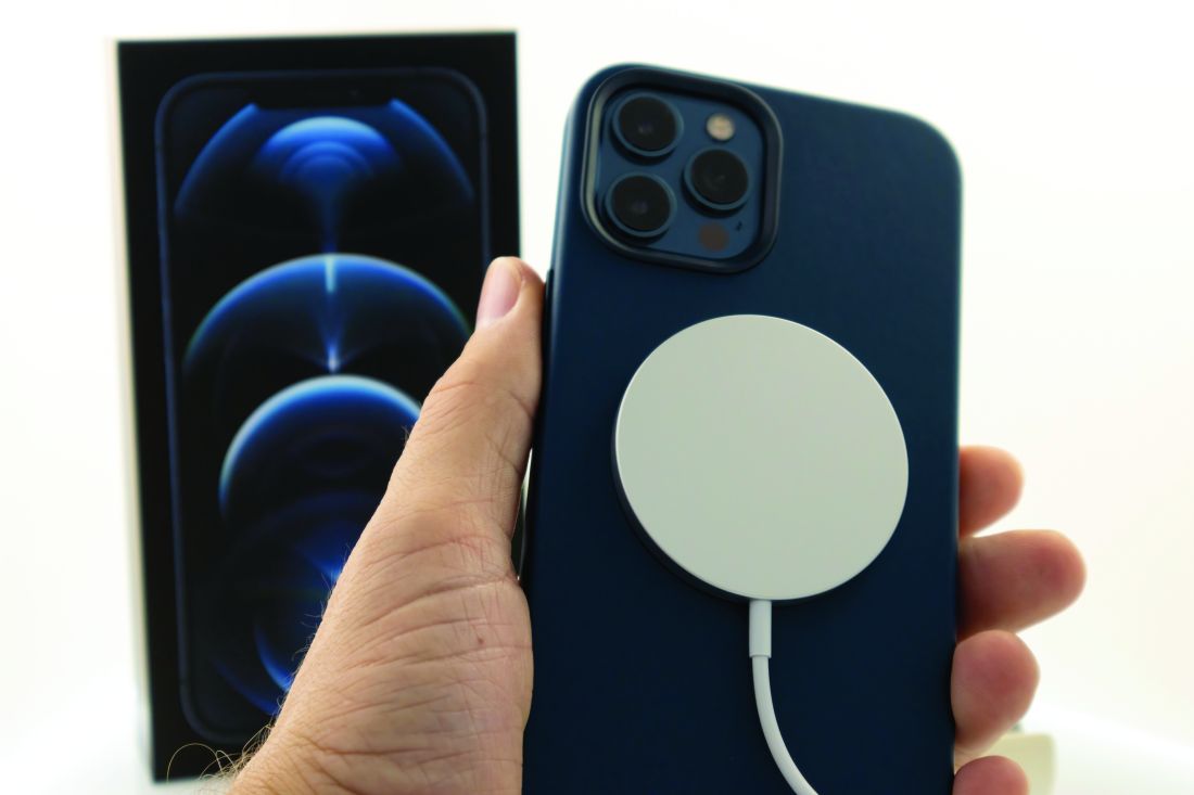
“Once the iPhone was brought close to the ICD over the left chest area, immediate suspension of ICD therapies was noted which persisted for the duration of the test,” reported the investigating team led by Joshua C. Greenberg, MD, who is an electrophysiology fellow at Henry Ford Hospital, Detroit. The results were published in Heart Rhythm.
The American Heart Association has already cautioned that magnetic fields can inhibit the pulse generators for ICDs and pacemakers. On the AHA website, there is a list of devices and their potential for functional interference, but cell phones and other common devices are identified as posing a low risk.
The most recent iPhone and perhaps other advanced smartphones appear to be different. According to the authors of a study that tested the iPhone 12, this model has a circular array of magnets around a central charging coil. This array interacts with Apple’s proprietary MagSafe technology, which accelerates charging. The magnets also serve to orient the phone on the charger and enable other MagSafe accessories.
The authors of the new study were concerned that this array of magnets might be sufficiently strong to interfere with ICDs or other devices at risk. In a previously published study, the strength of a magnetic field sufficient to interfere with implantable cardiac devices was estimated to be at least 10 gauss.
Tests were performed on a patient wearing a Medtronic ICD.
“Once the iPhone was brought close to the ICD over the left chest area, immediate suspension of ICD therapies was noted,” according to the authors of the study. The functional loss of the ICS persisted for the duration of proximity. It was reproduced multiple times and with multiple phone positions.
Previous studies have provided evidence that earlier models do not share this risk. In a study testing the iPhone 6 and an Apple Watch in 148 patients with various types of implantable electronic devices, including pacemakers, cardioverter defibrillators, resynchronization defibrillators, and resynchronization pacemakers, only one instance of interference was observed in 1,352 tests.
With wand telemetry, iPhone-induced interferences could be detected with the iPhone 6 in 14% of the patients, but these did not appear to be clinically meaningful, and this type of interference could not be detected with the Apple Watch, according to the report. The single observed interaction, which was between an iPhone 6 and a dual-chamber pacemaker, suggested device-device interactions are uncommon.
More recently, a woman with a single-chamber Medtronic ICD who went to sleep wearing an Apple Watch was awoken by warning beeps from her cardiac device, according to a case report published online. The Apple watch became the prime suspect in causing the ICD warning when proximity of the watch reproduced the warning during clinical examination. However, the magnetic interference was ultimately found to be emanating from the wristband, not the watch.
This case prompted additional studies with Fitbit and other Apple Watch wristbands. Both wristbands contain magnets used to track heart rate. Both were found capable of deactivating ICDs at distances of approximately 2 cm. On the basis of these results, the authors concluded that patients should be counseled about the risk posed by wristbands used in fitness tracking, concluding that they should be kept at least 6 inches away from ICDs and not worn while sleeping.
On their website, Apple maintains a page that specifically warns about the potential for interactions between iPhone 12s and medical devices . Although there is an acknowledgment that the iPhone12 contains more magnets than prior iPhone models, it is stated that iPhone 12 models are “not expected to pose a greater risk of magnetic interference to medical devices than prior iPhone models.” Nevertheless, the Apple instructions advise keeping the iPhone and MagSafe accessories more than 6 inches away from medical devices.
Dr. Greenberg and coinvestigators concluded that the iPhone 12 does pose a greater risk to the dysfunction of ICDs and other medical devices because of the more powerful magnets. As a result, the study brings forward “an important public health issue concerning the newer generation iPhone 12.”
Well aware of this issue and this study, Bruce L. Wilkoff, MD, director of cardiac pacing and tachyarrhythmia devices, Cleveland Clinic, agreed. He said the focus should not be restricted to the iPhone 12 series but other wearable devices as alluded to in the study.
“Pacemakers and implantable defibrillators are designed to respond to magnets for important reasons, but magnets have many common uses,” he said. These can change the function of the implantable cardiac devise, but “it is temporary and only when placed in close proximity.”
The solution is simple. “Patients should be careful to avoid locating these objects near these devices,” Dr. Wilkoff said.
However, the first step is awareness. According to the study authors, devices with magnets powerful enough to impair function of implantable devices, such as the iPhone 12 “can potentially inhibit lifesaving therapy.”
Patients should be counseled and provided with practical steps, according to the authors. This includes keeping these devices out of pockets near implantable devices. They called for more noise from makers of smartphones and other devices with strong enough magnets to alter pacemaker and ICD function, and they advised physicians to draw awareness to this issue.
Dr. Greenberg reported no potential conflicts of interest.
Patients with an implantable cardioverter defibrillator (ICD) should be warned that some newer models of smartphones equipped with magnets, such as the iPhone 12, can disable their device, inhibiting its lifesaving functions, according to investigators who tested and confirmed this effect.
“Once the iPhone was brought close to the ICD over the left chest area, immediate suspension of ICD therapies was noted which persisted for the duration of the test,” reported the investigating team led by Joshua C. Greenberg, MD, who is an electrophysiology fellow at Henry Ford Hospital, Detroit. The results were published in Heart Rhythm.
The American Heart Association has already cautioned that magnetic fields can inhibit the pulse generators for ICDs and pacemakers. On the AHA website, there is a list of devices and their potential for functional interference, but cell phones and other common devices are identified as posing a low risk.
The most recent iPhone and perhaps other advanced smartphones appear to be different. According to the authors of a study that tested the iPhone 12, this model has a circular array of magnets around a central charging coil. This array interacts with Apple’s proprietary MagSafe technology, which accelerates charging. The magnets also serve to orient the phone on the charger and enable other MagSafe accessories.
The authors of the new study were concerned that this array of magnets might be sufficiently strong to interfere with ICDs or other devices at risk. In a previously published study, the strength of a magnetic field sufficient to interfere with implantable cardiac devices was estimated to be at least 10 gauss.
Tests were performed on a patient wearing a Medtronic ICD.
“Once the iPhone was brought close to the ICD over the left chest area, immediate suspension of ICD therapies was noted,” according to the authors of the study. The functional loss of the ICS persisted for the duration of proximity. It was reproduced multiple times and with multiple phone positions.
Previous studies have provided evidence that earlier models do not share this risk. In a study testing the iPhone 6 and an Apple Watch in 148 patients with various types of implantable electronic devices, including pacemakers, cardioverter defibrillators, resynchronization defibrillators, and resynchronization pacemakers, only one instance of interference was observed in 1,352 tests.
With wand telemetry, iPhone-induced interferences could be detected with the iPhone 6 in 14% of the patients, but these did not appear to be clinically meaningful, and this type of interference could not be detected with the Apple Watch, according to the report. The single observed interaction, which was between an iPhone 6 and a dual-chamber pacemaker, suggested device-device interactions are uncommon.
More recently, a woman with a single-chamber Medtronic ICD who went to sleep wearing an Apple Watch was awoken by warning beeps from her cardiac device, according to a case report published online. The Apple watch became the prime suspect in causing the ICD warning when proximity of the watch reproduced the warning during clinical examination. However, the magnetic interference was ultimately found to be emanating from the wristband, not the watch.
This case prompted additional studies with Fitbit and other Apple Watch wristbands. Both wristbands contain magnets used to track heart rate. Both were found capable of deactivating ICDs at distances of approximately 2 cm. On the basis of these results, the authors concluded that patients should be counseled about the risk posed by wristbands used in fitness tracking, concluding that they should be kept at least 6 inches away from ICDs and not worn while sleeping.
On their website, Apple maintains a page that specifically warns about the potential for interactions between iPhone 12s and medical devices . Although there is an acknowledgment that the iPhone12 contains more magnets than prior iPhone models, it is stated that iPhone 12 models are “not expected to pose a greater risk of magnetic interference to medical devices than prior iPhone models.” Nevertheless, the Apple instructions advise keeping the iPhone and MagSafe accessories more than 6 inches away from medical devices.
Dr. Greenberg and coinvestigators concluded that the iPhone 12 does pose a greater risk to the dysfunction of ICDs and other medical devices because of the more powerful magnets. As a result, the study brings forward “an important public health issue concerning the newer generation iPhone 12.”
Well aware of this issue and this study, Bruce L. Wilkoff, MD, director of cardiac pacing and tachyarrhythmia devices, Cleveland Clinic, agreed. He said the focus should not be restricted to the iPhone 12 series but other wearable devices as alluded to in the study.
“Pacemakers and implantable defibrillators are designed to respond to magnets for important reasons, but magnets have many common uses,” he said. These can change the function of the implantable cardiac devise, but “it is temporary and only when placed in close proximity.”
The solution is simple. “Patients should be careful to avoid locating these objects near these devices,” Dr. Wilkoff said.
However, the first step is awareness. According to the study authors, devices with magnets powerful enough to impair function of implantable devices, such as the iPhone 12 “can potentially inhibit lifesaving therapy.”
Patients should be counseled and provided with practical steps, according to the authors. This includes keeping these devices out of pockets near implantable devices. They called for more noise from makers of smartphones and other devices with strong enough magnets to alter pacemaker and ICD function, and they advised physicians to draw awareness to this issue.
Dr. Greenberg reported no potential conflicts of interest.
FROM HEART RHYTHM
Tough pain relief choices in the COVID-19 pandemic
More people with fever and body aches are turning to NSAIDs to ease symptoms, but the drugs have come under new scrutiny as investigators work to determine whether they are a safe way to relieve the pain of COVID-19 vaccination or symptoms of the disease.
Early on in the pandemic, French health officials warned that NSAIDs, such as ibuprofen, could worsen coronavirus disease, and they recommended switching to acetaminophen instead.
The National Health Service in the United Kingdom followed with a similar recommendation for acetaminophen.
But the European Medicines Agency took a different approach, reporting “no scientific evidence” that NSAIDs could worsen COVID-19. The U.S. Food and Drug Administration also opted not to take a stance.
The debate prompted discussion on social media, with various reactions from around the world. It also inspired Craig Wilen, MD, PhD, from Yale University, New Haven, Conn., and associates to examine the effect of NSAIDs on COVID-19 infection and immune response. Their findings were published online Jan.20 in the Journal of Virology.
“It really bothered me that non–evidence-based decisions were driving the conversation,” Dr. Wilen said. “Millions of people are taking NSAIDs every day and clinical decisions about their care shouldn’t be made on a hypothesis.”
One theory is that NSAIDs alter susceptibility to infection by modifying ACE2. The drugs might also change the cell entry receptor for SARS-CoV-2, alter virus replication, or even modify the immune response.
British researchers, also questioning the safety of NSAIDs in patients with COVID-19, delved into National Health Service records to study two large groups of patients, some of whom were taking the pain relievers.
“We were watching the controversy and the lack of evidence and wanted to contribute,” lead investigator Angel Wong, PhD, from the London School of Hygiene and Tropical Medicine, said in an interview.
And with nearly 11 million NSAID prescriptions dispensed in primary care in England alone in the past 12 months, the inconsistency was concerning.
The team compared COVID-19–related deaths in two groups: one group of more than 700,000 people taking NSAIDs, including patients with rheumatoid arthritis and osteoarthritis; and another of almost 3.5 million people not on the medication.
NSAIDs work by inhibiting cyclooxygenase-1 and COX-2 enzymes in the body, which are crucial for the generation of prostaglandins. These lipid molecules play a role in inflammation and are blocked by NSAIDs.
The investigators found no evidence of a harmful effect of NSAIDs on COVID-19-related deaths; their results were published online Jan. 21 in the Annals of the Rheumatic Diseases.
The results, they pointed out, are in line with a Danish study that also showed no evidence of a higher risk for severe COVID-19 outcomes with NSAID use.
“It’s reassuring,” Dr. Wong said, “that patients can safely continue treatment.”
More new evidence
Dr. Wilen’s team found that SARS-CoV-2 infection stimulated COX-2 expression in human and mice cells. However, suppression of COX-2 by two commonly used NSAIDs, ibuprofen and meloxicam, had no effect on ACE2 expression, viral entry, or viral replication.
In their mouse model of SARS-CoV-2 infection, the investigators saw that NSAIDs impaired the production of proinflammatory cytokines and neutralizing antibodies. The findings suggest that NSAIDs influence COVID-19 outcomes by dampening the inflammatory response and production of protective antibodies, rather than modifying susceptibility to infection or viral replication.
Understanding the effect of NSAIDs on cytokine production is critical, Dr. Wilen pointed out, because they might be protective early in COVID-19 but pathologic at later stages.
Timing is crucial in the case of other immunomodulatory drugs. For example, dexamethasone lowers mortality in COVID-19 patients on respiratory support but is potentially harmful for those with milder disease.
There still is a lot to learn, Dr. Wilen acknowledged. “We may be seeing something similar going on with NSAIDs, where the timing of treatment is important.”
A version of this article first appeared on Medscape.com.
Maternal COVID antibodies cross placenta, detected in newborns
Antibodies against SARS-CoV-2 cross the placenta during pregnancy and are detectable in most newborns born to mothers who had COVID-19 during pregnancy, according to findings from a study presented Jan. 28 at the meeting sponsored by the Society for Maternal-Fetal Medicine.
“I think the most striking finding is that we noticed a high degree of neutralizing response to natural infection even among asymptomatic infection, but of course a higher degree was seen in those with symptomatic infection,” Naima Joseph, MD, MPH, of Emory University, Atlanta, said in an interview.
“Our data demonstrate maternal capacity to mount an appropriate and robust immune response,” and maternal protective immunity lasted at least 28 days after infection, Dr. Joseph said. “Also, we noted higher neonatal cord blood titers in moms with higher titers, which suggests a relationship, but we need to better understand how transplacental transfer occurs as well as establish neonatal correlates of protection in order to see if and how maternal immunity may also benefit neonates.”
The researchers analyzed the amount of IgG and IgM antibodies in maternal and cord blood samples prospectively collected at delivery from women who tested positive for COVID-19 at any time while pregnant. They used enzyme-linked immunosorbent assay to assess for antibodies for the receptor binding domain of the SARS-CoV-2 spike protein.
The 32 pairs of mothers and infants in the study were predominantly non-Hispanic Black (72%) and Hispanic (25%), and 84% used Medicaid as their payer. Most of the mothers (72%) had at least one comorbidity, most commonly obesity, hypertension, and asthma or pulmonary disease. Just over half the women (53%) were symptomatic while they were infected, and 88% were ill with COVID-19 during the third trimester. The average time from infection to delivery was 28 days.
All the mothers had IgG antibodies, 94% had IgM antibodies, and 94% had neutralizing antibodies against SARS-CoV-2. Among the cord blood samples, 91% had IgG antibodies, 9% had IgM antibodies, and 25% had neutralizing antibodies.
“It’s reassuring that, so far, the physiological response is exactly what we expected it to be,” Judette Louis, MD, MPH, an associate professor of ob.gyn. and the ob.gyn. department chair at the University of South Florida, Tampa, said in an interview. “It’s what we would expect, but it’s always helpful to have more data to support that. Otherwise, you’re extrapolating from what you know from other conditions,” said Dr. Louis, who moderated the oral abstracts session.
Symptomatic infection was associated with significantly higher IgG titers than asymptomatic infection (P = .03), but no correlation was seen for IgM or neutralizing antibodies. In addition, although mothers who delivered more than 28 days after their infection had higher IgG titers (P = .05), no differences existed in IgM or neutralizing response.
Infants’ cord blood titers were significantly lower than their corresponding maternal samples, independently of symptoms or latency from infection to delivery (P < .001), Dr. Joseph reported.
“Transplacental efficiency in other pathogens has been shown to be correlated with neonatal immunity when the ratio of cord to maternal blood is greater than 1,” Dr. Joseph said in her presentation. Their data showed “suboptimal efficiency” at a ratio of 0.81.
The study’s small sample size and lack of a control group were weaknesses, but a major strength was having a population at disproportionately higher risk for infection and severe morbidity than the general population.
Implications for maternal COVID-19 vaccination
Although the data are not yet available, Dr. Joseph said they have expanded their protocol to include vaccinated pregnant women.
“The key to developing an effective vaccine [for pregnant people] is in really characterizing adaptive immunity in pregnancy,” Dr. Joseph told SMFM attendees. “I think that these findings inform further vaccine development in demonstrating that maternal immunity is robust.”
The World Health Organization recently recommended withholding COVID-19 vaccines from pregnant people, but the SMFM and American College of Obstetricians and Gynecologists subsequently issued a joint statement reaffirming that the COVID-19 vaccines authorized by the FDA “should not be withheld from pregnant individuals who choose to receive the vaccine.”
“One of the questions people ask is whether in pregnancy you’re going to mount a good response to the vaccine the way you would outside of pregnancy,” Dr. Louis said. “If we can demonstrate that you do, that may provide the information that some mothers need to make their decisions.” Data such as those from Dr. Joseph’s study can also inform recommendations on timing of maternal vaccination.
“For instance, Dr. Joseph demonstrated that, 28 days out from the infection, you had more antibodies, so there may be a scenario where we say this vaccine may be more beneficial in the middle of the pregnancy for the purpose of forming those antibodies,” Dr. Louis said.
Consensus emerging from maternal antibodies data
The findings from Dr. Joseph’s study mirror those reported in a study published online Jan. 29 in JAMA Pediatrics. That study, led by Dustin D. Flannery, DO, MSCE, of Children’s Hospital of Philadelphia, also examined maternal and neonatal levels of IgG and IgM antibodies against the receptor binding domain of the SARS-CoV-2 spike protein. They also found a positive correlation between cord blood and maternal IgG concentrations (P < .001), but notably, the ratio of cord to maternal blood titers was greater than 1, unlike in Dr. Joseph’s study.
For their study, Dr. Flannery and colleagues obtained maternal and cord blood sera at the time of delivery from 1471 pairs of mothers and infants, independently of COVID status during pregnancy. The average maternal age was 32 years, and just over a quarter of the population (26%) were Black, non-Hispanic women. About half (51%) were White, 12% were Hispanic, and 7% were Asian.
About 6% of the women had either IgG or IgM antibodies at delivery, and 87% of infants born to those mothers had measurable IgG in their cord blood. No infants had IgM antibodies. As with the study presented at SMFM, the mothers’ infections included asymptomatic, mild, moderate, and severe cases, and the degree of severity of cases had no apparent effect on infant antibody concentrations. Most of the women who tested positive for COVID-19 (60%) were asymptomatic.
Among the 11 mothers who had antibodies but whose infants’ cord blood did not, 5 had only IgM antibodies, and 6 had significantly lower IgG concentrations than those seen in the other mothers.
In a commentary about the JAMA Pediatrics study, Flor Munoz, MD, of the Baylor College of Medicine, Houston, suggested that the findings are grounds for optimism about a maternal vaccination strategy to protect infants from COVID-19.
“However, the timing of maternal vaccination to protect the infant, as opposed to the mother alone, would necessitate an adequate interval from vaccination to delivery (of at least 4 weeks), while vaccination early in gestation and even late in the third trimester could still be protective for the mother,” Dr. Munoz wrote.
Given the interval between two-dose vaccination regimens and the fact that transplacental transfer begins at about the 17th week of gestation, “maternal vaccination starting in the early second trimester of gestation might be optimal to achieve the highest levels of antibodies in the newborn,” Dr. Munoz wrote. But questions remain, such as how effective the neonatal antibodies would be in protecting against COVID-19 and how long they last after birth.
No external funding was used in Dr. Joseph’s study. Dr. Joseph and Dr. Louis have disclosed no relevant financial relationships. The JAMA Pediatrics study was funded by the Children’s Hospital of Philadelphia. One coauthor received consultancy fees from Sanofi Pasteur, Lumen, Novavax, and Merck unrelated to the study. Dr. Munoz served on the data and safety monitoring boards of Moderna, Pfizer, Virometix, and Meissa Vaccines and has received grants from Novavax Research and Gilead Research.
A version of this article first appeared on Medscape.com.
Dr. Fauci sees ‘wake-up call’ in emergence of new virus variants
New data on COVID-19 vaccines should serve as a “wake-up call” about the need to stop the spread of the SARS-CoV-2 virus among people and thus deprive it of opportunities to evolve its defenses, the top federal expert on infectious diseases said.
“The virus will continue to mutate and will mutate for its own selective advantage,” said Anthony S. Fauci, MD, director of the National Institute of Allergy and Infectious Diseases, at a Friday news conference organized by the White House.
The continued transmission of SARS-CoV-2 “gives the virus the chance to adapt to the forces, in this case the immune response, that’s trying to get rid of it,” Dr. Fauci said. “That’s where you get mutations.”
Federal health officials are working to boost the U.S. supply of COVID-19 vaccines, even as signals emerge about the extent that the virus is already evolving.
Data released this week about the Janssen/Johnson & Johnson (J&J) and Novavax COVID-19 vaccines in late-stage development provides further evidence that they may not protect as well against emerging variants, Dr. Fauci said.
“Mutations that lead to different lineage do have clinical consequences,” he said, while also emphasizing that the emerging vaccines appear to confer broad protection. Dr. Fauci earlier in the day addressed the “messaging challenge” for clinicians and researchers in discussing the results of the J&J vaccine trial, which appear to fall short of those reported for the two vaccines already approved and in use in the United States. He noted the benefits of possibly soon having more authorized vaccines to combat COVID-19. But continued community spread of the infection will foster conditions that can undermine the vaccines’ effectiveness.
“Even though the long-range effect in the sense of severe disease is still handled reasonably well by the vaccines, this is a wake-up call to all of us,” Dr. Fauci said.
Pharmaceutical scientists and executives and government health officials will need to work together to continue to develop vaccines that can outwit the emerging variants, he said.
On Jan. 29, J&J reported that its highly anticipated single-dose vaccine had shown its worst results in South Africa where many cases of COVID-19 were caused by infection with a SARS-CoV-2 variant from the B.1.351 lineage. The overall efficacy was 66% globally, 72% in the United States, and 57% in South Africa against moderate to severe SARS-CoV-2, J&J said.
Novavax on Jan. 28 reported an efficacy rate for its COVID-19 vaccine of 49.4% from a clinical trial conducted in South Africa, compared with an 89.3% rate from a U.K. study. There already have been attempts to estimate how well the Pfizer/BioNTech and Moderna vaccines can handle new variants of the virus. They both have been granted emergency-use authorization by the U.S. Food and Drug Administration.
‘Genomic surveillance’
The Centers for Disease Control and Prevention on Thursday reported the first U.S.-documented cases of the B.1.351 variant of SARS-CoV-2 in South Carolina. On Jan. 26, the first confirmed U.S. case of a highly transmissible Brazilian coronavirus variant was detected in Minnesota, state health officials said.
The CDC’s stepped-up “genomic surveillance” will help keep clinicians and researchers aware of how SARS-CoV-2 is changing, Dr. Fauci said.
Speaking at the same White House news conference, CDC director Rochelle Walensky, MD, MPH, said the two South Carolina cases of the B.1.351 variant were reported in different parts of the state and not believed to be epidemiologically linked. The people involved “did not have any travel history,” she added.
The SARS-CoV-2 mutations were expected to emerge at some point, as with any virus, but their appearance underscores the need for people to remain vigilant about precautions that can stop its spread, Dr. Walensky said.
She and Dr. Fauci both stressed the need for continued use of masks and social distancing and urged people to get COVID-19 vaccines as they become available. Continued community spread of the virus allows this global health threat to keep replicating, and thus increases its chances to thwart medical interventions, Dr. Fauci said.
“The virus has a playing field, as it were, to mutate,” Dr. Fauci said. “If you stop that and stop the replication, the viruses cannot mutate if they don’t replicate.”
A version of this article first appeared on Medscape.com.
The COVID-19 virus may prompt the body to attack itself
An international team of researchers studying COVID-19 has made a startling and pivotal discovery: The virus appears to cause the body to make weapons to attack its own tissues.
The finding could unlock a number of COVID-19’s clinical mysteries. They include the puzzling collection of symptoms that can come with the infection; the persistence of symptoms in some people for months after they clear the virus, a phenomenon dubbed long COVID-19; and why some children and adults have a serious inflammatory syndrome, called multisystem inflammatory syndrome in children (MIS-C) or MIS in adults (MIS-A), after their infections.
“It suggests that the virus might be directly causing autoimmunity, which would be fascinating,” says lead study author Paul Utz, MD, who studies immunology and autoimmunity at Stanford (Calif.) University.
The study also deepens the question of whether other respiratory viruses might also break the body’s tolerance to itself, setting people up for autoimmune diseases like multiple sclerosis, rheumatoid arthritis, and lupus later in life.
Dr. Utz said he and his team are next going to study flu patients to see if that virus might also cause this phenomenon.
“My prediction is that it isn’t going to be specific just to SARS-CoV-2. I’m willing to bet that we will find this with other respiratory viruses,” he said.
The study comes on the heels of a handful of smaller, detailed investigations that have come to similar conclusions.
The study included data from more than 300 patients from four hospitals: two in California, one in Pennsylvania, and another in Germany.
Researchers used blood tests to study their immune responses as their infections progressed. Researchers looked for autoantibodies – weapons of the immune system that go rogue and launch an attack against the body’s own tissues. They compared these autoantibodies with those found in people who were not infected with the virus that causes COVID.
As previous studies have found, autoantibodies were more common after COVID – 50% of people hospitalized for their infections had autoantibodies, compared with less than 15% of those who were healthy and uninfected.
Some people with autoantibodies had little change in them as their infections progressed. That suggests the autoantibodies were there to begin with, possibly allowing the infection to burn out of control in the body.
“Their body is set up to get bad COVID, and it’s probably caused by the autoantibodies,” Dr. Utz said.
But in others, about 20% of people who had them, the autoantibodies became more common as the infection progressed, suggesting they were directly related to the viral infection, instead of being a preexisting condition.
Some of these were antibodies that attack key components of the immune system’s weapons against the virus, like interferon. Interferons are proteins that help infected cells call for reinforcements and can also interfere with a virus’s ability to copy itself. Taking them out is a powerful evasive tactic, and previous studies have shown that people who are born with genes that cause them to have lower interferon function, or who make autoantibodies against these proteins, appear to be at higher risk for life-threatening COVID infections.
“It seems to give the virus a powerful advantage,” said study author, John Wherry, PhD, who directs the Institute for Immunology at the University of Pennsylvania, Philadelphia. “Now your immune system, instead of having a tiny little hill to climb, is staring at Mount Everest. That really is devious.”
In addition to those that sabotage the immune system, some people in the study had autoantibodies against muscles and connective tissues that are seen in some rare disorders.
Dr. Utz said they started the study after seeing COVID patients with strange collections of symptoms that looked more like autoimmune diseases than viral infections – skin rashes, joint pain, fatigue, aching muscles, brain swelling, dry eyes, blood that clots easily, and inflamed blood vessels.
“One thing that’s very important to note is that we don’t know if these patients are going to go on to develop autoimmune disease,” Dr. Utz said. “I think we’ll be able to answer that question in the next 6-12 months as we follow the long haulers and study their samples.”
Dr. Utz said it will be important to study autoantibodies in long haulers to see if they can identify exactly which ones seem to be at work in the condition. If you can catch them early, it might be possible to treat those at risk for enduring symptoms with drugs that suppress the immune system.
What this means, he said, is that COVID will be with us for a long, long time.
“We have to realize that there’s going to be long-term damage from this virus for the survivors. Not just the long haulers, but all the people who have lung damage and heart damage and everything else. We’re going to be studying this virus and it’s badness for decades,” Dr. Utz said.
A version of this article first appeared on WebMD.com.
Neprilysin, corin singled out for potential to guide heart failure therapy
Although not correlated with each other, increased levels of circulating according to prospective analysis involving 1,009 HF patients.
This implies that these enzymes might have value for individualizing care, including treatment of patients in heart failure with preserved ejection fraction (HFpEF), reported a team of investigators led by D.H. Frank Gommans, MD, PhD, department of cardiology, Radboud University Medical Center, Nijmegen, the Netherlands.
When followed for up to 7 years and after adjustment for differences in sex and age, the highest risk for the primary composite endpoint of cardiovascular (CV) death and heart failure hospitalization was observed in those with both high soluble neprilysin (sNEP) and high soluble corin (sCOR). The lowest risk was observed in the group with low levels of both enzymes.
The data suggest that monitoring these enzymes might provide “a step toward individualized CHF patient management,” Dr. Gommans reported in JACC Heart Failure, the adjusted hazard ratio for elevated sNEP and sCOR translated into a greater than 50% increase in the composite primary endpoint relative to low levels of both (HR, 1.56; P = .003). After a “comprehensive multivariable analysis,” the increased risk remained substantial and significant (HR, 1.41; P = .03).
In the natriuretic peptide pathway, which has long been recognized as a mediator of vasodilation, venous compliance, diuresis, and other processes dysregulated in heart failure, NEP and COR are “key mediators,” according to the investigators, who cited previously published studies. More attention has turned to these enzymes as potential biomarkers in the context of the PARADIGM trial, which associated an angiotensin-receptor neprilysin inhibitor (ARNI) with a survival benefit in CHF.
The observational study consisted of CHF patients attending a heart failure clinic and who were ARNI naive at inclusion. On the basis of circulating enzyme measurements undertaken from blood samples employing standard techniques, they were stratified into four groups. Those with low levels of both enzymes served as the reference. They were compared with those with low sNEP and high sCOR, those with high sNEP and low sCOR, and those with high levels of both enzymes.
Over the course of a median 4.5 years of follow-up, there were 511 deaths, of which 54% were from a CV cause. There were also 331 heart failure hospitalizations. In all, 449 patients reached the primary composite endpoint.
When compared with the group with low sNEP and low sCOR, an elevation in either enzyme was associated with a numerically but not significantly greater hazard ratio for the primary composite endpoint. The lack of correlation in the elevation of these two enzymes suggests each provides different prognostic information, although it appears that both must be considered together to predict outcomes.
Clinically, stratification of these enzymes might be most useful in HFpEF patients. Relative to the separation of event curves in the CHF patients with reduced ejection fraction (HFrEF), the divergence in the event curves for HFpEF were greater. In addition, event curves separated from the reference in HFpEF patients but not the HFrEF patients if either enzyme was elevated.
Asked if these data hold particular promise for monitoring and individualizing therapy in HFpEF patients, Dr. Gommans said yes. Although he cautioned that this was an observational study and that the differences between the HFpEF and HFrEF should be considered exploratory, he agreed that components of the natriuretic peptide pathway have particular potential to provide new prognostic information and individualize care in HFpEF, where therapeutic options remain limited.
Stratification of natriuretic peptide enzymes in this group might “present as an interesting alternative to ejection fraction” for prognosis and the consideration of treatment choices, he suggested.
Although further validation of the prognostic importance of sNEP and sCOR is needed, according to Dr. Gommans, he foresees the potential of therapeutic trials based on elevated levels of these enzymes. For example, he speculated that these levels might distinguish HFpEF patients who could benefit from a first-line ARNI.
In an accompanying editorial, significant doubts were expressed about simple measurements of sNEP and sCOR concentrations to predict clinical course or guide treatment decisions. The authors of the editorial agreed this is an important area of study but warned of its complexity.
“Concentrations of circulating neprilysin have been shown to correlate poorly with neprilysin activity. Thus the rate of natriuretic peptide degradation by neprilysin cannot be determined solely by measuring circulating levels,” cautioned Peder L. Myhre, MD, PhD, who is a cardiology fellow at Akershus University Hospital in Nordbyhagen, Norway, and postdoc researcher at the University of Oslo.
“Accordingly, concentrations of neprilysin and corin cannot alone be used to predict response to therapies interacting with these peptides,” he added. He agreed that neprilysin and corin might be appropriate biomarkers in CHF, but he thinks the focus must be on their enzymatic activity, not their circulating levels.
“Measuring the enzymatic activity may be a feasible strategy, but this remains to be seen,” he said.
Dr. Gommans reported a financial relationship with Novartis. Dr. Myhre reported financial relationships with Amgen, Novartis, and Novo Nordisk.
FROM JACC HEART FAILURE