User login
Mitchel is a reporter for MDedge based in the Philadelphia area. He started with the company in 1992, when it was International Medical News Group (IMNG), and has since covered a range of medical specialties. Mitchel trained as a virologist at Roswell Park Memorial Institute in Buffalo, and then worked briefly as a researcher at Boston Children's Hospital before pivoting to journalism as a AAAS Mass Media Fellow in 1980. His first reporting job was with Science Digest magazine, and from the mid-1980s to early-1990s he was a reporter with Medical World News. @mitchelzoler
VIDEO: New stroke guideline embraces imaging-guided thrombectomy
LOS ANGELES – When a panel organized by the American Heart Association’s Stroke Council recently revised the group’s guideline for early management of acute ischemic stroke, they were clear on the overarching change they had to make: Incorporate recent evidence collected in two trials that established brain imaging as the way to identify patients eligible for clot removal treatment by thrombectomy, a change in practice that has made this outcome-altering intervention available to more patients.
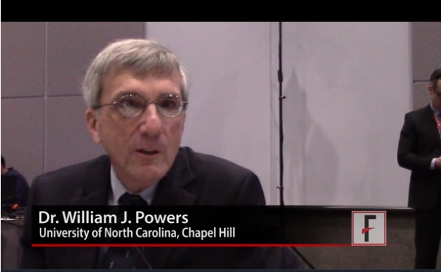
“The major take-home message [of the new guideline] is the extension of the time window for treating acute ischemic stroke,” said William J. Powers, MD, chair of the guideline group (Stroke. 2018 Jan 24. doi: 10.1161/STR.0000000000000158).
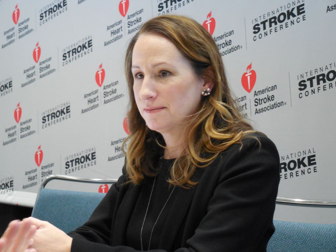
Based on recently reported results from the DAWN (N Engl J Med. 2018;378[1]:11-21) and DEFUSE 3 (N Engl J Med. 2018 Jan 24. doi: 10.1056/NEJMoa1713973) trials “we know that there are patients out to 24 hours from their stroke onset who may benefit” from thrombectomy. “This is a major, major change in how we view care for patients with stroke,” Dr. Powers said in a video interview. “Now there’s much more time. Ideally, we’ll see smaller hospitals develop the ability to do the imaging” that makes it possible to select acute ischemic stroke patients eligible for thrombectomy despite a delay of up to 24 hours from their stroke onset to the time of thrombectomy, said Dr. Powers, professor and chair of neurology at the University of North Carolina, Chapel Hill.
The big priority for the stroke community now that this major change in patient selection was incorporated into a U.S. practice guideline will be acting quickly to implement the steps needed to make this change happen, Dr. Powers and others said.
The new guideline will mean “changes in process and systems of care,” agreed Jeffrey L. Saver, MD, professor of neurology and director of the stroke unit at the University of California, Los Angeles. The imaging called for “will be practical at some primary stroke centers but not others,” he said, although most hospitals certified to provide stroke care as primary stroke centers or acute stroke–ready hospitals have a CT scanner that could provide the basic imaging needed to assess many patients. (CT angiography and perfusion CT are more informative for determining thrombectomy eligibility.) But interpretation of the brain images to distinguish patients eligible for thrombectomy from those who aren’t will likely happen at comprehensive stroke centers that perform thrombectomy or by experts using remote image reading.
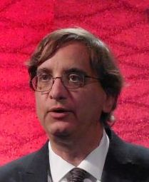
Dr. Saver expects that the new guideline will translate most quickly into changes in the imaging and transfer protocols that the Joint Commission may now require from hospitals certified as primary stroke centers or acute stroke-ready hospitals, changes that could be in place sometime later in 2018, he predicted. These are steps “that would really help drive system change.”
Dr. Powers and Dr. Furie had no disclosures. Dr. Saver has received research support and personal fees from Medtronic-Abbott and Neuravia.
LOS ANGELES – When a panel organized by the American Heart Association’s Stroke Council recently revised the group’s guideline for early management of acute ischemic stroke, they were clear on the overarching change they had to make: Incorporate recent evidence collected in two trials that established brain imaging as the way to identify patients eligible for clot removal treatment by thrombectomy, a change in practice that has made this outcome-altering intervention available to more patients.
“The major take-home message [of the new guideline] is the extension of the time window for treating acute ischemic stroke,” said William J. Powers, MD, chair of the guideline group (Stroke. 2018 Jan 24. doi: 10.1161/STR.0000000000000158).
Based on recently reported results from the DAWN (N Engl J Med. 2018;378[1]:11-21) and DEFUSE 3 (N Engl J Med. 2018 Jan 24. doi: 10.1056/NEJMoa1713973) trials “we know that there are patients out to 24 hours from their stroke onset who may benefit” from thrombectomy. “This is a major, major change in how we view care for patients with stroke,” Dr. Powers said in a video interview. “Now there’s much more time. Ideally, we’ll see smaller hospitals develop the ability to do the imaging” that makes it possible to select acute ischemic stroke patients eligible for thrombectomy despite a delay of up to 24 hours from their stroke onset to the time of thrombectomy, said Dr. Powers, professor and chair of neurology at the University of North Carolina, Chapel Hill.
The big priority for the stroke community now that this major change in patient selection was incorporated into a U.S. practice guideline will be acting quickly to implement the steps needed to make this change happen, Dr. Powers and others said.
The new guideline will mean “changes in process and systems of care,” agreed Jeffrey L. Saver, MD, professor of neurology and director of the stroke unit at the University of California, Los Angeles. The imaging called for “will be practical at some primary stroke centers but not others,” he said, although most hospitals certified to provide stroke care as primary stroke centers or acute stroke–ready hospitals have a CT scanner that could provide the basic imaging needed to assess many patients. (CT angiography and perfusion CT are more informative for determining thrombectomy eligibility.) But interpretation of the brain images to distinguish patients eligible for thrombectomy from those who aren’t will likely happen at comprehensive stroke centers that perform thrombectomy or by experts using remote image reading.
Dr. Saver expects that the new guideline will translate most quickly into changes in the imaging and transfer protocols that the Joint Commission may now require from hospitals certified as primary stroke centers or acute stroke-ready hospitals, changes that could be in place sometime later in 2018, he predicted. These are steps “that would really help drive system change.”
Dr. Powers and Dr. Furie had no disclosures. Dr. Saver has received research support and personal fees from Medtronic-Abbott and Neuravia.
EXPERT ANALYSIS FROM ISC 2018
HRS: Consider ablation for asymptomatic atrial fib
ORLANDO – When the Heart Rhythm Society and several collaborating groups published in October 2017 the first revised consensus statement on atrial fibrillation ablation in 5 years, the document included a novel and perhaps unexpected suggestion: Ablation for asymptomatic atrial fibrillation “may be considered.”
This was “the first time” any group of experts suggested an indication potentially existed for ablating asymptomatic atrial fibrillation (AF), Hugh Calkins, MD, said at the annual International AF Symposium.
“You might say ‘are you out of your mind recommending ablation for asymptomatic AF?’ ” conceded Dr. Calkins, professor of medicine and director of the arrhythmia service at Johns Hopkins Medicine in Baltimore. But Dr. Calkins quickly added that this was a “soft” recommendation by being in the “may be considered” category, and he also noted that it received broad support from about 90% of the members of the statement’s 60-member writing group (Heart Rhythm. 2017 Oct;14[10]:e445-e494).
In addition, he personally believed that an amber light for this strategy made a lot of sense.

He also acknowledged that this recommendation is sort of buried in the text of the consensus statement and does not appear in any summary diagram “because the reviewers wanted us to hide it. Only those who are passionate about ablation know about it.
“Our goal was not to send a message that this is for everyone. It’s for very select patients and for very select operators after a very careful discussion” of the risks and potential benefits from performing the procedure on a truly asymptomatic patient.
The ideal candidate for this approach would be a relatively young patient, say someone in their 50s, who is identified as having AF incidentally, such as someone with an irregular pulse that’s found during a routine examination that leads to an ECG and definitive identification of AF despite the patient’s complete denial of having symptoms.
The next step, Dr. Calkins suggested, would be to treat the patient with an antiarrhythmic drug, such as amiodarone or flecainide, and with cardioversion and see whether this stops the AF and makes the patient feel better. If the patient reports improvement, it suggests the AF really is symptomatic and management could then proceed as with any case of symptomatic AF. But if the patient perceives no change and the AF then recurs in a persistent presentation despite drug treatment, the cardiologist could then discuss with the patient the pros and cons of an ablative procedure.
The pros for immediate ablation are that, when left unablated, the patient will face a substantially increased lifetime risk for stroke, dementia, and new onset heart failure, and after 2-3 years of continued persistent AF the left atrium would remodel and become much less likely to respond to ablation with little prospect for the patient ever returning to a normal sinus rhythm. “It’s either a rhythm control strategy now, or we’ll leave you in AF for the rest of your life,” Dr. Calkins explained. “If I were 50 years old and had asymptomatic AF, there’s no way I’d want to have AF for the rest of my life.” The risks from ablation are that the procedure has about a 68% success rate and about a 1% rate of complications.
“A patient with asymptomatic paroxysmal AF doesn’t have much to lose by waiting and seeing whether symptoms develop, but for the patient with persistent AF there is a penalty for allowing continuous AF, because after 2-3 years you won’t be able to successfully ablate it. In the past, we left patients with asymptomatic AF that way for the rest of their life, but now we know that if patients remain in AF over time, they will lose the option to have it ablated, and their risk of stroke, dementia, and heart failure will increase.”Dr. Calkins has been a consultant or adviser to or received honoraria from Abbott, AtriCure, Boehringer Ingelheim, Boston Scientific, iRhythm, Medtronic, Pfizer, St. Jude, and Toray, He has also received research funding from Boston Scientific and Medtronic.
This article was updated 2/9/18.
ORLANDO – When the Heart Rhythm Society and several collaborating groups published in October 2017 the first revised consensus statement on atrial fibrillation ablation in 5 years, the document included a novel and perhaps unexpected suggestion: Ablation for asymptomatic atrial fibrillation “may be considered.”
This was “the first time” any group of experts suggested an indication potentially existed for ablating asymptomatic atrial fibrillation (AF), Hugh Calkins, MD, said at the annual International AF Symposium.
“You might say ‘are you out of your mind recommending ablation for asymptomatic AF?’ ” conceded Dr. Calkins, professor of medicine and director of the arrhythmia service at Johns Hopkins Medicine in Baltimore. But Dr. Calkins quickly added that this was a “soft” recommendation by being in the “may be considered” category, and he also noted that it received broad support from about 90% of the members of the statement’s 60-member writing group (Heart Rhythm. 2017 Oct;14[10]:e445-e494).
In addition, he personally believed that an amber light for this strategy made a lot of sense.
He also acknowledged that this recommendation is sort of buried in the text of the consensus statement and does not appear in any summary diagram “because the reviewers wanted us to hide it. Only those who are passionate about ablation know about it.
“Our goal was not to send a message that this is for everyone. It’s for very select patients and for very select operators after a very careful discussion” of the risks and potential benefits from performing the procedure on a truly asymptomatic patient.
The ideal candidate for this approach would be a relatively young patient, say someone in their 50s, who is identified as having AF incidentally, such as someone with an irregular pulse that’s found during a routine examination that leads to an ECG and definitive identification of AF despite the patient’s complete denial of having symptoms.
The next step, Dr. Calkins suggested, would be to treat the patient with an antiarrhythmic drug, such as amiodarone or flecainide, and with cardioversion and see whether this stops the AF and makes the patient feel better. If the patient reports improvement, it suggests the AF really is symptomatic and management could then proceed as with any case of symptomatic AF. But if the patient perceives no change and the AF then recurs in a persistent presentation despite drug treatment, the cardiologist could then discuss with the patient the pros and cons of an ablative procedure.
The pros for immediate ablation are that, when left unablated, the patient will face a substantially increased lifetime risk for stroke, dementia, and new onset heart failure, and after 2-3 years of continued persistent AF the left atrium would remodel and become much less likely to respond to ablation with little prospect for the patient ever returning to a normal sinus rhythm. “It’s either a rhythm control strategy now, or we’ll leave you in AF for the rest of your life,” Dr. Calkins explained. “If I were 50 years old and had asymptomatic AF, there’s no way I’d want to have AF for the rest of my life.” The risks from ablation are that the procedure has about a 68% success rate and about a 1% rate of complications.
“A patient with asymptomatic paroxysmal AF doesn’t have much to lose by waiting and seeing whether symptoms develop, but for the patient with persistent AF there is a penalty for allowing continuous AF, because after 2-3 years you won’t be able to successfully ablate it. In the past, we left patients with asymptomatic AF that way for the rest of their life, but now we know that if patients remain in AF over time, they will lose the option to have it ablated, and their risk of stroke, dementia, and heart failure will increase.”Dr. Calkins has been a consultant or adviser to or received honoraria from Abbott, AtriCure, Boehringer Ingelheim, Boston Scientific, iRhythm, Medtronic, Pfizer, St. Jude, and Toray, He has also received research funding from Boston Scientific and Medtronic.
This article was updated 2/9/18.
EXPERT ANALYSIS FROM THE AF SYMPOSIUM 2018
VIDEO: Anticoagulant underprescribing common, jeopardizing atrial fib patients
ORLANDO – A high fraction of U.S. patients with atrial fibrillation receive an inappropriately low dosage of an anticoagulant for stroke prevention, often in a misguided attempt to avoid potential bleeding complications.
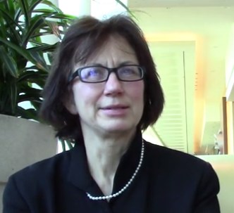
When physicians “reduce the dose to prevent a bleed they increase the risk for an ischemic stroke,” Elaine M. Hylek, MD, said in a video interview during the annual International AF Symposium.
Recent data on actual anticoagulant dosages prescribed to U.S. patients with atrial fibrillation show that “an unexpectedly high proportion of prescriptions for apixaban (Eliquis), dabigatran (Pradaxa), and rivaroxaban (Xarelto) are given at lower doses,” Dr. Hylek noted at the meeting. The lower-dose formulations with U.S. marketing are only appropriate for patients on apixaban with at least two of the following: serum creatinine 1.5 mg/dL or higher, age 80 years or older, and weight 60 kg or less; patients on dabigatran with moderate renal impairment or treated with dronedarone or systemic ketoconazole; or patients on rivaroxaban with a creatinine clearance of 15-50 mL/min.
For example, in the pivotal trial for apixaban only 5% of atrial fibrillation patients qualified for the lower dosage, yet recent data have shown that, in actual U.S. practice roughly a quarter of patients were on this lower dosage, said Dr. Hylek, professor of medicine at Boston University and director of the thrombosis and anticoagulation service at Boston Medical Center (Curr Med Res Opin. 2016 July;32[7]:1277-79). A second recent report showed that among U.S. patients with atrial fibrillation hospitalized for an ischemic stroke 84% had received inadequate anticoagulation with either subtherapeutic dosages of anticoagulant or no anticoagulant at all (JAMA. 2017 Mar 14;317[10]:1057-67).
Another manifestation of the underprescribing problem are patients with atrial fibrillation treated with aspirin only, an approach proven ineffective for preventing ischemic strokes in these patients, Dr. Hylek said.
Dr. Hylek has been an advisor to or has received honoraria from Bayer, Boehringer Ingelheim, Bristol-Myers Squibb, Doasense, Janssen, Medtronic, Pfizer, and Portola, and she has received research funding from Boehringer Ingelheim, Bristol-Myers Squibb, and Janssen.
[email protected]
On Twitter @mitchelzoler
ORLANDO – A high fraction of U.S. patients with atrial fibrillation receive an inappropriately low dosage of an anticoagulant for stroke prevention, often in a misguided attempt to avoid potential bleeding complications.
When physicians “reduce the dose to prevent a bleed they increase the risk for an ischemic stroke,” Elaine M. Hylek, MD, said in a video interview during the annual International AF Symposium.
Recent data on actual anticoagulant dosages prescribed to U.S. patients with atrial fibrillation show that “an unexpectedly high proportion of prescriptions for apixaban (Eliquis), dabigatran (Pradaxa), and rivaroxaban (Xarelto) are given at lower doses,” Dr. Hylek noted at the meeting. The lower-dose formulations with U.S. marketing are only appropriate for patients on apixaban with at least two of the following: serum creatinine 1.5 mg/dL or higher, age 80 years or older, and weight 60 kg or less; patients on dabigatran with moderate renal impairment or treated with dronedarone or systemic ketoconazole; or patients on rivaroxaban with a creatinine clearance of 15-50 mL/min.
For example, in the pivotal trial for apixaban only 5% of atrial fibrillation patients qualified for the lower dosage, yet recent data have shown that, in actual U.S. practice roughly a quarter of patients were on this lower dosage, said Dr. Hylek, professor of medicine at Boston University and director of the thrombosis and anticoagulation service at Boston Medical Center (Curr Med Res Opin. 2016 July;32[7]:1277-79). A second recent report showed that among U.S. patients with atrial fibrillation hospitalized for an ischemic stroke 84% had received inadequate anticoagulation with either subtherapeutic dosages of anticoagulant or no anticoagulant at all (JAMA. 2017 Mar 14;317[10]:1057-67).
Another manifestation of the underprescribing problem are patients with atrial fibrillation treated with aspirin only, an approach proven ineffective for preventing ischemic strokes in these patients, Dr. Hylek said.
Dr. Hylek has been an advisor to or has received honoraria from Bayer, Boehringer Ingelheim, Bristol-Myers Squibb, Doasense, Janssen, Medtronic, Pfizer, and Portola, and she has received research funding from Boehringer Ingelheim, Bristol-Myers Squibb, and Janssen.
[email protected]
On Twitter @mitchelzoler
EXPERT ANALYSIS FROM THE AF SYMPOSIUM 2018
VIDEO: Lean body mass linked to atrial fib etiology
ORLANDO – Results from a large Danish epidemiologic study published in 2017 upended the traditional view that obesity directly contributes to new onset atrial fibrillation by instead fingering lean body mass as the key body-habitus culprit.
“It’s a very different way of thinking about obesity” and it’s relationship to the etiology of atrial fibrillation, Stanley Nattel, MD, said in a video interview at the annual International AF Symposium. “I wouldn’t qualify it yet as a complete shift,” because the results came from a single study, “but the results are quite persuasive and very interesting,” said Dr. Nattel, professor of medicine and director of the electrophysiology research program at the Montreal Heart Institute.
The study he cited tracked the incidence of atrial fibrillation during median of 17 years in more than 55,000 Danish people who were aged 50-64 years old at baseline, and showed that lean body mass was the predominant anthropometric risk factor for new-onset atrial fibrillation (J Am Coll Cardiol. 2017 May;69[20]:2488-97). When the article appeared, it was accompanied by an editorial written by Dr. Nattel (J Am Coll Cardiol. 2017 May;69[20]2498-501).
“People had not thought a lot in the past” about lean body mass and atrial fibrillation, he noted.
If the finding is confirmed, it might make sense to target screening for atrial fibrillation to people with higher levels of lean body mass, Dr. Nattel suggested.
EXPERT ANALYSIS FROM THE AF SYMPOSIUM 2018

Mutations linked to checkpoint inhibitor response in RCC
in a derivation and validation study involving a total of 98 patients.
This finding “has important implications as a molecular tool for considering immunotherapy responsiveness” in patients with ccRCC and possibly patients with other cancer types, wrote Eliezer M. Van Allen, MD, of Dana Farber Cancer Institute in Boston and coauthors.
The derivation cohort included 35 patients with metastatic ccRCC treated with nivolumab in a prospective clinical trial. Genome sequencing of pretreatment tumor specimens showed that improved survival after treatment was significantly linked with truncating mutations in a gene, PBRM1, that codes for a protein in the SWI/SNF chromatin-remodeling complex. Patients in the derivation cohort who had these mutations were nearly 13-fold more likely to have clinical benefit from treatment, compared with those without these mutations.
The validation study included specimens and treatment-outcome results from 63 patients with metastatic ccRCC treated with either nivolumab or a different checkpoint inhibitor, such as atezolizumab (Tecentriq). In the validation study, PBRM1 mutations linked with a sixfold higher rate of clinical benefit from treatment.
The researchers noted that the types of mutations they identified as likely involved occur in more than 20% of all cancer types. Results from mouse studies have suggested that tumor cells with these types of mutations are more sensitive to T cell–mediated cytotoxicity, an observation that “lends a mechanistic basis” to the observed findings.
The study received funding in part from Bristol-Myers Squibb, the company that markets nivolumab (Obdivo). Several researchers involved in this study have received honoraria and research support from Bristol-Myers Squibb and from several other drug companies.
[email protected]
On Twitter @mitchelzoler
SOURCE: Miao D et al. Science. 2018 Jan 4. doi: 10.1126/science.aan5951
FROM SCIENCE
Key clinical point: Mutations in PBRM1 linked with better survival after immune checkpoint inhibitor therapy.
Major finding: Patients with a PBRM1 mutation were 6- to 13-fold more likely to have clinical benefit from checkpoint inhibitor treatment.
Study details: Derivation and validation studies that included 98 total patients with metastatic clear cell renal cell carcinoma.
Disclosures: The study received funding in part from Bristol-Myers Squibb, the company that markets nivolumab (Obdivo). Several researchers involved in this study have received honoraria and research support from Bristol-Myers Squibb and from several other drug companies.
Source: Miao D et al. Science. 2018 Jan 4. doi: 10.1126/science.aan5951.
Acute kidney injury linked with doubled inpatient VTEs
TORONTO – Hospitalized patients with acute kidney injury had more than double the inpatient rate of venous thromboembolism as had patients without acute kidney injury in a prospective, observational study of more than 6,000 hospitalized U.S. soldiers.
He offered four possible mechanisms to explain a link between AKI and VTE:
- Patients with AKI are in a hypercoagulable state.
- AKI alters the pharmacodynamics or pharmacokinetics of VTE prophylactic treatments.
- AKI is a marker of an illness that causes VTE.
- VTE leads to an increased rate of AKI rather than the other way around.
Dr. McMahon’s analysis also revealed that two other clinical conditions that are generally believed to raise VTE risk – obesity and impaired overall renal function identified with stagnant measures – did not correspond with a significantly elevated VTE rate in this study.
The data came from 6,552 adults hospitalized for at least 2 days at Walter Reed between September 2009 and March 2011. The study excluded patients with VTE at the time of admission and also those who had been treated with an anticoagulant at the time of admission. The patients averaged 55 years of age and were hospitalized for a median of 4 days. About 22% of patients received VTE prophylaxis with unfractionated heparin, about 41% received prophylaxis with low-molecular-weight heparin, and about 39% received no VTE prophylaxis (percentages total 102% because of rounding).
About 16% of the patients had been diagnosed with AKI at the time of admission, and an additional 8% developed AKI while hospitalized, defined as an increase in serum creatinine during hospitalization of at least 50% above baseline levels or an increase of more than 0.3 mg/dL above the level at time of admission. During hospitalization, 160 patients (2%) developed a new onset VTE.
In an analysis that adjusted for baseline differences in type of surgery, body mass index, sex, age, and prior hospitalizations during the prior 90 days, the results showed that patients with preexisting or new onset AKI had a 2.2-fold higher rate of VTE, compared with patients without AKI, and this difference was statistically significant, Dr. McMahon reported.
The analysis also showed a significant 62% relatively higher rate of VTE among soldiers hospitalized for a deployment-related event, as well as a significant 63% relatively lower VTE rate among patients not receiving medical prophylaxis, compared with patients receiving an anticoagulant. Dr. McMahon suggested that this lower rate of VTEs among patients not on prophylaxis reflected success in identifying which patients had an increased risk for VTE and hence received prophylaxis.
[email protected]
On Twitter @mitchelzoler
AT CHEST 2017
Key clinical point:
Major finding: Inpatients with AKI had an adjusted 2.2-fold higher rate of VTE, compared with other inpatients.
Data source: Prospective, observational data from 6,552 inpatients at a single U.S. military hospital.
Disclosures: Dr. McMahon had no disclosures.
Alcohol use, abuse rise after bariatric surgery
ORLANDO –
Following any of several methods of bariatric surgery, patients showed a statistically significant 8% higher rate of new onset alcohol abuse, and a relative 50% increased rate of significant alcohol use, compared with rates before surgery, Prandeet Wander, MD, said at the World Congress of Gastroenterology at ACG 2017.
Her meta-analysis identified prospective, retrospective, and cross-sectional studies of alcohol use that included more than 100 bariatric surgery patients and that had follow-up beyond 1 year. Patients could have undergone Roux-en-Y gastric bypass, sleeve gastrectomy, or laparoscopic adjustable gastric banding. Comparator populations had to be either the surgery patients prior to the procedure or the controls matched by age and body mass index.
The 28 included studies enrolled 15,714 patients who averaged 43 years old, with more than three quarters women. Follow-up averaged 2.6 years. The most common surgery was Roux-en-Y, used in 23 studies, followed by banding in 12 studies, and sleeves in 8 studies (some studies used more than one type of surgery).
Nineteen of the studies examined the prevalence of “significant alcohol abuse” following surgery in a total of 4,552 patients, with 23% of patients overall showing this behavior. Five studies, involving 2,698 patients, documented the rate of new-onset alcohol abuse after surgery, with an overall rate of 8% that was statistically significant. All five studies individually showed increased incidence of alcohol abuse, with rates that ranged from 4% to 8%.
The analysis that showed a relative 50% higher rate of “significant” alcohol use after surgery, compared with the same patients before their surgery used data from 11 studies with 3,370 patients. Five of these 11 studies individually showed a statistically significant increase in alcohol use, 1 showed a significant, 34% relative decrease, and the remaining 5 studies did not show statistically significant changes, with 3 studies trending toward an increased rate and two trending toward a decreased rate after surgery.
None of the 28 included studies had a randomized control arm, and the studies collectively ran in six countries, including the United States, and hence involved different societal norms of alcohol use. Changes in alcohol absorption and metabolism following bariatric surgery may play roles in the observed effects, as might undiagnosed depression or substance use by patients who undergo this surgery, Dr. Wander suggested.
SOURCE: Wander P et al. World Congress of Gastroenterology, abstract 10.
REPORTING FROM WORLD CONGRESS OF GASTROENTEROLOGY
Key clinical point: Following bariatric surgery patients have increased alcohol use and abuse.
Major finding: Alcohol abuse rose by 8%; significant alcohol use rose by a relative 50%.
Study details: Meta-analysis of 28 reports with 15,714 patients
Disclosures: Dr. Wander had no disclosures.
Source: Wander P et al. World Congress of Gastroenterology, abstract 10.
Alarm reductions don’t improve ICU response times
TORONTO – It will take more than a reduction in alarms to address the issue of alarm fatigue in the ICU; a change in the ICU staff culture is needed, suggests new research.
“It may take years to recondition clinicians [to realize] that alarms are actionable and must get a response,” Afua Kunadu, MD, said during her presentation on the study at the CHEST annual meeting. Results from prior studies had suggested that as many as 99% of clinical alarms do not result in clinical intervention, noted Dr. Kunadu, an internal medicine physician at Harlem Hospital Center in New York.

She described the program, which started in the 20-bed adult ICU of Harlem Hospital Center, following a 2014 National Patient Safety Goal issued by The Joint Commission to improve the safety of clinical alarm systems by reducing unneeded alarms and alarm fatigue. The Harlem Hospital task force that ran the program began with an audit of alarms that went off in the ICU and used the results to identify the three most common alarms: bedside cardiac monitors, infusion pumps, and mechanical ventilators. The task force arranged to reset the default settings on these devices to decrease alarm frequency and boost the clinical importance of each alarm that still sounded. Concurrently, they ran educational sessions about the new alarm thresholds, the anticipated drop in alarm number, and the increased urgency to respond to the remaining alarms very quickly for the ICU staff.
The raised thresholds effectively cut the number of alarms. The average number of alarms per patient per hour fell from 4.5 at baseline during September 2016 to about 2 after 1 month, during December 2016. Then the rate further declined to reach a steady nadir that stayed at about 1.3 alarms per patient per hour 4 months into the program.
But timely responses, measured as the percentage of alarm responses occurring within 60 seconds after the alarm went off, fell from 60% at 1 month into the program down to 12% after 4 months, Dr. Kunadu reported.
She had no disclosures.
[email protected]
On Twitter @mitchelzoler

TORONTO – It will take more than a reduction in alarms to address the issue of alarm fatigue in the ICU; a change in the ICU staff culture is needed, suggests new research.
“It may take years to recondition clinicians [to realize] that alarms are actionable and must get a response,” Afua Kunadu, MD, said during her presentation on the study at the CHEST annual meeting. Results from prior studies had suggested that as many as 99% of clinical alarms do not result in clinical intervention, noted Dr. Kunadu, an internal medicine physician at Harlem Hospital Center in New York.
She described the program, which started in the 20-bed adult ICU of Harlem Hospital Center, following a 2014 National Patient Safety Goal issued by The Joint Commission to improve the safety of clinical alarm systems by reducing unneeded alarms and alarm fatigue. The Harlem Hospital task force that ran the program began with an audit of alarms that went off in the ICU and used the results to identify the three most common alarms: bedside cardiac monitors, infusion pumps, and mechanical ventilators. The task force arranged to reset the default settings on these devices to decrease alarm frequency and boost the clinical importance of each alarm that still sounded. Concurrently, they ran educational sessions about the new alarm thresholds, the anticipated drop in alarm number, and the increased urgency to respond to the remaining alarms very quickly for the ICU staff.
The raised thresholds effectively cut the number of alarms. The average number of alarms per patient per hour fell from 4.5 at baseline during September 2016 to about 2 after 1 month, during December 2016. Then the rate further declined to reach a steady nadir that stayed at about 1.3 alarms per patient per hour 4 months into the program.
But timely responses, measured as the percentage of alarm responses occurring within 60 seconds after the alarm went off, fell from 60% at 1 month into the program down to 12% after 4 months, Dr. Kunadu reported.
She had no disclosures.
[email protected]
On Twitter @mitchelzoler
AT CHEST 2017
Key clinical point:
Major finding: Average alarms/patient/hour fell from 4.5 to 1.3, but the percentage of responses in less than 60 seconds fell from 60% to 12%.
Data source: An observational study at a single adult ICU in the United States.
Disclosures: Dr. Kunadu had no disclosures.
Ultrathin bronchoscopy plus radial EBUS unreliable at making diagnoses
TORONTO – Ultrathin bronchoscopy plus radial endobronchial ultrasound is not a great method for determining whether a suspicious lesion is cancerous or benign, suggests new research.
In this study of patients with CT-detected solid lung lesions, the researchers were able to make a diagnosis for only 49% of those whose nodules were evaluated using ultrathin bronchoscopy plus radial endobronchial ultrasound (EBUS).
“When you do CT-guided biopsies of lung lesions, the [diagnostic] yield is about 94%. So do the math” by comparing it to the roughly 50% yield from ultrathin bronchoscopy plus radial EBUS to decide whether the latter procedure is worth doing, she noted.
The study Dr. Tanner and her associates designed compared the diagnostic yield of ultrathin bronchoscopy plus radial EBUS with standard bronchoscopy and fluoroscopy in patients with CT-detected solid lung lesions 1.5-5.0 cm in size. It ran at five U.S. centers and randomized 221 patients: 85 evaluable patients were tested using the standard methods, and 112 evaluable patients were tested using ultrathin bronchoscopy plus radial EBUS. Patients averaged 65-68 years of age and were divided evenly between women and men. Their lesions averaged slightly more than 3 cm. The ultrathin device had a 4 mm wide diameter and had a 2 mm working channel.
The diagnostic yield was 38% among patients who underwent standard bronchoscopy and fluoroscopy, and 49% among those biopsied using ultrathin bronchoscopy and radial EBUS, Dr. Tanner reported. The between-group difference in yield fell short of being statistically significant.
Forty-six of the 53 patients who were not diagnosable using standard bronchoscopy and fluoroscopy crossed over to the investigational method, which produced a diagnosis for an additional seven patients (15% of the biopsied crossover patients).
The results showed that standard bronchoscopy plus fluoroscopy is “very poor” for distinguishing cancerous and benign pulmonary lesions, Dr. Tanner concluded. The yield from ultrathin bronchoscopy plus radial EBUS in her study was similar to the diagnostic yields reported in prior studies of guided bronchoscopy, even when also using radial EBUS, she added.
Given the limitations of ultrathin bronchoscopy plus radial EBUS, Dr. Tanner suggested that the best scenario for using this diagnostic method would be in patients who need a linear EBUS procedure for mediastinal lymph node staging. Such staging often requires a biopsy of the primary tumor to make a cancer diagnosis, and in such cases, “while you’re in the neighborhood, you could do bronchoscopy with an ultrathin scope,” she suggested.
The potential also exists to augment the diagnostic yield of ultrathin bronchoscopy by applying a navigational software platform and needle biopsy, two methods not included in the study, Dr. Tanner noted. “More studies should be done using this combination,” she said.
The study was funded by Olympus. Dr. Tanner has been a consultant to and has received research funding from Olympus. She has also been a consultant to Cook Medical, Integrated Diagnostics, Oncocyte, Veracyte, and Veran Medical Technologies, and she has also received research funding from Cook, Integrated Diagnostics, Oncocyte, Oncimmune, and Veracyte.
[email protected]
On Twitter @mitchelzoler
Although bronchoscopic tools are safe and accurate to evaluate both central and peripheral lung lesions, the diagnostic yield of the different available techniques is variable. In this study, a diagnostic yield of only 49% was achieved when ultrathin bronchoscopy with radial EBUS was performed for diagnosis of solid nodules. This yield is not much better than that obtained from conventional bronchoscopy with fluoroscopic guidance and much lower than the diagnostic yield from transthoracic needle biopsy. While there is no doubt that the advances in minimally invasive technologies for diagnosing lung nodules and diagnosing and staging lung cancer have revolutionized clinical practice, pulmonologists and thoracic surgeons need to recognize not only the utility but also the limitations of the available diagnostic procedures (as well as the cost). These technologies are complimentary and multidisciplinary discussions should facilitate selection of the best procedure for each individual case.

Although bronchoscopic tools are safe and accurate to evaluate both central and peripheral lung lesions, the diagnostic yield of the different available techniques is variable. In this study, a diagnostic yield of only 49% was achieved when ultrathin bronchoscopy with radial EBUS was performed for diagnosis of solid nodules. This yield is not much better than that obtained from conventional bronchoscopy with fluoroscopic guidance and much lower than the diagnostic yield from transthoracic needle biopsy. While there is no doubt that the advances in minimally invasive technologies for diagnosing lung nodules and diagnosing and staging lung cancer have revolutionized clinical practice, pulmonologists and thoracic surgeons need to recognize not only the utility but also the limitations of the available diagnostic procedures (as well as the cost). These technologies are complimentary and multidisciplinary discussions should facilitate selection of the best procedure for each individual case.
Although bronchoscopic tools are safe and accurate to evaluate both central and peripheral lung lesions, the diagnostic yield of the different available techniques is variable. In this study, a diagnostic yield of only 49% was achieved when ultrathin bronchoscopy with radial EBUS was performed for diagnosis of solid nodules. This yield is not much better than that obtained from conventional bronchoscopy with fluoroscopic guidance and much lower than the diagnostic yield from transthoracic needle biopsy. While there is no doubt that the advances in minimally invasive technologies for diagnosing lung nodules and diagnosing and staging lung cancer have revolutionized clinical practice, pulmonologists and thoracic surgeons need to recognize not only the utility but also the limitations of the available diagnostic procedures (as well as the cost). These technologies are complimentary and multidisciplinary discussions should facilitate selection of the best procedure for each individual case.
TORONTO – Ultrathin bronchoscopy plus radial endobronchial ultrasound is not a great method for determining whether a suspicious lesion is cancerous or benign, suggests new research.
In this study of patients with CT-detected solid lung lesions, the researchers were able to make a diagnosis for only 49% of those whose nodules were evaluated using ultrathin bronchoscopy plus radial endobronchial ultrasound (EBUS).
“When you do CT-guided biopsies of lung lesions, the [diagnostic] yield is about 94%. So do the math” by comparing it to the roughly 50% yield from ultrathin bronchoscopy plus radial EBUS to decide whether the latter procedure is worth doing, she noted.
The study Dr. Tanner and her associates designed compared the diagnostic yield of ultrathin bronchoscopy plus radial EBUS with standard bronchoscopy and fluoroscopy in patients with CT-detected solid lung lesions 1.5-5.0 cm in size. It ran at five U.S. centers and randomized 221 patients: 85 evaluable patients were tested using the standard methods, and 112 evaluable patients were tested using ultrathin bronchoscopy plus radial EBUS. Patients averaged 65-68 years of age and were divided evenly between women and men. Their lesions averaged slightly more than 3 cm. The ultrathin device had a 4 mm wide diameter and had a 2 mm working channel.
The diagnostic yield was 38% among patients who underwent standard bronchoscopy and fluoroscopy, and 49% among those biopsied using ultrathin bronchoscopy and radial EBUS, Dr. Tanner reported. The between-group difference in yield fell short of being statistically significant.
Forty-six of the 53 patients who were not diagnosable using standard bronchoscopy and fluoroscopy crossed over to the investigational method, which produced a diagnosis for an additional seven patients (15% of the biopsied crossover patients).
The results showed that standard bronchoscopy plus fluoroscopy is “very poor” for distinguishing cancerous and benign pulmonary lesions, Dr. Tanner concluded. The yield from ultrathin bronchoscopy plus radial EBUS in her study was similar to the diagnostic yields reported in prior studies of guided bronchoscopy, even when also using radial EBUS, she added.
Given the limitations of ultrathin bronchoscopy plus radial EBUS, Dr. Tanner suggested that the best scenario for using this diagnostic method would be in patients who need a linear EBUS procedure for mediastinal lymph node staging. Such staging often requires a biopsy of the primary tumor to make a cancer diagnosis, and in such cases, “while you’re in the neighborhood, you could do bronchoscopy with an ultrathin scope,” she suggested.
The potential also exists to augment the diagnostic yield of ultrathin bronchoscopy by applying a navigational software platform and needle biopsy, two methods not included in the study, Dr. Tanner noted. “More studies should be done using this combination,” she said.
The study was funded by Olympus. Dr. Tanner has been a consultant to and has received research funding from Olympus. She has also been a consultant to Cook Medical, Integrated Diagnostics, Oncocyte, Veracyte, and Veran Medical Technologies, and she has also received research funding from Cook, Integrated Diagnostics, Oncocyte, Oncimmune, and Veracyte.
[email protected]
On Twitter @mitchelzoler
AT CHEST 2017
Key clinical point:
Major finding: The diagnostic yield using ultrathin bronchoscopy with radial EBUS was 49%, while standard bronchoscopy had a 38% yield.
Data source: Multicenter, randomized study with 221 total patients and 197 evaluable patients.
Disclosures: The study was funded by Olympus. Dr. Tanner has been a consultant to and has received research funding from Olympus. She has also been a consultant to Cook Medical, Integrated Diagnostics, Oncocyte, Veracyte, and Veran Medical Technologies, and she has also received research funding from Cook, Integrated Diagnostics, Oncocyte, Oncimmune, and Veracyte.
Phrenic-nerve stimulator maintains benefits for 18 months
TORONTO – The implanted phrenic-nerve stimulation device that received Food and Drug Administration marketing approval in October 2017 for treating central sleep apnea has now shown safety and efficacy out to 18 months of continuous use in 102 patients.
After 18 months of treatment with the Remede System, patients’ outcomes remained stable and patients continued to see the improvements they had experienced after 6 and 12 months of treatment. These improvements included significant average reductions from baseline in apnea-hypopnea index and central apnea index and significant increases in oxygenation and sleep quality, Andrew C. Kao, MD, said at the CHEST annual meeting.
“We were concerned that there would be a degradation of the benefit [over time]. We are very happy that the benefit was sustained,” said Dr. Kao, a heart failure cardiologist at Saint Luke’s Health System in Kansas City, Mo.
Dr. Kao did not report an 18-month follow-up for the study’s primary endpoint, the percentage of patients after 6 months on treatment who had at least a 50% reduction from baseline in their apnea-hypopnea index. His report focused on the 6-, 12-, and 18-month changes relative to baseline for five secondary outcomes: central sleep apnea index, apnea-hypopnea index, arousal index, oxygen desaturation index, and time spent in REM sleep. For all five of these outcomes, the 102 patients showed an average, statistically significant improvement compared with baseline after 6 months on treatment that persisted virtually unchanged at 12 and 18 months.
For example, average central sleep apnea index fell from 27 events/hour at baseline to 5 per hour at 6, 12, and 18 months. Average apnea-hypopnea index fell from 46 events/hour at baseline to about 25 per hour at 6, 12, and 18 months. The average percentage of sleep spent in REM sleep improved from 12% at baseline to about 15% at 6, 12, and 18 months.
During 18 months of treatment following device implantation, four of the 102 patients had a serious adverse event. One patient required lead repositioning to relieve discomfort and three had an interaction with an implanted cardiac device. The effects resolved in all four patients without long-term impact. An additional 16 patients had discomfort that required an unscheduled medical visit, but these were not classified as serious episodes, and in 14 of these patients the discomfort resolved.
The Remede System phrenic-nerve stimulator received FDA marketing approval for moderate to severe central sleep apnea based on 6-month efficacy and 12-month safety data (Lancet. 2016 Sept 3;388[10048]:974-82). The Pivotal Trial of the Remede System enrolled 151 patients with an apnea-hypopnea index of at least 20 events/hour, about half of whom had heart failure. All patients received a device implant: In the initial intervention group of 73 patients, researchers turned on the device 1 month after implantation, and in the 78 patients randomized to the initial control arm, the device remained off for the first 7 months and then went active. The researchers followed up with 46 patients drawn from both the original treatment arm and 56 patients from the original control arm, at which point the patients had been receiving 18 months of treatment.
The Remede System pivotal trial was sponsored by Respicardia, which markets the phrenic-verse stimulator. Dr. Kao’s institution, Saint Luke’s Health System, received grant support from Respicardia.
[email protected]
On Twitter @mitchelzoler
AT CHEST 2017
Key clinical point:
Major finding: Average central apnea index improved from 27 events/hour at baseline to 5 events/hour after 6, 12, and 18 months of treatment.
Data source: 102 patients enrolled in the Pivotal Trial of the remede System were followed for 18 months of treatment.
Disclosures: The remede System pivotal trial was sponsored by Respicardia, which markets the phrenic-verse stimulator. Dr. Kao’s institution, Saint Luke’s Health System, received grant support from Respicardia.