User login
Mitchel is a reporter for MDedge based in the Philadelphia area. He started with the company in 1992, when it was International Medical News Group (IMNG), and has since covered a range of medical specialties. Mitchel trained as a virologist at Roswell Park Memorial Institute in Buffalo, and then worked briefly as a researcher at Boston Children's Hospital before pivoting to journalism as a AAAS Mass Media Fellow in 1980. His first reporting job was with Science Digest magazine, and from the mid-1980s to early-1990s he was a reporter with Medical World News. @mitchelzoler
VIDEO: Parabens named ‘nonallergen’ of the year
SAN DIEGO – With propylene glycol already declared 2018 Allergen of the Year in a published journal article, the news at the Allergen of the Year session of the American Contact Dermatitis Society was announcement of the 2019 pick, parabens.
From a skin perspective, parabens are “perhaps the safest” preservative, but despite that they have a bad public reputation Donald V. Belsito, MD, said in his Allergen of the Year talk during the Society’s annual meeting held the day before the annual meeting of the American Academy of Dermatology.
There is an unfounded public perception that parabens cause endocrine disruption. Naming parabens the “nonallergen” of the year for 2019 is an effort to dispel this myth, Dr. Belsito said in a video interview.
The public prejudice against parabens, exacerbated by many products that tout being paraben free, has helped cause a crisis because preservative systems in general have been under attack and facing restrictions. Dr. Belsito cited European limitations on the preservative methylisothiazolinone (Allergen of the Year in 2013) and withdrawal of formaldehyde (2015 Allergen of the Year) from many products.
Dr. Belsito also highlighted why propylene glycol received the nod as 2018’s Allergen of the Year (Dermatitis. 2018 Jan/Feb;29[1]:3-5). Propylene glycol is a very ubiquitous emulsifier found in cosmetics, foods, and both topical and oral medications. Caution is needed when running a patch test on the agent to distinguish an irritation reaction from an allergic reaction. Interpreting the test result correctly is very important, said Dr. Belsito, professor of dermatology at Columbia University in New York.
Parabens is the 20th Allergen of the Year named by the Society, an annual event since 2000.
Dr. Belsito has participated in the program since its start.
FROM ACDS 18
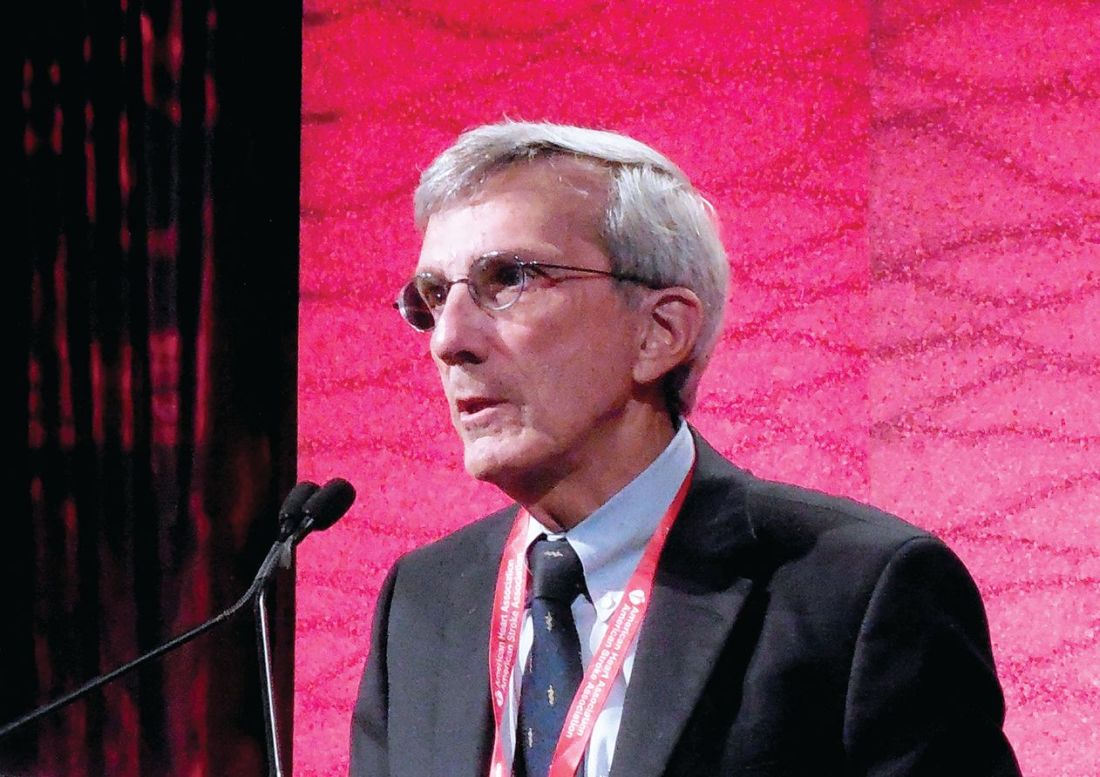
VIDEO: Stroke benefits from stem cells maintained for 2 years
LOS ANGELES – , Gary K. Steinberg, MD, said at the International Stroke Conference, sponsored by the American Heart Association.
Seeing sustained benefit out to 2 years was “quite surprising. We thought we’d lose the benefit,” Dr. Steinberg said in a video interview.
The findings “change our notion of what happens after a stroke. The damaged circuits can be resurrected,” said Dr. Steinberg, professor and chair of neurosurgery at Stanford (Calif.) University.
He reported long-term follow-up data for 18 chronic stroke patients who had received transplantation of allogeneic bone marrow–derived stem cells. The study’s primary efficacy endpoint, at 6 months after treatment, showed clinically meaningful improvements in several measures of stroke disability and function in 13 of the 18 patients (72%), including a rise of at least 10 points in the Fugl-Meyer total motor function score.
His new report on 2-year follow-up showed that these 6-month improvements continued. The average increase in Fugl-Meyer score over baseline was about 18 points at 6, 12, and 24 months of follow-up.
Based on the promise shown in this pilot study, Dr. Steinberg and his associates are running a randomized study with 156 patients. Enrollment recently completed, and the results should be available during the second half of 2019, Dr. Steinberg said.
SanBio funded the study. Dr. Steinberg has been a consultant or advisor to Qool Therapeutics, Peter Lazic US, and NeuroSave.
SOURCE: Steinberg K et al. International Stroke Conference 2018, Abstract LB14.
REPORTING FROM ISC 2018
Key clinical point: The stroke benefits from cell transplantation continued during 2-year follow-up.
Major finding: Among 18 treated patients, 13 (72%) had a sustained, clinically meaningful rise in their total motor function score.
Study details: Review of 18 patients who received intracranial cell transplantations at two U.S. sites.
Disclosures: SanBio funded the study. Dr. Steinberg has been a consultant or adviser to Qool Therapeutics, Peter Lazic US, and NeuroSave.
Source: Steinberg K et al. International Stroke Conference 2018, Abstract LB14.

Aspirin blunts early stroke risk from preeclampsia
LOS ANGELES – Women with a history of preeclampsia have a significantly increased risk for an early-onset stroke, but that risk is blunted in women taking aspirin, an epidemiologic analysis of data from more than 83,000 women in the California Teachers Study showed.
Among the 4,072 women in the study with a history of preeclampsia, 3,003 were not on aspirin, and during follow-up they had a greater than 1% incidence of stroke – ischemic and hemorrhagic combined – before turning 60 years of age. Their incidence rate was 40% higher than in the roughly 60,000 women without a preeclampsia history who were not taking aspirin, a statistically significant difference after adjustment for demographics, smoking, obesity, diabetes, and hypertension, Eliza C. Miller, MD, said at the International Stroke Conference, sponsored by the American Heart Association.![]()
Her analysis used data drawn from more than 133,000 women enrolled starting in 1995 in the California Teachers Study. She focused on 83,790 women who entered the study when they were younger than 60 years old, who had no history of stroke, and who provided data on their history of preeclampsia. The prevalence of a preeclampsia history was 4.9% overall, and 6.1% among women who had been pregnant at least once, an incidence rate similar to what has been found in other large populations of women, Dr. Miller said.
The average age of the women with preeclampsia was 44, and 46 for those without preeclampsia. The women with a history of preeclampsia also had a higher prevalence rate of obesity, hypertension, diabetes, and chronic kidney disease. Roughly a quarter of all women were regularly taking aspirin.
After adjustment of the data for demographic and clinical differences, women with a history of preeclampsia had a 20% higher overall rate of a subsequent stroke before reaching age 60 years, but this difference was not significant in an analysis that included both women taking aspirin and those not on the drug. When the analysis focused only on the women not on aspirin, the increased stroke rate linked with a preeclampsia history rose to 40% higher than in women without a preeclampsia history, a statistically significant difference. In contrast, among the quarter of women on aspirin, the two subgroups – with a preeclampsia history and without – had similar rates of incident strokes.
SOURCE: Miller E et al. International Stroke Conference 2018, A174 (Stroke. 2018 Jan;49[Suppl1]:A174).
REPORTING FROM ISC 2018
Key clinical point: Aspirin use dampened the rise in early-onset strokes seen after preeclampsia.
Major finding: Study details: A review of data collected from 83,790 women enrolled in the California Teachers Study.
Disclosures: Dr. Miller had no relevant financial disclosures.
Source: Miller E et al. International Stroke Conference 2018, A174 (Stroke. 2018 Jan;49[Suppl 1]:A174).
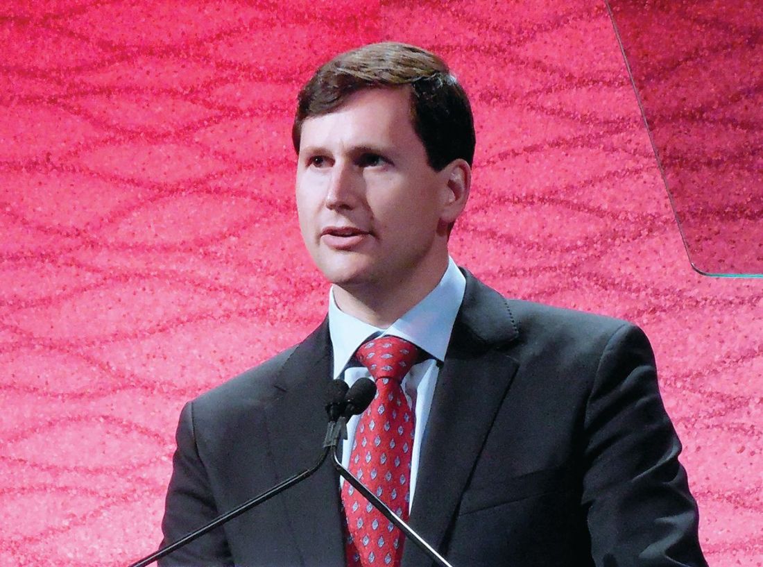
VIDEO: Rivaroxaban plus aspirin halves ischemic strokes
LOS ANGELES – Combined treatment with a low dosage of the anticoagulant rivaroxaban plus aspirin cut the incidence of ischemic strokes nearly in half, compared with aspirin alone, in a multicenter, randomized trial of more than 27,000 patients with stable atherosclerotic vascular disease.
This dramatic reduction in ischemic strokes as well as in all-cause strokes by adding low-dose rivaroxaban(Xarelto) occurred without any significant increase in hemorrhagic strokes but with a small increase in total major bleeding events, such as gastrointestinal bleeds, Mike Sharma, MD, said at the International Stroke Conference, sponsored by the American Heart Association.
“There was a consistent effect across all strata of stroke risk. For patients who had a prior stroke, it’s pretty clear to use rivaroxaban plus aspirin because it had a big benefit” with no increase in intracranial hemorrhages, Dr. Sharma said in a video interview.
“We think these results will fundamentally change how we approach stroke prevention,” added Dr. Sharma, a stroke neurologist in the Population Health Research Institute of McMaster University in Hamilton, Ont.
The results he reported came from a secondary analysis of data collected in the COMPASS (Rivaroxaban for the Prevention of Major Cardiovascular Events in Coronary or Peripheral Artery Disease) trial, which enrolled 27,395 patients with stable coronary or peripheral artery disease at 602 centers in 33 countries.
The primary outcome of the trial, reported in 2017, was the combined rate of cardiovascular death, MI, or stroke during an average 23 months of follow-up, which occurred in 4.1% of patients treated with 2.5 mg rivaroxaban twice daily plus 100 mg aspirin once daily, 4.9% of patients who received 5.0 mg rivaroxaban twice daily, and 5.4% in patients who received 100 mg aspirin daily, a statistically significant 24% relative risk reduction in the combined treatment group, compared with aspirin only. The rivaroxaban only–treated patients did not significantly differ from the control patients who received only aspirin (N Engl J Med. 2017 Oct 5;377[14]:1319-30). The main results showed a 1.2% increase in the rate of major bleeds in patients treated with rivaroxaban plus aspirin, compared with aspirin only, but the rate of nonfatal symptomatic intracranial hemorrhages was identical in the two treatment groups.
The new results Dr. Sharma reported at the conference focused on various measures of stroke. The rate of all strokes was 42% lower among the patients treated with rivaroxaban plus aspirin, compared with the aspirin alone patients, and ischemic strokes were 49% lower with the dual therapy, compared with aspirin only. Both differences were statistically significant. In contrast, the rivaroxaban alone regimen did not significantly reduce all-cause strokes. It did significantly reduce ischemic strokes, compared with aspirin only, but it also significantly increased hemorrhagic strokes, compared with aspirin only, an adverse effect not caused by the combination of low-dose rivaroxaban plus aspirin.
Rivaroxaban plus aspirin surpassed aspirin alone for preventing both mild and severe strokes and for preventing strokes both in patients with a history of a prior stroke and in those who never had a prior stroke. The stroke reduction produced by rivaroxaban plus aspirin was greatest in the highest risk patients – those with a prior stroke. On the combined regimen, these patients had an average stroke incidence of 0.7% per year, compared with an annual 3.4% rate among the patients on aspirin only, a 2.7% absolute reduction by using rivaroxaban plus aspirin that translated into a number needed to treat of 37 patients with a history of stroke to prevent one new stroke per year.
The 2017 report of the main COMPASS results included a net clinical benefit analysis that factored together the primary endpoint events and major bleeding events. The net rate of all these events was 4.7% with rivaroxaban plus aspirin and 5.9% with aspirin only, a statistically significant 20% relative risk reduction for all adverse outcomes with dual therapy. This net clinical benefit suggests that adding rivaroxaban has a cost-effective benefit. Assessment of rivaroxaban’s cost benefit in COMPASS is in process, Dr. Sharma said.
Rivaroxaban received Food and Drug Administration marketing approval in 2011 for preventing deep vein thrombosis and preventing stroke in patients with atrial fibrillation at dosages higher than what was used in COMPASS. The approved rivaroxaban dosage for preventing deep vein thrombosis is 10 mg/day, and 20 mg/day for preventing stroke in atrial fibrillation patients. The 2.5-mg formulation of rivaroxaban that was given twice daily had the best safety and efficacy in COMPASS, but it is not available now on the U.S. market, although it is available in Europe. Johnson & Johnson, which markets rivaroxaban globally with Bayer, submitted an application to the FDA in December for marketing approval of the 2.5-mg formulation in twice-daily dosing for use as in the COMPASS trial.
COMPASS was sponsored by Bayer, the company that markets rivaroxaban in collaboration with Johnson & Johnson. Dr. Sharma has been a consultant or adviser to Bayer, Bristol-Myers Squibb, Boehringer Ingelheim, and Daiichi-Sankyo.
SOURCE: Sharma M et al. ISC 2018, Abstract LB7.
REPORTING FROM ISC 2018
Key clinical point: Rivaroxaban plus aspirin cuts strokes in patients with stable atherosclerotic vascular disease.
Major finding: Rivaroxaban plus aspirin cut the rate of ischemic strokes by 49%, compared with aspirin only.
Study details: Secondary analysis from the COMPASS trial, a multicenter, randomized trial with 27,395 patients.
Disclosures: COMPASS was sponsored by Bayer, the company that markets rivaroxaban in collaboration with Johnson & Johnson. Dr. Sharma has been a consultant or adviser to Bayer, Bristol-Myers Squibb, Boehringer Ingelheim, and Daiichi-Sankyo.
Source: Sharma M et al. ISC 2018, Abstract LB7.
VIDEO: Retinal infarctions get missed as stroke harbingers
LOS ANGELES – Retinal infarctions are often going missed as important red flags for future ischemic strokes.
Among U.S. Medicare beneficiaries older than 65 years who had a retinal infarction (RI), “only one-third underwent adequate stroke risk factor evaluation,” Alexander E. Merkler, MD, reported in a poster presented at the International Stroke Conference sponsored by the American Heart Association. And fewer than 10% underwent assessment by a neurologist, based on a review of 5,688 of these older Medicare beneficiaries who had a RI sometime during 2009-2015.
The high-risk profile of these patients was affirmed by a 1% ischemic stroke incidence during the 90 days following their RI diagnosis, a rate roughly fourfold higher than in similar patients without a recent RI.
“A lot of people don’t recognize that a retinal infarction is a type of stroke,” Dr. Merkler said in a video interview. To test this hypothesis, Dr. Merkler and his associates examined the follow-up run on elderly Medicare beneficiaries following a RI diagnosis.”The guidelines recommend evaluating why these patients had a stroke [a retinal infarction] and treating risk factors to reduce the risk of a future stroke,” said Dr. Merkler, a neurologist at Weill Cornell Medicine in New York.
The review showed that 34% of the RI patients underwent cervical carotid imaging, 29% had heart rhythm monitoring, 23% underwent echocardiography, and 8% had assessment by a neurologist.
Dr. Merkler had no disclosures.
The video associated with this article is no longer available on this site. Please view all of our videos on the MDedge YouTube channel
SOURCE: Merkler A et al. ISC 2018 Abstract TMP76 (Stroke. 2018 Jan;49[Suppl 1]:ATMP76).
REPORTING FROM ISC 2018
Key clinical point: Retinal infarction patients often fail to undergo stroke assessment.
Major finding: One-third of Medicare beneficiaries with retinal infarction received adequate evaluation for stroke risk factors.
Study details: Review of 5,688 Medicare patients with a retinal infarction during 2009-2015.
Disclosures: Dr. Merkler had no disclosures.
Source: Merkler A et al. ISC 2018 Abstract TMP76 (Stroke. 2018 Jan;49[Suppl 1]:ATMP76).
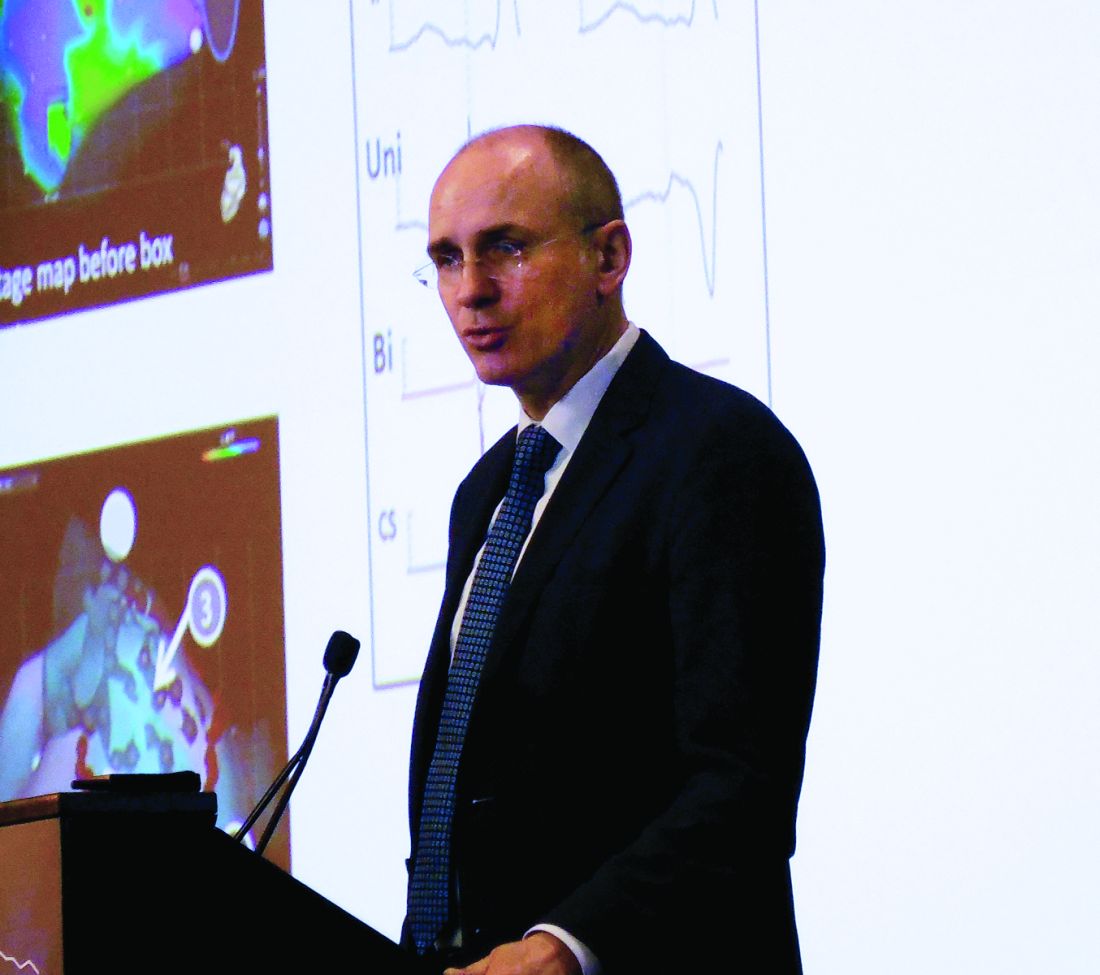
Tenecteplase surpasses alteplase for thrombolysing acute ischemic stroke
LOS ANGELES – Thrombolysis with tenecteplase beat alteplase on an acute imaging endpoint in patients with acute ischemic stroke who were on their way to also get thrombectomy in a randomized, multicenter study with 202 patients in Australia and New Zealand.
The results of the trial, called Tenecteplase Versus Alteplase Before Endovascular Therapy for Ischemic Stroke (EXTEND-IA TNK), showed that when the patients underwent their initial angiogram after receiving thrombolysis and before their thrombectomy procedure, the percentage of patients with robust blood flow – a Thrombolysis in Cerebral Infarction (TICI) score of 2b or 3 or no retrievable thrombus – was 22% in the patients treated with tenecteplase 0.25 mg/kg and 10% among those who received alteplase 0.9 mg/kg. After adjustment, the 12% incremental change in robust reperfusion with tenecteplase calculated into a 2.6-fold higher rate of robust reperfusion, statistically significant for both noninferiority and for superiority, Bruce C. Campbell, MBBS, PhD, said at the International Stroke Conference, sponsored by the American Heart Association.
Tenecteplase is a genetically-modified tissue plasminogen activator with enhanced fibrin specificity that increases the drug’s half life and allows for bolus administration, unlike alteplase, which needs continuous infusion (CNS Drugs. 2015 Oct;29[10]:811-8). Tenecteplase also has a U.S. wholesale price that is about $3,000 cheaper per vial than alteplase, said Dr. Campbell, professor of neurology at the University of Melbourne and head of hyperacute stroke at Royal Melbourne Hospital.
But further data are needed before tenecteplase is ready for routine use, conceded Dr. Campbell, an assessment other experts agreed with. Dr. Campbell cited two studies in progress that are comparing tenecteplase with alteplase in acute ischemic stroke patients not headed for endovascular thrombectomy, as well as a study he is leading that compares the tenecteplase dose he just tested, 0.25 mg/kg, with a higher dose, 0.40 mg/kg.
Several reports have appeared in recent years suggesting that treatment with tenecteplase seems to be at least as good as alteplase in ischemic stroke patients. For example, a randomized trial with 75 ischemic stroke patients selected by imaging at three Australian centers showed that treatment with tenecteplase produced a significant 24% improvement in the rate of arterial reperfusion and an average 5-point improvement in NIH Stroke Scale score (N Engl J Med. 2012;366[12]:1099-107). And results from the NOR-TEST study recently showed that among 1,100 patients randomized at 13 Norwegian centers, the primary outcome of a 90-day modified Rankin Scale score of 0-1 was achieved by 64% of the tenecteplase patients and by 63% of those who received alteplase (Lancet Neurol. 2017 Oct;16[10]:781-8).
The EXTEND-IA TNK trial ran during 2015-2017 at 18 hospitals. All enrolled patients received their thrombolytic treatment within 4.5 hours of their stroke onset, and underwent endovascular thrombectomy within 6 hours of onset. The safety outcomes of death, symptomatic intracranial hemorrhage, and parenchymal hematoma occurred at statistically similar rates in both treatment arms.
The study was investigator initiated and funded chiefly by the Australian government. Medtronic provided an unrestricted grant for trial infrastructure but had no role in study design conduct or analysis. Dr. Campbell and Dr. Powers had no disclosures.
SOURCE: Campbell B et al. ISC 2018, abstract LB2.
In the EXTEND-IA TNK study, tenecteplase appeared to act better than alteplase and has the extra advantage of being administered as a bolus injection. Alteplase is delivered as a drip, and it’s often hard to get patients with an intravenous infusion out of the hospital quickly when you have to transport the patient. You need a nurse in the ambulance monitoring the drip. With tenecteplase you administer the bolus and can then send the patient without an intravenous line.
Jeffrey L. Saver, MD, is professor of neurology and director of the stroke unit at the University of California, Los Angeles. He has received research support and personal fees from Medtronic-Abbott and Neuravia. He made these comments in an interview.
LOS ANGELES – Thrombolysis with tenecteplase beat alteplase on an acute imaging endpoint in patients with acute ischemic stroke who were on their way to also get thrombectomy in a randomized, multicenter study with 202 patients in Australia and New Zealand.
The results of the trial, called Tenecteplase Versus Alteplase Before Endovascular Therapy for Ischemic Stroke (EXTEND-IA TNK), showed that when the patients underwent their initial angiogram after receiving thrombolysis and before their thrombectomy procedure, the percentage of patients with robust blood flow – a Thrombolysis in Cerebral Infarction (TICI) score of 2b or 3 or no retrievable thrombus – was 22% in the patients treated with tenecteplase 0.25 mg/kg and 10% among those who received alteplase 0.9 mg/kg. After adjustment, the 12% incremental change in robust reperfusion with tenecteplase calculated into a 2.6-fold higher rate of robust reperfusion, statistically significant for both noninferiority and for superiority, Bruce C. Campbell, MBBS, PhD, said at the International Stroke Conference, sponsored by the American Heart Association.
Tenecteplase is a genetically-modified tissue plasminogen activator with enhanced fibrin specificity that increases the drug’s half life and allows for bolus administration, unlike alteplase, which needs continuous infusion (CNS Drugs. 2015 Oct;29[10]:811-8). Tenecteplase also has a U.S. wholesale price that is about $3,000 cheaper per vial than alteplase, said Dr. Campbell, professor of neurology at the University of Melbourne and head of hyperacute stroke at Royal Melbourne Hospital.
But further data are needed before tenecteplase is ready for routine use, conceded Dr. Campbell, an assessment other experts agreed with. Dr. Campbell cited two studies in progress that are comparing tenecteplase with alteplase in acute ischemic stroke patients not headed for endovascular thrombectomy, as well as a study he is leading that compares the tenecteplase dose he just tested, 0.25 mg/kg, with a higher dose, 0.40 mg/kg.
Several reports have appeared in recent years suggesting that treatment with tenecteplase seems to be at least as good as alteplase in ischemic stroke patients. For example, a randomized trial with 75 ischemic stroke patients selected by imaging at three Australian centers showed that treatment with tenecteplase produced a significant 24% improvement in the rate of arterial reperfusion and an average 5-point improvement in NIH Stroke Scale score (N Engl J Med. 2012;366[12]:1099-107). And results from the NOR-TEST study recently showed that among 1,100 patients randomized at 13 Norwegian centers, the primary outcome of a 90-day modified Rankin Scale score of 0-1 was achieved by 64% of the tenecteplase patients and by 63% of those who received alteplase (Lancet Neurol. 2017 Oct;16[10]:781-8).
The EXTEND-IA TNK trial ran during 2015-2017 at 18 hospitals. All enrolled patients received their thrombolytic treatment within 4.5 hours of their stroke onset, and underwent endovascular thrombectomy within 6 hours of onset. The safety outcomes of death, symptomatic intracranial hemorrhage, and parenchymal hematoma occurred at statistically similar rates in both treatment arms.
The study was investigator initiated and funded chiefly by the Australian government. Medtronic provided an unrestricted grant for trial infrastructure but had no role in study design conduct or analysis. Dr. Campbell and Dr. Powers had no disclosures.
SOURCE: Campbell B et al. ISC 2018, abstract LB2.
REPORTING FROM ISC 2018
Key clinical point: Tenecteplase thrombolysis produced better reperfusion than did alteplase.
Major finding: Robust reperfusion occurred in 22% of tenecteplase patients and in 10% of patients who received alteplase.
Study details: EXTEND-IA TNK, a multicenter, randomized trial with 202 patients.
Disclosures: The study was investigator initiated and funded chiefly by the Australian government. Medtronic provided an unrestricted grant for trial infrastructure but had no role in study design conduct or analysis. Dr. Campbell and Dr. Powers had no disclosures.
Source: Campbell B et al. ISC 2018, abstract LB2
LAA occlusion boosts anticoagulants’ protection
ORLANDO – When patients with atrial fibrillation have a history of cardioembolic events despite optimal anticoagulant therapy, treatment with left atrial appendage occlusion can substantially improve protection against future events, according to a multicenter review of 22 patients.
During the 2 years prior to undergoing left atrial appendage (LAA) occlusion, the 22 atrial fibrillation (AF) patients studied had a total of 44 cardioembolic events despite receiving “optimal” treatment with an oral anticoagulant, including nine patients with one event, six patients with two events, five patients with three events, and two patients with four events each, Xavier Freixa, MD, said at the annual International AF Symposium. In contrast, during a median follow-up of 1.8 years after their procedure additional events occurred in just two patients – one with a stroke, the other with a transient ischemic attack, while the remaining 20 patients remained free of any additional events.
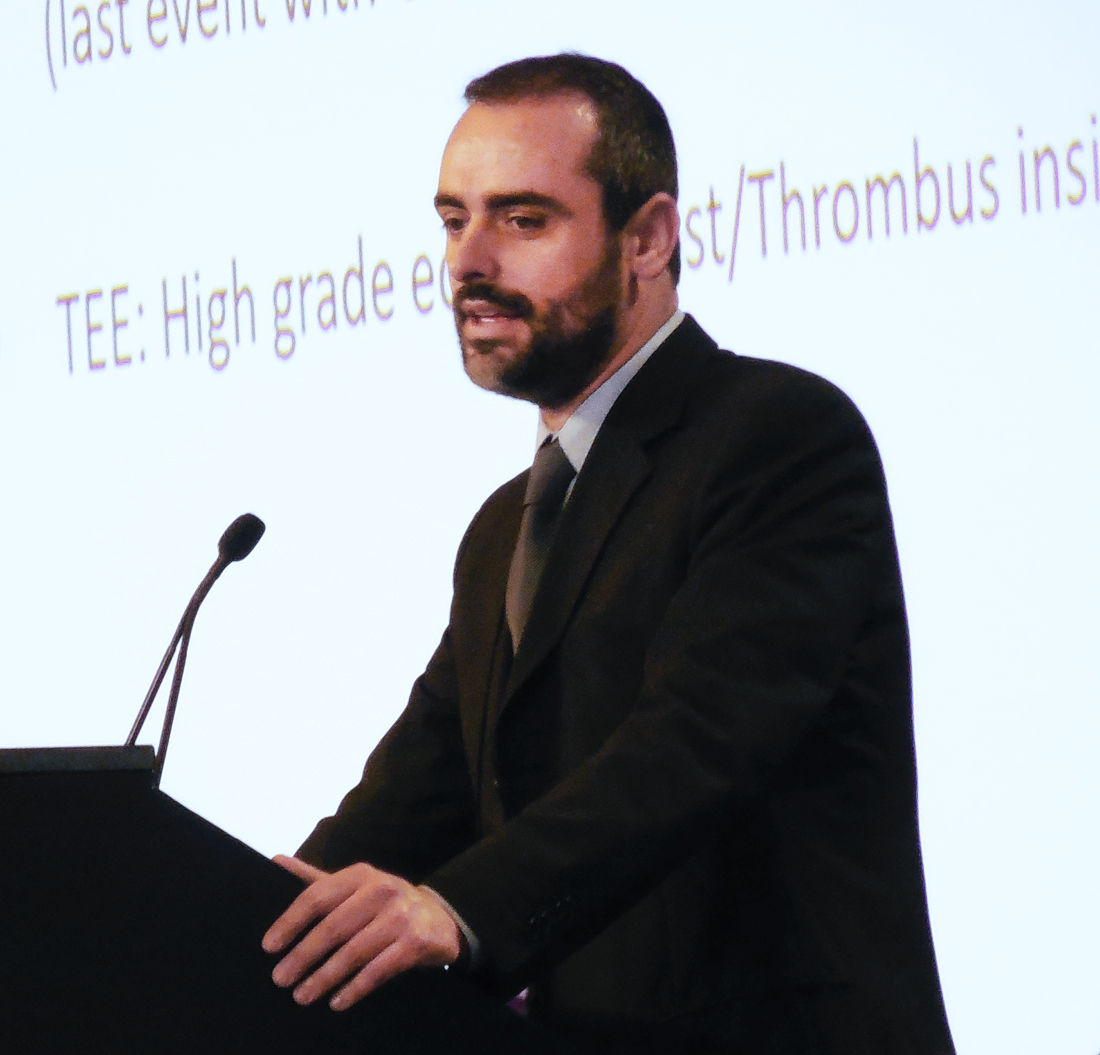
The analysis also revealed that the two patients who had cardioembolic events following their LAA occlusion had been withdrawn from oral anticoagulant treatment by their physicians, who had done this with a “false feeling of comfort,” said Dr. Freixa, an interventional cardiologist at the University Hospital Clinic of Barcelona. These two patients were among three patients maintained on dual-antiplatelet therapy rather than on an oral anticoagulant following LAA occlusion. The remaining 19 patients had remained on either warfarin, a novel oral anticoagulant, or both.
The study included patients from eight Spanish centers who underwent LAA occlusion during June 2009–June 2017, and included 14 with nonvalvular AF and 8 with valvular AF who had all undergone prior valve surgery. None of the 22 patients had a contraindication for treatment with an oral anticoagulant. They averaged about 69 years of age. Prior to their procedure, 18 had at least one stroke or transient ischemic attack, and the remaining 4 patients had at least one systemic embolism. Nineteen patients underwent occlusion with either an Amplatzer Cardiac Plug or Amplatzer Amulet device, two received a Watchman device, and one patient received a LAmbre device. All of the closure procedures were successful, with no complications.
“I think any device will do well for these patients as long as we occlude the LAA,” Dr. Freixa said.
[email protected]
On Twitter @mitchelzoler
SOURCE: Freixa X et al. AF Symposium 2018 Abstract 1821.
ORLANDO – When patients with atrial fibrillation have a history of cardioembolic events despite optimal anticoagulant therapy, treatment with left atrial appendage occlusion can substantially improve protection against future events, according to a multicenter review of 22 patients.
During the 2 years prior to undergoing left atrial appendage (LAA) occlusion, the 22 atrial fibrillation (AF) patients studied had a total of 44 cardioembolic events despite receiving “optimal” treatment with an oral anticoagulant, including nine patients with one event, six patients with two events, five patients with three events, and two patients with four events each, Xavier Freixa, MD, said at the annual International AF Symposium. In contrast, during a median follow-up of 1.8 years after their procedure additional events occurred in just two patients – one with a stroke, the other with a transient ischemic attack, while the remaining 20 patients remained free of any additional events.
The analysis also revealed that the two patients who had cardioembolic events following their LAA occlusion had been withdrawn from oral anticoagulant treatment by their physicians, who had done this with a “false feeling of comfort,” said Dr. Freixa, an interventional cardiologist at the University Hospital Clinic of Barcelona. These two patients were among three patients maintained on dual-antiplatelet therapy rather than on an oral anticoagulant following LAA occlusion. The remaining 19 patients had remained on either warfarin, a novel oral anticoagulant, or both.
The study included patients from eight Spanish centers who underwent LAA occlusion during June 2009–June 2017, and included 14 with nonvalvular AF and 8 with valvular AF who had all undergone prior valve surgery. None of the 22 patients had a contraindication for treatment with an oral anticoagulant. They averaged about 69 years of age. Prior to their procedure, 18 had at least one stroke or transient ischemic attack, and the remaining 4 patients had at least one systemic embolism. Nineteen patients underwent occlusion with either an Amplatzer Cardiac Plug or Amplatzer Amulet device, two received a Watchman device, and one patient received a LAmbre device. All of the closure procedures were successful, with no complications.
“I think any device will do well for these patients as long as we occlude the LAA,” Dr. Freixa said.
[email protected]
On Twitter @mitchelzoler
SOURCE: Freixa X et al. AF Symposium 2018 Abstract 1821.
REPORTING FROM THE AF SYMPOSIUM 2018
Key clinical point:
Major finding: Two of 22 patients had a cardioembolic event after left atrial appendage occlusion.
Study details: Review of 22 patients at eight Spanish centers with atrial fibrillation and a history of cardioembolic events despite oral anticoagulation.
Disclosures: Dr. Freixa has been a proctor for Abbott Medical.
Source: Freixa X et al. AF Symposium 2018 Abstract 1821.
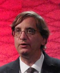
Atrial fibrosis weighed as key arrhythmia, stroke trigger
ORLANDO – , particularly strokes, and has also become the target for ablative strategies by some operators for more effective rhythm control in patients with atrial fibrillation.
“A strong and graded association exists between the severity of left atrial fibrosis severity and major cardiovascular adverse events, primarily due to stroke, transient ischemic attack, and heart failure, Nassir F. Marrouche, MD, said at the annual International AF Symposium.

Atrial fibrosis is quantified noninvasively by Dr. Marrouche and his associates with late gadolinium enhancement MRI. They published results of a retrospective study in 2017 showing that, during follow-up of 1,228 patients from their center with atrial fibrillation, the 5% of patients who had the highest level of left atrial fibrosis – 30% or more of left atrial tissue – had a significantly increased rate of cardiovascular events during follow-up, compared with the 35% of studied patients with fibrosis constituting less than 10% of their left atrium. The remaining 60% of studied patients had fibrotic tissue that formed 10%-29% of their total left atrial area.
This relationship was strongest when the analysis focused exclusively on the incidence of strokes or transient ischemic attacks. These events occurred nearly fourfold more often among patients with the highest amount of fibrosis, compared with those with the least (J Amer Coll Cardiol. 2017 Sep;70[11]:311-21).
Given that the finding came from a retrospective, single-center study, the clinical implication of fibrosis remains too uncertain to apply to current routine practice. But Dr. Marrouche strongly suspects that fibrotic tissue in the left atrium serves as a potent trigger of thrombosis.
“We think that even patients with a CHA2DS2-VASc score of zero who have a high level of fibrosis may need treatment with an anticoagulant, but we need a prospective study” to validate this, Dr. Marrouche said.
Another area for further research is to explore agents that may be able to prevent or reverse fibrosis in the left atrium. One candidate class of drugs are the angiotensin-converting enzyme inhibitors, he said in an interview.
Atrial fibrosis can have a variety of causes, including hypertension, inflammation, genetics, frequent high-intensity exercise, or other histories that produce regular intervals of extreme atrial stretch, he noted.
Dr. Marrouche is leading a multicenter trial that will test the efficacy of an ablation method for patients with atrial fibrillation tailored to isolating heavily fibrotic regions of the atria. The Efficacy of Delayed Enhancement MRI-Guided Ablation vs. Conventional Catheter Ablation of Atrial Fibrillation (DECAAFII) trial is comparing ablation aimed at fibrotic regions against conventional ablation in about 900 patients. Results are expected in another 2-3 years.
Although the usefulness of ablation aimed at isolating areas of fibrosis in the atrium remains unproven, this approach has already been embraced by some operators, including Hans Kottkamp, MD, a professor and electrophysiologist at Hirslanden Hospital in Zurich.
“Fibrosis is the fundamental histologic finding and the key pathophysiologic player in atrial fibrillation,” Dr. Kottkamp said in a separate talk at the meeting. “We suggest doing ablation based on the substrate, and not based on whether the atrial fibrillation is paroxysmal or persistent.” His group uses voltage mapping instead of MRI as their tool for quantifying the amount of fibrosis and pinpointing its location.
“The fibrosis is usually regionalized; it’s not everywhere” in the left atrium, Dr. Kottkamp noted, and the location is very individualized. “You need to look for it in each patient.”
In his experience, about 20% of patients with paroxysmal AF have enough atrial fibrosis to warrant using BIFA along with pulmonary vein isolation. “Among patients with persistent AF, the percentage with significant fibrosis rises to about two-thirds,” Dr. Kottkamp said in an interview. Combining BIFA with pulmonary vein isolation in these patients with higher fibrosis substantially boosts procedural success.
In a recent report, Dr. Kottkamp and his associates presented their experience using BIFA on 92 patients with AF and fibrotic atrial cardiomyopathy. The outcomes showed a relatively high level of success in resolving the AF after an average follow-up of 16 months, with a 69% success rate after a single procedure and an 83% success rate when selected patients received a second BIFA.
Overall, each patient in the series received an average of 1.2 BIFA ablations (J Cardiovasc Electrophysiol. 2017 Sep;28[9]:971-83).
Currently, a limited number of other groups uses BIFA or a similar fibrosis-based ablation approach, and these strategies have not yet undergone prospective assessment in a multicenter study. Despite that, Dr. Kottkamp said he was “confident it will become standard of care.”
Dr. Marrouche has been a consultant to Abbott, Biosense Webster, Biotronik, Boston Scientific, Cardiac Design, Marreck, Medtronic, Preventice, VytronUS, and Wavelet Health, and he has received research support from Abbott, Biosense Webster, Biotronik, Boston Scientific, GE Sciences, and VytronUS. Dr. Kottkamp has been a consultant to Biosense Webster and has been a consultant to and has an equity interest in Kardium.
ORLANDO – , particularly strokes, and has also become the target for ablative strategies by some operators for more effective rhythm control in patients with atrial fibrillation.
“A strong and graded association exists between the severity of left atrial fibrosis severity and major cardiovascular adverse events, primarily due to stroke, transient ischemic attack, and heart failure, Nassir F. Marrouche, MD, said at the annual International AF Symposium.
Atrial fibrosis is quantified noninvasively by Dr. Marrouche and his associates with late gadolinium enhancement MRI. They published results of a retrospective study in 2017 showing that, during follow-up of 1,228 patients from their center with atrial fibrillation, the 5% of patients who had the highest level of left atrial fibrosis – 30% or more of left atrial tissue – had a significantly increased rate of cardiovascular events during follow-up, compared with the 35% of studied patients with fibrosis constituting less than 10% of their left atrium. The remaining 60% of studied patients had fibrotic tissue that formed 10%-29% of their total left atrial area.
This relationship was strongest when the analysis focused exclusively on the incidence of strokes or transient ischemic attacks. These events occurred nearly fourfold more often among patients with the highest amount of fibrosis, compared with those with the least (J Amer Coll Cardiol. 2017 Sep;70[11]:311-21).
Given that the finding came from a retrospective, single-center study, the clinical implication of fibrosis remains too uncertain to apply to current routine practice. But Dr. Marrouche strongly suspects that fibrotic tissue in the left atrium serves as a potent trigger of thrombosis.
“We think that even patients with a CHA2DS2-VASc score of zero who have a high level of fibrosis may need treatment with an anticoagulant, but we need a prospective study” to validate this, Dr. Marrouche said.
Another area for further research is to explore agents that may be able to prevent or reverse fibrosis in the left atrium. One candidate class of drugs are the angiotensin-converting enzyme inhibitors, he said in an interview.
Atrial fibrosis can have a variety of causes, including hypertension, inflammation, genetics, frequent high-intensity exercise, or other histories that produce regular intervals of extreme atrial stretch, he noted.
Dr. Marrouche is leading a multicenter trial that will test the efficacy of an ablation method for patients with atrial fibrillation tailored to isolating heavily fibrotic regions of the atria. The Efficacy of Delayed Enhancement MRI-Guided Ablation vs. Conventional Catheter Ablation of Atrial Fibrillation (DECAAFII) trial is comparing ablation aimed at fibrotic regions against conventional ablation in about 900 patients. Results are expected in another 2-3 years.
Although the usefulness of ablation aimed at isolating areas of fibrosis in the atrium remains unproven, this approach has already been embraced by some operators, including Hans Kottkamp, MD, a professor and electrophysiologist at Hirslanden Hospital in Zurich.
“Fibrosis is the fundamental histologic finding and the key pathophysiologic player in atrial fibrillation,” Dr. Kottkamp said in a separate talk at the meeting. “We suggest doing ablation based on the substrate, and not based on whether the atrial fibrillation is paroxysmal or persistent.” His group uses voltage mapping instead of MRI as their tool for quantifying the amount of fibrosis and pinpointing its location.
“The fibrosis is usually regionalized; it’s not everywhere” in the left atrium, Dr. Kottkamp noted, and the location is very individualized. “You need to look for it in each patient.”
In his experience, about 20% of patients with paroxysmal AF have enough atrial fibrosis to warrant using BIFA along with pulmonary vein isolation. “Among patients with persistent AF, the percentage with significant fibrosis rises to about two-thirds,” Dr. Kottkamp said in an interview. Combining BIFA with pulmonary vein isolation in these patients with higher fibrosis substantially boosts procedural success.
In a recent report, Dr. Kottkamp and his associates presented their experience using BIFA on 92 patients with AF and fibrotic atrial cardiomyopathy. The outcomes showed a relatively high level of success in resolving the AF after an average follow-up of 16 months, with a 69% success rate after a single procedure and an 83% success rate when selected patients received a second BIFA.
Overall, each patient in the series received an average of 1.2 BIFA ablations (J Cardiovasc Electrophysiol. 2017 Sep;28[9]:971-83).
Currently, a limited number of other groups uses BIFA or a similar fibrosis-based ablation approach, and these strategies have not yet undergone prospective assessment in a multicenter study. Despite that, Dr. Kottkamp said he was “confident it will become standard of care.”
Dr. Marrouche has been a consultant to Abbott, Biosense Webster, Biotronik, Boston Scientific, Cardiac Design, Marreck, Medtronic, Preventice, VytronUS, and Wavelet Health, and he has received research support from Abbott, Biosense Webster, Biotronik, Boston Scientific, GE Sciences, and VytronUS. Dr. Kottkamp has been a consultant to Biosense Webster and has been a consultant to and has an equity interest in Kardium.
EXPERT ANALYSIS FROM AF SYMPOSIUM 2018
Heart attacks bring 12 weeks of higher stroke risk
LOS ANGELES – , based on a sample of Medicare beneficiaries.
The period of elevated stroke risk following an MI extends beyond the 30-day window that has traditionally been considered the interval of highest risk, Alexander E. Merkler, MD, said at the International Stroke Conference, sponsored by the American Heart Association.
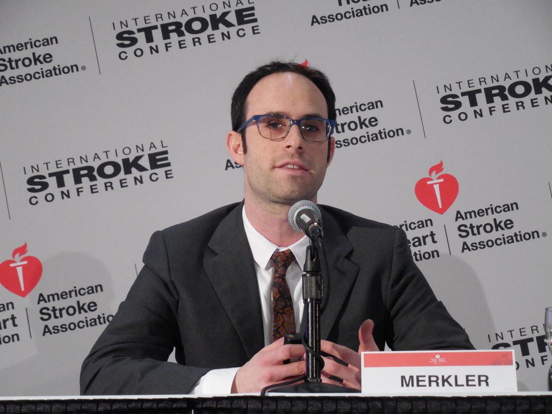
Beyond 12 weeks after MI discharge, the stroke incidence showed no significant difference compared with people without a recent MI history, said Dr. Merkler, a neurologist at Weill Cornell Medicine in New York.
He calculated these statistically significant elevated risk rates after adjusting for demographic measures, stroke risk factors, and the comorbidities included in the Charlson Comorbidity Index.
These increased stroke rates were independent of periprocedural strokes that might have happened during MI interventions, as the analysis excluded MI patients with a history of a stroke either before or during their MI hospitalization.
To run this analysis, Dr. Merkler and his associates used data collected in a 5% sample of Medicare beneficiaries who were at least 66 years old during 2008-2015. Among these 1.7 million people were 46,182 who were hospitalized for an MI.
Several factors associated with an acute MI likely contribute to an elevated stroke risk, including stasis in the heart and generation of microthrombi, and a possibly systemic proinflammatory state, Dr. Merkler suggested.
Dr. Merkler had no disclosures.
SOURCE: Merkler AE et al., International Stroke Conference abstract 172 (Stroke. 2018 Jan; 49[Suppl 1]:A172).
LOS ANGELES – , based on a sample of Medicare beneficiaries.
The period of elevated stroke risk following an MI extends beyond the 30-day window that has traditionally been considered the interval of highest risk, Alexander E. Merkler, MD, said at the International Stroke Conference, sponsored by the American Heart Association.
Beyond 12 weeks after MI discharge, the stroke incidence showed no significant difference compared with people without a recent MI history, said Dr. Merkler, a neurologist at Weill Cornell Medicine in New York.
He calculated these statistically significant elevated risk rates after adjusting for demographic measures, stroke risk factors, and the comorbidities included in the Charlson Comorbidity Index.
These increased stroke rates were independent of periprocedural strokes that might have happened during MI interventions, as the analysis excluded MI patients with a history of a stroke either before or during their MI hospitalization.
To run this analysis, Dr. Merkler and his associates used data collected in a 5% sample of Medicare beneficiaries who were at least 66 years old during 2008-2015. Among these 1.7 million people were 46,182 who were hospitalized for an MI.
Several factors associated with an acute MI likely contribute to an elevated stroke risk, including stasis in the heart and generation of microthrombi, and a possibly systemic proinflammatory state, Dr. Merkler suggested.
Dr. Merkler had no disclosures.
SOURCE: Merkler AE et al., International Stroke Conference abstract 172 (Stroke. 2018 Jan; 49[Suppl 1]:A172).
REPORTING FROM ISC 2018
Key clinical point: A patient’s stroke risk is elevated for 12 weeks following a myocardial infarction.
Major finding: The stroke rate was 2.7-fold, 2.0-fold, and 1.6-fold above background at 4, 8, and 12 weeks after an MI.
Study details: A review of 1.7 million Medicare beneficiaries during 2008-2015.
Disclosures: Dr. Merkler had no disclosures.
Source: Merkler AE et al., International Stroke Conference abstract 172 (Stroke. 2018 Jan;49[Suppl 1]:A172).
VIDEO: COMPASS shows stroke-clot aspiration noninferior to retrieval
LOS ANGELES – Clot removal in acute ischemic stroke patients using an aspiration catheter was noninferior to clot removal using the standard method, a stent retriever, in a multicenter, randomized trial with 270 patients.
“There is now level I evidence that stent retrievers and primary aspiration have equivalent outcomes in emergent large vessel occlusions,” J Mocco, MD, said at the International Stroke Conference, sponsored by the American Heart Association.
The results “will give physicians more choice,” Dr. Mocco said in a video interview. Until now, “many physicians felt that they had to use stent retrievers” because these devices had an much bigger evidence base of efficacy and safety. The new findings provide evidence that supports aspiration as an alternative strategy, said Dr. Mocco, professor of neurosurgery and director of the Cerebrovascular Center at Mount Sinai Medical Center in New York.
The results Dr. Mocco reported from the COMPASS trial (Comparison of Direct Aspiration vs. Stent Retriever as a First Approach) were the second time that clot aspiration was shown to produce outcomes similar to stent retrieval. Similar findings came from a French multicenter trial, ASTER, reported in 2017 (JAMA. 2017 Aug 1;318[5]:443-52).
The study ran at 15 U.S. centers during 2015-2017 and randomized patients with emergent large vessel occlusion strokes to receive initial treatment with either an aspiration catheter or a stent retriever. The specific type of catheter or stent used was left up to each operator. In 83% of the aspiration cases and 81% of the stent retriever cases, the initial device used produced a moderately high or better level of restored blood flow within the occluded artery, with thrombolysis in cerebral infarction (TICI) 2b flow or greater.
The study’s primary efficacy endpoint was the percentage of patients with a modified Rankin Scale score of 0-2 at 90 days after treatment, which occurred in 52% of patients treated with aspiration first and in 49% of those treated by retrieval first, which met the study’s prespecified criterion for noninferiority of the aspiration strategy, Dr. Mocco reported.
The results also showed suggestions of faster responses in the patients treated by aspiration first, although these did not reach statistical significance. The highest rate of restored blood flow within the occluded artery, TICI 3 flow, occurred in 34% of patients treated by aspiration first and in 23% of those treated with retrieval first. The average time to produce TICI 2b flow or greater was 22 minutes in the aspiration-first patients, compared with 33 minutes among retrieval-first patients.
“I prefer to start with aspiration first because it’s fast and efficient,” Dr. Mocco said. “I find encouragement in the nonsignificant faster rate of reperfusion” seen in the results.
The study’s safety endpoints, the rate of all-cause mortality after 90 days, all intracranial hemorrhages, and symptomatic intracranial hemorrhages, were all similar in the two study arms.
The COMPASS study was funded by Penumbra, but the company had no role in the trial’s design or analysis. Dr. Mocco has been a consultant to Cerebrotech Medical, EndoStream, Rebound Medical, Synchron, and Viseon and has investments in some of these companies. Dr. Sacco had no disclosures.
The video associated with this article is no longer available on this site. Please view all of our videos on the MDedge YouTube channel
SOURCE: Mocco J et al. ISC 2018, Abstract LB4.
REPORTING FROM ISC 2018
Key clinical point:
Major finding: The 90-day modified Rankin Scale score was 0-2 in 52% of aspiration patients and 49% of clot retrieval patients.
Study details: COMPASS, a multicenter, U.S. randomized trial with 270 patients.
Disclosures: The COMPASS study was funded by Penumbra, but the company had no role in the trial’s design or analysis. Dr. Mocco has been a consultant to Cerebrotech Medical, EndoStream, Rebound Medical, Synchron, and Viseon and has investments in some of these companies. Dr. Sacco had no disclosures.
Source: Mocco J et al. ISC 2018, Abstract LB4.