User login
VIDEO: Registry study will follow 4,000 fecal transplant patients for 10 years
CHICAGO – A 10-year registry study aims to gather clinical and patient-reported outcomes on 4,000 adult and pediatric patients who undergo fecal microbiota transplant in the United States, officials of the American Gastroenterological Association announced during Digestive Disease Week®.
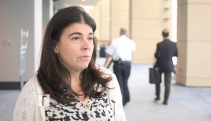
The AGA Fecal Microbiota Transplantation National Registry will be the first study to assess both short- and long-term patient outcomes associated with fecal microbiota transplant (FMT) in both adults and children, Colleen Kelly, MD, said in an video interview. Most subjects will have received FMT for recurrent or refractory Clostridium difficile infections – the only indication for which Food and Drug Administration currently allows independent clinician action. But the investigational uses of FMT are expanding rapidly, and patients who undergo the procedure during any registered study will be eligible for enrollment, said Dr. Kelly, co-chair of the study’s steering committee.
The video associated with this article is no longer available on this site. Please view all of our videos on the MDedge YouTube channel
The study’s primary objectives are short- and long-term safety outcomes, said Dr. Kelly of Brown University, Providence, R.I. While generally considered quite safe, short-term adverse events have been reported with FMT, and some of them have been serious – including one death from aspiration pneumonia in a patient who received donor stool via nasogastric tube (Clin Infect Dis. 2015 Mar;61[1]:136-7). Other adverse events are usually self-limited but can include low-grade fever, abdominal pain, distention, bloating, and diarrhea.
Researchers seek to illuminate many of the unknowns associated with this relatively new procedure. Scientists are only now beginning to unravel the myriad ways the human microbiome promotes both health and disease. Specific alterations, for example, have been associated with obesity and other conditions; there is concern that transplanting a new microbial population could induce a disease phenotype in a recipient who might not have otherwise been at risk.
With the planned cohort size and follow-up period, the study should be able to detect any unanticipated adverse events that occur in more than 1% of the population, Dr. Kelly said. It will include a comparator group of patients with recurrent or refractory C. difficile infection from a large insurance claims database to allow comparison between patients treated with FMT and those treated with antibiotics only.
The registry study also aims to discover which method or methods of transplant material delivery are best, she said. Right now, there are a number of methods (colonoscopy/sigmoidoscopy, enema, upper gastrointestinal endoscopy, nasogastric or nasoduodenal tube, and capsules), and no consensus on which is the best. As indications for FMT expand, there may be no single best method. The approach will probably be matched to the disorder being treated, and the study may help illuminate this as well.
For the first 2 years after a transplant, clinicians will follow patients and enter data into the registry. After that, an electronic patient-reported outcomes system will automatically contact the patient annually for follow-up information by email or text message. When patients enter their data, they can access educational material that will help keep them up-to-date on potential adverse events.
The study will also include a biobank of stool samples obtained during the procedures, hosted by the American Gut Project and the Microbiome Initiative at the University of California, San Diego. This arm of the project will analyze the microbiome of 3,000 stool samples from recipients, both before and after their transplant, as well as the corresponding donors whose material was used in the fecal transplant.
The registry study, a project of the AGA Center for Gut Microbiome Research and Education, is funded by a $3.3 million grant from the National Institute of Allergy and Infectious Diseases. It will be conducted in partnership with the Crohn’s and Colitis Foundation, Infectious Diseases Society, and the North American Society for Pediatric Gastroenterology, Hepatology, and Nutrition.
The registry study currently is accepting applications. Physicians who perform FMT for C. difficile infections, and centers that conduct FMT research for other potential indications, can fill out a short survey to indicate their interest.
Digestive Disease Week® is jointly sponsored by the American Association for the Study of Liver Diseases (AASLD), the American Gastroenterological Association (AGA) Institute, the American Society for Gastrointestinal Endoscopy (ASGE) and the Society for Surgery of the Alimentary Tract (SSAT).
This article was updated June 8, 2017.
[email protected]
On Twitter @Alz_gal
AT DDW
VIDEO: Indomethacin slashes post-ERCP pancreatitis risk in primary sclerosing cholangitis
CHICAGO – Rectal indomethacin reduced by 90% the risk of post-procedural pancreatitis in patients with primary sclerosing cholangitis.
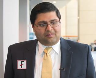
The anti-inflammatory has already been shown to reduce the risk of acute pancreatitis after endoscopic retrograde cholangiopancreatography (ERCP) in a general population, Nikhil Thiruvengadam, MD, said at the annual Digestive Disease Week®. Now, his retrospective study of almost 5,000 patients has shown the drug’s benefit in patients with primary sclerosing cholangitis (PSC), who are at particularly high risk of pancreatitis after the procedure.
The video associated with this article is no longer available on this site. Please view all of our videos on the MDedge YouTube channel
“A prior history of PEP and a difficult initial cannulation were significant risk factors for developing PEP,” he said. “Indomethacin significantly reduced this risk, and our findings suggest that future prospective trials studying pharmacological prophylaxis of PEP – including rectal indomethacin – should be powered to be able detect a difference in PSC patients, and they should be included in such studies.”
In 2016 Dr. Thiruvengadam and his colleagues showed that rectal indomethacin significantly reduced the risk of PEP by about 65% in a diverse group of patients, including those with malignant biliary obstruction (Gastroenterology. 2016;151:288–97). The new study used an expanded patient-cohort but focused on patients with PSC, as they require multiple ERCPs for diagnosis and stenting of strictures and cholangiocarcinoma screening and thus may be more affected by post-procedural pancreatitis.
The study comprised 4,764 patients who underwent ERCP at the University of Pennsylvania from 2007-2015; of these, 200 had PSC. Rectal indomethacin was routinely administered to patients beginning in June 2012. The primary outcome of the study was post-ERCP pancreatitis. The secondary outcome was the severity of post-ERCP pancreatitis.
PEP was about twice as common in the PSC group as in the overall cohort (6.5% vs. 3.8%). Moderate-severe PEP also was twice as common (4% vs. 2%).
Dr. Thiruvengadam broke down the cohort by indication for ERCP. These included PSC as well as liver transplant, choledocholithiasis, benign pancreatic disease, bile leaks, and ampullary adenoma. PSC patients had the highest risk of developing PEP – almost 3 times more than those without the disorder (OR 2.7).
Among PSC patients, age, gender, and total bilirubin were not associated with increased risk. A history of prior PEP increased the risk by 17 times, and a difficult initial cannulation that required a pre-cut sphincterotomy increased it by 15 times.
“Interestingly, dilation of a common bile duct stricture reduced the odds of developing PEP by 81%,” Dr. Thiruvengadam said.
He then examined the impact of rectal indomethacin on the study subjects. Overall, PEP developed in 5% of those who didn’t receive indomethacin and 2% of those who did. In the PSC group, PEP developed in 11% of those who didn’t get indomethacin and less than 1% of those who did.
Indomethacin was particularly effective at preventing moderate-severe PEP, Dr. Thiruvengadam noted. In the overall cohort, moderate-severe PEP developed in 3% of unexposed patients compared to 0.6% of those who received the drug. The difference was more profound in the PSC group: None of those treated with indomethacin developed moderate-severe PEP, which occurred in 9.3% of the unexposed group.
Generally, patients who have previously undergone a sphincterotomy are at lower risk for PEP, Dr. Thiruvengadam said, and this was reflected in the findings for the overall group: PEP developed in 3% of the untreated patients and 0.5% of the treated patients. Post-sphincterotomy patients with PSC, however, were still at an increased risk of PEP. Indomethacin significantly mitigated this – no patient who got the drug developed PEP, compared with 10.5% of those who didn’t get it.
A series of regression analyses confirmed the consistency of these findings. In an unadjusted model, rectal indomethacin reduced the risk of post-ERCP PEP by 91% in patients with PSC. A model that adjusted for common bile duct brushing, type of sedation, and common bile duct dilation found a 90% risk reduction. Another model that controlled for classic risk factors for PEP (age, gender, total bilirubin, history of PEP, pancreatic duct injection and cannulation, and pre-cut sphincterotomy) found a 94% risk reduction.
“We additionally performed a propensity score matched analysis to account for potential unmeasured differences between the two cohorts, and it also confirmed the results found and demonstrated that indomethacin significantly reduced the odds of developing PEP by 89%,” Dr. Thiruvengadam said.
He had no financial conflicts of interest to disclosures.
Digestive Disease Week® is jointly sponsored by the American Association for the Study of Liver Diseases (AASLD), the American Gastroenterological Association (AGA) Institute, the American Society for Gastrointestinal Endoscopy (ASGE), and the Society for Surgery of the Alimentary Tract (SSAT).
[email protected]
On Twitter @Alz_gal
AT DDW
Key clinical point:
Major finding: The anti-inflammatory reduced the risk in these patients by 90%.
Data source: A retrospective study of 4,764 patients with PSC who underwent ERCP at a single institution, Disclosures: Dr. Thiruvengadam had no financial disclosures.
VIDEO: Rifamycin matches ciprofloxacin’s efficacy in travelers’ diarrhea with less antibiotic resistance
CHICAGO – An investigational antibiotic was just as effective as ciprofloxacin at curing travelers’ diarrhea but was associated with a significantly lower rate of colonization with extended spectrum beta-lactam–resistant Escherichia coli, a phase III trial has determined.
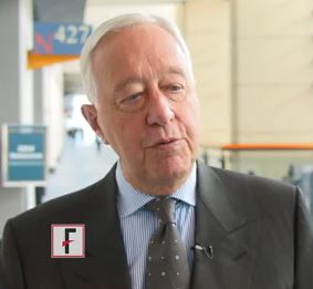
“Rifamycin was noninferior to ciprofloxacin on every endpoint in this trial,” Robert Steffen, MD, said at the annual Digestive Disease Week. “However, there was no increase in extended spectrum beta-lactamase–producing Enterobacteriaceae (ESBL-E) associated with rifamycin, and significantly less new acquisition of these pathogens than in the ciprofloxacin group.”
Rifamycin is a poorly absorbed, broad-spectrum antibiotic in the same chemical family as rifaximin. It’s designed, both molecularly and in packaging, to become active only in the lower ileum and colon with limited systemic absorption. The drug is approved in Europe for infectious colitis, Clostridium difficile, diverticulitis, and also as supportive treatment of inflammatory bowel diseases and hepatic encephalopathy.
The video associated with this article is no longer available on this site. Please view all of our videos on the MDedge YouTube channel
Subjects were randomized to 3 days of rifamycin 800 mg, or ciprofloxacin 1,000 mg. Follow-up visits occurred on days 2, 5, and 6, with a final follow-up by mail 4 weeks later. The primary endpoint was time to last unformed stool from the first dose of study medication. Secondary endpoints were clinical cure (24 hours with no clinical symptoms, fever, or watery stools, or 48 hours with no fever; and either no stools or only formed stools), need for rescue therapy, treatment failure, pathogen eradication in posttreatment stool, and the rate of ESBL-E colonization.
The time to last unformed stool was 43 hours in the rifamycin group and 37 hours in the ciprofloxacin group, which were not significantly different. The results were similar when broken down by infective organism, by gender, and by study location.
Rifamycin was also noninferior to ciprofloxacin in several secondary endpoints, including clinical cure (85% each), treatment failure (15% each), and need for rescue therapy (1% vs. 2.6%). The drugs were also virtually identical in the number of unformed stools per 24-hour interval, which fell precipitously from 5.5 on day 1, to 1 by day 5, and in complete resolution of gastrointestinal symptoms, which were about 75% resolved in each group by day 5.
Rifamycin was equally effective in eradicating all of the pathogens identified in the cohort. This included all pathogens in the E. coli group, all in the potentially invasive group (Shigella, Campylobacter, Salmonella, and Aeromonas), norovirus, giardia, and Cryptosporidium.
Treatment-emergent adverse events occurred in 12% of each group; none were serious. About 8% of each group experienced an adverse drug reaction.
Where the drugs did differ, and sharply so, was in antibiotic resistance, said Dr. Steffen, of the University of Zürich and the University of Texas School of Public Health, Houston. At baseline, about 16% of the group was infected with ESBL–E coli. At last follow-up, those species were present in 16% of the rifamycin group, but in 21% of the ciprofloxacin group. Similarly, there was less new ESBL–E. coli colonization in patients who had been negative at baseline (10% vs. 17%).
The findings are particularly important in light of the increasing worldwide emergence of antibiotic-resistant bacteria, Dr. Steffen said. In fact, new guidelines released April 29 by the International Society of Travel Medicine recommend that antibiotics be reserved for moderate to severe cases of traveler’s diarrhea and not be used at all in milder cases (J Travel Med. 2017 Apr 29;24[suppl. 1]:S57-S74).
“The widespread use of ciprofloxacin and other antibiotics for travelers’ diarrhea has contributed to the rise of these resistant bacteria,” Dr. Steffen said in an interview. “We need to rethink the way we use these drugs and to focus instead on drugs that are not systemically absorbed. If rifamycin is eventually approved for this indication, it would be a good alternative to systemic antibiotics, curing the acute illness, and not contributing as much to the emergence of these worrisome pathogens.”
Digestive Disease Week® is jointly sponsored by the American Association for the Study of Liver Diseases (AASLD), the American Gastroenterological Association (AGA) Institute, the American Society for Gastrointestinal Endoscopy (ASGE), and the Society for Surgery of the Alimentary Tract (SSAT).
In a video interview at the meeting, Dr. Steffen spoke about the trial and concerns about antibiotic resistance that are addressed in the new guidelines and by this new study.
Dr. Falk Pharma GmbH of Freiburg, Germany, is developing rifamycin and conducted the study. Dr. Steffen has received consulting and travel fees from the company.
[email protected]
On Twitter @alz_gal
AT DDW
Key clinical point:
Major finding: Clinical cure occurred in 85% of each group, but new beta-lactam–resistant E. coli colonization occurred in 16% of the rifamycin group and 21% of the ciprofloxacin group.
Data source: The randomized study comprised 835 subjects.
Disclosures: Dr. Falk Pharma GmbH of Freiburg, Germany, is developing the drug and sponsored the study. Dr. Steffen has received consulting and travel fees from the company.
Cushing’s appears to begin its cardiovascular effects during childhood
ORLANDO – Cushing’s disease may begin to exert its harmful cardiovascular effects quite early, a small pediatric study has found.
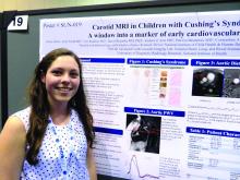
Children as young as 6 years old with the disorder already may show signs of cardiovascular remodeling, with stiffer aortas and higher aortic pulse-wave velocity than do age-matched controls, Hailey Blain and Maya Lodish, MD, said at the annual meeting of the Endocrine Society.
Cushing’s diseases has long been linked with increased cardiovascular risk in adults, but the study by Dr. Lodish and Ms. Blain is one of the first to examine the link in children. Their findings suggest that early cardiovascular risk factor management should be a routine part of these patients’ care, Dr. Lodish said in an interview.
“It’s very important to make sure that there is recognition of the cardiovascular risk factors that go along with this disease. Elevated levels of cholesterol, hypertension, and other risk factors that are in these individuals should be ameliorated as soon as possible from an early age and, most importantly, physicians should be diagnosing and treating children early, once they are identified as having Cushing’s disease. And, given that we are not sure whether these changes are reversible, we need to make sure these children are followed very closely.”
Indeed, Dr. Lodish has reason to believe that the changes may be long lasting or even permanent.
“We are looking at these children longitudinally and have 3-year data on some patients already. We want to see if they return to normal pulse wave velocity after surgical cure, or whether this is permanent remodeling. There is an implication already that it may be in a subset of individuals,” she said, citing her own 2009 study on hypertension in pediatric Cushing’s patients. “We looked at blood pressure at presentation, after surgical cure, and 1 year later. A significant portion of the kids still had hypertension at 1 year. This leads us to wonder if they will continue to be at risk for cardiovascular morbidity as adults.”
The patients had a mean 2.5-year history of Cushing’s disease Their mean midnight cortisol level was 18.8 mcg/dL and mean plasma adrenocorticotropic hormone level, 77.3 pg/mL. Five patients were taking antihypertensive medications. Low- and high-density lipoprotein levels were acceptable in all patients.
The cardiovascular measures were compared to an age-matched historical control group. In this comparison, patients had significantly higher pulse wave velocity compared with controls (mean 4 vs. 3.4 m/s). Pulse wave velocity positively correlated with both midnight plasma cortisol and 24-hour urinary free cortisol collections. In the three patients with long-term follow-up after surgical cure of Cushing’s, the pulse wave velocity did not improve, either at 6 months or 1 year after surgery. This finding echoes those of Dr. Lodish’s 2009 paper, suggesting that once cardiovascular remodeling sets in, the changes may be long lasting.
“The link between Cushing’s and cardiovascular remodeling is related to the other things that go along with the disease,” Dr. Lodish said. “The hypertension, the adiposity, and the high cholesterol all may contribute to arterial rigidity. It’s also thought to be due to an increase in connective tissue. The bioelastic function of the aorta may be affected by having Cushing’s.”
That connection also suggests that certain antihypertensives may be more beneficial to patients with Cushing’s disease, she added. “It might have an implication in what blood pressure drug you use. Angiotensin-converting enzyme inhibitors increase vascular distensibility and inhibit collagen formation and fibrosis. It is a pilot study and needs longitudinal follow up and additional patient accrual, however, finding signs of cardiovascular remodeling in young children with Cushing’s is intriguing and deserves further study.”
Neither Ms. Blain nor Dr. Lodish had any financial disclosures.
AT ENDO 2017
Key clinical point:
Major finding: Patients had significantly higher pulse wave velocity, compared with controls (mean 4 vs. 3.4 m/s).
Data source: The small cohort study comprises 10 patients and a series of age-matched historical controls.
Disclosures: Neither Dr. Lodish nor Ms. Blain have any financial disclosures.
Adjunctive rifampicin doesn’t improve any outcome in S. aureus bacteremia
VIENNA – When given in conjunction with an antibiotic, rifampicin didn’t improve treatment response or mortality in patients with Staphylococcus aureus bacteremia, either in an overall analysis or in any of 18 subgroups.
The only hints of benefit associated with the drug were decreases in treatment failure and recurrence, but the numbers needed to treat were excessive (29 and 26, respectively). They didn’t translate into any long-term survival benefit and couldn’t balance out other findings that rifampicin increased drug interactions and complicated treatment, Guy Thwaites, MD, said at the European Conference on Clinical Microbiology and Infectious Diseases.
He presented the results of the randomized, placebo-controlled ARREST (Adjunctive Rifampicin to Reduce Early Mortality From Staphylococcus aureus bacteremia) study. ARREST was conducted at 29 sites in the United Kingdom. It enrolled 758 adults with proven S. aureus bacteremia who were already on standard antibiotic therapy and switched them to either adjunctive rifampicin or placebo for 2 weeks. Clinicians could choose rifampicin in either 600 mg or 900 mg, oral or IV formulations, once or twice daily doses.
Patients were followed with clinical assessments and blood cultures for 12 weeks and for overall mortality for 102 weeks. The primary endpoint was bacteriologically confirmed treatment failure or recurrence by week 12.
Patients were a mean of 65 years old. Most infections (64%) were community acquired, with the remainder associated with a stay in a health care facility, and 6% were methicillin resistant. Serious comorbidities were common, including cancer (17%), chronic lung disease (12%), kidney disease (18%), and diabetes (30%).
The largest portion of infections (40%) had a deep focus, including native cardiac valve or joint, prosthetic cardiac valve or implant, and deep tissue infections. Other sites of infection were indwelling lines, skin/soft tissue, surgical sites, pneumonia, and urinary tract. For 18%, no specific focus was ever established.
Rifampicin was initiated a mean of 68 hours after main antibiotic therapy. Most patients (86%) received it orally, in the 900-mg dose (78%). The mean rifampicin treatment duration was 13 days.
Treatment failure rates through week 12 were practically identical for rifampicin and placebo (17.5% vs. 18.9%) in the overall analysis. Clinical failure or recurrence through week 12 was also similar for rifampicin and placebo (21.4% vs. 22.9%). Dr. Thwaites didn’t present all 18 subgroup analyses but said the results were similar no matter how patients were divided.
There was no significant difference in 12-week mortality for rifampicin vs. placebo (15.7% vs. 14.8%). There were 112 deaths, 56 in each group. Of these, 28 were directly related to the S. aureus infection. There was no difference in long-term survival measured at 102 weeks.
When an independent endpoint review committee examined some of the composite endpoints separately, it determined that rifampicin did confer a significant advantage in both bacterial and clinical recurrence. However, 29 patients needed to be treated to avoid a bacteriologic recurrence and 26 to avoid a clinical recurrence. Two cases of rifampicin resistance developed.
One-quarter of the group experienced serious adverse events. Dr. Thwaites didn’t review these but said they were evenly distributed between the groups. He also said that rifampicin was associated with an increase in drug-drug interactions, some of which required changing the backbone antibiotic.
There was a small, but nonsignificant, increase in acute kidney injury in the rifampicin group.
The study was funded by the National Institute of Health Research in the United Kingdom. Dr. Thwaites had no financial disclosures.
[email protected]
On Twitter @alz_gal
Key clinical point:
Major finding: Treatment failure was nearly identical with rifampicin and placebo (17.5% vs. 18.9%).
Data source: A placebo-controlled trial enrolling 758 adults.
Disclosures: The study was funded by the National Institute of Health Research in the United Kingdom. Dr. Thwaites had no financial disclosures.
For bone and joint infections, oral antibiotics match IV, cost less
VIENNA – Oral antibiotic therapy is just as effective as intravenous treatment in curing bone and joint infections, but costs about $3,500 less.
Treating these infections with oral agents also “improves patient autonomy, as it’s not necessary to have IV lines at home,” and represents a generally wiser use of powerful antibiotics, Matthew Scarborough, MD, said at the European Society of Clinical Microbiology and Infectious Diseases annual congress.
OVIVA (Oral vs. Intravenous Antibiotics for Bone and Joint Infection) was conducted at 26 sites in the United Kingdom. It randomized 1,054 adults with bone or joint infections to 6 weeks of either oral or intravenous treatment.
An important aspect of the trial was that both oral and IV treatment choices were made before randomization, Dr. Scarborough said. However, the decisions on what drug to use were left up to the treating physician and depended on the infection site and pathogen.
The primary outcome was definite treatment failure (bacteriologic, histologic, and clinical). Patients were followed for 1 year.
Patients were a median of 60 years old. All had surgical treatment before antibiotic therapy, including debridement and, in those with implants, removal of infected devices. The lower limb was involved in 81%, including hip, knee, and foot. The infection was in an upper limb in 10% and in the spine in 7%.
Staphylococcus aureus was present in 38% of cases, coagulase-negative staphylococci in 27%, and streptococci in 15%. Gram-negative bacteria were found in 22%.
For those patients randomized to IV therapy, glycopeptides and cephalosporins were most commonly employed (41% and 33%, respectively). For oral therapy, quinolones and penicillins were most common (37% and 16%). Most patients (74%) continued antibiotic treatment for more than 6 weeks. Forty patients were lost to follow-up.
In the primary intent-to-treat analysis, the failure rate was 13% for oral therapy and 14% for IV therapy, not a significant difference. Results were similar in the other analyses, including a modified intent to treat with only patients who had complete 1-year data, and a per-protocol analysis. All of the point prevalence numbers favored oral therapy, but crossed the null. Curves in the time-to-treatment-failure analysis were virtually superimposable, as were curves in time to discontinuation of therapy.
Another subgroup analysis examined treatment failure by infective organism; again, there were no significant treatment differences in any of the pathogen subgroups examined (S. aureus, coagulase-negative staph, streptococci species, and other gram-negative bacteria).
Nor did the type of antibiotic significantly affect failure rate, Dr. Scarborough noted. The median length of stay was 14 days for patients on IV treatment and 11 days for those taking oral medications. The incidence of serious adverse events was very similar – about 86% in each group.
On a visual analog scale that assessed health-related quality of life, patients taking oral treatment reported better mobility, self-care, and activity level, and less pain, discomfort, anxiety, and depression than those taking IV medications.
Cost represented the other significant difference between the groups. Over 1 year, the mean IV treatment cost was the equivalent of $17,152, and the mean oral treatment cost was $13,611 – a significant difference of $3,541.
“This represents a potential savings to the National Health Service of 16-25 million pounds sterling ($20.6 million-$32.3 million) per year,” Dr. Scarborough said. “All coming at no expense of good clinical outcomes.”
OVIVA was sponsored by the U.K. National Institute of Health Research. Dr. Scarborough had no financial disclosures.
[email protected]
On Twitter @alz_gal
AT ECCMID 2017
Key clinical point:
Major finding: At 1 year, cure rates were identical, but oral treatment cost about $3,500 less than IV treatment.
Data source: The study randomized 1,054 patients.
Disclosures: OVIVA was sponsored by the U.K. National Institute of Health Research. Dr. Scarborough had no financial disclosures.
Some data support botulinum toxin for psoriasis and rosacea
ORLANDO – Botulinum toxin may have a place in treating psoriasis and rosacea.
There is not a huge body of literature supporting the use of neuromodulators for these conditions, but a smattering of case reports have shown positive results and some clinicians are exploring their off label use, Erin Gilbert, MD, said at the annual meeting of the American Academy of Dermatology.
Her own interest was originally piqued when she began working with Nicole Ward, PhD, director of the morphology core of the Skin Diseases Research Center in the department of dermatology at Case Western Reserve University, Cleveland, who developed a transgenic mouse model of psoriasis. Dr. Ward discovered that transecting the thoracic-level cutaneous nerves at their entry site into back skin resulted in rapid and significant changes in the psoriatic phenotype (J Invest Dermatol. 2011 Jul;131[7]:1530–8). These included decreases of up to 40% in various immune cell populations and a 30% improvement in acanthosis relative to sham surgery sites on the same animals.
This gave rise to a new thought, Dr. Gilbert said. Could chemical denervation produce similar improvements?
Using the same mouse model, she and Dr. Ward evaluated the effect of injecting botulinum neurotoxin A (BoNT-A) 9 units/kg diluted in 1 ml saline at one site, and saline control at another site (J Invest Dermatol. 2012 Jul;132[7]:1927–30). The mice were euthanized at 2 and 6 weeks after treatment. The results were similar to those of the surgical denervation: At 6 weeks, a 25% reduction in acanthosis was observed relative to the control site, with decreases in immune cells and inflammatory markers.
BoNT-A inhibits the release of neurotransmitters by cleaving the SPAP25 protein, an inhibitor of acetylcholine, at the neuromuscular junction. This is the root of the toxin’s ability to relax muscle spasm and decrease hyperhidrosis. The investigators also suggested that BoNT-A inhibits nerve-derived release of calcitonin gene-related peptide and substance P – important peptides in pain and itch sensation.
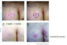
Dr. Gilbert and Dr. Ward also published a case report in which abobotulinumtoxinA was used off label to treat a recalcitrant psoriatic plaque in a 75-year-old woman (J Drugs Dermatol. 2014;13[11]:1407-8).
“This patient had psoriatic plaques concentrated on her trunk, arms, buttocks, and legs. She had been using strong topical corticosteroids for quite a long time with incomplete relief. I asked her to withdraw from all steroids for 3 months and then treated one lesion.”
The treated plaque was on the patient’s buttock. Dr. Gilbert injected a total of 30 units of abobotulinumtoxinA intradermally at eight points, about 1 cm apart. Within 3 weeks, there was complete remission of that plaque, sustained for 7 months. During this time, new lesions formed on other areas of her body. At 8 months, the treated plaque returned in the same place.
“Some of my patients had been completely recalcitrant to other therapies, and, following off label injection with neuromodulators, they have had life-changing results. In my experience, the key to consistently successful treatment is using adequate doses of toxin.”
This practice is supported by case reports in 2012 and 2015 (J Drugs Dermatol. 2012;11[12]:e76-e79; Dermatology 2015;230:299-301). Some investigators seem to think that, along with the anti-inflammatory and neurotransmitter effects, the toxin alters vascular tone.
Dr. Gilbert acknowledged that these treatments are expensive and cannot, in the case of psoriasis, be used in disseminated disease. However, she said that, for many patients, the relief is so profound and the benefit so long-lasting, that the expense is worth it. An argument in favor of this approach is that, where effective, BoNT-A could be used as a steroid-sparing agent and one that might reduce the need for systemic therapies.
“I will tell you that, sometimes, we get only partial relief and still need adjunctive therapies. Ultimately, neuromodulators may be especially useful for psoriatic plaques that are of cosmetic concern, such as those in the scalp or on the face. Limitations to their use include cost, the need for further studies, and safety concerns, such as muscle weakness.”
Dr. Gilbert had no relevant financial disclosures.
[email protected]
On Twitter @alz_gal
EXPERT ANALYSIS FROM AAD 17
For girls with Turner syndrome, experimental fertility preservation may offer the hope of a baby of their own
ORLANDO – Fertility preservation techniques pioneered in young cancer patients may someday allow some women with Turner syndrome to give birth to their own children, without relying on donated eggs.
Spontaneous conception and live birth are exceedingly rare among women with the genetic disorder. Until very recently, adoption was the only practical way for most to grow a family. In the last decade, however, fertility specialists in Europe and the United States have had good success with in vitro fertilization using donated eggs. Now, those clinicians are aiming for a higher goal: babies born from a patient’s own eggs, cryopreserved either individually or within whole ovarian tissue.
These are not pipe dreams, according to experts interviewed for this story. Autologous oocyte freezing is well established in healthy women and is now coming of age in cancer patients, with recent reports of live births. Ovarian tissue freezing and reimplantation is a much newer technique, also pioneered in cancer patients. To date, more than 70 live births have occurred from ovarian cortical tissue conservation and later transplantation in adults. Last December brought the first report of a live birth to a childhood cancer survivor who had prepubertal ovarian tissue frozen before chemotherapy. And a 2015 report detailed the case of a girl with primary ovarian failure secondary to sickle cell anemia treatment. At 25, she gave birth to a healthy child conceived from ovarian tissue removed when she was 14 years old.
Not all clinicians so enthusiastically embrace this future, however. A new set of consensus guidelines for the management of girls and women with Turner syndrome is in the works and will recommend a more conservative clinical approach, according to Nelly Mauras, MD, chief of endocrinology, diabetes, and metabolism at the Nemours Children’s Health System in Jacksonville, Fla.
A group of academic and patient advocacy stakeholders is cooperatively honing the document based on a meeting last summer in Cincinnati. These groups include the European Society of Endocrinology, the Endocrine Society, the U.S. Pediatric Endocrine Society, the European Society for Pediatric Endocrinology, Cardiology, and Reproductive Endocrinology, as well as Turner syndrome patient advocacy groups.
Dr. Mauras said the guideline will be “less discouraging” than the existing one issued in 2012 by the American Society of Reproductive Medicine. In its 2012 guidelines, the society identified Turner syndrome as a relative contraindication to pregnancy and an absolute contraindication in those with documented cardiac anomalies. However, greater experience has since been accrued in reproductive techniques of oocyte donation in Turner, with better outcomes.
The upcoming guidelines, however, will still recommend strongly against ovarian stimulation for fertility preservation for girls younger than 12, Dr. Mauras said, and will not recommend ovarian tissue conservation. “We just do not have the safety data and pregnancy outcomes that we need to give strong recommendations for these treatments,” she said. “These are still considered experimental treatments for girls with Turner syndrome.”
Turner syndrome, caused by a deletion of one X chromosome, throws a unique curve into the game – very early ovarian failure. Those with a complete deletion (45,X) begin losing their primordial ovarian follicles even before birth. Most will never experience spontaneous puberty; even if they do, their egg reserve is virtually gone soon after. Ovarian reserve may not be completely lost in girls with mosaicism, however, who have the X deletion in only a portion of their cells (46,XX/45,X). Some will enter puberty, and about 5% may even conceive spontaneously in younger years. But of the majority of Turner girls eventually experience complete ovarian failure.
This means that fertility preservation can’t be a wait-and-see issue, according to Kutluk Oktay, MD, PhD, a fertility specialist on the leading edge of this issue in the United States.
“We have to be proactive,” said Dr. Oktay, professor of obstetrics and gynecology at New York Medical College, Valhalla. “If we wait until girls are 12 or 13 to address this, a majority will have totally depleted their ovarian reserve by then. They will have no option other than an egg donor or adoption. We are suggesting that they should be screened as soon as they are diagnosed, and if they and their parents wish it, something should be done before it’s too late.”
Dr. Oktay is also the founder of fertilitypreservation.org, which specializes in advanced fertility treatments for cancer patients. He is one of a handful of physicians in the United States who advocate early oocyte harvesting in peripubertal girls and ovarian tissue harvesting in prepubertal girls with Turner syndrome. Last year, in conjunction with the Turner Syndrome Foundation, he and his colleagues published a set of guidelines for preserving fertility in these patients.
The paper recommends fertility assessment pathways for pre- and postpubertal girls. For both groups, Dr. Oktay employs serial assessments of ovarian reserve by monitoring several hormones, including follicle-stimulating hormone, luteinizing hormone, and antimüllerian hormone (AMH). Produced by primordial follicles, AMH declines as egg reserve declines over a lifetime, and is considered an accurate marker of ovarian reserve. When a girl experiences two consecutive AMH declines, egg depletion is probably accelerating. “This is the time to consider fertility preservation,” he said.
If a girl is peri- or postpubertal, the choice would probably be ovarian stimulation with the goal of retrieving mature oocytes. For prepubertal girls, the best choice is probably ovarian tissue cryopreservation. But because the egg reserve may already be spotty inside the ovary, he recommended freezing it en bloc, rather than preserving just cortical sections.
Because these techniques are only beginning to be used in young Turner patients, neither has been tested yet to see if it would result in a pregnancy. However, Dr. Oktay said, more than 80 babies have been born to women with other disorders who had ovaries frozen as adults. And European women with Turner syndrome have been successfully bearing children with donated oocytes for years.
“I don’t differentiate Europe from the U.S.,” he said. “We have no reason to believe Turner syndrome girls would be any different here than they are there.”
Pregnancy rates by egg donation in Turner syndrome patients are about half that typically seen in an otherwise healthy infertile woman, according to numerous sources; with a take-home baby rate of about 18%. There are numerous reasons for this. The miscarriage rate in Turner patients is about 44%. Women with Turner tend to have smaller uteri, with thinner endometrium, mostly because of the lack of estrogen.
But with careful management, those who do conceive can safely deliver a healthy baby, said Outi Hovatta, MD, a Finnish fertility specialist who has done extensive work in the area. “In Europe, we have been doing this quite liberally for years, and haven’t had a bad experience,” she said in an interview.
A 2013 review summarized both the success and the risks of these pregnancies. It examined obstetric and neonatal outcomes among 106 women with Turner syndrome who gave birth via egg donation from 1992 to 2011 in Sweden, Finland, and Denmark.
Most (70%) had a single embryo transferred, as virtually all guidelines recommend. Women with Turner are prone to cardiac and aortic defects that can worsen under the strain of pregnancy, or even present for the first time during gestation. Aortic dissection is a real threat; up to a third of patients with Turner experience it during adulthood, and it’s a major cause of death among them. In the Nordic series, 10 women (9%) had a known cardiac anomaly.
The multiple birth rate was 7%. More than a third (35%) developed a hypertensive disorder, including preeclampsia (20%). Four women had potentially life-threatening complications, including one with aortic dissection, one who developed mild regurgitation of the tricuspid and mitral valve, one with a mechanical heart valve who developed HELLP syndrome (hemolysis, elevated liver enzymes, low platelets), and one who underwent a postpartum hysterectomy because of severe hemorrhaging.
The infants, nevertheless, did well. The preterm birth rate was 8%, with 9% of the singleton infants having a low birth weight. About 4% had a major birth defect. Of the 131 born, three died (2.3%), including a set of extremely preterm twins.
Close follow-up and cross-specialty cooperation are what make these positive outcomes possible, said Dr. Hovatta, who is now a professor emeritus at the Karolinska Institute, Sweden.
“We do everything we can to exclude things that could cause bad outcomes.” That includes extremely rigorous cardiac testing before pregnancy and continuous monitoring during it. “If a woman has any sign of cardiac anomaly, she is advised not to become pregnant. If she shows any signs of aortic dilation, we follow her extremely carefully with experienced cardiologists.”
Like Dr. Oktay, Dr. Hovatta and her European colleagues make fertility preservation an early topic of conversation. Unlike in the United States, where many girls aren’t diagnosed with the disorder until they fail to enter puberty, almost all Turner girls in Europe are identified very early in childhood. They receive early growth hormone treatment, and there is frequent consultation with interdisciplinary specialists. Fertility is spoken of early and often.
Early oocyte retrieval is common, Dr. Hovatta said. “Yes, it’s possible to wait until puberty, but for so many girls, most of the eggs have disappeared by then, so we typically don’t wait. We start looking at that option around 11 years, which is the same time we think about cryopreserving ovarian tissue.”
However, she added, as in the United States, the outcomes of these procedures are still unknown. But the existing data in other populations, the ability to carefully shepherd women through a successful pregnancy, and the willingness of families to provide the option all support further exploring them. Dr. Hovatta was at the Cincinnati gathering last summer and said she did not agree with the conservative tone she heard. Dr. Oktay also does not agree.
“This evidence we have so far is good evidence,” he said. “Look, where we are right now with Turner girls is where we were 15 years ago with cancer patients. People thought, ‘They have cancer. They should just be worrying about surviving cancer, not about their fertility.’ Now fertility counseling is a very important part of cancer care. We have all these tools available to us for cancer patients who don’t want to lose their fertility. This accumulated experience that has already been applied in other medical conditions … why not use that for Turner syndrome? This is my point: With all the data out there about the potential benefits and the ways to manage the risks, we shouldn’t have to tell girls, ‘Well, you have to become menopausal, and maybe you can adopt someday.’ That doesn’t sit well with parents any more. We want these girls to thrive. Not just to survive, but to have as close to a normal quality of life as possible.”
WHO’s malaria pilot vaccine: No silver bullet, but a potential strike at malaria’s heart
EXPERT ANALYSIS FROM ECCMID 2017
VIENNA – The first malaria vaccine to enter a national pilot project is not a silver bullet against the disease that kills half a million every year, but it still might be powerful enough to significantly reduce global disease burden, and even impact transmission, according to infectious disease specialist Nick Beeching, MD.
The vaccine, RTS,S (Mosquirix; GlaxoSmithKline), will be tested in three African countries beginning next year, the World Health Organization announced on April 25. The pilot programs will target 720,000 children aged 5-17 months in high-risk areas of the three countries.
Even though it’s the first malaria vaccine to pass its pivotal phase III trial, RTS,S isn’t terribly effective by any standards, said Dr. Beeching of the Royal Liverpool (England) University.
April 25, 2017, is World Malaria Day, and Anthony S. Fauci, MD, and B. Fenton Hall, MD, PhD, of the U.S. National Institute of Allergy and Infectious Diseases, said in a statement, “Safe and effective vaccines are critical tools for future efforts to control, eliminate, and, ultimately, eradicate malaria. NIAID is supporting the development of numerous malaria vaccine candidates, 10 of which are in clinical trials. In 2015, an estimated 212 million new malaria cases and 429,000 deaths occurred. Nearly 90% of these cases were among children under the age of 5 years in Africa, where malaria claims the life of a child every 2 minutes.”
GSK has been working on this vaccine since 1985, according to the company’s RTS,S literature. It is a recombinant protein that targets the circumsporozoite protein of the Plasmodium falciparum parasite at an early stage, before it enters the liver and begins to embed in erythrocytes. The aim, Dr. Beeching said, was to develop an antigen that would mobilize the immune system from the moment a mosquito injected the sporozoites through a bite, “well before they have a chance to hide in the liver.”
The 2- and 3-year follow-up results of the phase III trial, conducted in 15,500 children, were published in the Lancet in 2015. RTS,S was administered as a three-dose series, plus a booster dose, beginning at 5 months of age. The primary immunizations were given with a minimum 4-week interval between doses, with the booster administered 18 months after the last dose.
The primary series reduced clinical cases by 26%. With the booster dose, cases were reduced by 39% overall. The vaccine averted 1,774 episodes of clinical malaria per 1,000 vaccinated children, and 983 cases per 1,000 vaccinated infants. But vaccine efficacy waned over time, disappearing completely in children who got only the three-dose series. The booster dose improved response stability somewhat; during the 12 months after the fourth dose, vaccine efficacy was about 25%.
Based on these results, GSK received approval from the European Medicines Agency in 2015, and the WHO recommended a large-scale implementation of the vaccine be carried out last year. GSK will provide the vaccine at no cost, and each country’s government will decide which regions to include in the pilot study.
This real-world use will put RTS,S to the ultimate test, Dr. Beeching said: “There is always the practical problem of how do you get four doses of vaccine into people. It’s easy to do in a clinical trial, but the operations and the logistics of getting it right on the ground are what really matter. We don’t know how good less than four doses would be, and we still don’t know how long the protective effect of the full series plus booster will last. I think there’s concern that it might wane with time.”
Still, he said, even a 39% reduction in disease burden is worth aggressively pursuing, not only because of the thousands of children’s lives that could be saved, but because unvaccinated children and adults could potentially be protected as well: “We could see a knock-on effect. By reducing the burden of malaria in children, it may also reduce transmission to other people who haven’t been vaccinated.”
The vaccine certainly won’t eradicate malaria, Dr. Beeching said. It needs to be viewed as an addition to WHO’s core vector control strategy, which includes insecticide-impregnated bed nets and mosquito eradication programs.
Cost is an unresolved issue. According to the Malaria Vaccine Initiative, which is partnering with GSK to launch RTS,S, the company won’t charge for the vaccine in the pilot project, and is committed to making sure the children who need it get it.
“In many African countries, childhood vaccines are provided at no cost to children or their families, thanks to existing international and national financing mechanisms,” the company said in a press release. “The RTS,S partnership anticipates that similar mechanisms would be implemented for a malaria vaccine. A shared goal is to have the cost of a malaria vaccine not be a barrier to access.
“GSK has previously stated that the price of RTS,S will cover the cost of manufacturing the vaccine together with a small return of around 5%, which will be reinvested in research and development for next-generation malaria vaccines or vaccines against other neglected tropical diseases.”
Finally, Dr. Beeching said, there’s no way to know to know how long any malaria vaccine would retain its effectiveness.
“Making a malaria vaccine has been a dream for years, and a tough one. The antigens change according to the stage of the parasite, and there is always continuous genetic variation. So there is a possibility of escape from vaccine coverage. These are very clever parasites,” he said.
Dr. Beeching has no financial interest in the vaccine.
[email protected]
On Twitter @Alz_gal
After the epidemic, Ebola’s destructive power still haunts survivors
VIENNA – The Ebola crisis may be over in Sierra Leone, but the suffering is not.
The last patient from the epidemic was discharged in February 2016, but 78% of survivors now appear to have one or more sequelae of the infection. Some problems are mild, but some are so debilitating that life may never be the same.
Janet Scott, MD, of the University of Liverpool (England), heads a task force studying Ebola’s lingering aftereffects. These fall into four categories, Dr. Scott said at the European Society of Clinical Microbiology and Infectious Diseases annual congress: musculoskeletal pain, headache, eye problems, and psychological disorders.
They add up to an enormous risk of disability – survivors are more than 200 times more likely than controls to express at least moderate disability.
Dr. Scott and her team of researchers are partnering with clinicians and data managers in the Ebola treatment unit in the 34th Regimental Military Hospital (MH34) in Freetown, Sierra Leone. In Sierra Leone alone, she said, there were nearly 9,000 cases and 3,500 deaths from the virus; about 5,000 patients survived. So far, Dr. Scott and her team have collected data on about 500 patients for whom they also provide free health care.
The project has six arms, each headed by an expert: clinical care, data collection, disability, neurology, ophthalmology, and psychiatry.
The team sees patients in a large tent sectioned by a plywood wall*. Wireless Internet access, which she said is “enormously expensive” in Sierra Leone, has been donated by Omline Business Communications*. It’s the team’s lifeline, allowing them to transmit data between Freetown and participating units around the world. Members also Skype regularly, talking with patients and with each other.
All patients who come into the clinic have an initial visit that includes collection of demographics, their Ebola and clinical history (including an exploration of comorbidities), a maternal health screening for women, vital signs and symptom assessment, medication dispensing, and a treatment plan.
Then they visit the specialists, either onsite or through local referral*. These specialist modules include joints, eyes, headache, ears, neurology, cardiac, respiratory, gastrointestinal, renal and urologic, reproductive health for both genders, and psychiatry.
Last year, Dr. Scott published initial data on 44 patients. At ECCMID 2017, she expanded that report to include 203 survivors. They spanned all ages, but about 67% were in their most productive adult years, aged 20-39 years.
Her findings are striking: About 78% report musculoskeletal pain, with many saying they have trouble walking even short distances, climbing stairs, or picking up their children.
Headache was the next most common problem, reported by nearly 40%. About 15% report ocular problems, which include anterior uveitis, cataracts – even in very young children – and retinal lesions. Abdominal and chest pain affect about 10% of the survivors.
Although she didn’t present specific numbers, Dr. Scott also said that many of the survivors experience psychological sequelae, including insomnia, anxiety, and depression. Whether this is related to viral pathology isn’t clear; it could be a not-unexpected response to the trauma of living through the epidemic.
“Many of these people have lost their entire family, and those that are left now shun them,” she said in a live video interview on Facebook. “It’s almost like a post-traumatic stress reaction.”
The other symptoms probably are related to the disease pathology, she observed. “Unfortunately, we don’t have all the clinical details of the acute phase for everyone, but for those for whom we do have details, we are seeing correlation between some of the problems with viral loads at admission, and even episodes of becoming unconscious during the acute illness.”
Patrick Howlett, MD, of the King’s Sierra Leone Partnership, Freetown, leads the neurology study. So far, the researchers have collected data on 19 patients with severe neurological consequences. Of those, 12 (63%) experienced a period of unconsciousness during their acute Ebola episode. In a comparator group of 21 with nonsevere neurologic sequelae, 33% had experienced unconsciousness.
Headache was present in nine (47%) of the patients. Migraine was the most common diagnosis. “We don’t have money for migraine medications, but fortunately, most of our migraine patients seem to be doing well on beta blockers,” Dr. Scott said.
CT scans were performed on 17 patients: three showed cerebral or cerebellar atrophy and two had confirmed stroke.
The brain injuries were severe in two, including a 42-year-old with extensive gliosis in the left middle cerebral artery region and a dilated left ventricle secondary to loss of volume in that hemisphere. A 12-year-old girl showed extensive parietal and temporal lobe atrophy. She is now so disabled that her family can’t care for her at home.
Other neurological problems include peripheral neuropathy, brachial plexus neuropathy, and asymmetric lower limb muscular atrophy.
Paul Steptoe, MD, an ophthalmic registrar from St. Paul’s Eye Unit at the Royal Liverpool Hospital, heads the eye study. He has observed dense cataracts, even in children, and anterior uveitis that has blinded some patients. There is concern about live virus persisting in vitreal fluid, but two eye taps have been negative, Dr. Scott said.
The most exciting recent finding, however, was made possible by the donation of a digital retinal camera, which “enabled us to get dozens of amazing images,” Dr. Scott said. With it, Dr. Steptoe conducted a case-control study of 81 Ebola survivors and 106 community controls. The findings of this study are potentially very, very important, Dr. Scott said.
“The first thing we found out is that retinal scarring is pervasive in our control patients,” she said. “There is just a lot of it out here in the community. But more interesting is that Dr. Steptoe seems to have identified a characteristic retinal lesion seen only in our survivors. It could be evidence of neurotropic aspects of the Ebola virus.”
The lesions occurred in 12 (15%) of the survivors and none of the controls. They are of a striking and consistent shape: straight-edged and sharply angulated. The lesions are only on the surface of the retina and do not penetrate into deeper levels. Nor do they interfere with vision. Dr. Steptoe has proposed that they take their angular shape from the retina’s underlying structures. His paper documenting this finding has been accepted and will be published shortly in the journal Emerging Infectious Diseases.
All of the post-Ebola sequelae add up to general disability for survivors, Dr. Scott said. Soushieta Jagadesh, of the* Liverpool School of Tropical Medicine, is conducting a disability survey. The comparison between 27 survivors and 54 community controls employed the Washington Group extended disability questionnaire. “We noted major limitations 1 year after discharge in mobility, vision, cognition, and affect,” Dr. Scott said.
The hazard ratios for these issues are enormous: Overall, compared with controls, survivors were 23 times more likely to have some level of disability. They were 94 times more likely to have walking limitations and 65 times more likely to have problems with stairs. Survivors were over 200 times more likely to have moderate disability than were their unaffected neighbors.
If funding for the project is renewed – and Dr. Scott admitted this is an “if,” not a “when” – caring for and studying these survivors will continue. Just in this one city, she said, the need is huge.
According to data from the U.S. Centers for Disease Control and Prevention, more than 17,000 patients in Sierra Leone, Liberia, and Guinea survived the 2014 Ebola outbreak.
If the assessments of Freetown survivors hold true across this population, thousands of survivors face life-limiting sequelae of the disease.
“We still have patients walk in every day with musculoskeletal pain, headaches, and ocular issues,” Dr. Scott said. “At the beginning of the epidemic, we were just focusing on containing it and reducing transmission. Now, we are faced with the long-term consequences.”
The Wellcome Trust supported the study. The authors have been awarded a grant from the Enhancing Research Activity in Epidemic Situations (ERAES) program, funded by the Wellcome Trust to support further research into the sequelae of Ebola virus disease.
The video associated with this article is no longer available on this site. Please view all of our videos on the MDedge YouTube channel
[email protected]
On Twitter @Alz_gal
*This story was updated May 2, 2017.
AT ECCMID 2017
