User login
Pediatric type 2 diabetes guidelines stress metformin
New clinical guidelines on type 2 diabetes mellitus in children and adolescents from the American Academy of Pediatrics advise physicians to start metformin in newly diagnosed patients with moderate hyperglycemia, while also promoting diet and exercise changes.
The guidelines are addressed to the primary care physician, as children and adolescents with diabetes are most likely to be treated in primary care and not by an endocrinologist, according to the authors of the guidelines, led by pediatric endocrinologist Dr. Kenneth C. Copeland, of the University of Oklahoma (Pediatrics 2013;131:364-82,e648-e664).
The guidelines note that the sharp rise in incidence of type 2 diabetes among children and adolescents in recent decades has occurred alongside a severe shortage of pediatric endocrinologists in the United States. The scarcity of specialists, added to the fact that children with type 2 diabetes are disproportionately ethnic minorities living in poverty, means that access to pediatric endocrinologists will be "difficult or, in some cases, impossible." The guidelines, then, are intended to help primary care physicians who find themselves "unequipped to treat adult diseases encountered in children," according to the authors of the technical report that accompanies the guidelines, led by epidemiologist and neonatologist Dr. Shelley C. Springer of Big Lake, Minn. (Pediatrics 2013 Jan. 28 [doi: 10.1542/peds.2012-3496]).
An earlier guideline on type 2 diabetes in children and adolescents, issued by the International Society for Pediatric and Adolescent Diabetes (Pediatr. Diabetes 2009;10:17-32), had recommended initiation of metformin for youth with moderate hyperglycemia only after diet and exercise had failed.
The new American Academy of Pediatrics (AAP) guidelines recommend starting metformin as first-line therapy for children and adolescents aged 10-18 years with HbA1c of 6.5% or higher, citing observational and randomized controlled trial evidence that metformin is more effective than lifestyle interventions alone. Metformin should be started at 500 mg daily and increased by 500 mg every 1-2 weeks, up to 2,000 mg daily.
The guidelines note that while metformin is the only antidiabetic agent besides insulin that is currently approved for children, it may be of more limited benefit than previously believed. "Since the completion of these guidelines, results of the TODAY trial (N. Engl. J. Med. 2012;366:2247-56) have become available and reveal that metformin alone is inadequate in effecting sustained glycemic control in the majority of youth with diabetes," the authors wrote, noting that other agents are likely to become "reasonable options for initial pharmacologic management" of children and adolescents with type 2 diabetes in the near future.
"The writing group for these guidelines continues to recommend metformin as first-line therapy in this age group but with close monitoring for glycemic deterioration and the early addition of insulin or another pharmacologic agent if needed," Dr. Copeland and his associates wrote.
The guidelines recommend that clinicians incorporate the Academy of Nutrition and Dietetics’ pediatric weight management guidelines in nutrition counseling at diagnosis and during ongoing management. Clinicians also should prescribe 1 hour a day of moderate to vigorous exercise, and the limiting of nonacademic "screen time," or time before computers, televisions, gaming devices, and the like, to less than 2 hours daily.
HbA1c concentrations should be measured every 3 months, and treatment intensified if targets are not being met.
Physicians should initiate treatment with insulin for children and adolescents who are ketotic or present with diabetic ketoacidosis; children in whom the distinction between type 1 diabetes and type 2 diabetes is unclear; and those whose HbA1c is greater than 9% or plasma glucose concentration is 250 mg/dL or higher.
Up to a quarter of adolescents with type 2 diabetes will present with ketoacidosis, the guidelines’ authors noted, and these patients should immediately be referred to an inpatient setting for treatment with insulin and fluid replacement under the care of a physician experienced in treating this complication.
Finger-stick monitoring should be used to measure blood glucose concentrations in patients who are taking insulin or other medications and have a risk of hypoglycemia; who are initiating or changing their diabetes treatment regimens; who have not met treatment goals; or who have intercurrent illnesses, according to the guidelines.
The AAP guidelines were produced by systematic literature review, with 58 studies included, and with the support of the American Diabetes Association, the Pediatric Endocrine Society, the American Academy of Family Physicians, and the Academy of Nutrition and Dietetics.
The guidelines’ lead author, Dr. Copeland, disclosed financial relationships with Novo Nordisk, Genentech, and Endo. Coauthor Dr. Janet Silverstein disclosed support from Pfizer, Novo Nordisk, Lilly, and other companies, and coauthor Dr. Kelly Roberta Moore disclosed a relationship with the Merck Company Foundation.
Type 2 diabetes is a multidimensional disorder that requires a multidimensional management approach. The new AAP guidelines on type 2 diabetes in children and adolescents codify the current practice of nearly all pediatric endocrinologists, which is to start metformin in addition to initiating critically important nutrition and exercise changes.
However, as the TODAY study has shown [N. Engl. J. Med.2012;366:2247-56], metformin is less effective in children than adults. Metabolically, the young patient with type 2 diabetes is the toughest to treat.
Dr. Jay Cohen |
Endocrinologists need an enhanced relationship with the Food and Drug Administration and pharmaceutical companies to design clinical trials (some are already in progress) with medications currently approved for use in adults: dipeptidyl peptidase-4 inhibitor (DPP-4)-based therapies and glucagon-like peptide-1 receptor agonist (GLP-1)-based therapies. These two medication classes offer significant potential benefits to the adolescent and pediatric population, and desperately need to be looked at so that they may at some point become standard of care with or without metformin.
Many endocrinologists in the United States are already using these medicines for challenging adolescent and pediatric type 2 diabetes. The next challenge is in getting safety data for this population so that the drugs can become available to real-world patients and their parents. Understanding the family dynamics of each patient and communicating with families in a culturally sensitive manner, along with utilizing social, educational, and even faith-based resources, are all critical elements required for long-term success and prevention of premature complications.
Jay Cohen, M.D., is medical director of The Endocrine Clinic, Memphis, Tenn. Dr. Cohen has received research funding and/or speaking fees from Bristol-Myers Squibb, Novo Nordisk, Mannkind, Eli Lilly, and Sanofi-Aventis.
New clinical guidelines on type 2 diabetes mellitus in children and adolescents from the American Academy of Pediatrics advise physicians to start metformin in newly diagnosed patients with moderate hyperglycemia, while also promoting diet and exercise changes.
The guidelines are addressed to the primary care physician, as children and adolescents with diabetes are most likely to be treated in primary care and not by an endocrinologist, according to the authors of the guidelines, led by pediatric endocrinologist Dr. Kenneth C. Copeland, of the University of Oklahoma (Pediatrics 2013;131:364-82,e648-e664).
The guidelines note that the sharp rise in incidence of type 2 diabetes among children and adolescents in recent decades has occurred alongside a severe shortage of pediatric endocrinologists in the United States. The scarcity of specialists, added to the fact that children with type 2 diabetes are disproportionately ethnic minorities living in poverty, means that access to pediatric endocrinologists will be "difficult or, in some cases, impossible." The guidelines, then, are intended to help primary care physicians who find themselves "unequipped to treat adult diseases encountered in children," according to the authors of the technical report that accompanies the guidelines, led by epidemiologist and neonatologist Dr. Shelley C. Springer of Big Lake, Minn. (Pediatrics 2013 Jan. 28 [doi: 10.1542/peds.2012-3496]).
An earlier guideline on type 2 diabetes in children and adolescents, issued by the International Society for Pediatric and Adolescent Diabetes (Pediatr. Diabetes 2009;10:17-32), had recommended initiation of metformin for youth with moderate hyperglycemia only after diet and exercise had failed.
The new American Academy of Pediatrics (AAP) guidelines recommend starting metformin as first-line therapy for children and adolescents aged 10-18 years with HbA1c of 6.5% or higher, citing observational and randomized controlled trial evidence that metformin is more effective than lifestyle interventions alone. Metformin should be started at 500 mg daily and increased by 500 mg every 1-2 weeks, up to 2,000 mg daily.
The guidelines note that while metformin is the only antidiabetic agent besides insulin that is currently approved for children, it may be of more limited benefit than previously believed. "Since the completion of these guidelines, results of the TODAY trial (N. Engl. J. Med. 2012;366:2247-56) have become available and reveal that metformin alone is inadequate in effecting sustained glycemic control in the majority of youth with diabetes," the authors wrote, noting that other agents are likely to become "reasonable options for initial pharmacologic management" of children and adolescents with type 2 diabetes in the near future.
"The writing group for these guidelines continues to recommend metformin as first-line therapy in this age group but with close monitoring for glycemic deterioration and the early addition of insulin or another pharmacologic agent if needed," Dr. Copeland and his associates wrote.
The guidelines recommend that clinicians incorporate the Academy of Nutrition and Dietetics’ pediatric weight management guidelines in nutrition counseling at diagnosis and during ongoing management. Clinicians also should prescribe 1 hour a day of moderate to vigorous exercise, and the limiting of nonacademic "screen time," or time before computers, televisions, gaming devices, and the like, to less than 2 hours daily.
HbA1c concentrations should be measured every 3 months, and treatment intensified if targets are not being met.
Physicians should initiate treatment with insulin for children and adolescents who are ketotic or present with diabetic ketoacidosis; children in whom the distinction between type 1 diabetes and type 2 diabetes is unclear; and those whose HbA1c is greater than 9% or plasma glucose concentration is 250 mg/dL or higher.
Up to a quarter of adolescents with type 2 diabetes will present with ketoacidosis, the guidelines’ authors noted, and these patients should immediately be referred to an inpatient setting for treatment with insulin and fluid replacement under the care of a physician experienced in treating this complication.
Finger-stick monitoring should be used to measure blood glucose concentrations in patients who are taking insulin or other medications and have a risk of hypoglycemia; who are initiating or changing their diabetes treatment regimens; who have not met treatment goals; or who have intercurrent illnesses, according to the guidelines.
The AAP guidelines were produced by systematic literature review, with 58 studies included, and with the support of the American Diabetes Association, the Pediatric Endocrine Society, the American Academy of Family Physicians, and the Academy of Nutrition and Dietetics.
The guidelines’ lead author, Dr. Copeland, disclosed financial relationships with Novo Nordisk, Genentech, and Endo. Coauthor Dr. Janet Silverstein disclosed support from Pfizer, Novo Nordisk, Lilly, and other companies, and coauthor Dr. Kelly Roberta Moore disclosed a relationship with the Merck Company Foundation.
Lymph node count may not be best predictor of colon cancer survival
Lymph node counts of 12 or higher, widely considered a key marker of surgical quality in colon cancer resection, had no significant effect on 5-year survival rates in settings where surgeons are audited and credentialed and where surgical techniques are standardized.
Lymph node (LN) count is one of several measures used to determine the extent of surgical resection and an indicator of clear surgical margins, which have been assumed to result in better survival outcomes for colon cancer patients. A new data analysis, however, of the COST (Clinical Outcomes of Surgical Therapy) trial suggests reevaluating the use of surgical surrogates such as 12 LNs and margins.
The COST trial, a large, multicenter randomized trial of colon cancer procedures, compared outcomes for laparoscopic and open techniques in treating colon adenocarcinoma. The trial collected data on a number of surgical variables, including tumor location and LN count. A total of 787 patients were included: 267 with stage I disease, 284 with stage II, and 236 with stage III. Their median age was 70 years, and 50% were male.
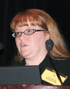
In the current study, Dr. Kellie L. Mathis of the Mayo Clinic in Rochester, Minn., and her colleagues found that 5-year overall and disease-free survival were not influenced by LN count of above or below 12 (Ann. Surg. 2012;257:102-7 [doi:10.1097/SLA.0b013e318260a8e6]). When they adjusted for age and cancer stage, LN count was seen as not predictive of overall or disease-free survival (P = .60).
Other surgical surrogates, including total bowel length, margins, or mesenteric length, likewise did not have a significant effect on survival (P greater than .05 for all), nor did tumor location (right, left, or sigmoid), surgical technique (laparoscopic or open), and sex. Only patient age and cancer stage were found to be predictive of survival.
"On the basis of abundant literature and the acceptance of the 12 LN count as a surgical quality surrogate by National Quality Forum, most would expect the 12 LN count or other surgical variables to be predictive of survival," Dr. Mathis and her associates wrote. However, they hypothesized that procedural standardization, monitoring, and credentialing may provide a better strategy for quality control.
Overall 5-year survival results from the COST trial, they noted, were 77.2% – better than national rates for comparable patient groups in the same time period. All enrolling surgeons underwent pretrial credentialing and had performed a minimum of 20 laparoscopic colon resections, for which they had submitted operative and pathology reports. All laparoscopic resections were video recorded, and videos were randomly audited by an external review committee.
If the observations in the current study can be validated by others, Dr. Mathis and her colleagues said, "we submit that now is the time to invest in the development of technical quality control programs that directly measure and monitor surgical procedures."
Dr. Mathis and her colleagues stated that they had no conflicts of interest related to their findings.
FROM ANNALS OF SURGERY
Major Finding: Lymph node count of 12 or higher was not associated with better 5-year survival rates in patients undergoing surgery to treat colon cancer.
Data Source: A secondary analysis of data from the COST trial comparing laparoscopic vs. open colectomy in 787patients with stages I-III colon cancer.
Disclosures: Dr. Mathis and colleagues stated that they had no conflicts of interest related to their findings.
Adhesiolysis: An underestimated morbidity risk
Adhesiolysis, or the removal of adhesions immediately after abdominal surgery, is a risk factor for postoperative surgical complications, longer hospital stays, readmissions, and increased costs, according to findings from a large prospective cohort study.
And yet, to date, "adhesiolysis at repeat surgery has received less attention than bowel obstruction and infertility in reports assessing the clinical and socioeconomic burden of postoperative adhesions," wrote Dr. Richard P. G. ten Broek of Radboud University Nijmegen (the Netherlands) Medical Center and his associates.
He and his colleagues evaluated 755 consecutive elective abdominal procedures at the medical center between June 2008 and June 2010, of which adhesiolysis was deemed necessary and performed in 475. In both groups, most procedures were open rather than laparoscopic. Detailed data on adhesiolysis were gathered with direct observation of the procedures by an unaffiliated observer (Ann. Surg. 2012 Sept. 25 [doi: 10.1097/SLA.0b013e31826f4969]).
Primary outcomes were the incidence of adhesions; adhesiolysis time; and the incidence of bowel defects, seromuscular injury, injuries to other organs and structures, and major surgery-related complications.
In the adhesiolysis group, 111 (23.4%) of procedures had one or more major complications, compared with 50 (17.6%) in the nonadhesiolysis group (P = .047).
Adhesiolysis was associated with a significantly higher risk of sepsis (odds ratio, 5.12; 95% confidence interval, 1.06-24.71), intra-abdominal complications (OR, 3.46; 95% CI, 1.49-8.05), and wound infections (OR, 2.45; 95% CI, 1.01-5.94).
Operative time was a mean 20 minutes longer for the adhesiolysis group, and mean inpatient costs were $18,579 per operation, compared with $14,063 in the nonadhesiolysis group (P less than .001). Readmission within 30 days of discharge was also higher in the adhesiolysis group.
Full-thickness bowel defects, either in the form of inadvertent enterotomy or delayed diagnosed perforation, were seen in 10.5% of procedures with adhesiolysis (and in 40% of operations in which adhesiolysis lasted more than 1 hour). By contrast, no bowel defects were reported for the 280 procedures in which adhesiolysis was not performed.
Bowel defects were associated with an increase in in-hospital mortality, from 1.6% among patients without defects to 8%, along with significantly more surgical interventions and longer hospital stays. Patients with bowel defects incurred mean inpatient costs of $43,784.
The study demonstrated the substantial clinical and socioeconomic burden of adhesiolysis, "particularly when a bowel defect occurs. All physicians treating patients with disorders of the abdominal cavity that might require surgery should be aware of the adverse effects of adhesiolysis," Dr. ten Broek and his colleagues wrote.
To date, they said, few clinicians appear to be taking the risks of adhesiolysis seriously. "Underestimation of the related morbidity and the passiveness of many physicians, who consider adhesiolysis an annoying but unavoidable part of redo surgery, account for the paucity of reports on the consequences of adhesiolysis," they wrote.
The investigators noted as weaknesses of their study the need for adhesiolysis in 60% of procedures in the cohort and the low number of laparoscopies – which, they said, could limit the generalizability of the study results.
The study was sponsored by Radboud University Nijmegen Medical Center. Dr. ten Broek and his associates stated that they had no conflicts of interest.
FROM ANNALS OF SURGERY
Major finding: Adhesiolysis was required in 62.9% of patients undergoing elective abdominal operations, and bowel defects were incurred in 10.5% of the adhesiolysis patients.
Data source: A prospective cohort study of 755 directly observed procedures in 715 patients at a surgical center in the Netherlands.
Disclosures: The study was sponsored by Radboud University Nijmegen Medical Center. Dr ten Broek and his colleagues stated that they had no conflicts of interest.
U.S. surgeon supply projected to drop 18% by 2028
The supply of surgeons in the United States will decrease by nearly one-fifth by 2028, likely resulting in shortages in all but a handful of specialties, a study has shown.
Currently proposed changes to increase surgeon training will not be enough to offset the number of surgeons who will retire, predicted Erin P. Fraher, Ph.D., of the department of surgery at the University of North Carolina at Chapel Hill, and colleagues. If this trend continues, the overall supply of full-time-equivalent surgeons will have decreased 18% between 2009 and 2028, with declines in all specialties except colorectal, pediatric, neurologic, and vascular surgery.
Such a drop could result in a workforce insufficient to meet the needs of the U.S. population – particularly in light of the expanded access to and increased usage of health care services projected for the future. It could also exacerbate problems related to the geographic distribution of surgeons, the investigators cautioned, "leading to delayed or lost access to time-sensitive surgical procedures, particularly in rural areas" (Ann. Surg. 2012 [doi:10.1097/SLA.0b013e31826fccfa]).
These projections are lower than those forecast in a 2008 report by the U.S. Health Resources and Services Administration for general, cardiothoracic, orthopedic, urologic, plastic, ophthalmologic, and obstetrics/gynecologic surgeons. However, they are brighter than previously published projections for vascular and pediatric surgeons (J. Vasc. Surg. 2009;50:946-52; J. Pediatr. Surg. 2009;44:1677-82). Predictions of a decline in cardiothoracic surgeons are consistent with some earlier findings (Circulation 2009;120:488-94).
For the current research, the investigators used a stock-and-flow model, which calculates current numbers of physicians, projected numbers of physicians graduating from medical school, and physicians reentering the workforce, and subtracts from these anticipated deaths, retirements, and career breaks. They then adjusted the resulting head count to full-time-equivalent work participation rates of surgeons by age, sex, and specialty.
Data were drawn from databases maintained by the American Medical Association, the American Board of Medical Specialties, the National Resident Match Program, the San Francisco match for plastic surgery and ophthalmology, the American Urological Association, the Health Resources and Services Administration, and the North Carolina Health Professions Data System.
Unlike static projection models, this model allows for real-time updating to take into account changes in data and policy decisions, which enhances the accuracy of its workforce projections, according to the investigators.
Surgeons’ participation in patient care declines somewhat after age 60, they noted, making age a key factor in determining full-time equivalents for each specialty. Another factor considered in modeling was the "feminization" of various specialties. By 2019, Dr. Fraher and colleagues predicted, half of general surgery residents will be female, compared with 95% of obstetrics and gynecology residents and 28% of orthopedic surgery residents.
Furthermore, an estimated 25% of general surgery cases that have traditionally been performed by surgical specialists will, in the future, need to be made up by general surgeons, the investigators said.
The most important driver of future supply estimates, they said, "is whether anticipated declines in full-time equivalent rates will occur as expected and whether these full-time equivalent decreases will be offset, at least partially, by productivity gains."
Dr. Fraher and colleagues called their findings "a snapshot of trends that may or may not develop depending on whether there are changes in graduate medical education training pathways, in the length of training, and in attrition from residency programs."
Current proposals to either cut or increase graduate medical education funding under Medicare focus largely on increasing the supply of primary care physicians, but results from this model suggest that it is equally important to ensure an adequate supply of surgeons in the future, the authors said. They emphasized that current published recommendations to boost graduate medical education by the Council of Graduate Medical Education, Congress, and others, if implemented, would not avert declines in surgical workforce supply during the period forecasted.
Dr. Fraher and colleagues’ study was sponsored by the American College of Surgeons. None of its authors declared conflicts of interest.
FROM ANNALS OF SURGERY
Major Finding: The overall availability of surgeons in the United States will have decreased 18% by 2028 if current trends continue, with exceptions for some specialties.
Data Source: Databases maintained by the American Medical Association and several other organizations.
Disclosures: The study was sponsored by the American College of Surgeons. None of the authors declared conflicts of interest.
Chronic Hepatitis C: New Cause for Optimism
Chronic hepatitis C virus infection is currently responsible for more U.S. deaths than HIV; is complicated to treat; is not vaccine preventable; and is a major driver of liver transplants, cirrhosis, chronic and end-stage liver disease, and liver cancer.
Although infection rates peaked more than a decade ago, rates of complications related to hepatitis C virus (HCV), such as cirrhosis, are projected to continue rising until 2020 or later (Gastroenterology 2010;138:513-21).
Amid this bad news, however, is emerging a remarkably promising treatment picture, said Dr. Marc Ghany of the liver diseases branch at the National Institute of Diabetes and Digestive and Kidney Diseases n Bethesda, Md.
At grand rounds at the NIH Clinical Center, Dr. Ghany told clinicians that, with the current Food and Drug Administration–approved regimens, "efficacy of therapy is improving and cure is now possible." The addition of direct-acting antiviral agents to regimens previously containing only peginterferon and ribavirin has been "a game-changer," Dr. Ghany said, with some patients achieving sustained virologic response (SVR) in as few as 12 weeks.
SVR is normally defined as testing negative for HCV-RNA 24 weeks after the end of treatment, Dr. Ghany said.
With chronic HCV infection, SVR is shorthand for cure, with nearly 100% of SVR patients having undetectable HCV-RNA over long-term follow-up (Clin. Infect. Dis. 2011;52:889-900). More importantly, "SVR is associated with improved outcomes," Dr. Ghany said, citing a large NIH multicenter study in which subjects who achieving SVR had vastly lower rates of decompensated liver disease, liver-related death, and liver transplantation. Liver cancer, while also reduced, was somewhat more persistent in the SVR population, meaning that people with SVR still need to be monitored for the development of cancer.
Triple therapies using peginterferon, ribavirin, and a direct-acting antiviral agent such as boceprevir or telaprevir, have increased the SVR rate to more than 75% among treatment-naive people with HCV genotype 1, the hardest of the six major genotypes to treat and the one responsible for the lion’s share of infections in the United States. With the earlier dual-therapy regimens, less than half of patients saw SVR, according to Dr. Ghany.
He described the current optimal therapy for HCV 1 infection as peginterferon alfa-2a 180 mcg weekly or peginterferon alfa-2b 1.5 mcg/kg weekly, plus oral ribavirin 800-1,400 mg in two divided doses daily, combined with either boceprevir 800 mg three times daily or telaprevir 750 mg every 8 hours.
Simple as they may sound, "these regimens are actually quite complex," Dr. Ghany said, with pill burdens as high as 18 a day. Nor are the agents interchangeable: the boceprevir and telaprevir regimens are administered very differently and must be adjusted within specific time frames according to patient response, he said.
Moreover, he said, "the side effects are substantial." Anemia (including severe anemia), neutropenia, and dysgeusia are all significantly higher with the boceprevir triple regimen than with the dual regimens, and telaprevir is associated with severe body rashes that only resolve when treatment is stopped. Cases of drug rash with eosinophilia and systemic symptoms (DRESS) and Stevens-Johnson syndrome also have been reported with telaprevir, he said.
Both regimens come with specific stopping rules for nonresponse, futility, and resistance prevention. These must be followed to the letter, Dr. Ghany said. Neither telaprevir nor boceprevir should ever be used as monotherapy, as resistance can develop in as little as 4 days. Clinicians should carefully consider the potential for drug interactions, particularly among transplant recipients or HIV-coinfected patients.
Optimal treatment times have not been established for people with cirrhosis, and one observational study showed significantly higher adverse effect rates for people with cirrhosis being treated with these regimens than were seen in phase III trials.
Dr. Ghany said that the decision whether to treat or observe can be difficult, and clinicians must consider the likelihood of disease progression, response, contraindications, disease stage, comorbidities, and side effects when working with a patient to choose the right course. Given the toxicity of some regimens, it might do to wait with some patients until safer therapies become available, he said.
Several all-oral regimens are now being tested that could eliminate the need for interferon. An interferon-free regimen is the "holy grail" of HCV treatment, Dr. Ghany said, pointing to a recent study of 21 patients in which SVR occurred after 12 weeks in 4 of 11 (36%) HCV-1 patients treated with a combination of two direct-acting antivirals. In a second group of 10 patients receiving these plus peginterferon and ribavirin, all had an SVR at 12 weeks after stopping therapy.
Many more drug combinations are currently being studied and are showing similar rates of efficacy. Though the current studies are small, "it’s hard to argue when you get 100% response rates," Dr. Ghany said.
Although toxicity remains a looming problem – some experimental agents have already been withdrawn on toxicity concerns – Dr. Ghany said he remains optimistic that a safer yet equally or more potent combination will emerge.
Future therapies also are likely to come with shorter treatment times and to be "interferon- and perhaps even ribavirin-free," he said, suggesting it’s not inconceivable that one day soon "we will be treating HCV with a single coformulated pill, perhaps for a duration of only 12 weeks."
Dr. Ghany disclosed no conflicts of interest.
EXPERT ANALYSIS FROM GRAND ROUNDS AT THE NIH CLINICAL CENTER
Chronic hepatitis C: new cause for optimism
Chronic hepatitis C virus infection is currently responsible for more U.S. deaths than HIV; is complicated to treat; is not vaccine preventable; and is a major driver of liver transplants, cirrhosis, chronic and end-stage liver disease, and liver cancer.
Although infection rates peaked more than a decade ago, rates of complications related to hepatitis C virus (HCV), such as cirrhosis, are projected to continue rising until 2020 or later (Gastroenterology 2010;138:513-21).
Amid this bad news, however, is emerging a remarkably promising treatment picture, said Dr. Marc Ghany of the liver diseases branch at the National Institute of Diabetes and Digestive and Kidney Diseases n Bethesda, Md.
At grand rounds at the NIH Clinical Center, Dr. Ghany told clinicians that, with the current Food and Drug Administration–approved regimens, "efficacy of therapy is improving and cure is now possible." The addition of direct-acting antiviral agents to regimens previously containing only peginterferon and ribavirin has been "a game-changer," Dr. Ghany said, with some patients achieving sustained virologic response (SVR) in as few as 12 weeks.
SVR is normally defined as testing negative for HCV-RNA 24 weeks after the end of treatment, Dr. Ghany said.
With chronic HCV infection, SVR is shorthand for cure, with nearly 100% of SVR patients having undetectable HCV-RNA over long-term follow-up (Clin. Infect. Dis. 2011;52:889-900). More importantly, "SVR is associated with improved outcomes," Dr. Ghany said, citing a large NIH multicenter study in which subjects who achieving SVR had vastly lower rates of decompensated liver disease, liver-related death, and liver transplantation. Liver cancer, while also reduced, was somewhat more persistent in the SVR population, meaning that people with SVR still need to be monitored for the development of cancer.
Triple therapies using peginterferon, ribavirin, and a direct-acting antiviral agent such as boceprevir or telaprevir, have increased the SVR rate to more than 75% among treatment-naive people with HCV genotype 1, the hardest of the six major genotypes to treat and the one responsible for the lion’s share of infections in the United States. With the earlier dual-therapy regimens, less than half of patients saw SVR, according to Dr. Ghany.
He described the current optimal therapy for HCV 1 infection as peginterferon alfa-2a 180 mcg weekly or peginterferon alfa-2b 1.5 mcg/kg weekly, plus oral ribavirin 800-1,400 mg in two divided doses daily, combined with either boceprevir 800 mg three times daily or telaprevir 750 mg every 8 hours.
Simple as they may sound, "these regimens are actually quite complex," Dr. Ghany said, with pill burdens as high as 18 a day. Nor are the agents interchangeable: the boceprevir and telaprevir regimens are administered very differently and must be adjusted within specific time frames according to patient response, he said.
Moreover, he said, "the side effects are substantial." Anemia (including severe anemia), neutropenia, and dysgeusia are all significantly higher with the boceprevir triple regimen than with the dual regimens, and telaprevir is associated with severe body rashes that only resolve when treatment is stopped. Cases of drug rash with eosinophilia and systemic symptoms (DRESS) and Stevens-Johnson syndrome also have been reported with telaprevir, he said.
Both regimens come with specific stopping rules for nonresponse, futility, and resistance prevention. These must be followed to the letter, Dr. Ghany said. Neither telaprevir nor boceprevir should ever be used as monotherapy, as resistance can develop in as little as 4 days. Clinicians should carefully consider the potential for drug interactions, particularly among transplant recipients or HIV-coinfected patients.
Optimal treatment times have not been established for people with cirrhosis, and one observational study showed significantly higher adverse effect rates for people with cirrhosis being treated with these regimens than were seen in phase III trials.
Dr. Ghany said that the decision whether to treat or observe can be difficult, and clinicians must consider the likelihood of disease progression, response, contraindications, disease stage, comorbidities, and side effects when working with a patient to choose the right course. Given the toxicity of some regimens, it might do to wait with some patients until safer therapies become available, he said.
Several all-oral regimens are now being tested that could eliminate the need for interferon. An interferon-free regimen is the "holy grail" of HCV treatment, Dr. Ghany said, pointing to a recent study of 21 patients in which SVR occurred after 12 weeks in 4 of 11 (36%) HCV-1 patients treated with a combination of two direct-acting antivirals. In a second group of 10 patients receiving these plus peginterferon and ribavirin, all had an SVR at 12 weeks after stopping therapy.
Many more drug combinations are currently being studied and are showing similar rates of efficacy. Though the current studies are small, "it’s hard to argue when you get 100% response rates," Dr. Ghany said.
Although toxicity remains a looming problem – some experimental agents have already been withdrawn on toxicity concerns – Dr. Ghany said he remains optimistic that a safer yet equally or more potent combination will emerge.
Future therapies also are likely to come with shorter treatment times and to be "interferon- and perhaps even ribavirin-free," he said, suggesting it’s not inconceivable that one day soon "we will be treating HCV with a single coformulated pill, perhaps for a duration of only 12 weeks."
Dr. Ghany disclosed no conflicts of interest.
EXPERT ANALYSIS FROM GRAND ROUNDS AT THE NIH CLINICAL CENTER
Fatty Liver Disease: The Silent Epidemic
Nonalcoholic fatty liver disease is an "an underdiagnosed epidemic resulting from overnutrition," according to a leading researcher of the genetics and pathogenesis of NAFLD.
In grand rounds at the NIH Clinical Center, Dr. Yaron Rotman told clinicians that an increasing number of Americans have ultrasound evidence of NAFLD, which is defined as greater than 5.5% fat in liver that cannot be attributed to another cause, such as alcohol consumption.
A significant portion of patients with NAFLD, Dr. Rotman said, will develop nonalcoholic steatohepatitis (NASH), a more serious, related condition in which fat accumulation is accompanied by hepatic injury that can progress to liver cirrhosis. NASH can only be confirmed by biopsy: None of the noninvasive markers is reliable enough to be used in clinical practice. Moreover, there is no approved therapy for NAFLD and NASH, save for measures indicated in end-stage liver disease, said Dr. Rotman of the liver diseases branch of the National Institute of Diabetes and Digestive and Kidney Diseases (NIDDK).
"This is an epidemic with hard implications – people will die from this," he said. "We need prevention, more understanding of mechanisms, noninvasive diagnosis, and improved therapies."
The prevalence of NAFLD has increased with rising obesity rates, and is now the most common cause of elevated liver enzymes in the United States, although many NAFLD patients have normal enzymes.
Current estimates of NAFLD prevalence range from 20% to 30% in the general population and from 67% to 75% in obese individuals (Hepatology 2010;52:894-903). African Americans appear to have some protection from NAFLD and NASH compared with the population at large, likely due to genetic factors that have yet to be discovered (Hepatology 2004;40:1387-95). Children and nonobese people may also present with NAFLD and NASH, and genetic factors are suspected to play a role.
Symptoms of NAFLD and NASH can include vague right-upper quadrant abdominal discomfort and fatigue, but unless patients present with advanced liver disease, they tend to be discovered incidentally while being evaluated for something else, Dr. Rotman said in a separate interview. "The progression of disease doesn’t go hand in hand with progression of symptoms – this is not slope, it’s a cliff. Oftentimes, there are no symptoms until the liver disease is advanced."
Statins and fibrates have been found to be largely ineffective, and pioglitazone, an insulin sensitizer, has demonstrated benefit but has not led to improvement in fibrosis and carries potential side effects. Other treatments being tried include the antioxidant vitamin E, which has been shown to improve the histological features of NASH at 800 IU daily (N. Engl. J. Med. 2010;362:1675-85). Loss of 7%-10% of body weight has also been shown to procure histological improvement in patients with NASH (Hepatology 2010;51:121-9).
Dr. Rotman’s team at the NIDDK has been working to uncover the mechanisms of NAFLD and NASH, trying to unlock the mystery of why some patients will progress to NASH while others will not.
Dr. Rotman said that research is beginning to point to free fatty acids, not triglycerides, as the source of liver injury. The current theory of NASH pathogenesis, he said, is that "you have increased fatty acid delivery to the liver, some of which will be shunted to triglycerides, and the excess will spill over into toxic metabolites or reactive oxygen species, which will themselves cause liver damage. These can actually increase insulin resistance and enter into a vicious cycle."
The question of why some people appear to be protected – they have excess fat in their liver but never develop NASH – "we think is related to a strong genetic component," Dr. Rotman said.
Dr. Rotman’s team is working to find single nucleotide polymorphisms (SNPs), or genetic markers, of interest for NAFLD and NASH, looking for those with implications not just for disease but for severity of disease. In one study, 894 adults and 223 children with histologically confirmed NAFLD were genotyped for six SNPs associated with hepatic fat or liver enzymes in genomewide association studies.
In the adults, three SNPs, all on chromosome 22, showed associations with NASH. One SNP, PNPLA3, was associated with steatosis, inflammation, Mallory-Denk bodies, and fibrosis, and two others in the same region had similar associations.
In the children with NASH, there was no SNP associated with histological severity; however, the rs738409 G allele was seen associated with an earlier presentation of disease (Hepatology 2010;52:894-903).
In a separate study using the same cohort, the researchers found a different SNP, HSD17B13, associated with increased liver fat but decreased injury, inflammation, and fibrosis, as well as lower enzymes.
Dr. Rotman and his colleagues are also currently conducting a small study seeking the optimal dose of the antioxidant vitamin E in patients with NASH.
Gene studies, so far, account for only a fraction of fat in the liver, he noted. "They are polygenic disorders affected by many genes, with each gene making a small contribution," he said in an interview. "The bottom line is that as we learn more, we’ll find more of these genetic contributions – these are genes we don’t know anything about, and they’ll teach us much about normal physiology as well."
For clinicians treating patients with NAFLD, the question of whether and when to biopsy can be difficult, Dr. Rotman continued, not least because management strategy is largely the same whether or not there is histological evidence of NASH.
"In the research setting, biopsy is recommended because it’s the only way you can learn about the disease," he said. "In the clinical setting, the predominant reason I’ll send a patient to an invasive procedure is if it’s going to change management. Since there is no approved treatment for NAFLD and almost every patient with NAFLD will have to change their lifestyle – lose weight, exercise, and eat a healthy diet – it is not necessary to biopsy routinely."
However, he said, a biopsy "assures that you are not missing a completely different disorder, and there may be some benefit to be able to stage and give a prognosis. With some patients, a scary biopsy result can help them focus on changing their lifestyle."
Dr. Rotman disclosed no conflicts of interest.
NIH Clinical Center, Dr. Yaron Rotman, fat in liver, alcohol consumption, nonalcoholic steatohepatitis, NASH, fat accumulation, hepatic injury, liver cirrhosis, National Institute of Diabetes and Digestive and Kidney Diseases, NIDDK,
Nonalcoholic fatty liver disease is an "an underdiagnosed epidemic resulting from overnutrition," according to a leading researcher of the genetics and pathogenesis of NAFLD.
In grand rounds at the NIH Clinical Center, Dr. Yaron Rotman told clinicians that an increasing number of Americans have ultrasound evidence of NAFLD, which is defined as greater than 5.5% fat in liver that cannot be attributed to another cause, such as alcohol consumption.
A significant portion of patients with NAFLD, Dr. Rotman said, will develop nonalcoholic steatohepatitis (NASH), a more serious, related condition in which fat accumulation is accompanied by hepatic injury that can progress to liver cirrhosis. NASH can only be confirmed by biopsy: None of the noninvasive markers is reliable enough to be used in clinical practice. Moreover, there is no approved therapy for NAFLD and NASH, save for measures indicated in end-stage liver disease, said Dr. Rotman of the liver diseases branch of the National Institute of Diabetes and Digestive and Kidney Diseases (NIDDK).
"This is an epidemic with hard implications – people will die from this," he said. "We need prevention, more understanding of mechanisms, noninvasive diagnosis, and improved therapies."
The prevalence of NAFLD has increased with rising obesity rates, and is now the most common cause of elevated liver enzymes in the United States, although many NAFLD patients have normal enzymes.
Current estimates of NAFLD prevalence range from 20% to 30% in the general population and from 67% to 75% in obese individuals (Hepatology 2010;52:894-903). African Americans appear to have some protection from NAFLD and NASH compared with the population at large, likely due to genetic factors that have yet to be discovered (Hepatology 2004;40:1387-95). Children and nonobese people may also present with NAFLD and NASH, and genetic factors are suspected to play a role.
Symptoms of NAFLD and NASH can include vague right-upper quadrant abdominal discomfort and fatigue, but unless patients present with advanced liver disease, they tend to be discovered incidentally while being evaluated for something else, Dr. Rotman said in a separate interview. "The progression of disease doesn’t go hand in hand with progression of symptoms – this is not slope, it’s a cliff. Oftentimes, there are no symptoms until the liver disease is advanced."
Statins and fibrates have been found to be largely ineffective, and pioglitazone, an insulin sensitizer, has demonstrated benefit but has not led to improvement in fibrosis and carries potential side effects. Other treatments being tried include the antioxidant vitamin E, which has been shown to improve the histological features of NASH at 800 IU daily (N. Engl. J. Med. 2010;362:1675-85). Loss of 7%-10% of body weight has also been shown to procure histological improvement in patients with NASH (Hepatology 2010;51:121-9).
Dr. Rotman’s team at the NIDDK has been working to uncover the mechanisms of NAFLD and NASH, trying to unlock the mystery of why some patients will progress to NASH while others will not.
Dr. Rotman said that research is beginning to point to free fatty acids, not triglycerides, as the source of liver injury. The current theory of NASH pathogenesis, he said, is that "you have increased fatty acid delivery to the liver, some of which will be shunted to triglycerides, and the excess will spill over into toxic metabolites or reactive oxygen species, which will themselves cause liver damage. These can actually increase insulin resistance and enter into a vicious cycle."
The question of why some people appear to be protected – they have excess fat in their liver but never develop NASH – "we think is related to a strong genetic component," Dr. Rotman said.
Dr. Rotman’s team is working to find single nucleotide polymorphisms (SNPs), or genetic markers, of interest for NAFLD and NASH, looking for those with implications not just for disease but for severity of disease. In one study, 894 adults and 223 children with histologically confirmed NAFLD were genotyped for six SNPs associated with hepatic fat or liver enzymes in genomewide association studies.
In the adults, three SNPs, all on chromosome 22, showed associations with NASH. One SNP, PNPLA3, was associated with steatosis, inflammation, Mallory-Denk bodies, and fibrosis, and two others in the same region had similar associations.
In the children with NASH, there was no SNP associated with histological severity; however, the rs738409 G allele was seen associated with an earlier presentation of disease (Hepatology 2010;52:894-903).
In a separate study using the same cohort, the researchers found a different SNP, HSD17B13, associated with increased liver fat but decreased injury, inflammation, and fibrosis, as well as lower enzymes.
Dr. Rotman and his colleagues are also currently conducting a small study seeking the optimal dose of the antioxidant vitamin E in patients with NASH.
Gene studies, so far, account for only a fraction of fat in the liver, he noted. "They are polygenic disorders affected by many genes, with each gene making a small contribution," he said in an interview. "The bottom line is that as we learn more, we’ll find more of these genetic contributions – these are genes we don’t know anything about, and they’ll teach us much about normal physiology as well."
For clinicians treating patients with NAFLD, the question of whether and when to biopsy can be difficult, Dr. Rotman continued, not least because management strategy is largely the same whether or not there is histological evidence of NASH.
"In the research setting, biopsy is recommended because it’s the only way you can learn about the disease," he said. "In the clinical setting, the predominant reason I’ll send a patient to an invasive procedure is if it’s going to change management. Since there is no approved treatment for NAFLD and almost every patient with NAFLD will have to change their lifestyle – lose weight, exercise, and eat a healthy diet – it is not necessary to biopsy routinely."
However, he said, a biopsy "assures that you are not missing a completely different disorder, and there may be some benefit to be able to stage and give a prognosis. With some patients, a scary biopsy result can help them focus on changing their lifestyle."
Dr. Rotman disclosed no conflicts of interest.
NIH Clinical Center, Dr. Yaron Rotman, fat in liver, alcohol consumption, nonalcoholic steatohepatitis, NASH, fat accumulation, hepatic injury, liver cirrhosis, National Institute of Diabetes and Digestive and Kidney Diseases, NIDDK,
NIH Clinical Center, Dr. Yaron Rotman, fat in liver, alcohol consumption, nonalcoholic steatohepatitis, NASH, fat accumulation, hepatic injury, liver cirrhosis, National Institute of Diabetes and Digestive and Kidney Diseases, NIDDK,
EXPERT ANALYSIS FROM GRAND ROUNDS AT THE NIH CLINICAL CENTER
FEBSTAT Study Hints at Predictors of Epilepsy
While febrile seizures are common in early childhood and generally benign, prolonged febrile seizures of 30 minutes or longer have been associated with a greatly increased risk of later epilepsy, particularly temporal lobe epilepsy.
To better understand the pathogenesis of epilepsy among children presenting with febrile status epilepticus (FSE) and to guide the development of interventions that could prevent it following a first episode of FSE, a group of clinical researchers has been working with a cohort of 199 children aged 1 month to 6 years (median age, 15.8 months) who presented with FSE between 2003 and 2010.
The study, being conducted at five clinical sites in the United States under funding from the National Institutes of Health (NIH), is designed with a considerable follow-up, with data collection planned for 1, 5, 10, and 15 years after the initial acute episode.
But even with the first-year results yet to be published, the Consequences of Prolonged Febrile Seizures in Childhood, or FEBSTAT, study has already generated some important findings.
Human herpes viruses 6 and 7 are strongly associated with FSE, the FEBSTAT team revealed this June, with HHV-6b or 7 infections occurring in 32% of 169 children in the cohort (Epilepsia 2012;53:1481-8). Whether there is a relationship between HHV infection at baseline and subsequent epilepsy will take years to establish.
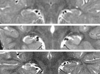
In July, the FEBSTAT investigators published results of a study showing that 11.5% of 191 children in the cohort had MRI (magnetic resonance imaging) evidence of an acute injury to one hippocampus following FSE (Neurology 2012;79:871-7), compared with none in a control group of children presenting with benign febrile seizures. A substantial number of cases also had developmental abnormalities of the hippocampus (10.5%); but some children in the control group also had abnormalities (2.1%).
In November, the researchers revealed that of acute EEG findings following FSE, 45.2% were abnormal, with the most common abnormality being focal slowing or attenuation. Epileptiform abnormalities also were seen in 6.5% of the full cohort of 199 children (Neurology 2012 Nov. 7 [doi: 10.1212/WNL.0b013e3182759766]). The EEG findings are highly associated with MRI evidence of acute hippocampal injury, and may also reveal injuries too subtle to be picked up by MRI imaging.
FEBSTAT principal investigator Dr. Shlomo Shinnar, professor of neurology, pediatrics, and epidemiology at Montefiore Medical Center and Albert Einstein College of Medicine in New York, said in an interview that MRI and EEG are used in tandem to offer the most complete picture possible.
"Sometimes the EEG will show you’re evolving into epilepsy when the MRI doesn’t; sometimes the MRI will show changes before the EEG. We want multimodal assessments, so whenever we do an MRI we do an EEG. EEG is noninvasive and minimal risk; it turns out to be quite predictive and it is very easy to get."
At this point in the FEBSTAT study, the MRI and EEG study results cannot fully clarify whether FSE results in damage to the hippocampus and later epilepsy, or whether children with hippocampal abnormalities are more likely to present with FSE. However, it is already clear that while children with hippocampal abnormalities may be more susceptible to prolonged febrile seizure, these can result in acute injury.
The researchers conduct MRIs and EEGs on all subjects at the predetermined follow-up points, which will help reveal evidence of injuries or abnormalities that may have been undetectable at baseline. At 5 years, investigators collect extensive data on cognition, memory, executive function, attention, and behavior and psychiatric morbidity, in addition to imaging. "Our hypothesis is that when 5-year follow-up data are complete, kids with smaller hippocampi will have memory impairment," said Dr. Shinnar, lead author on the MRI study and coauthor on the EEG and HHV studies.
Dr. Shinnar said that one goal of the prospective cohort study is to identify biomarkers that will be useful in indicating preventive therapies for the children most at risk. Right now, he said, prophylaxis would be limited to antiseizure drugs; however, "the hope is that if we can identify that if you have this MRI or that EEG and are determined to be at high risk, you can find some agent that you can give for a couple weeks and prevent the development of epilepsy."
"The NIH has just funded a planning grant in which they’re testing agents in animals to prevent epileptogenesis," Dr. Shinnar said. "We are going to provide them some data from FEBSTAT to assist the modeling. There is a lot of interest in developing an agent that will prevent epilepsy, but of the candidate agents, none has so far passed muster."
The HHV study offered another clue to a potential route of intervention. HHV-6b and HHV-7, both found to be common in FSE, have potential for reactivation after primary infection. Evidence of infection with HHV-6b or HHV-7 was present in 85% of the cohort, with viremia in 32%.
What the FEBSTAT team still doesn’t know, Dr. Shinnar said, "is whether HHV-6b is a common cause of high fever in young children and therefore of febrile status, but that nothing about it could cause further epilepsy down the road; or whether it is a common cause of fever but because of the properties of virus is more likely to cause you to have status. We’re adequately powered to answer these questions, but you’ll have to tune in 10 years from now." A long-term aim of the FEBSTAT study is to determine whether FSE due to HHV-6 or HHV-7 infection is more likely to result in hippocampal injury, hippocampal sclerosis, and temporal lobe epilepsy than FSE without HHV-6 or HHV-7 infection.
The long follow-up of the cohort, which is powered to withstand some attrition over time, should prove one of its greatest strengths. The first 10-year data will be collected next year, and hopefully will reveal even more about the role of hippocampal injury, hippocampal abnormality, and HHV-6b and 7 in the pathogenesis of epilepsy.
"God and NIH willing," Dr. Shinnar said, he’ll be around for the 15-year results, by which time many of his earliest recruits will be headed off to college.
Dr. Shinnar disclosed past service on an advisory board for King Pharmaceuticals, and past financial relationships with Questcor, Sunovion, Eisai, Neuronex, and UCB. Several of Dr. Shinnar’s coinvestigators on the EEG and MRI studies also disclosed past and ongoing financial relationships with pharmaceutical manufacturers.
Dr. Yu-Tze Ng comments: The FEBSTAT study team should be congratulated for working their way toward determining the acute and long-term consequences of FSE in childhood. Their preliminary results for MRI scans and EEGs possibly performed in the post-ictal period (within 72 hours of the FSE episode) suggest the evolution of mesial temporal sclerosis (MTS) in a small but significant number of affected children. To date, 17 (8.5%) children had definitely abnormal T2 signal in the hippocampus and a further 5 (2.5%) had equivocal findings. Significant focal non-epileptiform findings were seen in 60 (30.2%) acute EEGs. Perhaps not unexpectedly, epileptiform discharges were noted in only 13 (6.5%) tracings. It will be of particular interest to watch the (possibly expected) evolution of more MTS cases from this cohort of patients. However, that may be offset with expected normalization of many of the EEGs and also possible resolution of MRI T2 signal changes, some of which may simply have been (sub-)acute post-ictal findings.
To date, PubMed reveals eight major publications related to FEBSTAT.
Besides examining post-ictal MRI and EEG results, other issues being studied include the association between FSE and human herpesvirus (HHV) 6 and 7 infections, although it is a long way from being proved causative. About one-third of the children in the study had HHV infection: predominantly HHV-6b, less frequently HHV-7, and none with HHV-6a. Possibly more significant was the FEBSTAT group’s recent finding that 136 children with FSE who underwent lumbar puncture had essentially normal cerebrospinal fluid results without pleocytosis (J. Pediatr. 2012 Sept. 14 [doi:10.1016/j.jpeds.2012.08.008]). Hence, caregivers should not ignore a "positive" lumbar puncture result in a patient with FSE.
There are a few clinical and pragmatic limitations to the conclusions that can be drawn so far. The FEBSTAT study is predominantly concentrating on patients with FSE, not those with simple febrile seizures. Although febrile seizures may affect nearly 1 in 20 children, less than a third (and probably closer to only 10%-20%) have complex febrile seizures. Hence, for current and future extrapolations, the majority of practitioners will probably not be fully aware of this very significant difference in the subset group of patients who are being studied.
Although temporal lobe epilepsy (TLE) is the commonest form of adult epilepsy, and MTS is found predominantly in refractory TLE patients, only a minority of these patients report having had febrile seizures and nearly always the subtle distinction of FSE is not made.
The authors also acknowledge that it remains to be proven that some of these findings are incidental and not causative (pre-FSE MRIs were not available), because MTS in children certainly exists (J. Child Neurol. 2006;21:512-7).
We await further results from this cohort of FSE patients.
It is possible that more questions rather than answers may arise. The next major step will be to try to develop preventive interventions for febrile seizures and TLE from the information gathered.
Yu-Tze Ng, M.D., is director of epilepsy and chair of child neurology at the University of Oklahoma Health Sciences Center, Oklahoma City. He serves on the scientific advisory board for Lundbeck and receives honoraria for speaking engagements or educational activities from Lundbeck, UCB Pharma, and Cyberonics. He is not involved in the FEBSTAT study.
While febrile seizures are common in early childhood and generally benign, prolonged febrile seizures of 30 minutes or longer have been associated with a greatly increased risk of later epilepsy, particularly temporal lobe epilepsy.
To better understand the pathogenesis of epilepsy among children presenting with febrile status epilepticus (FSE) and to guide the development of interventions that could prevent it following a first episode of FSE, a group of clinical researchers has been working with a cohort of 199 children aged 1 month to 6 years (median age, 15.8 months) who presented with FSE between 2003 and 2010.
The study, being conducted at five clinical sites in the United States under funding from the National Institutes of Health (NIH), is designed with a considerable follow-up, with data collection planned for 1, 5, 10, and 15 years after the initial acute episode.
But even with the first-year results yet to be published, the Consequences of Prolonged Febrile Seizures in Childhood, or FEBSTAT, study has already generated some important findings.
Human herpes viruses 6 and 7 are strongly associated with FSE, the FEBSTAT team revealed this June, with HHV-6b or 7 infections occurring in 32% of 169 children in the cohort (Epilepsia 2012;53:1481-8). Whether there is a relationship between HHV infection at baseline and subsequent epilepsy will take years to establish.
In July, the FEBSTAT investigators published results of a study showing that 11.5% of 191 children in the cohort had MRI (magnetic resonance imaging) evidence of an acute injury to one hippocampus following FSE (Neurology 2012;79:871-7), compared with none in a control group of children presenting with benign febrile seizures. A substantial number of cases also had developmental abnormalities of the hippocampus (10.5%); but some children in the control group also had abnormalities (2.1%).
In November, the researchers revealed that of acute EEG findings following FSE, 45.2% were abnormal, with the most common abnormality being focal slowing or attenuation. Epileptiform abnormalities also were seen in 6.5% of the full cohort of 199 children (Neurology 2012 Nov. 7 [doi: 10.1212/WNL.0b013e3182759766]). The EEG findings are highly associated with MRI evidence of acute hippocampal injury, and may also reveal injuries too subtle to be picked up by MRI imaging.
FEBSTAT principal investigator Dr. Shlomo Shinnar, professor of neurology, pediatrics, and epidemiology at Montefiore Medical Center and Albert Einstein College of Medicine in New York, said in an interview that MRI and EEG are used in tandem to offer the most complete picture possible.
"Sometimes the EEG will show you’re evolving into epilepsy when the MRI doesn’t; sometimes the MRI will show changes before the EEG. We want multimodal assessments, so whenever we do an MRI we do an EEG. EEG is noninvasive and minimal risk; it turns out to be quite predictive and it is very easy to get."
At this point in the FEBSTAT study, the MRI and EEG study results cannot fully clarify whether FSE results in damage to the hippocampus and later epilepsy, or whether children with hippocampal abnormalities are more likely to present with FSE. However, it is already clear that while children with hippocampal abnormalities may be more susceptible to prolonged febrile seizure, these can result in acute injury.
The researchers conduct MRIs and EEGs on all subjects at the predetermined follow-up points, which will help reveal evidence of injuries or abnormalities that may have been undetectable at baseline. At 5 years, investigators collect extensive data on cognition, memory, executive function, attention, and behavior and psychiatric morbidity, in addition to imaging. "Our hypothesis is that when 5-year follow-up data are complete, kids with smaller hippocampi will have memory impairment," said Dr. Shinnar, lead author on the MRI study and coauthor on the EEG and HHV studies.
Dr. Shinnar said that one goal of the prospective cohort study is to identify biomarkers that will be useful in indicating preventive therapies for the children most at risk. Right now, he said, prophylaxis would be limited to antiseizure drugs; however, "the hope is that if we can identify that if you have this MRI or that EEG and are determined to be at high risk, you can find some agent that you can give for a couple weeks and prevent the development of epilepsy."
"The NIH has just funded a planning grant in which they’re testing agents in animals to prevent epileptogenesis," Dr. Shinnar said. "We are going to provide them some data from FEBSTAT to assist the modeling. There is a lot of interest in developing an agent that will prevent epilepsy, but of the candidate agents, none has so far passed muster."
The HHV study offered another clue to a potential route of intervention. HHV-6b and HHV-7, both found to be common in FSE, have potential for reactivation after primary infection. Evidence of infection with HHV-6b or HHV-7 was present in 85% of the cohort, with viremia in 32%.
What the FEBSTAT team still doesn’t know, Dr. Shinnar said, "is whether HHV-6b is a common cause of high fever in young children and therefore of febrile status, but that nothing about it could cause further epilepsy down the road; or whether it is a common cause of fever but because of the properties of virus is more likely to cause you to have status. We’re adequately powered to answer these questions, but you’ll have to tune in 10 years from now." A long-term aim of the FEBSTAT study is to determine whether FSE due to HHV-6 or HHV-7 infection is more likely to result in hippocampal injury, hippocampal sclerosis, and temporal lobe epilepsy than FSE without HHV-6 or HHV-7 infection.
The long follow-up of the cohort, which is powered to withstand some attrition over time, should prove one of its greatest strengths. The first 10-year data will be collected next year, and hopefully will reveal even more about the role of hippocampal injury, hippocampal abnormality, and HHV-6b and 7 in the pathogenesis of epilepsy.
"God and NIH willing," Dr. Shinnar said, he’ll be around for the 15-year results, by which time many of his earliest recruits will be headed off to college.
Dr. Shinnar disclosed past service on an advisory board for King Pharmaceuticals, and past financial relationships with Questcor, Sunovion, Eisai, Neuronex, and UCB. Several of Dr. Shinnar’s coinvestigators on the EEG and MRI studies also disclosed past and ongoing financial relationships with pharmaceutical manufacturers.
Perioperative Fish Oil Supplements Did Not Cut AF Risk
Perioperative supplementation with fish oil–derived fatty acids did not prevent postoperative atrial fibrillation in patients undergoing cardiac surgery, according to results from a large, randomized controlled trial.
Atrial fibrillation (AF) affects approximately one-third of patients who undergo cardiac surgery. Animal studies have suggested that the long-chain n-3-polyunsaturated fatty acids (PUFAs) found in fish oil have antiarrhythmic activity, and clinical trials have shown that habitual consumption of fish oil favorably affects physiological pathways that influence AF as well as overall cardiac mortality risk (J. Am. Coll. Cardiol. 2011;58:2047-67).
The new study, published online Nov. 5 in JAMA (doi:10.1001/jama.2012.28733) and presented simultaneously at the annual scientific sessions of the American Heart Association, demonstrated that fish oil supplementation beginning 2-5 days prior to cardiac surgery and continuing until hospital discharge did not reduce risk of postoperative atrial fibrillation.
For their research, Dr. Dariush Mozaffarian of the Harvard School of Public Health, Boston, and Dr. Roberto Marchioli of Consorzio Mario Negri Sud in Santa Maria Imbaro, Italy, and their colleagues randomized to fish oil capsules or placebo 1,516 patients who were scheduled for cardiac surgery at 28 centers in the United States, Italy, and Argentina.
Patients took 10 capsules, each 1-g capsule containing at least 840 mg of n-3-PUFAs, eicosapentaenoic acid (approximately 465 mg) plus docosahexaenoic acid (approximately 375 mg) as ethyl esters (Omacor) or matched placebo (olive oil) over 3-5 days before surgery, followed by two capsules per day until discharge or day 10 post surgery, whichever came first. The subjects’ average age was 64 years; 72% were men. Patients who took fish oil regularly were excluded from the study.
Centers were encouraged to use continuous electrocardiographic monitoring for at least 5 days post surgery. Twelve-lead ECGs were recommended daily and more frequently at the discretion of the treating physicians for symptoms or clinically suspected arrhythmia. Confirmatory rhythm strips or 12-lead ECGs were collected for all postoperative arrhythmias of at least 30 seconds’ duration.
AF episodes lasting 30 seconds or longer occurred after surgery in 233 (30.7%) controls and in 227 (30%) subjects given fish oil (odds ratio, 0.96 [95% confidence interval, 0.77-1.20]; P = .74). The two groups did not differ significantly in the number of AF episodes or the incidence of sustained AF episodes. Also, there was no significant difference in the risk of clinical bleeding, even though more than half of the study patients were also taking aspirin or other anticoagulants, the investigators reported.
Though the investigators hypothesized that they would see stronger efficacy of treatment among patients who consumed less than two servings a week of oily fish or those who had lower (<4%) plasma-PUFA levels at enrollment, no significant differences were found in those subgroups, they wrote.
Patients were identified and assigned to receive fish oil capsules for different durations (ranging from 2 to 5 days) prior to surgery, which could have resulted in the shorter durations being less effective, the investigators acknowledged. Subgroup analyses, however, did not detect significantly greater risk reduction associated with more days of fish oil before surgery.
The researchers noted that current best-practice guidelines for preventing postoperative AF were recommended to all participating surgical centers, "which could have reduced the influence of any additional therapy on risk of postoperative AF." Further, the supplement doses may have been too low to produce a benefit, even though phospholipid n-3-PUFA levels increased by an average of 40% by the time of surgery, "providing novel evidence that even short-term supplementation significantly influences circulating levels," they said.
Because of the known benefits of n-3-PUFAs on cardiovascular risk factors and physiologic pathways, "a more promising strategy may be long-term consumption to reduce the primary incidence of AF among ambulatory elderly adults with hypertension or other risk factors; such an approach should be tested in appropriately designed and powered clinical trials," the investigators wrote in their analysis.
The study was funded with grants from the National Institutes of Health, GlaxoSmithKline, Sigma Tau, and Pronova BioPharma, which also provided the capsules used in the study. None of the investigators declared conflicts of interest.
FROM JAMA
Major Finding: Atrial fibrillation lasting 30 seconds or longer occurred after cardiac surgery in 233 (30.7%) controls and 227 (30%) patients who started fish oil supplements 2-5 days before surgery (OR 0.96 [95% CI, 0.77-1.20]; P = .74).
Data Source: OPERA (Omega-3 Fatty Acids for Prevention of Post-operative Atrial Fibrillation) was a randomized, placebo-controlled trial enrolling more than 1,500 patients scheduled for cardiac surgery at 28 centers in three countries.
Disclosures: The study was funded with grants from the National Institutes of Health, GlaxoSmithKline, Sigma Tau, and Pronova BioPharma, which also provided the capsules used in the study. None of the investigators declared conflicts of interest.
Tattoo Removal: The Promise of Pico
For more than 2 decades, dermatologists have tackled tattoo removal with Q-switched lasers, which work by heating and destroying the target chromophore, allowing pigment to be released into the extracellular space.
But Q-switched laser treatments, while effective, also are time consuming and costly, with eight or more clinical visits needed, spaced a month or more apart, before treatment is complete, according to Dr. Nazanin Saedi, director of laser surgery and cosmetic dermatology at Jefferson Medical College in Philadelphia.
New methods can help optimize clinical results from Q-switched treatments, allowing for fewer visits before the removal is complete, said Dr. Saedi. Picosecond technology – or "pico," as it is commonly called for short – is also promising.
Picosecond lasers pulse at a trillionth of a second (compared with the billionth of a second pulsed by Q-switched lasers). By delivering the energy in a shorter amount of time, picosecond lasers require less energy to clear pigment, and damage to surrounding tissue may be minimized, Dr. Saedi said in an interview.
Dr. Saedi was an investigator on one recent nonrandomized clinical trial enrolling 15 patients with dark tattoos. Using a picosecond laser, the investigators produced tattoo clearance of 75% or greater in an average of 4.25 sessions, or about half the time associated with Q-switched lasers (Arch. Dermatol. 2012;148:820-3).
Further studies are now attempting to determine whether pico is "color blind," and therefore could work on different colors of tattoo ink, Dr. Saedi said. This would represent another advantage, because with conventional Q-switched lasers, wavelengths need to be changed for each targeted color.
Because the pico laser is still being studied and has not yet been approved by the Food and Drug Administration, clinicians may want to know how they can maximize use of currently approved technologies.
Dr. Saedi said that combining Q-switched lasers with a fractionated ablative or nonablative device offers some promise, citing a small, nonrandomized study that showed less blistering, shortened recovery time, and less treatment-induced hypopigmentation, compared with published results from standard treatment (Dermatol. Surg. 2011;37:97-9).
Other options for optimizing results include offering up to four consecutive treatments with Q-switched lasers, spaced 20 minutes apart. But realistically, Dr. Saedi said, "what works in a study may not be practical in the clinic."
SDEF and this news organization are owned by Frontline Medical Communications.
Dr. Saedi did not declare any financial conflicts related to her talk.
EXPERT ANALYSIS FROM SDEF LAS VEGAS DERMATOLOGY SEMINAR
