User login
Fecal Immunochemical Testing, Colonoscopy Found Similar for Detecting Advanced Cancers
Fecal immunochemical testing with a low hemoglobin threshold for colonoscopy resembled one-time, primary colonoscopy for detecting advanced neoplasias in the first-degree relatives of colorectal cancer patients, investigators reported in the November issue of Gastroenterology.
Annual fecal immunochemical testing (FIT), followed by colonoscopy if hemoglobin levels met or exceeded 10 mcg per gram of feces, detected all cases of colorectal cancer (CRC) and 61% of advanced adenomas in the study population, said Dr. Enrique Quintero and Dr. Maria Carrillo at the Universidad de la Laguna in Spain and their associates.
But one-time colonoscopy was better than FIT for detecting all neoplasms as a whole in first-degree relatives of patients with CRC, the researchers reported. Based on the findings, initial screening with FIT should be considered when access to colonoscopy is limited, especially if patients are more likely to accept FIT than colonoscopy, the investigators said (Gastroenterology [doi: 10.1053/j.gastro.2014.08.004]).
Courtesy: American Gastroenterological Association
The trial included 1,918 first-degree relatives of patients with CRC. In all, 782 relatives were randomized to one-time colonoscopy, while 784 were assigned to annual FIT for 3 years, the researchers reported. Advanced neoplasia was detected in 3.9% of the FIT group and in 5.8% of the primary colonoscopy group, the investigators said (odds ratio, 1.56; 95% confidence interval, 0.95-2.56; P = .08). Rates of detection of advanced neoplasia also were similar between the FIT and primary colonoscopy groups when participants were stratified by age, sex, age of family member with CRC, type of familial relationship, and number of relatives with CRC, the researchers reported. However, primary colonoscopy identified significantly more nonadvanced adenomas (19.8%) than did FIT (5.4%), they added (OR, 4.71; 95% CI, 3.22-6.89; P less than .001).
Participants with negative FIT results were invited to undergo colonoscopy at the end of the study, the researchers said. Follow-up colonoscopies in these relatives showed that FIT had missed 39% of advanced adenomas but no cases of CRC, they reported. To detect one case of advanced CRC, only 4 relatives in the FIT group needed to undergo colonoscopy, compared with 18 members in the primary colonoscopy group, they added. “A potential benefit of FIT over primary colonoscopy in familial CRC screening is that it may save a substantial number of unnecessary colonoscopies, thus preventing harm and lowering costs,” the investigators concluded.
Ethical concerns prevented the researchers from assessing the efficacy of FIT for more than 3 years, they said. In addition, participants knew they could opt out of their assigned screening method before providing informed consent, which could have biased rates of detection of advanced CRC, the researchers noted. However, these rates did not significantly differ between diagnostic groups, they said. The study did not look at sessile serrated or traditional serrated polyps, because the study was designed when these polyps were still considered hyperplastic and nonmalignant, the investigators noted.
Future studies should evaluate the acceptance of FIT-based screening and its effects on mortality in familial CRC, the researchers concluded.
Their study was supported by grants from Fundación Canaria para la Investigación Sanitaria, Caja de Canarias, and Departmento de Medicina Interna de la Universidad de La Laguna. They reported having no conflicts of interest.
Fecal immunochemical testing, colonoscopy found similar for detecting advanced cancers
Fecal immunochemical testing with a low hemoglobin threshold for colonoscopy resembled one-time, primary colonoscopy for detecting advanced neoplasias in the first-degree relatives of colorectal cancer patients, investigators reported in the November issue of Gastroenterology.
Annual fecal immunochemical testing (FIT), followed by colonoscopy if hemoglobin levels met or exceeded 10 mcg per gram of feces, detected all cases of colorectal cancer (CRC) and 61% of advanced adenomas in the study population, said Dr. Enrique Quintero and Dr. Maria Carrillo at the Universidad de la Laguna in Spain and their associates.
But one-time colonoscopy was better than FIT for detecting all neoplasms as a whole in first-degree relatives of patients with CRC, the researchers reported. Based on the findings, initial screening with FIT should be considered when access to colonoscopy is limited, especially if patients are more likely to accept FIT than colonoscopy, the investigators said (Gastroenterology [doi: 10.1053/j.gastro.2014.08.004]).
Courtesy: American Gastroenterological Association
The trial included 1,918 first-degree relatives of patients with CRC. In all, 782 relatives were randomized to one-time colonoscopy, while 784 were assigned to annual FIT for 3 years, the researchers reported. Advanced neoplasia was detected in 3.9% of the FIT group and in 5.8% of the primary colonoscopy group, the investigators said (odds ratio, 1.56; 95% confidence interval, 0.95-2.56; P = .08). Rates of detection of advanced neoplasia also were similar between the FIT and primary colonoscopy groups when participants were stratified by age, sex, age of family member with CRC, type of familial relationship, and number of relatives with CRC, the researchers reported. However, primary colonoscopy identified significantly more nonadvanced adenomas (19.8%) than did FIT (5.4%), they added (OR, 4.71; 95% CI, 3.22-6.89; P less than .001).
Participants with negative FIT results were invited to undergo colonoscopy at the end of the study, the researchers said. Follow-up colonoscopies in these relatives showed that FIT had missed 39% of advanced adenomas but no cases of CRC, they reported. To detect one case of advanced CRC, only 4 relatives in the FIT group needed to undergo colonoscopy, compared with 18 members in the primary colonoscopy group, they added. “A potential benefit of FIT over primary colonoscopy in familial CRC screening is that it may save a substantial number of unnecessary colonoscopies, thus preventing harm and lowering costs,” the investigators concluded.
Ethical concerns prevented the researchers from assessing the efficacy of FIT for more than 3 years, they said. In addition, participants knew they could opt out of their assigned screening method before providing informed consent, which could have biased rates of detection of advanced CRC, the researchers noted. However, these rates did not significantly differ between diagnostic groups, they said. The study did not look at sessile serrated or traditional serrated polyps, because the study was designed when these polyps were still considered hyperplastic and nonmalignant, the investigators noted.
Future studies should evaluate the acceptance of FIT-based screening and its effects on mortality in familial CRC, the researchers concluded.
Their study was supported by grants from Fundación Canaria para la Investigación Sanitaria, Caja de Canarias, and Departmento de Medicina Interna de la Universidad de La Laguna. They reported having no conflicts of interest.
Colonoscopy is the preferred screening method for first-degree relatives (FDRs) of colorectal cancer patients. But evidence supporting the use of colonoscopy in this high-risk population remains indirect, with no randomized trials showing a reduction in CRC incidence or mortality. Recently, screening with fecal immunochemical testing in average-risk populations has gained widespread adoption, mainly because of its low cost, ease of use, and moderate sensitivity and high specificity for CRC. However, despite the fact that FIT is an accepted screening strategy in the average-risk population, little is known regarding FIT’s ability to detect advanced neoplasia in FDRs of CRC patients.
Dr. Quintero and his colleagues are to be congratulated for performing a randomized trial comparing the efficacy of repeated annual FIT versus a one-time colonoscopy in detecting advanced neoplasia in FDRs of CRC patients. The results of their study clearly show that annual FIT is equally effective in detecting advanced neoplasia, compared with a one-time colonoscopy after 3 years.
However, despite the study’s statistical significance in demonstrating equivalence between the two screening modalities, there was still a marked absolute difference in detecting advanced neoplasia between the two tests. Furthermore, the usefulness of FIT screening as an alternative to colonoscopy in this high-risk population will depend on patient uptake. The current study was unable to address this issue because participants knew they could opt out of their assigned strategy and still participate in the study, which was seen in the high crossover rate from the FIT group to the colonoscopy group.
These issues aside, the Quintero study provides important information about alternative screening modalities for the detection of advanced neoplasia in FDRs of CRC patients, and paves the way for future clinical studies.
Dr. Jeffrey Lee is assistant clinical professor of medicine, division of gastroenterology, University of California, San Francisco. He has no conflicts of interest.
Fecal immunochemical testing with a low hemoglobin threshold for colonoscopy resembled one-time, primary colonoscopy for detecting advanced neoplasias in the first-degree relatives of colorectal cancer patients, investigators reported in the November issue of Gastroenterology.
Annual fecal immunochemical testing (FIT), followed by colonoscopy if hemoglobin levels met or exceeded 10 mcg per gram of feces, detected all cases of colorectal cancer (CRC) and 61% of advanced adenomas in the study population, said Dr. Enrique Quintero and Dr. Maria Carrillo at the Universidad de la Laguna in Spain and their associates.
But one-time colonoscopy was better than FIT for detecting all neoplasms as a whole in first-degree relatives of patients with CRC, the researchers reported. Based on the findings, initial screening with FIT should be considered when access to colonoscopy is limited, especially if patients are more likely to accept FIT than colonoscopy, the investigators said (Gastroenterology [doi: 10.1053/j.gastro.2014.08.004]).
Courtesy: American Gastroenterological Association
The trial included 1,918 first-degree relatives of patients with CRC. In all, 782 relatives were randomized to one-time colonoscopy, while 784 were assigned to annual FIT for 3 years, the researchers reported. Advanced neoplasia was detected in 3.9% of the FIT group and in 5.8% of the primary colonoscopy group, the investigators said (odds ratio, 1.56; 95% confidence interval, 0.95-2.56; P = .08). Rates of detection of advanced neoplasia also were similar between the FIT and primary colonoscopy groups when participants were stratified by age, sex, age of family member with CRC, type of familial relationship, and number of relatives with CRC, the researchers reported. However, primary colonoscopy identified significantly more nonadvanced adenomas (19.8%) than did FIT (5.4%), they added (OR, 4.71; 95% CI, 3.22-6.89; P less than .001).
Participants with negative FIT results were invited to undergo colonoscopy at the end of the study, the researchers said. Follow-up colonoscopies in these relatives showed that FIT had missed 39% of advanced adenomas but no cases of CRC, they reported. To detect one case of advanced CRC, only 4 relatives in the FIT group needed to undergo colonoscopy, compared with 18 members in the primary colonoscopy group, they added. “A potential benefit of FIT over primary colonoscopy in familial CRC screening is that it may save a substantial number of unnecessary colonoscopies, thus preventing harm and lowering costs,” the investigators concluded.
Ethical concerns prevented the researchers from assessing the efficacy of FIT for more than 3 years, they said. In addition, participants knew they could opt out of their assigned screening method before providing informed consent, which could have biased rates of detection of advanced CRC, the researchers noted. However, these rates did not significantly differ between diagnostic groups, they said. The study did not look at sessile serrated or traditional serrated polyps, because the study was designed when these polyps were still considered hyperplastic and nonmalignant, the investigators noted.
Future studies should evaluate the acceptance of FIT-based screening and its effects on mortality in familial CRC, the researchers concluded.
Their study was supported by grants from Fundación Canaria para la Investigación Sanitaria, Caja de Canarias, and Departmento de Medicina Interna de la Universidad de La Laguna. They reported having no conflicts of interest.
Key clinical point: Screening with fecal immunochemical testing was comparable to one-time colonoscopy for detecting advanced neoplasias in relatives of colorectal cancer patients.
Major finding: In all, 3.9% of the FIT group and 5.8% of the primary colonoscopy group had advanced neoplasia (odds ratio, 1.56; 95% confidence interval, 0.95-2.56; P = .08).
Data source: Randomized controlled study of 1,918 first-degree relatives of patients with colorectal cancer.
Disclosures: The research was supported by grants from Fundación Canaria para la Investigación Sanitaria, Caja de Canarias, and Departmento de Medicina Interna de la Universidad de La Laguna. The investigators reported having no conflicts of interest.
Hepatitis E virus mutation linked to ribavirin resistance
A mutation in the hepatitis E virus might contribute to ribavirin treatment failure in transplant recipients with chronic HEV infection, according to a small study published in the November issue of Gastroenterology.
The study is the first to find in vitro evidence for a virulence mutation in HEV, said Dr. Yannick Debing at the University of Leuven in Belgium, Dr. Annett Gisa at the Hanover Medical School in Germany, and their associates.
Courtesy: American Gastroenterological Association
The researchers studied 15 solid-organ transplant recipients who received ribavirin monotherapy for chronic HEV infection. Thirteen patients were successfully treated, but two with genotype 3c infections failed therapy. Viral RNA levels in both of these patients initially dropped and then rose, suggesting emerging resistance to ribavirin therapy, the investigators said (Gastroenterology 2014 Aug. 30 [doi: 10.1053/j.gastro.2014.08.040]).
When the researchers sequenced HEV genomes from the nonresponders, they found a G1634R mutation in HEV viral polymerase that conferred greater replication capacity compared with wild-type HEV, even after in vitro ribavirin treatment, they reported. “It may be interesting to assess the possible use of position 1634 as a prognostic marker, and accordingly to adjust the dose and duration of ribavirin therapy based on the presence of the G1634R variant,” Dr. Debing and his associates said.
The mutation consisted of a G-to-A nucleotide substitution in the C-terminal region of viral polymerase, the investigators said. By comparing HEV sequences in GenBank (an open-access collection of publicly available nucleotide sequences and their proteins), they found that G1634 was the most common amino acid sequence in genotype 3 HEV, while the K1634 sequence predominated in genotypes 1 and 4.
A subgenomic replicon of genotype 3 HEV with the 1634R mutation showed no increase in ribavirin sensitivity, compared with the wild-type replicon (50% effective concentration [EC50] values, 5.1 ± 4.1 mcM and 5.1 ± 3.7 mcM, respectively), the investigators said. But when they tested the 1634R variant in human hepatoma cells, they observed a 3.4-fold increase in luminescence signaling, compared with cells transfected with capped replicon RNA from wild-type HEV (P = .04). Greater luminescence indicated that the 1634R mutation increased viral replication, they said.
In addition, an HEV replicon with the K1634 mutation showed 2.7 times more luminescence compared with cells transfected with wild-type viral RNA (P = .07), the researchers reported.
The investigators next studied the effects of the 1634 R/K mutations on the full-length HEV genome. They introduced HEV with the two variants into human hepatoma cells, measured the amounts of viral RNA released, and found that both variants replicated at higher levels than did wild-type virus. When they treated these cells with 10- or 25-mcM ribavirin, replication levels dropped for the 1634 R/K variants and for wild-type virus, but remained at least twice as high for the variants, they said.
“The increased replication capacity of the mutant may have contributed to the persistent disease courses despite [ribavirin] treatment, although other patient- and virus-related factors could have contributed as well,” the investigators said.
The research was supported by the Research Foundation–Flanders, KU Leuven University, the Robert Koch Institute, Geconcerteerde Onderzoeksactie, the German Federal Ministry for Education and Research, and EU FP7 project SILVER. The authors reported having no conflicts of interest.
Key clinical point: A virulence mutation in the hepatitis E virus (HEV) was associated with ribavirin treatment failure.
Major finding: The mutation was associated with a 3.4-fold increase in luminescence signals, compared with wild-type HEV (P = .04), indicating greater viral replication.
Data source: Study of 15 solid-organ transplant recipients with chronic HEV infection.
Disclosures: The research was supported by the Research Foundation–Flanders, KU Leuven University, the Robert Koch Institute, Geconcerteerde Onderzoeksactie, the German Federal Ministry for Education and Research, and EU FP7 project SILVER. The authors reported having no conflicts of interest.
Topical steroid might improve mucosal integrity in eosinophilic esophagitis
Topical steroid therapy improved some indicators of mucosal integrity in patients with eosinophilic esophagitis, but proton pump inhibitor therapy did not, according to two studies reported in the November issue of Clinical Gastroenterology and Hepatology.
The first study found that topical fluticasone therapy at a dose of 880 mcg twice daily for 2 months helped correct esophageal spongiosis, or dilated intercellular space, in patients with eosinophilic esophagitis (EoE). Spongiosis scores for treated patients were significantly lower than for untreated patients (0.4 vs. 1.3; P = .016), said Dr. David Katzka at the Mayo Clinic in Rochester, Minn. and his associates (Clin. Gastroenterol. Hepatol. 2014 [doi:10.1016/j.cgh.2014.02.039]).
In the study, histologic analyses also showed that improved spongiosis scores in treated patients correlated with increased density of two tight junction proteins, filaggrin (P = .001) and zonula occludens-3 (P = .016), said the investigators. These proteins might help regulate antigenic penetration of the esophageal mucosa and also could permit migration of white blood cells, they said. “Loss of tight junction regulators and dilation of intercellular spaces appear to be involved in the pathophysiology of EoE and could be targets for treatment,” the researchers concluded. But they also noted that their study did not examine the same patients before and after steroid therapy and did not look at desmosomes, intercellular junctions that past research has suggested might be affected in EoE.
For the second study, Dr. Bram van Rhijn and his associates at the Academic Medical Center in the Netherlands compared endoscopies of 16 patients with dysphagia and suspected (unconfirmed) EoE with 11 controls, both at baseline and after 8 weeks of high-dose esomeprazole treatment. Esophageal mucosal integrity was “severely impaired” in patients with confirmed EoE and in those with proton pump inhibitor–responsive eosinophilia (PPRE), the researchers said (Clin. Gasteroenterol. Hepatol. 2014 [doi:10.1016/j.cgh.2014.02.037]).
In both forms of disease, molecules as large as 40,000 daltons were able to pass through the compromised esophageal mucosa, Dr. Bram van Rhijn and his associates reported. “This size is similar to the size of most plant and animal food allergens to which EoE patients are sensitized,” they added. Esophageal permeability might increase the rate of immune exposure to allergens, thereby mediating EoE and PPRE, they said.
On mucosal functional tests, both EoE and PPRE were associated with reduced transepithelial electrical resistance and lower electrical tissue impedance, most notably in patients with EoE (P less than .001 for both, compared with controls), the investigators reported. Proton pump inhibitor treatment partially reversed these changes in patients with PPRE but showed no effect for patients with EoE, they said. This finding suggests that acid reflux might play a role in PPRE, but not in EOE, they concluded.
Dr. Katzka and his associates disclosed no funding sources and reported having no conflicts of interest. Dr. Rhijn and his associates were supported by the Netherlands Organization for Scientific Research. Two of Dr. Rhijn’s coauthors reported financial relationships with AstraZeneca, Endostim, Medical Measurement Systems, Shire, and GlaxoSmithKline.
In the past year, the topic of mucosal integrity in eosinophilic esophagitis has garnered growing attention. Epithelial permeability defects have been described in the pathogenesis of GI disorders, including inflammatory bowel disease and celiac sprue, as well as allergic disorders such as atopic dermatitis. In EoE, both experimental as well as clinical studies have shown an eosinophil-predominant inflammatory response to specific antigens, particularly common food allergens. Increased permeability may predispose genetically susceptible individuals to swallowed allergen penetration through the esophageal epithelium. Beneath the epithelial barrier, antigens have access to antigen presenting cells, including dendritic cells, leading to both allergic sensitization and perpetuation of the TH-2 chronic inflammatory response.

|
| Dr. Ikuo Hirano |
The article by Dr. Katzka and his colleagues supports the concept of epithelial barrier defects in EoE through the demonstration of reduced immunohistochemical expression of filaggrin, zonula occludens-3, and claudin-1, important tight junction proteins. Expression was increased in EoE patients treated with topical steroids. Similarly, the study by Dr. van Rhijn and his associates identified impaired mucosal integrity in EoE by a variety of techniques that included electron microscopic demonstration of dilated intercellular spaces, electrical tissue impedance as an in vivo biomarker, and in vitro transepithelial molecular flux in an Ussing chamber. Furthermore, they found that proton pump inhibitor therapy partially restored mucosal permeability defects to a greater degree in patients with PPI-responsive esophageal eosinophilia, compared with patients with EoE. These two studies substantiate studies from the Cincinnati group that previously identified reduced mRNA expression of filaggrin in esophageal mucosal biopsies as well as reduced expression of the intercellular adhesion molecule, desmoglein 1.
In spite of these novel data, the exact role of altered esophageal epithelial permeability in the pathogenesis of EoE is yet unclear. The reversibility of the defect with medical therapy argues against defective cell junction proteins as an intrinsic abnormality. Furthermore, the location of antigen presentation in EoE may occur through other routes such as the small intestine, nasal epithelium, or skin. In the meantime, these studies provide an important advance in our understanding of EoE and open the door to novel therapeutic approaches.
Dr. Ikuo Hirano, AGAF, is professor of medicine at Northwestern University, Chicago. He reported no conflicts of interest.
In the past year, the topic of mucosal integrity in eosinophilic esophagitis has garnered growing attention. Epithelial permeability defects have been described in the pathogenesis of GI disorders, including inflammatory bowel disease and celiac sprue, as well as allergic disorders such as atopic dermatitis. In EoE, both experimental as well as clinical studies have shown an eosinophil-predominant inflammatory response to specific antigens, particularly common food allergens. Increased permeability may predispose genetically susceptible individuals to swallowed allergen penetration through the esophageal epithelium. Beneath the epithelial barrier, antigens have access to antigen presenting cells, including dendritic cells, leading to both allergic sensitization and perpetuation of the TH-2 chronic inflammatory response.
|
|
| Dr. Ikuo Hirano |
The article by Dr. Katzka and his colleagues supports the concept of epithelial barrier defects in EoE through the demonstration of reduced immunohistochemical expression of filaggrin, zonula occludens-3, and claudin-1, important tight junction proteins. Expression was increased in EoE patients treated with topical steroids. Similarly, the study by Dr. van Rhijn and his associates identified impaired mucosal integrity in EoE by a variety of techniques that included electron microscopic demonstration of dilated intercellular spaces, electrical tissue impedance as an in vivo biomarker, and in vitro transepithelial molecular flux in an Ussing chamber. Furthermore, they found that proton pump inhibitor therapy partially restored mucosal permeability defects to a greater degree in patients with PPI-responsive esophageal eosinophilia, compared with patients with EoE. These two studies substantiate studies from the Cincinnati group that previously identified reduced mRNA expression of filaggrin in esophageal mucosal biopsies as well as reduced expression of the intercellular adhesion molecule, desmoglein 1.
In spite of these novel data, the exact role of altered esophageal epithelial permeability in the pathogenesis of EoE is yet unclear. The reversibility of the defect with medical therapy argues against defective cell junction proteins as an intrinsic abnormality. Furthermore, the location of antigen presentation in EoE may occur through other routes such as the small intestine, nasal epithelium, or skin. In the meantime, these studies provide an important advance in our understanding of EoE and open the door to novel therapeutic approaches.
Dr. Ikuo Hirano, AGAF, is professor of medicine at Northwestern University, Chicago. He reported no conflicts of interest.
Topical steroid therapy improved some indicators of mucosal integrity in patients with eosinophilic esophagitis, but proton pump inhibitor therapy did not, according to two studies reported in the November issue of Clinical Gastroenterology and Hepatology.
The first study found that topical fluticasone therapy at a dose of 880 mcg twice daily for 2 months helped correct esophageal spongiosis, or dilated intercellular space, in patients with eosinophilic esophagitis (EoE). Spongiosis scores for treated patients were significantly lower than for untreated patients (0.4 vs. 1.3; P = .016), said Dr. David Katzka at the Mayo Clinic in Rochester, Minn. and his associates (Clin. Gastroenterol. Hepatol. 2014 [doi:10.1016/j.cgh.2014.02.039]).
In the study, histologic analyses also showed that improved spongiosis scores in treated patients correlated with increased density of two tight junction proteins, filaggrin (P = .001) and zonula occludens-3 (P = .016), said the investigators. These proteins might help regulate antigenic penetration of the esophageal mucosa and also could permit migration of white blood cells, they said. “Loss of tight junction regulators and dilation of intercellular spaces appear to be involved in the pathophysiology of EoE and could be targets for treatment,” the researchers concluded. But they also noted that their study did not examine the same patients before and after steroid therapy and did not look at desmosomes, intercellular junctions that past research has suggested might be affected in EoE.
For the second study, Dr. Bram van Rhijn and his associates at the Academic Medical Center in the Netherlands compared endoscopies of 16 patients with dysphagia and suspected (unconfirmed) EoE with 11 controls, both at baseline and after 8 weeks of high-dose esomeprazole treatment. Esophageal mucosal integrity was “severely impaired” in patients with confirmed EoE and in those with proton pump inhibitor–responsive eosinophilia (PPRE), the researchers said (Clin. Gasteroenterol. Hepatol. 2014 [doi:10.1016/j.cgh.2014.02.037]).
In both forms of disease, molecules as large as 40,000 daltons were able to pass through the compromised esophageal mucosa, Dr. Bram van Rhijn and his associates reported. “This size is similar to the size of most plant and animal food allergens to which EoE patients are sensitized,” they added. Esophageal permeability might increase the rate of immune exposure to allergens, thereby mediating EoE and PPRE, they said.
On mucosal functional tests, both EoE and PPRE were associated with reduced transepithelial electrical resistance and lower electrical tissue impedance, most notably in patients with EoE (P less than .001 for both, compared with controls), the investigators reported. Proton pump inhibitor treatment partially reversed these changes in patients with PPRE but showed no effect for patients with EoE, they said. This finding suggests that acid reflux might play a role in PPRE, but not in EOE, they concluded.
Dr. Katzka and his associates disclosed no funding sources and reported having no conflicts of interest. Dr. Rhijn and his associates were supported by the Netherlands Organization for Scientific Research. Two of Dr. Rhijn’s coauthors reported financial relationships with AstraZeneca, Endostim, Medical Measurement Systems, Shire, and GlaxoSmithKline.
FROM CLINICAL GASTROENTEROLOGY AND HEPATOLOGY
Key clinical point: Topical steroids seemed to improve mucosal integrity in patients with eosinophilic esophagitis, but proton pump inhibitor therapy did not.
Major finding: Mean spongiosis score was significantly lower among treated vs. untreated patients (0.4 vs. 1.3; P = .016).
Data source: Immunohistochemistry, histology, endoscopy, and mucosal functional analyses of 57 subjects in two separate studies.
Disclosures: Dr. Katzka and associates disclosed no funding sources and reported having no conflicts of interest. Dr. Rhijn and associates were supported by the Netherlands Organization for Scientific Research. Two of Dr. Rhijn’s coauthors reported financial relationships with AstraZeneca, Endostim, Medical Measurement Systems, Shire, and GlaxoSmithKline.

Thiopurine for IBD linked to almost sixfold increase in lymphoma risk
Patients with inflammatory bowel disease who took thiopurines for at least a year had an almost sixfold greater rate of lymphoma than did the general population, researchers reported in the November issue of Clinical Gastroenterology and Hepatology.
But lymphoma risk appeared to return to baseline after patients stopped taking thiopurines, suggesting that immunosuppression – not DNA damage – was the root cause of cancer in these patients, said Dr. David Kotlyar at the National Cancer Institute and his associates (Clin. Gastroenterol. Hepatol. [doi:10.1016/j.cgh.2014.05.015]). The researchers carried out a meta-analysis of 18 cohort studies of IBD that they identified by querying MEDLINE, Embase, the Cochrane databases, and other sources. In all, the studies identified 91 patients with lymphoma that occurred after a median of 18 months of exposure to azathioprine or 6-mercaptopurine (range, 1-109 months), the investigators reported.
In a subanalysis of eight population studies, patients who were currently taking thiopurines had a 5.7-fold greater incidence of lymphoma compared with the general population (95% confidence interval, 3.22-10.1), the researchers said. In contrast, patients who had never used thiopurines or who no longer used the drugs did not have an increased lymphoma risk, they said. In addition, standardized incidence ratios (SIRs) in referral center studies were as high as 9.16 (95% CI, 5.03-17.6), the investigators reported. The IBD seen in referral centers tends to be more severe than that in the population overall, which could explain the discrepancy in SIRs between referral and population studies, they said.
The study also found that men who took thiopurines had a more than threefold greater incidence of lymphoma, compared with the general population (SIR, 3.60; 95% CI, 2.68-4.83), while women had an almost twofold increase in estimated risk (SIR, 1.76; 95% CI, 1.08-2.87). Results of age stratifications varied depending on how the researchers defined age groups, but the absolute risk of lymphoma in men under 30 years of age was substantially higher than expected, they said. However, only two studies included raw data on age, and findings should be interpreted with caution, they added.
“It is important to emphasize that although the relative risks of lymphoma in active users are moderate, there remains a very low absolute risk of lymphoma for any given patient,” said Dr. Kotlyar and his associates. “For patients of all ages and genders, the risk of lymphoma needs to be weighed against the potential benefits of therapy. Further work is needed to understand how this trade-off of potential benefit and harm varies by age, particularly in the era of combination immunosuppression therapy.”
Dr. Kotlyar is funded by the National Institutes of Health. Four of the coauthors reported financial relationships with Pfizer, Centocor, Shire Pharmaceuticals Group, AstraZeneca, GlaxoSmithKline, and many others. The remaining authors reported no conflicts of interest.
Key clinical point: Thiopurine substantially increased the risk of lymphoma in patients with inflammatory bowel disease.
Major finding: Patients with IBD who took thiopurines had a 5.7-fold greater incidence of lymphoma, compared with the general population.
Data source: Review of 18 studies of lymphoma and thiopurine exposure in patients with IBD.
Disclosures: Dr. Kotlyar is funded by the National Institutes of Health. Four of the reported financial relationships with Pfizer, Centocor, Shire Pharmaceuticals Group, AstraZeneca, GlaxoSmithKline, and many others. The remaining authors reported no conflicts of interest.
Dexmethylphenidate XR-guanfacine combo deemed ‘cardiovascularly safe’
SAN DIEGO – Extended-release dexmethylphenidate combined with guanfacine caused no adverse cardiovascular effects among children with attention-deficit/hyperactivity disorder, according to results from a double-blind, randomized trial of 207 patients presented at the annual meeting of the American Academy of Child and Adolescent Psychiatry.
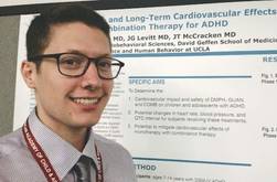
On electrocardiography, a QTc interval prolongation of more than 500 ms raises concerns about arrhythmia and sudden death, “but this study showed that this did not happen with combination treatment,” lead investigator Dr. Gregory Sayer said in an interview. “In fact, there were no clinically meaningful cardiovascular effects.
“The take-home message is that combination therapy with dexmethylphenidate extended release and guanfacine immediate release is a cardiovascularly safe option for patients who might require dual therapy.”
Stimulants for ADHD were previously thought to carry the risk of sudden cardiac death, noted Dr. Sayer, a third-year psychiatry resident at the University of California, Los Angeles. Although further analyses revealed that these concerns were unfounded, clinicians have continued to question the milder cardiovascular effects of stimulants for ADHD, as well as the effects of combining stimulants with alpha-2 agonists such as guanfacine, which can improve cognitive function in ADHD, he said.
Therefore, Dr. Sayer and his associates monitored pulse, blood pressure, and electrocardiograms in children with ADHD who were 7-14 years old and had been randomized in a double-blinded fashion to immediate-release guanfacine (1-3 mg/day), extended-release dexmethylphenidate (5-20 mg/day), or both. They measured vital signs and ECGs at baseline, at the end of the 8-week dose-optimization period, and then monthly during a 12-month, open-label maintenance phase, they reported.
Guanfacine and dexmethylphenidate had “opposing cardiovascular effects,” although none of these effects were clinically significant, the researchers said. During titration, guanfacine monotherapy lowered children’s pulse and blood pressure; dexmethylphenidate increased pulse, blood pressure, and QTc interval; and combination therapy increased diastolic blood pressure alone, they said. “The combination group’s parameters fell between the ranges for both monotherapy groups, and there were no significant QTc changes in the combination therapy group,” Dr. Sayer added.
“Combination treatment may buffer long-term cardiovascular effects of guanfacine and stimulant monotherapy, possibly reducing risk from the small but significant changes resulting from either single treatment,” he and his associates concluded.
During the yearlong maintenance phase, cardiovascular measures remained stable except for a borderline significant increase in pulse in the guanfacine group (P = .06) and a decrease in systolic blood pressure in the dexmethylphenidate group (P < .0001, the researchers reported.
The study is part of a larger trial that is examining the effects of combination therapy on ADHD symptoms and cognitive effects, such as working memory, Dr. Sayer noted. In the current study, the researchers did not continuously monitor vital signs, such as with a Holter monitor, so they could not eliminate the possibility of cardiac phenomena occurring between monitoring points, he added.
The National Institute of Mental Health funded the study. Dr. Sayer declared having no conflicts of interest. Two coauthors reported financial or advisory relationships with Akili Interactive Labs, Sunovion, and other companies.
SAN DIEGO – Extended-release dexmethylphenidate combined with guanfacine caused no adverse cardiovascular effects among children with attention-deficit/hyperactivity disorder, according to results from a double-blind, randomized trial of 207 patients presented at the annual meeting of the American Academy of Child and Adolescent Psychiatry.
On electrocardiography, a QTc interval prolongation of more than 500 ms raises concerns about arrhythmia and sudden death, “but this study showed that this did not happen with combination treatment,” lead investigator Dr. Gregory Sayer said in an interview. “In fact, there were no clinically meaningful cardiovascular effects.
“The take-home message is that combination therapy with dexmethylphenidate extended release and guanfacine immediate release is a cardiovascularly safe option for patients who might require dual therapy.”
Stimulants for ADHD were previously thought to carry the risk of sudden cardiac death, noted Dr. Sayer, a third-year psychiatry resident at the University of California, Los Angeles. Although further analyses revealed that these concerns were unfounded, clinicians have continued to question the milder cardiovascular effects of stimulants for ADHD, as well as the effects of combining stimulants with alpha-2 agonists such as guanfacine, which can improve cognitive function in ADHD, he said.
Therefore, Dr. Sayer and his associates monitored pulse, blood pressure, and electrocardiograms in children with ADHD who were 7-14 years old and had been randomized in a double-blinded fashion to immediate-release guanfacine (1-3 mg/day), extended-release dexmethylphenidate (5-20 mg/day), or both. They measured vital signs and ECGs at baseline, at the end of the 8-week dose-optimization period, and then monthly during a 12-month, open-label maintenance phase, they reported.
Guanfacine and dexmethylphenidate had “opposing cardiovascular effects,” although none of these effects were clinically significant, the researchers said. During titration, guanfacine monotherapy lowered children’s pulse and blood pressure; dexmethylphenidate increased pulse, blood pressure, and QTc interval; and combination therapy increased diastolic blood pressure alone, they said. “The combination group’s parameters fell between the ranges for both monotherapy groups, and there were no significant QTc changes in the combination therapy group,” Dr. Sayer added.
“Combination treatment may buffer long-term cardiovascular effects of guanfacine and stimulant monotherapy, possibly reducing risk from the small but significant changes resulting from either single treatment,” he and his associates concluded.
During the yearlong maintenance phase, cardiovascular measures remained stable except for a borderline significant increase in pulse in the guanfacine group (P = .06) and a decrease in systolic blood pressure in the dexmethylphenidate group (P < .0001, the researchers reported.
The study is part of a larger trial that is examining the effects of combination therapy on ADHD symptoms and cognitive effects, such as working memory, Dr. Sayer noted. In the current study, the researchers did not continuously monitor vital signs, such as with a Holter monitor, so they could not eliminate the possibility of cardiac phenomena occurring between monitoring points, he added.
The National Institute of Mental Health funded the study. Dr. Sayer declared having no conflicts of interest. Two coauthors reported financial or advisory relationships with Akili Interactive Labs, Sunovion, and other companies.
Key clinical point: Combination therapy with dexmethylphenidate extended release and guanfacine immediate release is a cardiovascularly safe option for patients who might require dual therapy.
Major finding: There were no clinically meaningful cardiovascular changes in any treatment group during the acute titration and maintenance phases.
Data source: Double-blind, randomized, parallel-group, fixed-flexible dosing study with 12-month open-label follow-up of 207 children and adolescents with ADHD.
Disclosures: The National Institute of Mental Health funded the study. Dr. Sayer declared having no conflicts of interest. Two coauthors reported financial or advisory relationships with Akili Interactive Labs, Sunovion, and other companies.
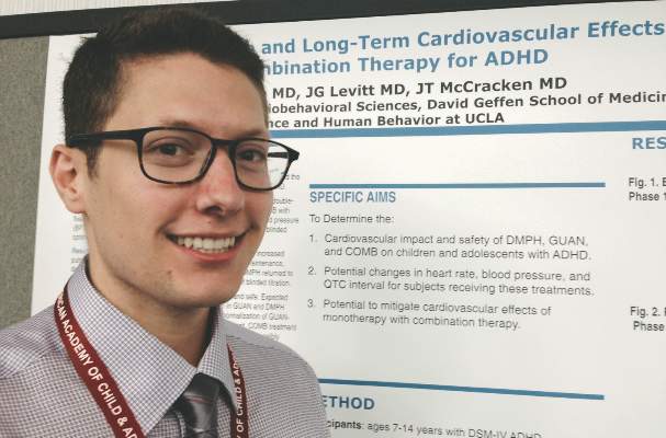
How to know when a recurrent infection signals something more
SAN DIEGO – Children who parents describe as “always sick” often are immunologically normal, but knowing when to suspect otherwise is crucial, according to Dr. Meg Fisher.
“When parents tell you their child is sick all the time, they are. They’re literally just recovering from one thing when they pick up the next,” said Dr. Fisher, a pediatric infectious diseases specialist at Monmouth Medical Center in West Orange, N.J.

Children under 2 years of age typically acquire 4-10 symptomatic respiratory infections per year – and up to 13 if they are in day care, Dr. Fisher said. These children also normally develop one to four gastrointestinal infections per year, she added. And children older than 2 years average four to eight respiratory infections and up to two gastrointestinal infections annually, she said at the annual meeting of the American Academy of Pediatrics .
Certain types of infections always merit a closer look, Dr. Fisher emphasized. Invasive infections, recurrent meningitis, excessive episodes of respiratory disease, chronic diarrhea, recurrent urinary tract infections, and recurrent skin and soft tissue infections can indicate immunodeficient disorders, inherited diseases such as cystic fibrosis, or anatomic abnormalities that predispose children to serious infections, she said. “The site of recurrent infections is really going to help you determine which subspecialist you need,” she added.
Culture results also provide clues about underlying conditions. “If you ever recover Pseudomonas aeruginosa from the respiratory tract of a child, you should automatically think about cystic fibrosis,” Dr. Fisher said. Pseudomonas is often isolated from stool samples, but is not normally found in a child’s respiratory tract, she explained.
“Unusual organisms and abnormal laboratory results should always trigger a follow-up evaluation,” Dr. Stuart Abramson said in a related presentation. The most common primary immunodeficiency disorders include selective IgA deficiency – which affects 1 in 500 children – IgG2deficiency, transient hypogammaglobulinemia of infancy, and DiGeorge anomaly, said Dr. Abramson, who is director of allergy and immunology services at Shannon Medical Center in San Angelo, Tex. Less common primary immunodeficiencies affect 1 in 50,000 to 1 in 200,000 children and include B-cell disorders, T-cell disorders, phagocytic disorders, and complement disorders, he said at the meeting.
Of the four stages of testing for primary immunodeficiencies, only the first stage needs to be done by the primary care pediatrician; testing at the first stage includes taking a history and physical examination, complete blood count and differential, and quantitative immunoglobulin levels for IgA, IgG, and IgM, said Dr. Fisher. A child who needs further testing “might be better served by an immunologist,” she added.
Pediatricians should consider first-stage testing if a patient presents with one or more warning signs of a primary immunodeficiency in children, Dr. Abramson said.
Those warning signs are:
• Four or more new ear infections within 1 year.
• Two or more serious sinus infections within 1 year.
• Two or more months on antibiotics with little effect.
• Two or more pneumonias within 1 year.
• Failure of an infant to gain weight or grow normally.
• Recurrent, deep abscesses of the skin or organs.
• Persistent oral thrush or cutaneous mycoses.
• Need for intravenous antibiotics to clear infections.
• Two or more deep-seated infections, including septicemia.
• A family history of primary immunodeficiency.
(This list was assembled by the Jeffrey Modell Foundation.)
Dr. Fisher and Dr. Abramson declared no relevant financial relationships.
SAN DIEGO – Children who parents describe as “always sick” often are immunologically normal, but knowing when to suspect otherwise is crucial, according to Dr. Meg Fisher.
“When parents tell you their child is sick all the time, they are. They’re literally just recovering from one thing when they pick up the next,” said Dr. Fisher, a pediatric infectious diseases specialist at Monmouth Medical Center in West Orange, N.J.
Children under 2 years of age typically acquire 4-10 symptomatic respiratory infections per year – and up to 13 if they are in day care, Dr. Fisher said. These children also normally develop one to four gastrointestinal infections per year, she added. And children older than 2 years average four to eight respiratory infections and up to two gastrointestinal infections annually, she said at the annual meeting of the American Academy of Pediatrics .
Certain types of infections always merit a closer look, Dr. Fisher emphasized. Invasive infections, recurrent meningitis, excessive episodes of respiratory disease, chronic diarrhea, recurrent urinary tract infections, and recurrent skin and soft tissue infections can indicate immunodeficient disorders, inherited diseases such as cystic fibrosis, or anatomic abnormalities that predispose children to serious infections, she said. “The site of recurrent infections is really going to help you determine which subspecialist you need,” she added.
Culture results also provide clues about underlying conditions. “If you ever recover Pseudomonas aeruginosa from the respiratory tract of a child, you should automatically think about cystic fibrosis,” Dr. Fisher said. Pseudomonas is often isolated from stool samples, but is not normally found in a child’s respiratory tract, she explained.
“Unusual organisms and abnormal laboratory results should always trigger a follow-up evaluation,” Dr. Stuart Abramson said in a related presentation. The most common primary immunodeficiency disorders include selective IgA deficiency – which affects 1 in 500 children – IgG2deficiency, transient hypogammaglobulinemia of infancy, and DiGeorge anomaly, said Dr. Abramson, who is director of allergy and immunology services at Shannon Medical Center in San Angelo, Tex. Less common primary immunodeficiencies affect 1 in 50,000 to 1 in 200,000 children and include B-cell disorders, T-cell disorders, phagocytic disorders, and complement disorders, he said at the meeting.
Of the four stages of testing for primary immunodeficiencies, only the first stage needs to be done by the primary care pediatrician; testing at the first stage includes taking a history and physical examination, complete blood count and differential, and quantitative immunoglobulin levels for IgA, IgG, and IgM, said Dr. Fisher. A child who needs further testing “might be better served by an immunologist,” she added.
Pediatricians should consider first-stage testing if a patient presents with one or more warning signs of a primary immunodeficiency in children, Dr. Abramson said.
Those warning signs are:
• Four or more new ear infections within 1 year.
• Two or more serious sinus infections within 1 year.
• Two or more months on antibiotics with little effect.
• Two or more pneumonias within 1 year.
• Failure of an infant to gain weight or grow normally.
• Recurrent, deep abscesses of the skin or organs.
• Persistent oral thrush or cutaneous mycoses.
• Need for intravenous antibiotics to clear infections.
• Two or more deep-seated infections, including septicemia.
• A family history of primary immunodeficiency.
(This list was assembled by the Jeffrey Modell Foundation.)
Dr. Fisher and Dr. Abramson declared no relevant financial relationships.

Screening did not increase mental health consults
SAN DIEGO – Only 1% of families that filled out psychosocial screening questionnaires during medical appointments later sought free mental health consultations, the same rate as for families that were not screened, investigators reported.
“Unless large controlled trials are able to show a process and an outcome benefit, it may be preferable to invest in providing mental health treatment” instead of screening, concluded lead investigator Brianna J. Lewis of the Mount Sinai School of Medicine, New York, and her associates. The researchers presented the findings at the annual meeting of the American Academy of Child and Adolescent Psychiatry.
The study was a post-hoc analysis of data on 3,143 patient encounters at a pediatric allergy clinic in New York City between March and September 2013. Two to five days a week, the investigators had asked children aged 8 years and older and their parents to fill out one-page questionnaires about problems such as distress, anxiety, bullying, and quality-of-life issues. They did not screen patients on the other days, “creating a naturalistic opportunity to compare between screened and nonscreened cohorts,” they added. Because screening was part of regular care, participants did not need to provide informed consent, which eliminated the possibility of selection bias, the researchers said.
In all, 6.1% of families who underwent screening were referred to a mental health consultation, but only 1% followed up, even though consults were offered for free and without third-party billing, the researchers said. The follow-up rate also was 1% for the 1,972 families that were not screened. Among the families who pursued a follow-up consult, 56% of the screened group and 67% of the unscreened group received a psychiatric diagnosis (P = 0.26), Ms. Lewis and her associates reported.
Past studies by the investigators showed that screening children and adults during medical care appointments is “hard to justify,” they noted.
The Jaffe Family Foundation, Pine/Segal Family, and Vanech Family Foundation supported the research. The investigators declared no conflicts of interest.
Key clinical point: Psychosocial screening did not increase mental health consultations.
Major finding: Only 1% of families followed up for free mental health consultations after screening – the same rate as for families that were not screened.
Data source: Post-hoc review of a 7-month screening program in a pediatric food allergy clinic.
Disclosures: The Jaffe Family Foundation, Pine/Segal Family, and Vanech Family Foundation supported the research. The investigators declared no conflicts of interest.
How and when to screen for hypertrophic cardiomyopathy
SAN DIEGO – Pediatricians should screen patients for hypertrophic cardiomyopathy if a first- or second-degree relative has been diagnosed with the condition or has a history of sudden death, said Dr. Kevin Shannon, a pediatric cardiologist at the University of California, Los Angeles, Medical Center.
“Anybody with a family history of HCM, including uncles, cousins, parents, and siblings, should be screened,” he emphasized. “Even if they don’t have obstructive HCM, they will often have a murmur, most commonly at the upper sternal border.” The murmur is typically late-peaking and worsens during Valsalva, he added.
HCM is the most common cause of sudden death in athletes and is the most common genetic cardiac disease, affecting about 1 in 500 young adults (Circulation 1995;92:785-9). The autosomal dominant inheritance pattern shows varying degrees of penetrance, translating to a range of phenotypes, Dr. Shannon said at the annual meeting of the American Academy of Pediatrics. “Even if your family member didn’t have hypertrophy until age 30, you can get it at age 5, and the opposite can also be true,” he noted.
HCM can be a silent disease until it causes sudden death. However, pediatricians should ask patients about exercise tolerance and should consider HCM in patients who describe a substantial drop in sports performance, Dr. Shannon said. An athlete with HCM might say that he or she always finished the field lap first and is now second to last, and a soccer player with a field position might say, “Now I finish the runs after the goalies,” he said.
A resting electrocardiogram is not sufficient to exclude HCM, Dr. Shannon emphasized. Because up to 10% of patients with HCM have normal ECGs, children whose parents or grandparents have HCM should have an echocardiogram every 5 years until puberty, and then every year until they turn 20 years old or finish growing, he said. But ECGs also are important because they may become abnormal before the echocardiogram does, Dr. Shannon said. If the ECG becomes abnormal, pediatricians should tell parents that the echocardiogram is likely to become abnormal and should advise them to steer the child away from competitive athletics, he said.
Twenty-four hour ambulatory (Holter) ECGs also are part of risk stratification in patients with HCM, Dr. Shannon said. The American College of Cardiology and the American Heart Association recommend Holter monitoring for the initial evaluation of patients with HCM and for patients with HCM who develop palpitations or lightheadedness (J. Am. Coll. Cardiol. 2011;58:2703-38).
Dr. Shannon reported no conflicts of interest.
Avoid misdiagnosing pediatric viral myocarditis
SAN DIEGO– Pediatricians are at risk of misdiagnosing myocarditis despite its severity. That’s because children tend to present with abdominal symptoms and a history of a recent viral illness that lacked signs of cardiac involvement, Dr. Kevin Shannon said.
“Typically, they have a bout of flu, they seem to be getting better, and then they start vomiting or having stomach pain again,” he said at the annual meeting of the American Academy of Pediatrics.
Viruses ranging from adenovirus to varicella have been implicated in myocarditis in children (J. Clin. Microbiol. 2010;48:642-5; Pediatr. Cardiol. 2011;32:1241-3). Pediatricians should watch for patients who were recently ill and are now presenting with an apparent relapse and tachycardia that is worse than how they appear overall, said Dr. Shannon, a pediatric cardiologist at the University of California, Los Angeles, Medical Center. “A lot of these children will seem more ill than their vomiting will suggest,” he added. “They’ll have a heart rate of 180 [beats per minute] that is out proportion to how they look.”
Fluid therapy does not improve tachycardia and may even worsen it, indicating that dehydration is not the underlying cause, said Dr. Shannon. Children with viral myocarditis also often have acute upper-right quadrant pain as a result of hepatic distension, he said.
Laboratory findings can be very helpful. Cardiac troponin T is almost always elevated in children with myocarditis (Pediatr. Emerg. Care 2012;28:1173-8), and erythrocyte sedimentation rate also may be high. Electrocardiography can show a variety of focal or diffuse abnormalities, none of which are pathognomonic for the condition, Dr. Shannon said. Focal abnormalities can mimic an ST segment elevation myocardial infarction (STEMI), he added.
On chest x-ray, the heart margins also are often normal because the heart has not yet enlarged to compensate for impaired function, said Dr. Shannon. “This is a poorly functioning, normal-sized heart,” he added. Chest films often will reveal interstitial edema that might be misinterpreted as interstitial pneumonia, in keeping with the child’s recent illness.
Treatment of acquired myocarditis is based on supportive care, said Dr. Shannon, adding that use of immunomodulators in children with myocarditis is controversial. “If they have low blood pressure, they need volume, even if their heart rate gets higher,” he added. “If they don’t tolerate fluid therapy, they need inotropes and sometimes intubation.”
Dr. Shannon reported no conflicts of interest.