User login
Sleep apnea may correlate with anxiety, depression in patients with PCOS
a study suggests.

This finding could have implications for screening and treatment, Diana Xiaojie Zhou, MD, said at the American Society for Reproductive Medicine’s 2020 annual meeting, held virtually this year.
“Routine OSA screening in women with PCOS should be considered in the setting of existing depression and anxiety,” said Dr. Zhou, a reproductive endocrinology and infertility fellow at the University of California, San Francisco. “Referral for OSA diagnosis and treatment in those who screen positive may have added psychological benefits in this population, as has been seen in the general population.”
Patients with PCOS experience a range of comorbidities, including higher rates of psychological disorders and OSA, she said.
OSA has been associated with depression and anxiety in the general population, and research indicates that treatment, such as with continuous positive airway pressure (CPAP), may have psychological benefits, such as reduced depression symptoms.
PCOS guidelines recommend screening for OSA to identify and alleviate symptoms such as fatigue that may to contribute to mood disorders. “However, there is a lack of studies assessing the relationship between OSA and depression and anxiety specifically in women with PCOS,” Dr. Zhou said.
A cross-sectional study
To evaluate whether OSA is associated with depression and anxiety in women with PCOS, Dr. Zhou and colleagues conducted a cross-sectional study of all women seen at a multidisciplinary PCOS clinic at university between June 2017 and June 2020.
Participants had a diagnosis of PCOS clinically confirmed by the Rotterdam criteria. Researchers determined OSA risk using the Berlin questionnaire, which is divided into three domains. A positive score in two or more domains indicates a high risk of OSA.
The investigators used the Patient Health Questionnaire-9 (PHQ-9) to assess depression symptoms, and they used the Generalized Anxiety Disorder-7 (GAD-7) to assess anxiety symptoms.
Researchers used two-sided t-test, chi-square test, and Fisher’s exact test to evaluate for differences in patient characteristics. They performed multivariate logistic regression analyses to determine the odds of moderate to severe symptoms of depression (that is, a PHQ-9 score of 10 or greater) and anxiety (a GAD-7 score of 10 or greater) among patients with a high risk of OSA, compared with patients with a low risk of OSA. They adjusted for age, body mass index, free testosterone level, and insulin resistance using the Homeostatic Model Assessment of Insulin Resistance (HOMA-IR).
The researchers examined data from 201 patients: 125 with a low risk of OSA and 76 with a high risk of OSA. The average age of the patients was 28 years.
On average, patients in the high-risk OSA group had a greater body mass index (37.9 vs. 26.5), a higher level of free testosterone (6.5 ng/dL vs. 4.5 ng/dL), and a higher HOMA-IR score (7 vs. 3.1), relative to those with a low risk of OSA. In addition, a greater percentage of patients with a high risk of OSA experienced oligomenorrhea (84.9% vs. 70.5%).
The average PHQ-9 score was significantly higher in the high-risk OSA group (12 vs. 8.3), as was the average GAD-7 score (8.9 vs. 6.1).
In univariate analyses, having a high risk of OSA increased the likelihood of moderate or severe depression or anxiety approximately threefold.
In multivariate analyses, a high risk of OSA remained significantly associated with moderate or severe depression or anxiety, with an odds ratio of about 2.5. “Of note, BMI was a statistically significant predictor in the univariate analyses, but not so in the multivariate analyses,” Dr. Zhou said.
Although the investigators assessed OSA, depression, and anxiety using validated questionnaires, a study with clinically confirmed diagnoses of those conditions would strengthen these findings, she said.
Various possible links
Investigators have proposed various links between PCOS, OSA, and depression and anxiety, Dr. Zhou noted. Features of PCOS such as insulin resistance, obesity, and hyperandrogenemia increase the risk of OSA. “The sleep loss and fragmentation and hypoxia that define OSA then serve to increase sympathetic tone and oxidative stress, which then potentially can lead to an increase in depression and anxiety,” Dr. Zhou said.
The results suggests that treating OSA “may have added psychological benefits for women with PCOS and highlights the broad health implications of this condition,” Marla Lujan, PhD, chair of the ASRM’s androgen excess special interest group, said in a society news release.
“The cause of PCOS is still not well understood, but we do know that 1 in 10 women in their childbearing years suffer from PCOS,” said Dr. Lujan, of Cornell University, Ithaca, N.Y. “In addition to infertility, PCOS is also associated with type 2 diabetes and cardiovascular complications such as hypertension and abnormal blood lipids.”
In a discussion following Dr. Zhou’s presentation, Alice D. Domar, PhD, said the study was eye opening.
Dr. Domar, director of integrative care at Boston IVF and associate professor of obstetrics, gynecology, and reproductive biology at Harvard Medical School, Boston, said that she does not typically discuss sleep apnea with patients. “For those of us who routinely work with PCOS patients, we are always looking for more information.”
Although PCOS guidelines mention screening for OSA, Dr. Zhou expects that few generalists who see PCOS patients or even subspecialists actually do.
Nevertheless, the potential for intervention is fascinating, she said. And if treating OSA also reduced a patient’s need for psychiatric medications, there could be added benefit in PCOS due to the metabolic side effects that accompany some of the drugs.
Dr. Zhou and Dr. Lujan had no relevant disclosures. Dr. Domar is a co-owner of FertiCalm, FertiStrong, and Aliz Health Apps, and a speaker for Ferring, EMD Serono, Merck, and Abbott.
SOURCE: Zhou DX et al. ASRM 2020. Abstract O-146.
a study suggests.
This finding could have implications for screening and treatment, Diana Xiaojie Zhou, MD, said at the American Society for Reproductive Medicine’s 2020 annual meeting, held virtually this year.
“Routine OSA screening in women with PCOS should be considered in the setting of existing depression and anxiety,” said Dr. Zhou, a reproductive endocrinology and infertility fellow at the University of California, San Francisco. “Referral for OSA diagnosis and treatment in those who screen positive may have added psychological benefits in this population, as has been seen in the general population.”
Patients with PCOS experience a range of comorbidities, including higher rates of psychological disorders and OSA, she said.
OSA has been associated with depression and anxiety in the general population, and research indicates that treatment, such as with continuous positive airway pressure (CPAP), may have psychological benefits, such as reduced depression symptoms.
PCOS guidelines recommend screening for OSA to identify and alleviate symptoms such as fatigue that may to contribute to mood disorders. “However, there is a lack of studies assessing the relationship between OSA and depression and anxiety specifically in women with PCOS,” Dr. Zhou said.
A cross-sectional study
To evaluate whether OSA is associated with depression and anxiety in women with PCOS, Dr. Zhou and colleagues conducted a cross-sectional study of all women seen at a multidisciplinary PCOS clinic at university between June 2017 and June 2020.
Participants had a diagnosis of PCOS clinically confirmed by the Rotterdam criteria. Researchers determined OSA risk using the Berlin questionnaire, which is divided into three domains. A positive score in two or more domains indicates a high risk of OSA.
The investigators used the Patient Health Questionnaire-9 (PHQ-9) to assess depression symptoms, and they used the Generalized Anxiety Disorder-7 (GAD-7) to assess anxiety symptoms.
Researchers used two-sided t-test, chi-square test, and Fisher’s exact test to evaluate for differences in patient characteristics. They performed multivariate logistic regression analyses to determine the odds of moderate to severe symptoms of depression (that is, a PHQ-9 score of 10 or greater) and anxiety (a GAD-7 score of 10 or greater) among patients with a high risk of OSA, compared with patients with a low risk of OSA. They adjusted for age, body mass index, free testosterone level, and insulin resistance using the Homeostatic Model Assessment of Insulin Resistance (HOMA-IR).
The researchers examined data from 201 patients: 125 with a low risk of OSA and 76 with a high risk of OSA. The average age of the patients was 28 years.
On average, patients in the high-risk OSA group had a greater body mass index (37.9 vs. 26.5), a higher level of free testosterone (6.5 ng/dL vs. 4.5 ng/dL), and a higher HOMA-IR score (7 vs. 3.1), relative to those with a low risk of OSA. In addition, a greater percentage of patients with a high risk of OSA experienced oligomenorrhea (84.9% vs. 70.5%).
The average PHQ-9 score was significantly higher in the high-risk OSA group (12 vs. 8.3), as was the average GAD-7 score (8.9 vs. 6.1).
In univariate analyses, having a high risk of OSA increased the likelihood of moderate or severe depression or anxiety approximately threefold.
In multivariate analyses, a high risk of OSA remained significantly associated with moderate or severe depression or anxiety, with an odds ratio of about 2.5. “Of note, BMI was a statistically significant predictor in the univariate analyses, but not so in the multivariate analyses,” Dr. Zhou said.
Although the investigators assessed OSA, depression, and anxiety using validated questionnaires, a study with clinically confirmed diagnoses of those conditions would strengthen these findings, she said.
Various possible links
Investigators have proposed various links between PCOS, OSA, and depression and anxiety, Dr. Zhou noted. Features of PCOS such as insulin resistance, obesity, and hyperandrogenemia increase the risk of OSA. “The sleep loss and fragmentation and hypoxia that define OSA then serve to increase sympathetic tone and oxidative stress, which then potentially can lead to an increase in depression and anxiety,” Dr. Zhou said.
The results suggests that treating OSA “may have added psychological benefits for women with PCOS and highlights the broad health implications of this condition,” Marla Lujan, PhD, chair of the ASRM’s androgen excess special interest group, said in a society news release.
“The cause of PCOS is still not well understood, but we do know that 1 in 10 women in their childbearing years suffer from PCOS,” said Dr. Lujan, of Cornell University, Ithaca, N.Y. “In addition to infertility, PCOS is also associated with type 2 diabetes and cardiovascular complications such as hypertension and abnormal blood lipids.”
In a discussion following Dr. Zhou’s presentation, Alice D. Domar, PhD, said the study was eye opening.
Dr. Domar, director of integrative care at Boston IVF and associate professor of obstetrics, gynecology, and reproductive biology at Harvard Medical School, Boston, said that she does not typically discuss sleep apnea with patients. “For those of us who routinely work with PCOS patients, we are always looking for more information.”
Although PCOS guidelines mention screening for OSA, Dr. Zhou expects that few generalists who see PCOS patients or even subspecialists actually do.
Nevertheless, the potential for intervention is fascinating, she said. And if treating OSA also reduced a patient’s need for psychiatric medications, there could be added benefit in PCOS due to the metabolic side effects that accompany some of the drugs.
Dr. Zhou and Dr. Lujan had no relevant disclosures. Dr. Domar is a co-owner of FertiCalm, FertiStrong, and Aliz Health Apps, and a speaker for Ferring, EMD Serono, Merck, and Abbott.
SOURCE: Zhou DX et al. ASRM 2020. Abstract O-146.
FROM ASRM 2020
Studies gauge toll of pausing fertility treatment during pandemic
More than 60% of patients at a center for reproductive medicine in Utah who had fertility treatments canceled because of the COVID-19 pandemic opted to resume treatment once the suspension was lifted about 7 weeks later.
At another fertility center in New York, a survey found that 96% of respondents who had a cycle canceled because of the pandemic found it upsetting, and 22% found it extremely upsetting, with extremely upsetting defined as equivalent to the loss of a child.
The indefinite time frame for resuming treatment when the New York survey was conducted may have been a major source of distress for patients, one of the researchers said at the American Society for Reproductive Medicine’s 2020 annual meeting, held virtually this year.
“They don’t know when they might have that chance again,” said Jenna M. Turocy, MD, of Columbia University Fertility Center, New York.
COVID-19 guidelines published by ASRM on March 17 recommended the suspension of new treatment cycles, including ovulation induction, intrauterine inseminations, and in vitro fertilization (IVF).
An ASRM COVID-19 task force has since supported “the measured resumption of fertility care following the easing of restrictions,” said Paul C. Lin, MD, president of the Society for Assisted Reproductive Technology and a member of the task force.
“Over the past several months, significant knowledge has been gained regarding the COVID-19 virus and its impact on patients and the medical system,” he said in a news release about the two studies that assessed the pandemic’s effects.
Certain precautions remain. “It has become clear that we will need to be practicing COVID-19 protocols at least until an effective and safe vaccine or broadly effective treatment becomes widely available,” Dr. Lin said.
Desire to proceed during a pandemic
The center continued to offer IVF cycles for oncofertility patients on an urgent basis.
In early May, patients whose cycles had been suspended had the option to receive treatment.
“Upon reopening, every patient received standardized counseling from their primary IVF physician,” Lauren Verrilli, MD, a reproductive endocrinology and infertility fellow at the University of Utah, Salt Lake City, said at the virtual meeting.
Doctors explained that much remained unknown about COVID-19 in pregnancy, and that it was unclear whether the clinic would need to shut down again. In addition, patients had to undergo COVID-19 testing.
To identify factors associated with proceeding with treatment after the suspension, the researchers compared patients who resumed treatment with patients who did not.
Their analysis included 278 patients who had planned an IVF cycle or frozen embryo transfer (FET) prior to the shutdown. The researchers examined factors such as age, parity, anti-Müllerian hormone, antral follicle count, history of prior IVF cycles or FET, number of frozen blastocysts, gamete source, and use of a gestational carrier.
In all, 62% of patients opted to receive treatment once restrictions were lifted, including 69 of the 133 (52%) patients with planned fresh cycles and 104 of the 145 (72%) patients with planned FET cycles.
Among those with planned fresh cycles, those who opted to resume treatment tended to be older than those who did not resume treatment, with a median age of 37 years versus 35 years, but the difference was not statistically significant.
Among patients with planned FET cycles, those who did not resume treatment were more likely to have a gestational carrier, compared with those who resumed treatment (7% vs. 1%). In some cases, gestational carriers lived in another state and the pandemic complicated travel arrangements, which contributed to delays, Dr. Verrilli said.
The analysis did not include information about income or socioeconomic status, which may play a role in patients’ decisions, Dr. Verrilli said.
Emotional impact of indefinite delay
Fertility treatment is often time sensitive, particularly for patients with advanced reproductive age or diminished ovarian reserve, and indefinite postponement of fertility treatment potentially could lead some patients to lose the ability to conceive with their own gametes, Dr. Turocy said.
In early April, Dr. Turocy and colleagues surveyed patients at their academic fertility center in New York City to assess patients’ reactions to the ASRM recommendations. They decided to conduct the study after they realized that treatment cancellations were having a significant emotional effect.
Investigators emailed an 18-item survey to more than 3,000 patients.
In all, 518 patients completed the survey, a response rate of 17%. Patients had an average age of 37 years (range, 23-52 years), and 92% were female. About 24% had children, and 66% had received at least one fertility treatment.
Half had a cycle canceled because of the COVID-19 pandemic, including timed intercourse cycles (5%), intrauterine insemination cycles (23%), IVF cycles with a planned fresh embryo transfer (10%), IVF with all frozen embryos (27%), egg freeze cycles (3%), and FET cycles (30%).
In response to survey questions about whether they agreed with ASRM recommendations, “the reactions were mixed,” Dr. Turocy said.
About 36% of patients agreed all fertility cycles should be canceled, 22% were unsure, and 43% disagreed with the recommendation. Patients who had a cycle canceled were slightly more likely to agree with the cancellations (40% agreed) than those who did not have a cycle canceled (30% agreed), Dr. Turocy said.
Most respondents would have preferred an option to start a treatment cycle in consultation with their doctor. Half “would have chosen to start a new cycle during the height of the pandemic in New York City,” Dr. Turocy said.
Patient opinions may vary by region and depend on the severity of COVID-19 outbreaks there, and they also might change over time, Dr. Turocy suggested. In addition, the opinions and characteristics of patients who responded to the anonymous survey may differ from those of patients who did not respond.
Dr. Verrilli, Dr. Turocy, and Dr. Lin had no relevant financial disclosures.
FROM ASRM 2020
Low threshold to biopsy atypical lesions may ID vulvar melanoma early, experts say
Having a low threshold to biopsy atypical pigmented lesions on the vulva may identify melanoma early, according to a lecture at virtual conference on diseases of the vulva and vagina, hosted by the International Society for the Study of Vulvovaginal Disease.
Pigmented brown or black vulvar lesions occur in approximately 10% of women, and they may be normal and benign.
“Often we will see a pigmented lesion on the vulva and think that there is nothing to worry about,” said Melissa Mauskar, MD.
Lesions could be angiokeratomas, petechiae, purpura, melanosis, and nevi, for example. Seborrheic keratoses can mimic melanoma. “If it looks odd, don’t be afraid to biopsy it,” said Dr. Mauskar, assistant professor of dermatology and obstetrics and gynecology at the University of Texas Southwestern Medical Center in Dallas.
Characteristics of melanoma, covered by the mnemonic ABCDE, include asymmetry, borders that are irregular, coloring that is uneven, diameter greater than 7 mm, and evolution over time.
When biopsying a lesion because of concerns about melanoma, the goal is to remove the whole lesion at once, Dr. Mauskar said.
In a recent U.S. population-based study of more than 1,800 patients with malignant melanoma of the vulva or vagina (including 1,400 patients with vulvar melanoma and 463 patients with vaginal melanoma), median disease-specific survival was 99 months for vulvar melanoma and 19 months for vaginal melanoma.
Patients with vaginal melanoma were more likely than patients with vulvar melanoma to have nodular lesions. The American Joint Committee on Cancer staging system predicts vulvar melanoma outcomes, the researchers found. In addition, lymph node status and mitotic rate were important predictors of survival.
A wide local excision is the mainstay of therapy for melanoma. Other therapeutic advances are “changing the survival curves for these patients, especially when we can find things early,” Dr. Mauskar said.
Photographing lesions can help doctors monitor them over time, she added.
It is important for dermatologists to include the vulva in skin exams and for gynecologists to have a low threshold to biopsy atypical pigmented lesions, Dr. Mauskar said. “Having a very low threshold for biopsy ... will increase our chances of finding these lesions when they are more at the superficial spreading phase as opposed to the nodular phase,” she said.
Capturing the depth of a tumor within the confines of a biopsy may help accurately stage malignant melanoma, Jason Reutter, MD, a pathologist in Hickory, N.C., said in a separate presentation. He suggested trying to get around the lesion with a punch biopsy if possible. A shave biopsy may be advantageous for larger macular lesions. To diagnose one melanoma, doctors may have to biopsy many lesions, Dr. Reutter noted.
At one institution, the number of skin biopsies needed to diagnose skin cancer ranged from 2.82 to 6.55, depending on the type of clinician, according to a recent study. The number of biopsies needed to detect one melanoma was greater – between 14 and 54 – depending on type of clinician.
For larger lesions, scouting biopsies of different areas may be the best approach, Dr. Reutter said.
Dr. Mauskar and Dr. Reutter had no relevant financial conflicts of interest.
FROM A CONFERENCE ON DISEASES OF THE VULVA AND VAGINA
Poor image quality may limit televulvology care
Seeing patients with vulvar problems via telemedicine can lead to efficient and successful care, but there are challenges and limitations with this approach, doctors are finding.
Image quality is one key factor that determines whether a clinician can assess and manage a condition remotely, said Aruna Venkatesan, MD, chief of dermatology and director of the genital dermatology clinic at Santa Clara Valley Medical Center in San Jose, Calif. Other issues may be especially relevant to televulvology, including privacy concerns.
“Who is helping with the positioning? Who is the photographer? Is the patient comfortable with having photos taken of this part of their body and submitted, even if they know it is submitted securely? Because they might not be,” Dr. Venkatesan said in a lecture at a virtual conference on diseases of the vulva and vagina, hosted by the International Society for the Study of Vulvovaginal Disease.
When quality photographs from referring providers are available, Dr. Venkatesan has conducted virtual new consultations. “But sometimes I will do a virtual telemedicine visit as the first visit and then figure out, okay, this isn’t really sufficient. I need to see them in person.”
Melissa Mauskar, MD, assistant professor of dermatology and obstetrics and gynecology at the University of Texas Southwestern Medical Center, Dallas, described a case early on during the COVID-19 pandemic that illustrates a limitation of virtual visits.
A patient sent in a photograph that appeared to show lichen sclerosus. “There looked like some classic lichen sclerosus changes,” Dr. Mauskar said during a discussion at the meeting. “But she was having a lot of pain, and after a week, her pain still was not better.”
Dr. Mauskar brought the patient into the office and ultimately diagnosed a squamous cell carcinoma. “What I thought was a normal erosion was actually an ulcerated plaque,” she said.
Like Dr. Venkatesan, Dr. Mauskar has found that image quality can be uneven. Photographs may be out of focus. Video visits have been a mixed bag. Some are successful. Other times, Dr. Mauskar has to tell the patient she needs to see her in the office.
Certain clinical scenarios require a vaginal exam, Dr. Venkatesan noted. Although some type of assessment may be possible if a patient is with a primary care provider during the telemedicine visit, the examination may not be equivalent. Doctors also should anticipate where a patient might go to have a biopsy if one is necessary.
Another telemedicine caveat pertains to patient counseling. When using store-and-forward telemedicine systems, advising patients in a written report can be challenging. “Is there an easy way ... to counsel patients how to apply their topical medications?” Dr. Venkatesan said.
Excellent care is possible
Vulvology is a small part of Dr. Venkatesan’s general dermatology practice, which has used telemedicine extensively since the pandemic.
In recent years, Dr. Venkatesan’s clinic began encouraging providers in their health system to submit photographs with referrals. “That has really paid off now because we have been able to help provide a lot of excellent quality care for patients without them having to come in,” she said. “We may be able to say: ‘These are excellent photos. We know what this patient has. We can manage it. They don’t need to come see us in person.’ ” That could be the case for certain types of acne, eczema, and psoriasis.
In other cases, they may be able to provide initial advice remotely but still want to see the patient. For a patient with severe acne, “I may be able to tell the referring doctor: ‘Please start the patient on these three medicines. It will take 2 months for those medicines to start working and then we will plan to have an in-person dermatology visit.’ ” In this case, telemedicine essentially replaces one in-person visit.
If photographs are poor, the differential diagnosis is broad, a procedure is required, the doctor needs to touch the lesion, or more involved history taking or counseling are required, the patient may need to go into the office.
Beyond its public health advantages during a pandemic, telemedicine can improve access for patients who live far away, lack transportation, or are unable to take time off from work. It also can decrease patient wait times. “Once we started doing some telemedicine work … we went from having a 5-month wait time for patients to see us in person to a 72-hour wait time for providing some care for patients if they had good photos as part of their referral,” Dr. Venkatesan said.
Telemedicine has been used in inpatient and outpatient dermatology settings. Primary care providers who consult with dermatologists using a store-and-forward telemedicine system may improve their dermatology knowledge and feel more confident in their ability to diagnose and manage dermatologic conditions, research indicates.
In obstetrics and gynecology, telemedicine may play a role in preconception, contraception, and medical abortion care, prenatal visits, well-woman exams, mental health, and pre- and postoperative counseling, a recent review suggests.
Image quality is key
“Quality of the image is so critical for being able to provide good care, especially in such a visual exam field as dermatology,” Dr. Venkatesan said.
To that end, doctors have offered recommendations on how to photograph skin conditions. A guide shared by the mobile telehealth system company ClickMedix suggests focusing on the area of importance, capturing the extent of involvement, and including involved and uninvolved areas.
Good lighting and checking the image resolution can help, Dr. Venkatesan offered. Nevertheless, patients may have difficulty photographing themselves. If a patient is with their primary care doctor, “we are much more likely to be able to get good quality photos,” she said.
Dr. Venkatesan is a paid consultant for DirectDerm, a store-and-forward teledermatology company. Dr. Mauskar had no relevant disclosures.
FROM THE ISSVD BIENNIAL CONFERENCE
Report may inform first dietary guidelines for Americans from birth to 24 months
The U.S. Department of Agriculture and the Department of Health & Human Services aim to release new dietary guidelines by the end of 2020.
An advisory committee submitted to the agencies a scientific report that examines relationships between diet and health at various life stages. Four chapters focus on dietary considerations for infants and toddlers, and two chapters focus on diet during pregnancy and lactation.
The report may inform the development of the new guidelines. The advisory committee’s recommendations include introducing infants to foods that are rich in zinc and iron at about age 6 months and having women who are lactating eat sources of omega-3 and omega-6 fatty acids, such as fish, to improve the fatty acid status of infants.
Ahead of the release of the 2020-2025 Dietary Guidelines for Americans, Joan Younger Meek, MD, discussed parts of the scientific report at the annual meeting of the American Academy of Pediatrics, held virtually this year.
While the 2015-2020 guidelines use ChooseMyPlate to help people implement the recommendations, it is not known how the new guidelines will be presented to the public, she said. “Many of you will remember the pyramids earlier and different food groups before that.”
Promote healthy dietary patterns
The advisory committee’s report notes that diet in the first years of life contributes to long-term health and shapes taste preferences, said Dr. Meek, professor of clinical sciences at Florida State University, Orlando. Human milk or infant formula are primary sources of nutrition until approximately 6 months, when families may introduce complementary foods and beverages. Between 6 months and 24 months, children transition to the typical family diet.
Dr. Meek highlighted some of the advisory committee’s findings and recommendations.
- Infants who are ever breastfed have a reduced risk of overweight or obesity, type 1 diabetes, and asthma. Likewise, longer duration of breastfeeding is associated with lower risk of type 1 diabetes and asthma, and exclusive breastfeeding is associated with lower risk of type 1 diabetes.
- Complementary foods and beverages should not be introduced before age 4 months. Limited evidence indicates that their introduction before 4 months may be associated with increased odds of overweight or obesity. Introducing complementary foods or beverages at 4 or 5 months, compared with 6 months, is not associated with long-term advantages or disadvantages.
- Introducing peanut and egg after age 4 months may reduce the risk of food allergies.
- From age 12 months to 24 months, children should consume a variety of nutrient-rich protein sources from animals – including meat, poultry, seafood, eggs, and dairy – plus nuts, seeds, fruits, vegetables, and grains.
- The report prioritizes oils over solid fats, and whole grains over refined grains. It also discourages added sugars, particularly from sugar-sweetened beverages. Other sources of added sugars include sweets, baked goods, and sweetened dairy products.
The report acknowledges that dietary guidelines should accommodate cultural preferences and cost considerations.
Recommendations during pregnancy
Healthy dietary patterns before or during pregnancy may modestly reduce the odds of gestational diabetes, hypertensive disorders of pregnancy, and preterm birth, according to the report.
The report recommends that during pregnancy women consume 8-12 ounces per week of seafood with high levels of omega-3 fatty acids and low levels of methylmercury, consistent with existing recommendations.
Egg and milk consumption during pregnancy does not influence the risk of food allergy, asthma, or atopic disease in the child, according to the report.
The advisory committee recommended universal folic acid supplementation during pregnancy.
Addressing a gap

The Agricultural Act of 2014 required that infants and toddlers and women who are pregnant or lactating be included in the 2020-2025 guidelines. Covering these populations in the scientific report was a substantial undertaking, said Kathryn Dewey, PhD, of the Institute for Global Nutrition at the University of California, Davis. Dr. Dewey chaired the subcommittee on birth to 24 months for the 2020 Dietary Guidelines Advisory Committee.
“Given that this age group had not been covered before, we could not rely on previous dietary guidelines’ reports,” Dr. Dewey said in an interview.
Outlining food patterns for infants and toddlers proved challenging. The committee explored models that considered various scenarios including children who consumed human milk, children who consumed formula, and those with vegetarian diets. Future research should clarify dietary reference intakes for these age groups, Dr. Dewey said.
Dr. Dewey sees the committee’s report on dietary guidance for birth to 24 months as a starting point and not necessarily an exhaustive look at the subject.
For one, the committee focused more on what to feed infants and toddlers rather than on how to feed them. Information about how to feed children is considered more in depth in a 2020 report from the National Academies of Sciences, Engineering, and Medicine. That report summarizes existing guidance from various organizations on feeding infants and children from birth to 24 months. Dr. Dewey chaired the committee that created the National Academies report.
Sharing the new USDA and HHS guidelines after they are released could be the next important step. “The public does not necessarily know about the guidelines or they do not necessarily seek them out unless there is a very well-constructed strategy for dissemination and implementation,” Dr. Dewey said.
To that end, health care providers can play a role, Dr. Meek said. “Be aware of changes in guidance, adopt those new recommendations, and then advocate those with our patients as well as with the public at large.”
Dr. Meek and Dr. Dewey had no relevant financial disclosures.
The U.S. Department of Agriculture and the Department of Health & Human Services aim to release new dietary guidelines by the end of 2020.
An advisory committee submitted to the agencies a scientific report that examines relationships between diet and health at various life stages. Four chapters focus on dietary considerations for infants and toddlers, and two chapters focus on diet during pregnancy and lactation.
The report may inform the development of the new guidelines. The advisory committee’s recommendations include introducing infants to foods that are rich in zinc and iron at about age 6 months and having women who are lactating eat sources of omega-3 and omega-6 fatty acids, such as fish, to improve the fatty acid status of infants.
Ahead of the release of the 2020-2025 Dietary Guidelines for Americans, Joan Younger Meek, MD, discussed parts of the scientific report at the annual meeting of the American Academy of Pediatrics, held virtually this year.
While the 2015-2020 guidelines use ChooseMyPlate to help people implement the recommendations, it is not known how the new guidelines will be presented to the public, she said. “Many of you will remember the pyramids earlier and different food groups before that.”
Promote healthy dietary patterns
The advisory committee’s report notes that diet in the first years of life contributes to long-term health and shapes taste preferences, said Dr. Meek, professor of clinical sciences at Florida State University, Orlando. Human milk or infant formula are primary sources of nutrition until approximately 6 months, when families may introduce complementary foods and beverages. Between 6 months and 24 months, children transition to the typical family diet.
Dr. Meek highlighted some of the advisory committee’s findings and recommendations.
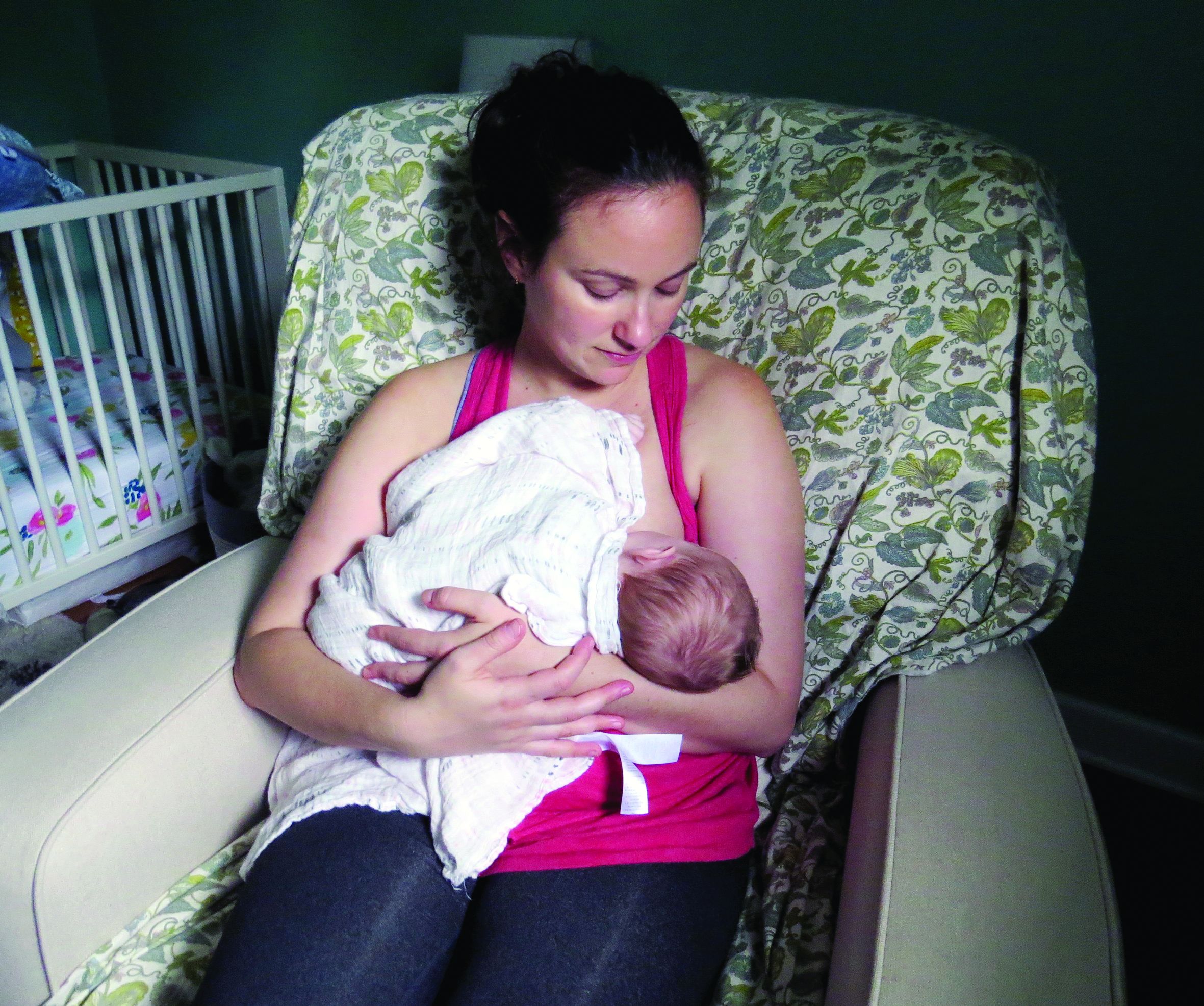
- Infants who are ever breastfed have a reduced risk of overweight or obesity, type 1 diabetes, and asthma. Likewise, longer duration of breastfeeding is associated with lower risk of type 1 diabetes and asthma, and exclusive breastfeeding is associated with lower risk of type 1 diabetes.
- Complementary foods and beverages should not be introduced before age 4 months. Limited evidence indicates that their introduction before 4 months may be associated with increased odds of overweight or obesity. Introducing complementary foods or beverages at 4 or 5 months, compared with 6 months, is not associated with long-term advantages or disadvantages.
- Introducing peanut and egg after age 4 months may reduce the risk of food allergies.
- From age 12 months to 24 months, children should consume a variety of nutrient-rich protein sources from animals – including meat, poultry, seafood, eggs, and dairy – plus nuts, seeds, fruits, vegetables, and grains.
- The report prioritizes oils over solid fats, and whole grains over refined grains. It also discourages added sugars, particularly from sugar-sweetened beverages. Other sources of added sugars include sweets, baked goods, and sweetened dairy products.
The report acknowledges that dietary guidelines should accommodate cultural preferences and cost considerations.
Recommendations during pregnancy
Healthy dietary patterns before or during pregnancy may modestly reduce the odds of gestational diabetes, hypertensive disorders of pregnancy, and preterm birth, according to the report.
The report recommends that during pregnancy women consume 8-12 ounces per week of seafood with high levels of omega-3 fatty acids and low levels of methylmercury, consistent with existing recommendations.
Egg and milk consumption during pregnancy does not influence the risk of food allergy, asthma, or atopic disease in the child, according to the report.
The advisory committee recommended universal folic acid supplementation during pregnancy.
Addressing a gap
The Agricultural Act of 2014 required that infants and toddlers and women who are pregnant or lactating be included in the 2020-2025 guidelines. Covering these populations in the scientific report was a substantial undertaking, said Kathryn Dewey, PhD, of the Institute for Global Nutrition at the University of California, Davis. Dr. Dewey chaired the subcommittee on birth to 24 months for the 2020 Dietary Guidelines Advisory Committee.
“Given that this age group had not been covered before, we could not rely on previous dietary guidelines’ reports,” Dr. Dewey said in an interview.
Outlining food patterns for infants and toddlers proved challenging. The committee explored models that considered various scenarios including children who consumed human milk, children who consumed formula, and those with vegetarian diets. Future research should clarify dietary reference intakes for these age groups, Dr. Dewey said.
Dr. Dewey sees the committee’s report on dietary guidance for birth to 24 months as a starting point and not necessarily an exhaustive look at the subject.
For one, the committee focused more on what to feed infants and toddlers rather than on how to feed them. Information about how to feed children is considered more in depth in a 2020 report from the National Academies of Sciences, Engineering, and Medicine. That report summarizes existing guidance from various organizations on feeding infants and children from birth to 24 months. Dr. Dewey chaired the committee that created the National Academies report.
Sharing the new USDA and HHS guidelines after they are released could be the next important step. “The public does not necessarily know about the guidelines or they do not necessarily seek them out unless there is a very well-constructed strategy for dissemination and implementation,” Dr. Dewey said.
To that end, health care providers can play a role, Dr. Meek said. “Be aware of changes in guidance, adopt those new recommendations, and then advocate those with our patients as well as with the public at large.”
Dr. Meek and Dr. Dewey had no relevant financial disclosures.
The U.S. Department of Agriculture and the Department of Health & Human Services aim to release new dietary guidelines by the end of 2020.
An advisory committee submitted to the agencies a scientific report that examines relationships between diet and health at various life stages. Four chapters focus on dietary considerations for infants and toddlers, and two chapters focus on diet during pregnancy and lactation.
The report may inform the development of the new guidelines. The advisory committee’s recommendations include introducing infants to foods that are rich in zinc and iron at about age 6 months and having women who are lactating eat sources of omega-3 and omega-6 fatty acids, such as fish, to improve the fatty acid status of infants.
Ahead of the release of the 2020-2025 Dietary Guidelines for Americans, Joan Younger Meek, MD, discussed parts of the scientific report at the annual meeting of the American Academy of Pediatrics, held virtually this year.
While the 2015-2020 guidelines use ChooseMyPlate to help people implement the recommendations, it is not known how the new guidelines will be presented to the public, she said. “Many of you will remember the pyramids earlier and different food groups before that.”
Promote healthy dietary patterns
The advisory committee’s report notes that diet in the first years of life contributes to long-term health and shapes taste preferences, said Dr. Meek, professor of clinical sciences at Florida State University, Orlando. Human milk or infant formula are primary sources of nutrition until approximately 6 months, when families may introduce complementary foods and beverages. Between 6 months and 24 months, children transition to the typical family diet.
Dr. Meek highlighted some of the advisory committee’s findings and recommendations.
- Infants who are ever breastfed have a reduced risk of overweight or obesity, type 1 diabetes, and asthma. Likewise, longer duration of breastfeeding is associated with lower risk of type 1 diabetes and asthma, and exclusive breastfeeding is associated with lower risk of type 1 diabetes.
- Complementary foods and beverages should not be introduced before age 4 months. Limited evidence indicates that their introduction before 4 months may be associated with increased odds of overweight or obesity. Introducing complementary foods or beverages at 4 or 5 months, compared with 6 months, is not associated with long-term advantages or disadvantages.
- Introducing peanut and egg after age 4 months may reduce the risk of food allergies.
- From age 12 months to 24 months, children should consume a variety of nutrient-rich protein sources from animals – including meat, poultry, seafood, eggs, and dairy – plus nuts, seeds, fruits, vegetables, and grains.
- The report prioritizes oils over solid fats, and whole grains over refined grains. It also discourages added sugars, particularly from sugar-sweetened beverages. Other sources of added sugars include sweets, baked goods, and sweetened dairy products.
The report acknowledges that dietary guidelines should accommodate cultural preferences and cost considerations.
Recommendations during pregnancy
Healthy dietary patterns before or during pregnancy may modestly reduce the odds of gestational diabetes, hypertensive disorders of pregnancy, and preterm birth, according to the report.
The report recommends that during pregnancy women consume 8-12 ounces per week of seafood with high levels of omega-3 fatty acids and low levels of methylmercury, consistent with existing recommendations.
Egg and milk consumption during pregnancy does not influence the risk of food allergy, asthma, or atopic disease in the child, according to the report.
The advisory committee recommended universal folic acid supplementation during pregnancy.
Addressing a gap
The Agricultural Act of 2014 required that infants and toddlers and women who are pregnant or lactating be included in the 2020-2025 guidelines. Covering these populations in the scientific report was a substantial undertaking, said Kathryn Dewey, PhD, of the Institute for Global Nutrition at the University of California, Davis. Dr. Dewey chaired the subcommittee on birth to 24 months for the 2020 Dietary Guidelines Advisory Committee.
“Given that this age group had not been covered before, we could not rely on previous dietary guidelines’ reports,” Dr. Dewey said in an interview.
Outlining food patterns for infants and toddlers proved challenging. The committee explored models that considered various scenarios including children who consumed human milk, children who consumed formula, and those with vegetarian diets. Future research should clarify dietary reference intakes for these age groups, Dr. Dewey said.
Dr. Dewey sees the committee’s report on dietary guidance for birth to 24 months as a starting point and not necessarily an exhaustive look at the subject.
For one, the committee focused more on what to feed infants and toddlers rather than on how to feed them. Information about how to feed children is considered more in depth in a 2020 report from the National Academies of Sciences, Engineering, and Medicine. That report summarizes existing guidance from various organizations on feeding infants and children from birth to 24 months. Dr. Dewey chaired the committee that created the National Academies report.
Sharing the new USDA and HHS guidelines after they are released could be the next important step. “The public does not necessarily know about the guidelines or they do not necessarily seek them out unless there is a very well-constructed strategy for dissemination and implementation,” Dr. Dewey said.
To that end, health care providers can play a role, Dr. Meek said. “Be aware of changes in guidance, adopt those new recommendations, and then advocate those with our patients as well as with the public at large.”
Dr. Meek and Dr. Dewey had no relevant financial disclosures.
FROM AAP 2020
Fertility delay varied with contraceptive method in study
according to a new prospective cohort study.
Women who used hormonal intrauterine devices, copper intrauterine devices, and implants had the shortest delays, based on the same research project, which involved analyzing data from approximately 18,000 women in North America and Denmark.
“Most research on the use of contraceptives and fertility has focused on the effect of oral contraceptives on fecundability,” and data on the association between fertility and other contraceptive methods are limited, wrote Jennifer J. Yland, MS, of Boston University School of Public Health and colleagues.
“Given the increasing popularity of long acting reversible contraceptive methods and other alternatives to oral contraceptives, more research into their short- and long-term effects on fertility is needed,” the researchers noted.
In the study, which was published in the BMJ, the researchers reviewed data from a total of 17,954 women from three cohort studies of individuals planning pregnancies between 2007 and 2019. Participants reported their contraceptive use and typical menstrual cycle at baseline, then responded to questionnaires every 2 months for up to a year or until pregnancy.
On average, users of injectable contraceptives had the longest delay in return of normal fertility (five to eight menstrual cycles), compared with four cycles for patch contraceptives, three cycles for oral and ring contraceptives, and two cycles for hormonal and copper intrauterine devices and implants.
A total of 10,729 pregnancies were reported within 66,759 menstrual cycles; approximately 77% of the women conceived within 12 months, and 56% conceived within 6 months.
Oral contraceptives were the most common method of contraception (38%), followed by barrier methods (31%), natural methods (15%), and long-acting reversible contraceptives (13%). Intrauterine devices were the most frequently used of long-acting reversible contraceptives (8% hormonal, 4% copper).
The time until fertility returned after discontinuing contraceptives was not associated with duration of contraceptive use.
The study findings were limited by several factors including the potential misclassification of menstrual cycles and the use of self-reports for the time of contraceptive discontinuation, especially for users of injectable contraceptives, the researchers noted.
However, the results were strengthened by the large study size and show “little or no lasting effect” of long-term use of any of the reported contraceptive methods on fertility, the researchers noted. “Understanding the comparative effects of different contraceptives on fecundity is essential for family planning, counseling for contraception, and management of infertility,” they said.
Comparison of contraceptives can inform counseling
The study is important because the use of long-acting reversible contraceptive (LARC) methods (IUDs, implants, patches, and injectable contraceptives) has become increasingly common worldwide, corresponding author Jennifer J. Yland, MS, said in an interview. “Many women are concerned about the potential effects of contraception on future fertility. However, previous research on this topic has focused mostly on oral contraceptives,” she said.
Ms. Yland said that the findings on oral and injectable contraceptives were consistent with previous publications. However, “we were surprised to find that women who had recently used the hormonal IUD had a shorter time to pregnancy, compared with women who used barrier methods,” she said.
The take-home message for clinicians is that delays in the return to normal fertility were temporary for all hormonal contraceptive methods, Ms. Yland emphasized. “However, delays in the return of fertility after discontinuing certain hormonal methods, such as injectables, were considerably longer than that shown for oral contraceptives. These findings should be taken into account when women are considering contraceptive choice in the context of family planning and infertility management,” she noted.
“Future research should evaluate the potential associations between recent use of hormonal contraceptives and perinatal outcomes,” she added.
Managing expectations helps patients plan
“The question of return to fertility is one that many patients who use contraception have unless they have completed their child bearing,” said Sarah W. Prager, MD, of the University of Washington, Seattle, in an interview. “For patients who want to plan a pregnancy, knowing what to expect in terms of return to fertility is important so they can make sure they are in the space and place they want to be with their health, life, job, and partner,” she said.
Dr. Prager said she was not surprised by the study findings because they agree with previously published data. “Overall, except for the injection, people using any form of contraception are back to their baseline fertility within a few months,” she noted. “It also makes perfect sense for return to fertility to be longer with the injection, as it is designed to prevent pregnancy for 16 weeks after the injection is given. Unlike all the other methods, it cannot be removed from the body once given,” she said.
“Clinicians should continue to advise patients that their return to baseline fertility is relatively rapid with any contraception other than the Depo-Provera injection,” said Dr. Prager. “There are no data to support a benefit in switching from an IUD or implant to a combination hormonal method (pills, patch, ring) before starting to try to conceive,” she said.
“This study tries to account for differences in baseline fertility for people using the different methods, but since the choice of method was not randomized, there could still be baseline differences that were not measured or accounted for,” Dr. Prager noted. “A randomized study would certainly eliminate some of these biases; however, I don’t think the differences found in this study are so profound as to require such study,” she said. “Generally speaking, almost 80% of people using any form of contraception were able to conceive within 1 year of trying, which has been the stated fertility data for decades,” she said.
The study was supported by the Eunice Kennedy Shriver National Institute of Child Health and Human Development, National Institutes of Health. Lead author Ms. Yland had no financial conflicts to disclose. Dr. Prager had no financial conflicts to disclose.
SOURCE: Yland JJ et al. BMJ. 2020 Nov 12. doi: 10.1136/bmj.m3966.
FROM THE BMJ
Rare event: Iatrogenic injury during cervical cancer screening
Cervical cancer screening is a routine procedure, but in rare instances, there can be medical complications. A new study finds that, compared with women who have normal results, women who are diagnosed with an invasive malignancy have an increased risk for iatrogenic injuries.
Researchers in Sweden analyzed data on more than 3 million women who had undergone cervical cancer screening. The team found that 42 iatrogenic injuries that required at least 2 days of hospitalization occurred during the diagnostic work-up of women who had an abnormal screening test.
“Although cervical cancer screening is one of the most successful cancer prevention programs ... our research indicates that women with invasive cervical cancer experienced medical complications and psychological stress during their diagnostic work-up, although at a very low level,” commented corresponding author Qing Shen, PhD, from the department of medical epidemiology and biostatistics at the Karolinska Institute in Stockholm, Sweden.
The study was published in Cancer Epidemiology, Biomarkers & Prevention.
“Injuries can occur with diagnostic evaluation for cervical cancer,” commented Kecia Gaither, MD, MPH, FACOG, director of perinatal services at Lincoln Medical and Mental Health Center, New York City Health and Hospitals System, who was not involved in the study.
“Given the fact that neovascularization occurs with cancers, a large biopsy in such a circumstance could lead to a hematoma or excessive blood loss. It rarely occurs but most certainly is possible,” said Dr. Gaither.
Also weighing in with comments, Cathy Popadiuk, MD, FRCS, an associate professor of medicine in the department of obstetrics and gynecology at Memorial University of Newfoundland, St. John’s, said the findings are reflective of the real-world and North American experience.
“There are indeed rare bad things that can happen during diagnostic work-up of abnormal pap smears, and usually this is with associated other disease, such as actual cancer or fibroids that can also bleed and may be in the cervix, etc.,” she told this news organization. “And definitely, when there is undetected cancer, this can bleed, requiring transfusion and hospital admission.”
Dr. Popadiuk pointed out that Sweden may be more liberal in admitting patients to hospital, whereas in North America, “we are trying to move away from inpatient care.” She added, “When you are getting these relatively minor procedures, you don’t expect something bad to happen in the clinic.”
Women may become anxious, and admission is the easiest way to arrange for care such as transfusions or observation for more bleeding, she noted. In addition, vaginal packing may be needed to control hemorrhage, and “with vaginal packing, women are unable to void and need a Foley catheter, and that, again, cannot be managed at home easily,” she explained. “After bleeding settles, the vaginal pack is removed, often the next day.” This may be why some women are admitted to hospital.
Increased risk of injury
In a previous study, Dr. Shen and colleagues found there was an increased risk for injuries during the period before and after a diagnosis of any cancer (BMJ. 2016;354:i4218). Those findings suggested the interval between first suspicion of cancer and diagnosis or initiation of treatment might be a high-risk time for injuries in cancer care.
In this latest study, they assessed whether there was a similar increase in injury risk among patients screened for cervical cancer. Using the Swedish Total Population Register, they identified 3,016,307 women who had undergone cervical screening during the period 2001-2012.
The final analysis included 1,853,510 women whose pap smear results were normal; 22,435 women who were diagnosed with cervical intraepithelial neoplasia (CIN); 20,692 women with CIN2; 36,542 women with CIN3 or adenocarcinoma in situ (AIS); and 5,189 women with invasive cervical cancer.
The team found that, among women who had an abnormal screening test, 42 iatrogenic injuries occurred that required at least 2 days of hospital admission. The highest risk was among women diagnosed with invasive cancer. The risk was also increased among women diagnosed with CIN3/AIS, but not among women with lower grades of CIN.
The most common types of iatrogenic injuries were hemorrhage or hematoma and infections. Among all groups of women, the incidence rate of injuries that were caused by medical procedures and care was greater than that of injuries caused by drugs or biological substances.
A total of 91 noniatrogenic injuries that required at least 1 day of hospitalization were identified. The risk was increased among women with invasive cervical cancer but not for women with other cervical abnormalities. The most common type of noniatrogenic injury was unintentional injuries.
The study was sponsored by the Swedish Cancer Society and the Swedish Research Council for Health, Working Life and Welfare. One author received a Karolinska Institute Senior Researcher Award and a Strategic Research Area in Epidemiology Award, and one author received a grant from the China Scholarship Council. Dr. Shen has disclosed no relevant financial relationships. Dr. Popadiuk has received personal fees and nonfinancial support as a member of the OncoSim Initiative from the Canadian Partnership Against Cancer.
A version of this article originally appeared on Medscape.com.
Denosumab favored over alendronate for BMD protection in glucocorticoid-induced osteoporosis
Denosumab boosted bone mineral density (BMD) over 12 months to a greater extent than did alendronate in a randomized, 12-month study. The investigator-initiated research compared BMD at the lumbar spine and elsewhere among people with systemic lupus erythematosus (SLE) and other autoimmune conditions. Long-term glucocorticoid therapy places some people in this group at higher risk for adverse effects of bone density loss.

“Glucocorticoids remain the mainstay of treatment of rheumatic diseases, but [they are] a major risk factor for osteoporosis and fracture,” study author Chi Chiu Mok, MD, said in an interview.
Compared with baseline, adults randomly assigned to denosumab had a 3.5% increase in lumbar spine BMD at 12 months, compared with 2.5% among those taking alendronate, a significant difference. Dr. Mok, a consultant and honorary associate professor in the department of medicine and nuclear medicine at Tuen Mun Hospital in Hong Kong, presented the study results at the virtual annual meeting of the American College of Rheumatology.
“Given the knowledge that denosumab is more effective than alendronate in raising spinal BMD in chronic users of GCs without increasing adverse events, this drug may be considered as an alternative first-line therapy in higher-risk patients and in those who are contraindicated for the oral bisphosphonates,” he said.
Cost considerations
Denosumab is a human monoclonal antibody administered as a subcutaneous injection, available under the brand names Prolia and Xgeva. Alendronate is an oral agent available as both generic and brand name formulations.
“Yes, denosumab is more expensive, more costly than oral alendronate, but our study shows efficacy is better for steroid users,” Dr. Mok said in answer to a question about cost disparity between the two agents during his presentation at the meeting. “For patients who are contraindicated or have low compliance for bisphosphonate, or are high-risk patients, I recommend first-line use of denosumab.”
Researchers previously studied these agents, including a smaller study by Dr. Mok and colleagues that showed a BMD benefit after switching people on an oral bisphosphonate to denosumab. However, he said, “There is a paucity of data regarding comparative efficacy of denosumab and the bisphosphonates in long-term steroid users.”
To explore any differences in a larger patient population, the investigators randomly assigned adults with SLE and other autoimmune conditions to the two treatments: denosumab 60 mg subcutaneoulsy every 6 months or oral alendronate 70 mg/week. All patients also received 3,000 mg calcium and 1,000 IU vitamin D3 (cholecalciferol) each day.
After three discontinuations in denosumab cohort and four in the alendronate group, the researchers evaluated 69 people taking denosumab and 70 others taking alendronate. The discontinuations were caused by noncompliance, Dr. Mok said, not by adverse events.
Adverse events were reported, but the rate did not differ significantly between groups. Dr. Mok highlighted some notable differences, including more minor infections and arthralgias reported in the denosumab cohort. Chest discomfort was reported in one denosumab recipient versus no patients in the alendronate group. Dyspepsia/upper GI symptoms and dizziness/vertigo occurred more often in the alendronate group.
Women were 96% of the study population, and mean age was 50 years. A majority, 81%, had underlying SLE. Other diagnoses included rheumatoid arthritis, myositis, antineutrophil cytoplasmic antibody–associated vasculitis, and polymyalgia rheumatica. The mean dose of prednisolone at study entry was 5.1 mg/day.
Key BMD and biomarker findings
BMD increased significantly in the spine, hip, and femoral neck in both treatment groups by 12 months. However, after adjustment for baseline BMD and covariates including age, menopause, and history of fracture, the gains in the denosumab group were significantly higher.
The increase in lumbar spine BMD at 12 months of 3.5% in the denosumab group versus 2.5% in the alendronate group was statistically significant (P = .045). Less significant was a 0.9% increase at the hip in the denosumab patients versus 1.6% in the alendronate group (P = .10), as well as femoral neck BMD gains of 1% in the denosumab group versus 1.5% in the alendronate group (P = .86).
Furthermore, “denosumab was more potent in suppressing the bone markers at 12 months,” Dr. Mok said.
Specifically, the percentage decrease in serum PINP (procollagen type I N-terminal propeptide) levels in the denosumab group was significantly greater than in the alendronate group (P = .001). Likewise, the decrease in CTX (C-terminal telopeptide of type I collagen) was significantly greater in the denosumab cohort versus the alendronate cohort (P < .001).
“Dr. Mok’s study was a well-controlled investigation. The superiority of denosumab was impressive, especially given the small group sizes of 69 and 70,” session comoderator Gregg Silverman, MD, professor in the department of internal medicine and the department of pathology at New York University, said when asked for comment.
“However, bone density measurements may not tell the whole story. These results support a bigger and much larger-scale study to confirm that rates of fracture on denosumab are also reduced.”
No new symptomatic fractures occurred in either group during the study. The investigators are evaluating for any new radiologic fractures, with results pending.
Dr. Mok said “results of our study in Asian patients are largely confirmatory” of a previous 2018 comparison study and a 2019 comparison study, each sponsored by Amgen.
A small sample size, short duration of treatment, and the open-label design were limitations of the study.
The trial was an investigator-initiated study. Dr. Mok and colleagues had no relevant financial disclosures. Dr. Silverman had no relevant financial disclosures.
SOURCE: Mok CC et al. Arthritis Rheumatol. 2020;72(suppl 10). ACR 2020, Abstract 1442.
Denosumab boosted bone mineral density (BMD) over 12 months to a greater extent than did alendronate in a randomized, 12-month study. The investigator-initiated research compared BMD at the lumbar spine and elsewhere among people with systemic lupus erythematosus (SLE) and other autoimmune conditions. Long-term glucocorticoid therapy places some people in this group at higher risk for adverse effects of bone density loss.
“Glucocorticoids remain the mainstay of treatment of rheumatic diseases, but [they are] a major risk factor for osteoporosis and fracture,” study author Chi Chiu Mok, MD, said in an interview.
Compared with baseline, adults randomly assigned to denosumab had a 3.5% increase in lumbar spine BMD at 12 months, compared with 2.5% among those taking alendronate, a significant difference. Dr. Mok, a consultant and honorary associate professor in the department of medicine and nuclear medicine at Tuen Mun Hospital in Hong Kong, presented the study results at the virtual annual meeting of the American College of Rheumatology.
“Given the knowledge that denosumab is more effective than alendronate in raising spinal BMD in chronic users of GCs without increasing adverse events, this drug may be considered as an alternative first-line therapy in higher-risk patients and in those who are contraindicated for the oral bisphosphonates,” he said.
Cost considerations
Denosumab is a human monoclonal antibody administered as a subcutaneous injection, available under the brand names Prolia and Xgeva. Alendronate is an oral agent available as both generic and brand name formulations.
“Yes, denosumab is more expensive, more costly than oral alendronate, but our study shows efficacy is better for steroid users,” Dr. Mok said in answer to a question about cost disparity between the two agents during his presentation at the meeting. “For patients who are contraindicated or have low compliance for bisphosphonate, or are high-risk patients, I recommend first-line use of denosumab.”
Researchers previously studied these agents, including a smaller study by Dr. Mok and colleagues that showed a BMD benefit after switching people on an oral bisphosphonate to denosumab. However, he said, “There is a paucity of data regarding comparative efficacy of denosumab and the bisphosphonates in long-term steroid users.”
To explore any differences in a larger patient population, the investigators randomly assigned adults with SLE and other autoimmune conditions to the two treatments: denosumab 60 mg subcutaneoulsy every 6 months or oral alendronate 70 mg/week. All patients also received 3,000 mg calcium and 1,000 IU vitamin D3 (cholecalciferol) each day.
After three discontinuations in denosumab cohort and four in the alendronate group, the researchers evaluated 69 people taking denosumab and 70 others taking alendronate. The discontinuations were caused by noncompliance, Dr. Mok said, not by adverse events.
Adverse events were reported, but the rate did not differ significantly between groups. Dr. Mok highlighted some notable differences, including more minor infections and arthralgias reported in the denosumab cohort. Chest discomfort was reported in one denosumab recipient versus no patients in the alendronate group. Dyspepsia/upper GI symptoms and dizziness/vertigo occurred more often in the alendronate group.
Women were 96% of the study population, and mean age was 50 years. A majority, 81%, had underlying SLE. Other diagnoses included rheumatoid arthritis, myositis, antineutrophil cytoplasmic antibody–associated vasculitis, and polymyalgia rheumatica. The mean dose of prednisolone at study entry was 5.1 mg/day.
Key BMD and biomarker findings
BMD increased significantly in the spine, hip, and femoral neck in both treatment groups by 12 months. However, after adjustment for baseline BMD and covariates including age, menopause, and history of fracture, the gains in the denosumab group were significantly higher.
The increase in lumbar spine BMD at 12 months of 3.5% in the denosumab group versus 2.5% in the alendronate group was statistically significant (P = .045). Less significant was a 0.9% increase at the hip in the denosumab patients versus 1.6% in the alendronate group (P = .10), as well as femoral neck BMD gains of 1% in the denosumab group versus 1.5% in the alendronate group (P = .86).
Furthermore, “denosumab was more potent in suppressing the bone markers at 12 months,” Dr. Mok said.
Specifically, the percentage decrease in serum PINP (procollagen type I N-terminal propeptide) levels in the denosumab group was significantly greater than in the alendronate group (P = .001). Likewise, the decrease in CTX (C-terminal telopeptide of type I collagen) was significantly greater in the denosumab cohort versus the alendronate cohort (P < .001).
“Dr. Mok’s study was a well-controlled investigation. The superiority of denosumab was impressive, especially given the small group sizes of 69 and 70,” session comoderator Gregg Silverman, MD, professor in the department of internal medicine and the department of pathology at New York University, said when asked for comment.
“However, bone density measurements may not tell the whole story. These results support a bigger and much larger-scale study to confirm that rates of fracture on denosumab are also reduced.”
No new symptomatic fractures occurred in either group during the study. The investigators are evaluating for any new radiologic fractures, with results pending.
Dr. Mok said “results of our study in Asian patients are largely confirmatory” of a previous 2018 comparison study and a 2019 comparison study, each sponsored by Amgen.
A small sample size, short duration of treatment, and the open-label design were limitations of the study.
The trial was an investigator-initiated study. Dr. Mok and colleagues had no relevant financial disclosures. Dr. Silverman had no relevant financial disclosures.
SOURCE: Mok CC et al. Arthritis Rheumatol. 2020;72(suppl 10). ACR 2020, Abstract 1442.
FROM ACR 2020
Treatment sequence with romosozumab influences osteoporosis outcomes
Timing is everything when it comes to the use of the anabolic agent romosozumab (Evenity) for the treatment of advanced osteoporosis, a review of clinical trials suggests.

In four studies with treatment sequences in which romosozumab was administered either before or following the use of an antiresorptive agent, initial treatment with 1 year of romosozumab produced substantial bone mineral density (BMD) gains in the total hip and lumbar spine.
Transition from romosozumab to a potent resorptive agent, either alendronate or denosumab (Prolia) augmented the initial gains, reported Felicia Cosman, MD, professor of clinical medicine at Columbia University, New York.
Romosozumab was the third approved agent in its class, following teriparatide in 2002, and abaloparatide (Tymlos) in 2017, both of which have been shown to produce rapid reductions in fracture risk and large improvements in BMD when they were administered up front, followed by an antiresorptive agent.
“But since romosozumab has a very different mechanism of action compared to both teriparatide and abaloparatide, we didn’t know if treatment sequence would be as important for this agent as it was for teriparatide,” she said during a press briefing prior to her presentation of the data in an oral abstract session at the virtual annual meeting of the American College of Rheumatology.
Two-for-one
Romosozumab is unique in that it both increases bone formation and decreases bone resorption, and has been shown in treatment-naive postmenopausal women with osteoporosis to significantly improve BMD and reduce fracture risk, compared with either placebo or alendronate. Romosozumab has also been studied as sequential therapy in patients treated initially with either alendronate or denosumab.
To see whether treatment sequence could have differential effects on clinical outcomes for patients with osteoporosis, Dr. Cosman and colleagues looked at results from four clinical trials, using levels of bone turnover markers (procollagen type I N-terminal propeptide [PINP] and beta-isomer of the C-terminal telopeptide of type I collagen [beta-CTX]) and BMD gains in the total hip and spine as outcomes.
The two trials of romosozumab in treatment-naive women were the ARCH trial comparing romosozumab with alendronate in a double-blind phase for 1 year, followed by 1 year of open-label alendronate, and the FRAME trial, in which romosozumab was compared with placebo in a 1-year double-blind phase, followed by 1-year of open-label denosumab.
The two trials of romosozumab in women treated initially with antiresorptive agents were the STRUCTURE trial in which patients on oral bisphosphonates for at least 3 years or alendronate 70 mg weekly for 1 year were randomized to receive either romosozumab or teriparatide, and a phase 2 trial (NCT00896532) that included a 24-month romosozumab or placebo treatment phase followed by rerandomization to a 12-month extension phase with denosumab or placebo, followed by a 12-month retreatment phase with romosozumab, followed by a 24-month follow-on phase with zoledronic acid or no intervention.
Total hip BMD gains
In the ARCH trial, total hip BMD increased 6.2% with 1 year of romosozumab, and a cumulative total of 7.1% with the 2-year romosozumab/alendronate sequence. In the FRAME trial, patients gained 6.8% in total hip BMD after 1 year of romosozumab and a total of 8.8% after 2 years of romosozumab followed by denosumab.
In contrast, in the STRUCTURE trial, patients treated for 1 year or longer with alendronate and then with 1 year of romosozumab had a 2.9% BMD gain in the total hip. In the phase 2 trial, 1 year of romosozumab following 1 year of denosumab yielded a 0.9% BMD gain, for a total gain of 3.8% with the denosumab sequence.
Lumbar spine BMD gains
In ARCH, lumbar spine BMD increased 13.7% with 1 year of romosozumab, and a total of 15.2% with the 2-year sequence of romosozumab followed by alendronate. Similarly, in FRAME, patients gained 13.3% in BMD after a year of romosozumab, and total of 17.6% by the end of the 2-year romosozumab/denosumab sequence.
In contrast, in STRUCTURE, patients who had previously been on alendronate for at least 1 year had a gain of 9.8% after 1 year of romosozumab, and in the phase 2 study, patients who had been on denosumab for 1 year had an increase in lumbar spine BMD of 5.3% after 1 year on romosozumab, and a total gain of 11.5% at the end of the 2-year sequence.
Serum PINP and beta-CTX
Looking at the markers of bone turnover, the investigators saw that, in both ARCH and FRAME, PINP peaked at over 80% of baseline at 1 month, and then continued to steadily decline past 1 year. The beta-CTX nadir was 40%-50% below baseline at 1 year.
At the end of year 2, the PINP nadir was –67% with follow-on alendronate, and –69% with denosumab, and the beta-CTX nadir was –72% and –92%, respectively.
In the two trials where romosozumab was the follow-on therapy, however, the trends were distinctly different. In STRUCTURE, for example, PINP peaked at 141% of baseline at 1 month, and then returned toward baseline, whereas beta-CTX remained largely unchanged.
In the phase 2 trial, PINP peaked at 28% above baseline at 9 months, and then only slightly declined, and beta-CTX peaked at 211% at the end of 1 year of romosozumab.
Best used up front
“This study is important, because it suggests that for the three bone-building drugs that the best effects will really be attained on bone strength if the agents are used as initial therapy in very-high-risk patients. Those are people who have sustained fractures within the preceding 2 years, who had multiple fractures at any point in their adulthood, and who present with very low BMD, particularly if they have any associated clinical risk factors such as family history or other underlying diseases or medications that have detrimental effects on bone,” Dr. Cosman said at the briefing.
Marcy Bolster, MD, from the division of rheumatology, allergy, and immunology at Massachusetts General Hospital, Boston, and associate professor of medicine at Harvard Medical School in Boston, who was not involved in the study, commented that the study provides important information for clinicians who treat patients with osteoporosis.

“We have an increasing number of medications available for use in the treatment of patients with osteoporosis, and as we consider the importance of reducing fracture risk, the duration of therapy, the timing of a bisphosphonate holiday, it is essential that we consider any advantages to the order or sequence of our medications,” she said when asked for comment.
“This study provides evidence supporting the concept of the ‘anabolic window’ in which there is a demonstrated advantage in treating patients with an anabolic agent prior to treatment with an antiresorptive agent, and while gains in bone mineral density were achieved with either order of medication use, the gains were more dramatic with treatment with romosozumab as the first agent,” she added.
Dr. Bolster also noted it will be important to demonstrate reduction in fracture risk as well as gain in BMD.
The study was sponsored by Amgen, Astellas, and UCB. Dr. Cosman disclosed grants/research support from Amgen, and consulting fees and speaker activities for Amgen and Radius Health. Dr. Bolster disclosed relationships with AbbVie, Corbus, Cumberland, Gilead, Johnson & Johnson, and Pfizer.
SOURCE: Cosman F et al. Arthritis Rheumatol. 2020;72(suppl 10), Abstract 1973.
Timing is everything when it comes to the use of the anabolic agent romosozumab (Evenity) for the treatment of advanced osteoporosis, a review of clinical trials suggests.
In four studies with treatment sequences in which romosozumab was administered either before or following the use of an antiresorptive agent, initial treatment with 1 year of romosozumab produced substantial bone mineral density (BMD) gains in the total hip and lumbar spine.
Transition from romosozumab to a potent resorptive agent, either alendronate or denosumab (Prolia) augmented the initial gains, reported Felicia Cosman, MD, professor of clinical medicine at Columbia University, New York.
Romosozumab was the third approved agent in its class, following teriparatide in 2002, and abaloparatide (Tymlos) in 2017, both of which have been shown to produce rapid reductions in fracture risk and large improvements in BMD when they were administered up front, followed by an antiresorptive agent.
“But since romosozumab has a very different mechanism of action compared to both teriparatide and abaloparatide, we didn’t know if treatment sequence would be as important for this agent as it was for teriparatide,” she said during a press briefing prior to her presentation of the data in an oral abstract session at the virtual annual meeting of the American College of Rheumatology.
Two-for-one
Romosozumab is unique in that it both increases bone formation and decreases bone resorption, and has been shown in treatment-naive postmenopausal women with osteoporosis to significantly improve BMD and reduce fracture risk, compared with either placebo or alendronate. Romosozumab has also been studied as sequential therapy in patients treated initially with either alendronate or denosumab.
To see whether treatment sequence could have differential effects on clinical outcomes for patients with osteoporosis, Dr. Cosman and colleagues looked at results from four clinical trials, using levels of bone turnover markers (procollagen type I N-terminal propeptide [PINP] and beta-isomer of the C-terminal telopeptide of type I collagen [beta-CTX]) and BMD gains in the total hip and spine as outcomes.
The two trials of romosozumab in treatment-naive women were the ARCH trial comparing romosozumab with alendronate in a double-blind phase for 1 year, followed by 1 year of open-label alendronate, and the FRAME trial, in which romosozumab was compared with placebo in a 1-year double-blind phase, followed by 1-year of open-label denosumab.
The two trials of romosozumab in women treated initially with antiresorptive agents were the STRUCTURE trial in which patients on oral bisphosphonates for at least 3 years or alendronate 70 mg weekly for 1 year were randomized to receive either romosozumab or teriparatide, and a phase 2 trial (NCT00896532) that included a 24-month romosozumab or placebo treatment phase followed by rerandomization to a 12-month extension phase with denosumab or placebo, followed by a 12-month retreatment phase with romosozumab, followed by a 24-month follow-on phase with zoledronic acid or no intervention.
Total hip BMD gains
In the ARCH trial, total hip BMD increased 6.2% with 1 year of romosozumab, and a cumulative total of 7.1% with the 2-year romosozumab/alendronate sequence. In the FRAME trial, patients gained 6.8% in total hip BMD after 1 year of romosozumab and a total of 8.8% after 2 years of romosozumab followed by denosumab.
In contrast, in the STRUCTURE trial, patients treated for 1 year or longer with alendronate and then with 1 year of romosozumab had a 2.9% BMD gain in the total hip. In the phase 2 trial, 1 year of romosozumab following 1 year of denosumab yielded a 0.9% BMD gain, for a total gain of 3.8% with the denosumab sequence.
Lumbar spine BMD gains
In ARCH, lumbar spine BMD increased 13.7% with 1 year of romosozumab, and a total of 15.2% with the 2-year sequence of romosozumab followed by alendronate. Similarly, in FRAME, patients gained 13.3% in BMD after a year of romosozumab, and total of 17.6% by the end of the 2-year romosozumab/denosumab sequence.
In contrast, in STRUCTURE, patients who had previously been on alendronate for at least 1 year had a gain of 9.8% after 1 year of romosozumab, and in the phase 2 study, patients who had been on denosumab for 1 year had an increase in lumbar spine BMD of 5.3% after 1 year on romosozumab, and a total gain of 11.5% at the end of the 2-year sequence.
Serum PINP and beta-CTX
Looking at the markers of bone turnover, the investigators saw that, in both ARCH and FRAME, PINP peaked at over 80% of baseline at 1 month, and then continued to steadily decline past 1 year. The beta-CTX nadir was 40%-50% below baseline at 1 year.
At the end of year 2, the PINP nadir was –67% with follow-on alendronate, and –69% with denosumab, and the beta-CTX nadir was –72% and –92%, respectively.
In the two trials where romosozumab was the follow-on therapy, however, the trends were distinctly different. In STRUCTURE, for example, PINP peaked at 141% of baseline at 1 month, and then returned toward baseline, whereas beta-CTX remained largely unchanged.
In the phase 2 trial, PINP peaked at 28% above baseline at 9 months, and then only slightly declined, and beta-CTX peaked at 211% at the end of 1 year of romosozumab.
Best used up front
“This study is important, because it suggests that for the three bone-building drugs that the best effects will really be attained on bone strength if the agents are used as initial therapy in very-high-risk patients. Those are people who have sustained fractures within the preceding 2 years, who had multiple fractures at any point in their adulthood, and who present with very low BMD, particularly if they have any associated clinical risk factors such as family history or other underlying diseases or medications that have detrimental effects on bone,” Dr. Cosman said at the briefing.
Marcy Bolster, MD, from the division of rheumatology, allergy, and immunology at Massachusetts General Hospital, Boston, and associate professor of medicine at Harvard Medical School in Boston, who was not involved in the study, commented that the study provides important information for clinicians who treat patients with osteoporosis.
“We have an increasing number of medications available for use in the treatment of patients with osteoporosis, and as we consider the importance of reducing fracture risk, the duration of therapy, the timing of a bisphosphonate holiday, it is essential that we consider any advantages to the order or sequence of our medications,” she said when asked for comment.
“This study provides evidence supporting the concept of the ‘anabolic window’ in which there is a demonstrated advantage in treating patients with an anabolic agent prior to treatment with an antiresorptive agent, and while gains in bone mineral density were achieved with either order of medication use, the gains were more dramatic with treatment with romosozumab as the first agent,” she added.
Dr. Bolster also noted it will be important to demonstrate reduction in fracture risk as well as gain in BMD.
The study was sponsored by Amgen, Astellas, and UCB. Dr. Cosman disclosed grants/research support from Amgen, and consulting fees and speaker activities for Amgen and Radius Health. Dr. Bolster disclosed relationships with AbbVie, Corbus, Cumberland, Gilead, Johnson & Johnson, and Pfizer.
SOURCE: Cosman F et al. Arthritis Rheumatol. 2020;72(suppl 10), Abstract 1973.
FROM ACR 2020
.
Pregnancy can be safe with interstitial lung disease
Pregnant women with interstitial lung disease (ILD) related to autoimmune disease may not need to terminate their pregnancies if they have close monitoring before, during, and after pregnancy with a multidisciplinary team of physicians, new research suggests.
Senior author Megan Clowse, MD, MPH, associate professor of medicine in the division of rheumatology at Duke University, Durham, N.C., explained during a press conference at the virtual annual meeting of the American College of Rheumatology that women with ILD are often advised by obstetricians or rheumatologists to avoid conception or terminate their pregnancies, though evidence for that has been based on small studies of 9-15 patients that have had mixed results.
“Many of these pregnancies were delivered 20-30 years ago, definitely with different rheumatic and obstetric care than we can provide now,” she said. “It’s really time to rethink our approach to interstitial lung disease and pregnancy.”
This study showed that while adverse pregnancy outcomes are common in these women, overall maternal morbidity and mortality are low.
ILD may be a secondary disease in people who have scleroderma, lupus, and sarcoidosis.
Largest study to date
This Pfizer-sponsored retrospective study of 67 pregnant women is the largest to date, and it analyzed 94 pregnancies (including five sets of twins).
Sarah Rae Easter, MD, maternal-fetal medicine doctor in the department of obstetrics and gynecology at Brigham and Women’s Hospital, Boston, called the work “exciting” as the researchers were able to look back at a large number of cases for a rare condition for more than 20 years.
“Their data provides much-needed evidence to provide some reassurance for women affected by this type of pulmonary disease regarding the relative safety of pregnancy,” she said in an interview.
Study spanned 23 years
The researchers reviewed pregnancy records in patients diagnosed with ILD secondary to autoimmune disease at Duke University Health System from January 1996 to July 2019.
They classified the severity of ILD based on two standard breathing tests – forced vital capacity and diffusion capacity for carbon monoxide.
Overall, 69% of the women were diagnosed with sarcoidosis and the remaining 31% had a connective tissue disease associated with ILD (CTD-ILD). Of those measured for ILD severity, 11% were severe, 25% were moderate, 50% were mild, and 14% were normal. Their average maternal age was 32.1 and 83% were Black.
While 70% of the pregnancies resulted in live births, 9% were terminated. The remainder resulted in miscarriage or stillbirth.
Researchers reported a 15% rate of preeclampsia, a 34% rate of the composite measure PROMISSE-Adverse Pregnancy Outcome (APO), and a 15% rate of PROMISSE-APO SEVERE. Patients with severe disease had the highest rates of PROMISSE-APO (P = .03 across groups).
(PROMISSE stands for the Predictors of Pregnancy Outcome: Biomarkers in Antiphospholipid Antibody Syndrome and Systemic Lupus Erythematosus study.)
None of the women died
Dr. Clowse said it was a pleasant surprise to find that none of the women died, though patients with severe ILD had more adverse outcomes. Only 2.1% were treated in an intensive care unit during or soon after delivery. In 4.2%, ILD patients had significant shortness of breath due to fluid volume overload around the time of delivery.
For the women who had normal-to-moderate lung disease, Dr. Clowse said, “they really had remarkably good outcomes, really pretty comparable to the general population. About 15% delivered preterm and about 20% suffered a pregnancy loss.”
Dr. Easter, who was not involved with the study, noted the large number of Black women in the cohort.
“Focusing in on improving outcomes for Black and Brown women related to pregnancy in our country is a much-needed undertaking,” Dr. Easter said.
Being able to quote percentages from this research, based on a good-sized study “at least gives people a benchmark about what kind of risk they are willing to assume for themselves,” she said.
For providers, being able to place this rare disease within the spectrum of other diseases where there is more data is also very helpful, she said.
Dr. Clowse said in an interview that the preponderance of Black women in the study was a surprise but may be explained by two factors: Sarcoidosis is seen more frequently in Black women and in the study area in North Carolina there is a large population of Black women.
“Also, our patients with more severe lupus, the ones who are more likely to have interstitial lung disease, are often Black and that’s likely contributing as well,” she said.
Multidisciplinary teams advised
Dr. Clowse emphasized that women with ILD need multidisciplinary teams in pregnancy and should be managed at tertiary care centers where there is a full complement of obstetric and internal medicine experts.
“We do recommend evaluating the severity of their lungs and their heart disease around the time of pregnancy and during pregnancy if they have shortness of breath,” she said.
“We currently recommend that these patients with moderate or severe disease stay in the hospital for up to a week, just for monitoring,” she said.
Dr. Easter said having that kind of access to a large academic healthcare center should be an important part of the decision-making.
Patients need to think about whether they would have access to care similar to what the researchers are describing when they are making the decision to pursue or continue pregnancy, she said.
The study was sponsored by Pfizer Inc. Dr. Clowse reported relationships with UCB, GlaxoSmithKline, AstraZeneca, and Pfizer. Dr. Easter has disclosed no relevant financial relationships.
A version of this article originally appeared on Medscape.com.