User login
Drugs Targeting Osteoarthritis Pain: What’s in Development?
WASHINGTON — Investigational treatments aimed specifically at reducing pain in knee osteoarthritis (OA) are moving forward in parallel with disease-modifying approaches.
“We still have very few treatments for the pain of osteoarthritis…It worries me that people think the only way forward is structure modification. I think while we’re waiting for some drugs to be structure modifying, we still need more pain relief. About 70% of people can’t tolerate or shouldn’t be on a [nonsteroidal anti-inflammatory drug], and that leaves a large number of people with pain,” Philip Conaghan, MBBS, PhD, Chair of Musculoskeletal Medicine at the University of Leeds in England, said in an interview.
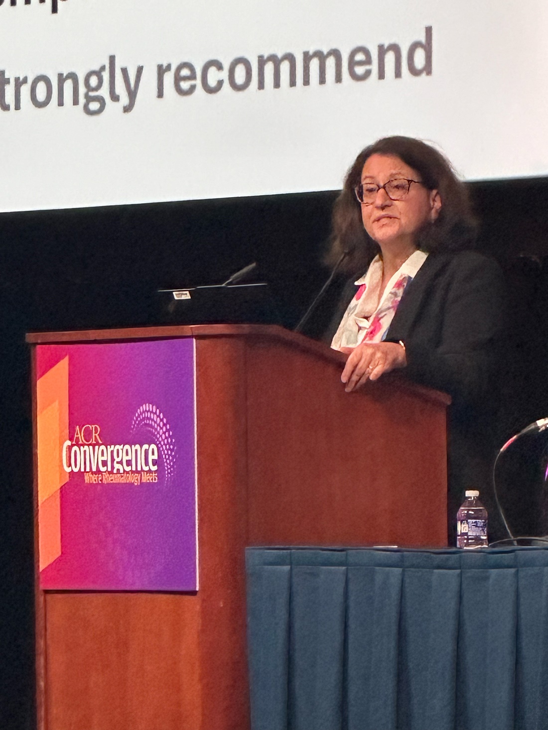
At the annual meeting of the American College of Rheumatology, Conaghan, who is also honorary consultant rheumatologist for the Leeds Teaching Hospitals NHS Trust, presented new data for two novel approaches, both targeting peripheral nociceptive pain signaling.
In a late-breaking poster, he presented phase 2 trial data on RTX-GRT7039 (resiniferatoxin [RTX]), an agonist of the transient receptor potential vanilloid 1 that is a driver of OA pain. The trial investigated the efficacy and safety of a single intra-articular injection of RTX-GRT7039 in people with knee OA.
And separately, in a late-breaking oral abstract session, Conaghan presented phase 2 trial safety and efficacy data for another investigational agent called LEVI-04, a first-in-class neurotrophin receptor fusion protein (p75NTR-Fc) that supplements the endogenous protein and provides analgesia via inhibition of NT-3 activity.
“I think both have potential to provide good pain relief, through slightly different mechanisms,” Conaghan said in an interview.
Asked to comment, session moderator Gregory C. Gardner, MD, emeritus professor in the Division of Rheumatology at the University of Washington, Seattle, said in an interview: “I think the results are really exciting terms of the ability to control pain to a significant degree in patients with osteoarthritis.”
However, Gardner also said, “The molecules can be very expensive ... so who do we give them to? Will insurance companies pay for this simply for OA pain? They improve function ... so clearly, [they] will be a boon to treating osteoarthritis, but do we give them to people with only more advanced forms of osteoarthritis or earlier on?”
Moreover, Gardner said, “One of my concerns about treating osteoarthritis is I don’t want to do too good of a job treating pain in somebody who has a biomechanically abnormal joint. ... You’ve got a knee that’s worn out some of the cartilage, and now you feel like you can go out and play soccer again. That’s not a good thing. That joint will wear out very quickly, even though it doesn’t feel pain.”
Another OA expert, Matlock Jeffries, MD, director of the Arthritis Research Unit at the Oklahoma Medical Research Foundation, Oklahoma City, said in an interview, “I think we don’t focus nearly enough on pain, and that’s [partly] because the [Food and Drug Administration] has defined endpoints for knee OA trials that are radiographic. ... Patients do not care what their joint space narrowing is. They care what their pain is. And joint space changes and pain do not correlate in knee OA. ... About 20% or 30% of patients who have completely normal x-rays have a lot of pain…I hope that we’ll have some new OA pain therapeutics in the future because that’s what patients actually care about.”
But Jeffries noted that it will be very important to ensure that these agents don’t produce significant side effects, as had been seen previously in several large industry-sponsored trials of drugs targeting nerve growth factors.
“The big concern that we have in the field ... is that the nerve growth factor antibody trials were all stopped because there was a low but persistent risk of rapidly progressive OA in a small percent of patients. I think one of the questions in the field is whether targeting other things having to do with OA pain is going to result in similar bad outcomes. I think the answer is probably not, but that’s one thing that people do worry about, and they never really figured out why the [rapidly progressive OA] was happening.”
‘Potential to Provide Meaningful and Sustained Analgesia’
The phase 2 trial of RTX-GRT7039, funded by manufacturer Grünenthal, enrolled 40 patients with a baseline visual analog pain score (VAS) of > 40 mm on motion for average joint pain in the target knee over the past 2 days with or without analgesic medication and Kellgren-Lawrence grades 2-4.
They were randomized to receive a single intra-articular injection of 2 mg or 4 mg RTX-GRT7039 within 1 minute after receiving 5 mL ropivacaine (0.5%) or 4 mg or 8 mg RTX-GRT7039 administered 15 minutes after 5 mL ropivacaine pretreatment, or equivalent placebo treatments plus ropivacaine.
Plasma samples were collected for up to 2 hours, and VAS pain scores were collected for up to 3 hours post injection.
Reductions in VAS scores from baseline in the treated knee were seen in all RTX treatment groups as early as day 8 post injection and were maintained up to 6 months, while no reductions in VAS pain on motion scores were seen in the placebo group.
At 3 months, the absolute baseline-adjusted reductions in VAS scores were similar for RTX 2 mg (–39.75), RTX 4 mg (–40.20), and RTX 8 mg (–30.25), while the reduction in the placebo group was just –8.50. At 6 months, the mean absolute reduction in VAS score was numerically greater in the RTX 2-mg (–46.49), RTX 4-mg (–43.40), and RTX 8-mg (–38.60) groups vs the group that received RTX 4 mg within 1 minute after receiving ropivacaine (–22.00).
At both 3 and 6 months, a higher proportion of patients receiving any dose of RTX-GRT7039 achieved ≥ 50% and ≥ 70% reduction in pain on motion, compared with those who received placebo. All RTX-GRT7039 treatment groups reported a greater improvement in Western Ontario and McMaster Universities Osteoarthritis Index (WOMAC) total score than the placebo group at both 3 and 6 months.
Rates of treatment-emergent adverse events were similar between the RTX groups (85.7%-90.9%) and placebo (85.7%) and slightly lower in the group that received RTX 4 mg within 1 minute of receiving ropivacaine (60.0%).
There was a trend toward greater procedural/injection site pain in the RTX treatment groups, compared with placebo, most commonly arthralgia (37.5%), headache (17.5%), and back pain (10%). This tended to peak around 0.5 hours post injection and resolve by 1.5-3.0 hours.
No treatment-related serious adverse events occurred, and no treatment-emergent adverse events led to discontinuation or death.
“This early-phase trial indicates that RTX-GRT7039 has the potential to provide meaningful and sustained analgesia for patients with knee OA pain,” Conaghan and colleagues wrote in their poster.
The drug is now being evaluated in three phase 3 trials (NCT05248386, NCT05449132, and NCT05377489).
LEVI-04: Modulation of NT-3 Appears to Work Safely
LEVI-04 was evaluated in a phase 2, 20-week, 13-center (Europe and Hong Kong) randomized, double-blind, placebo-controlled trial in 518 people with knee OA who had WOMAC pain subscale scores ≥ 20, mean average daily pain numeric rating scale score of 4-9, and radiographic Kellgren-Lawrence grade ≥ 2.
They were randomized to a total of five infusions of placebo or 0.3 mg/kg, 0.1 mg/kg, or 2 mg/kg LEVI-04 from baseline through week 16, with safety follow-up to week 30.
The primary endpoint, change in WOMAC pain from baseline to weeks 5 and 17, was met for all three doses. At 17 weeks, those were –2.79, –2.89, and –3.08 for 0.3 mg, 1.0 mg, and 2 mg, respectively, vs –2.28 for placebo (all P < .05).
Secondary endpoints, including WOMAC physical function, WOMAC stiffness, and Patient Global Assessment, and > 50% pain responders, were also all met at weeks 5 and 17. More than 50% of the LEVI-04–treated patients reported ≥ 50% reduction in pain, and > 25% reported ≥ 75% reduction at weeks 5 and 17.
“So, this modulation of NT-3 is working,” Conaghan commented.
There were no increased incidences of severe adverse events, treatment-emergent adverse events, or joint pathologies, including rapidly progressive OA, compared with placebo.
There were more paresthesias reported with the active drug, 2-4 vs 1 with placebo. “That says to me that the drug is working and that it’s having an effect on peripheral nerves, but luckily these were all mild or moderate and didn’t lead to any study withdrawal or discontinuation,” Conaghan said.
Phase 3 trials are in the planning stages, he noted.
Other Approaches to Treating OA Pain
Other approaches to treating OA pain have included methotrexate, for which Conaghan was also a coauthor on one paper that came out earlier in 2024. “This presumably works by treating inflammation, but it’s not clear if that is within-joint inflammation or systemic inflammation,” he said in an interview.
Another approach, using the weight loss drug semaglutide, was presented in April 2024 at the 2024 World Congress on Osteoarthritis annual meeting and published in October 2024 in The New England Journal of Medicine
The trial involving RTX-GRT7039 was funded by Grünenthal, and some study coauthors are employees of the company. The trial involving LEVI-04 was funded by Levicept, and some study coauthors are employees of the company. Conaghan is a consultant and/or speaker for Eli Lilly, Eupraxia Pharmaceuticals, Formation Bio, Galapagos, Genascence, GlaxoSmithKline, Grünenthal, Janssen Pharmaceuticals, Kolon TissueGene, Levicept, Medipost, Moebius, Novartis, Pacira, Sandoz, Stryker Corporation, and Takeda. Gardner and Jeffries had no disclosures.
A version of this article appeared on Medscape.com.
WASHINGTON — Investigational treatments aimed specifically at reducing pain in knee osteoarthritis (OA) are moving forward in parallel with disease-modifying approaches.
“We still have very few treatments for the pain of osteoarthritis…It worries me that people think the only way forward is structure modification. I think while we’re waiting for some drugs to be structure modifying, we still need more pain relief. About 70% of people can’t tolerate or shouldn’t be on a [nonsteroidal anti-inflammatory drug], and that leaves a large number of people with pain,” Philip Conaghan, MBBS, PhD, Chair of Musculoskeletal Medicine at the University of Leeds in England, said in an interview.
At the annual meeting of the American College of Rheumatology, Conaghan, who is also honorary consultant rheumatologist for the Leeds Teaching Hospitals NHS Trust, presented new data for two novel approaches, both targeting peripheral nociceptive pain signaling.
In a late-breaking poster, he presented phase 2 trial data on RTX-GRT7039 (resiniferatoxin [RTX]), an agonist of the transient receptor potential vanilloid 1 that is a driver of OA pain. The trial investigated the efficacy and safety of a single intra-articular injection of RTX-GRT7039 in people with knee OA.
And separately, in a late-breaking oral abstract session, Conaghan presented phase 2 trial safety and efficacy data for another investigational agent called LEVI-04, a first-in-class neurotrophin receptor fusion protein (p75NTR-Fc) that supplements the endogenous protein and provides analgesia via inhibition of NT-3 activity.
“I think both have potential to provide good pain relief, through slightly different mechanisms,” Conaghan said in an interview.
Asked to comment, session moderator Gregory C. Gardner, MD, emeritus professor in the Division of Rheumatology at the University of Washington, Seattle, said in an interview: “I think the results are really exciting terms of the ability to control pain to a significant degree in patients with osteoarthritis.”
However, Gardner also said, “The molecules can be very expensive ... so who do we give them to? Will insurance companies pay for this simply for OA pain? They improve function ... so clearly, [they] will be a boon to treating osteoarthritis, but do we give them to people with only more advanced forms of osteoarthritis or earlier on?”
Moreover, Gardner said, “One of my concerns about treating osteoarthritis is I don’t want to do too good of a job treating pain in somebody who has a biomechanically abnormal joint. ... You’ve got a knee that’s worn out some of the cartilage, and now you feel like you can go out and play soccer again. That’s not a good thing. That joint will wear out very quickly, even though it doesn’t feel pain.”
Another OA expert, Matlock Jeffries, MD, director of the Arthritis Research Unit at the Oklahoma Medical Research Foundation, Oklahoma City, said in an interview, “I think we don’t focus nearly enough on pain, and that’s [partly] because the [Food and Drug Administration] has defined endpoints for knee OA trials that are radiographic. ... Patients do not care what their joint space narrowing is. They care what their pain is. And joint space changes and pain do not correlate in knee OA. ... About 20% or 30% of patients who have completely normal x-rays have a lot of pain…I hope that we’ll have some new OA pain therapeutics in the future because that’s what patients actually care about.”
But Jeffries noted that it will be very important to ensure that these agents don’t produce significant side effects, as had been seen previously in several large industry-sponsored trials of drugs targeting nerve growth factors.
“The big concern that we have in the field ... is that the nerve growth factor antibody trials were all stopped because there was a low but persistent risk of rapidly progressive OA in a small percent of patients. I think one of the questions in the field is whether targeting other things having to do with OA pain is going to result in similar bad outcomes. I think the answer is probably not, but that’s one thing that people do worry about, and they never really figured out why the [rapidly progressive OA] was happening.”
‘Potential to Provide Meaningful and Sustained Analgesia’
The phase 2 trial of RTX-GRT7039, funded by manufacturer Grünenthal, enrolled 40 patients with a baseline visual analog pain score (VAS) of > 40 mm on motion for average joint pain in the target knee over the past 2 days with or without analgesic medication and Kellgren-Lawrence grades 2-4.
They were randomized to receive a single intra-articular injection of 2 mg or 4 mg RTX-GRT7039 within 1 minute after receiving 5 mL ropivacaine (0.5%) or 4 mg or 8 mg RTX-GRT7039 administered 15 minutes after 5 mL ropivacaine pretreatment, or equivalent placebo treatments plus ropivacaine.
Plasma samples were collected for up to 2 hours, and VAS pain scores were collected for up to 3 hours post injection.
Reductions in VAS scores from baseline in the treated knee were seen in all RTX treatment groups as early as day 8 post injection and were maintained up to 6 months, while no reductions in VAS pain on motion scores were seen in the placebo group.
At 3 months, the absolute baseline-adjusted reductions in VAS scores were similar for RTX 2 mg (–39.75), RTX 4 mg (–40.20), and RTX 8 mg (–30.25), while the reduction in the placebo group was just –8.50. At 6 months, the mean absolute reduction in VAS score was numerically greater in the RTX 2-mg (–46.49), RTX 4-mg (–43.40), and RTX 8-mg (–38.60) groups vs the group that received RTX 4 mg within 1 minute after receiving ropivacaine (–22.00).
At both 3 and 6 months, a higher proportion of patients receiving any dose of RTX-GRT7039 achieved ≥ 50% and ≥ 70% reduction in pain on motion, compared with those who received placebo. All RTX-GRT7039 treatment groups reported a greater improvement in Western Ontario and McMaster Universities Osteoarthritis Index (WOMAC) total score than the placebo group at both 3 and 6 months.
Rates of treatment-emergent adverse events were similar between the RTX groups (85.7%-90.9%) and placebo (85.7%) and slightly lower in the group that received RTX 4 mg within 1 minute of receiving ropivacaine (60.0%).
There was a trend toward greater procedural/injection site pain in the RTX treatment groups, compared with placebo, most commonly arthralgia (37.5%), headache (17.5%), and back pain (10%). This tended to peak around 0.5 hours post injection and resolve by 1.5-3.0 hours.
No treatment-related serious adverse events occurred, and no treatment-emergent adverse events led to discontinuation or death.
“This early-phase trial indicates that RTX-GRT7039 has the potential to provide meaningful and sustained analgesia for patients with knee OA pain,” Conaghan and colleagues wrote in their poster.
The drug is now being evaluated in three phase 3 trials (NCT05248386, NCT05449132, and NCT05377489).
LEVI-04: Modulation of NT-3 Appears to Work Safely
LEVI-04 was evaluated in a phase 2, 20-week, 13-center (Europe and Hong Kong) randomized, double-blind, placebo-controlled trial in 518 people with knee OA who had WOMAC pain subscale scores ≥ 20, mean average daily pain numeric rating scale score of 4-9, and radiographic Kellgren-Lawrence grade ≥ 2.
They were randomized to a total of five infusions of placebo or 0.3 mg/kg, 0.1 mg/kg, or 2 mg/kg LEVI-04 from baseline through week 16, with safety follow-up to week 30.
The primary endpoint, change in WOMAC pain from baseline to weeks 5 and 17, was met for all three doses. At 17 weeks, those were –2.79, –2.89, and –3.08 for 0.3 mg, 1.0 mg, and 2 mg, respectively, vs –2.28 for placebo (all P < .05).
Secondary endpoints, including WOMAC physical function, WOMAC stiffness, and Patient Global Assessment, and > 50% pain responders, were also all met at weeks 5 and 17. More than 50% of the LEVI-04–treated patients reported ≥ 50% reduction in pain, and > 25% reported ≥ 75% reduction at weeks 5 and 17.
“So, this modulation of NT-3 is working,” Conaghan commented.
There were no increased incidences of severe adverse events, treatment-emergent adverse events, or joint pathologies, including rapidly progressive OA, compared with placebo.
There were more paresthesias reported with the active drug, 2-4 vs 1 with placebo. “That says to me that the drug is working and that it’s having an effect on peripheral nerves, but luckily these were all mild or moderate and didn’t lead to any study withdrawal or discontinuation,” Conaghan said.
Phase 3 trials are in the planning stages, he noted.
Other Approaches to Treating OA Pain
Other approaches to treating OA pain have included methotrexate, for which Conaghan was also a coauthor on one paper that came out earlier in 2024. “This presumably works by treating inflammation, but it’s not clear if that is within-joint inflammation or systemic inflammation,” he said in an interview.
Another approach, using the weight loss drug semaglutide, was presented in April 2024 at the 2024 World Congress on Osteoarthritis annual meeting and published in October 2024 in The New England Journal of Medicine
The trial involving RTX-GRT7039 was funded by Grünenthal, and some study coauthors are employees of the company. The trial involving LEVI-04 was funded by Levicept, and some study coauthors are employees of the company. Conaghan is a consultant and/or speaker for Eli Lilly, Eupraxia Pharmaceuticals, Formation Bio, Galapagos, Genascence, GlaxoSmithKline, Grünenthal, Janssen Pharmaceuticals, Kolon TissueGene, Levicept, Medipost, Moebius, Novartis, Pacira, Sandoz, Stryker Corporation, and Takeda. Gardner and Jeffries had no disclosures.
A version of this article appeared on Medscape.com.
FROM ACR 2024
Triple Therapy Now Advised for Lupus Nephritis in Updated Guideline
WASHINGTON — A new guideline for management of lupus nephritis (LN) was unveiled at the annual meeting of the American College of Rheumatology (ACR), updating the 2012 LN guideline to recommend a more aggressive first-line approach to treating the disease.
“The biggest differences are that we are recommending what we’re calling triple therapy, where we incorporate the glucocorticoid therapy with baseline conventional immunosuppressants, usually mycophenolate with cyclophosphamide, and the addition of one of the newer agents more recently approved by the FDA [Food and Drug Administration] — belimumab, voclosporin, or another CNI [calcineurin inhibitor],” said Lisa Sammaritano, MD, director of the Rheumatology Reproductive Health Program of the Barbara Volcker Center for Women and Rheumatic Diseases at the Hospital for Special Surgery and professor of clinical medicine at Weill Cornell Medical College, both in New York City.
“This is a bit of a change from not only our previous guideline but some of the other guidelines out there, and it is based on the fact that we have very convincing evidence that starting with triple therapy yields to better long-term outcomes for our patients than starting with only two agents and waiting to see if they respond before escalating therapy,” she said. Other key updates include recommending use of pulse glucocorticoid therapy with a lower dose and more rapid steroid taper and treating patients with the recommended therapy for 3-5 years.
The guiding principles of the guideline are not only to preserve kidney function and minimize morbidity and mortality but also to ensure collaborative care with nephrology, to utilize shared decision-making that includes patients’ values and preferences, to reduce healthcare disparities, and to consider pediatric and geriatric populations. The guidelines are based on a quantitative synthesis of 105 studies that yielded 7 strong recommendations, 21 conditional recommendations, and 13 good practice statements — those commonly accepted as beneficial or practical advice even if there is little direct evidence to support them. The voting panel of 19 members included not only 3 nephrologists and 2 pediatric rheumatologists but also 2 patient representatives with LN.
The recommendations are just that, “a recommendation, not an order,” Sammaritano said, and strong recommendations are those “where we think, unequivocally, almost everybody should follow that recommendation. When we feel that we cannot make a strong recommendation, then we call our recommendation conditional, and it is conditional on looking at different things,” she said.
“Patients are different, especially lupus patients, and so one lupus nephritis patient may have different clinical characteristics, different thoughts about what therapy will work for them in their lives, or what therapy they really do not want to pursue,” Sammaritano said. “Maybe they can’t conceive of coming to the hospital once a month for intravenous therapy. Maybe they’re concerned about pill burden, which is something that our patient panel really emphasized to us. So, conditional recommendation means this voting panel thought that this was the best overall for most patients and most circumstances, recognizing there will still be a significant number of people, clinicians and patients, who may feel differently for that particular situation. So, that’s where you know the patient-clinician discussion can help with decision-making.”
What Are the Recommendations?
All patients with systemic lupus erythematosus (SLE) are strongly recommended to undergo proteinuria screening every 6-12 months or at the time of a flare. Those suspected of having LN should receive a prompt kidney biopsy and treatment with glucocorticoids while awaiting the biopsy and results. Two conditional recommendations for kidney biopsy include patients with SLE with unexplained impaired kidney function or a protein to creatinine ratio > 0.5 g/g, and patients with LN with a suspected flare after initial response or a lack of response or worsening after 6 months of therapy.
The guidelines include a strong recommendation for all patients with SLE to receive hydroxychloroquine and a conditional recommendation for all patients with elevated proteinuria (> 0.5 g/g) to receive renin-angiotensin-aldosterone system inhibitors (RAAS-I). Dosages in patients with LN with decreased glomerular filtration rate (GFR) should be adjusted as needed.
Sammaritano then reviewed the specifics on medication treatment. The glucocorticoid therapy in all patients with LN should begin with Pulse IV Therapy at 250-1000 mg/d for 1-3 days, followed by oral prednisone ≤ 0.5 mg/kg per day up to 40 mg/d, then tapered to a target dose > 5 mg/d within 6 months. The justification for this course comes from a 2024 systematic review finding pulse followed by oral glucocorticoids maximized complete renal response while minimizing toxicities, Sammaritano said.
“We have all become acutely aware of the very high risk of prolonged high dose of glucocorticoids for our patients,” she said, “and importantly, our patient panel participants strongly emphasized their preference for minimizing glucocorticoids dose.”
In addition to the recommendation of all patients receiving hydroxychloroquine and RAAS-I, first-line treatment of active, new-onset, or flaring LN should begin with triple therapy — glucocorticoids with two additional immunosuppressive agents. For patients with class III/IV LN, triple therapy includes the glucocorticoids course with a mycophenolic acid analog (MPAA) and either belimumab or a CNI. Conditional recommendations support MPAA with belimumab for significant extrarenal manifestations and MPAA with CNI for proteinuria ≥ 3 g/g.
An alternative triple therapy for class III/IV is glucocorticoids with low-dose cyclophosphamide and belimumab, but MPAA at 2-3 g/d is preferred over cyclophosphamide. The preferred regimen for cyclophosphamide is derived from the Euro-Lupus Nephritis Trial: Intravenous 500 mg every 2 weeks for six doses and then MPAA. Sammaritano noted that there are some limited data on using cyclophosphamide with belimumab, but “we do not specifically recommend cyclophosphamide with a CNI as one of our options because this combination has not been studied in randomized controlled trials.”
There are less data supporting class V recommendations, Sammaritano said, but for those with proteinuria of at least 1 g/g, the panel still recommends triple therapy with glucocorticoids, a MPAA, and a CNI. A CNI is preferred over belimumab because of its stabilizing effects on the podocyte cytoskeleton. Two alternative triple therapies for class V–only patients are glucocorticoids with belimumab and either low-dose cyclophosphamide or MPAA.
Dual therapy is only recommended if triple therapy is not available or not tolerated. The voting panel chose to recommend triple therapy over dual therapy with escalation for two reasons. First, the BLISS-LN and AURORA 1 trials showed improved outcomes with initial triple therapy over initial dual therapies.
Second, “nephron loss proceeds throughout a person’s lifetime even for those who do not have lupus nephritis, and every case of lupus nephritis or every period of time with uncontrolled lupus nephritis changes the course of that decline for the worse,” Sammaritano said. “So, we feel we can’t wait for nephron loss to implement what has been shown to be the most efficacious therapy. We want to gain rapid control of inflammation using the most effective regimen to prevent further damage and flare and maintain survival.”
Therapy is conditionally recommended for at least 3-5 years because “not only do we want to gain rapid control of disease activity [but we also] want to maintain control of disease activity until there’s sustained inactive disease,” Sammaritano said. “Repeat kidney biopsies show that immunologic activity persists in the kidneys for several years, and the withdrawal of immunosuppression when there is histologic activity predisposes patients to flare.” But immunosuppressive therapy can be tapered over time as determined by renal disease activity and medication tolerability.
For patients with refractory disease, consider additional factors that could be affecting the disease, such as adherence, the presence of other diagnoses, or advanced chronicity.
“If true refractory nephritis is present,” she said, “we recommend escalation to a more intensive regimen,” including the addition of anti-CD20 agents, combination therapy with three immunosuppressives, or referral for investigational therapy.
“We also emphasize the importance of other adjunctive therapies preventing comorbidities, such as cardiovascular disease, changes in bone health, or infection risk,” she said. In older patients, avoid polypharmacy as much as possible and be mindful of age-related GFR, she added.
A strong recommendation supported monitoring patients with LN and proteinuria at least every 3 months if they have not achieved complete renal response and every 3-6 months after sustained complete renal response.
Last, in patients with LN and end-stage kidney disease (ESKD), the voting panel strongly recommends transplant over dialysis and conditionally recommends proceeding to the transplant without requiring a complete clinical or serologic remission as long as no other organs are involved. In patients with LN at risk for ESKD, the guideline conditionally recommends consideration of a preemptive transplant, and patients on dialysis or post transplant are strongly recommended to regularly follow up with rheumatology.
Gabriel Kirsch, MD, a resident rheumatologist at the University of Florida College of Medicine, Jacksonville, said he found the guidelines helpful, “especially the guidance on the dichotomy between using belimumab and voclosporin and the clinical and patient preference that help you make that decision.”
Kirsch had hoped, however, to hear more about the impact of therapeutic drug monitoring of hydroxychloroquine on LN outcomes. He also noted a clinical scenario he’s come across that wasn’t addressed.
“When you’re checking GFR on these folks, a lot of our eGFR calculators are creatinine based, and creatinine at the extremes of muscle mass can be inaccurate,” such as getting artificially low creatinine readings from pediatric patients because of their low muscle mass or from patients with muscle atrophy caused by a lot of glucocorticoid exposure. “I was hoping for some more guidance on that,” he said.
Ellen Ginzler, MD, MPH, chief of rheumatology at SUNY Health Science Center in Brooklyn, New York, said the guidelines were pretty much what she expected them to be. She agreed with the panel’s advice that, when deciding between belimumab or voclosporin, “if it’s pure proteinuria, then you add voclosporin. If the patient has extra renal manifestations, you go with belimumab first.”
“They really made it quite clear that, despite the fact that people really want to reduce the amount of immunosuppression — and I agree you should taper steroids quickly — you really need to keep the immunosuppression for a prolonged period of time because all of the studies that have been done for years show that the longer you’re on immunosuppression after you achieve remission or a low disease activity state, the better your chance of not flaring,” Ginzler said. “Rapid tapering or discontinuation really increases the risk of flare.”
Sammaritano, Kirsch, and Ginzler had no disclosures. No external funding was used.
A version of this article appeared on Medscape.com.
FROM ACR 2024
PET/CT Imaging Study Reveals Differing Views on How to Manage Incidental Findings
Disparate views on managing incidental imaging findings made during clinical research — particularly for unclear results — signal a need for standardized guidance, according to recent survey results.
Respondents were split on whether it was the site primary investigator’s responsibility to decide which incidental findings should be reported back to the patient, and the most commonly cited challenges included adequately explaining these findings and the follow-up required. These issues were most present when dealing with nonspecific incidental findings or findings of unclear importance, said lead author Jane S. Kang, MD, a bioethicist and associate professor of medicine in the Division of Rheumatology at Columbia University Irving Medical Center, New York City.
“It can be difficult to have a clear approach” when it comes to these situations that are not black and white, and it is hard to get a clear answer, she said in an interview.
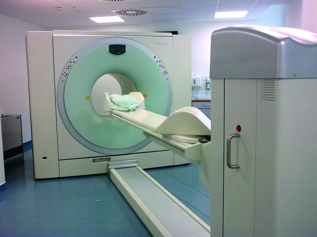
The survey included responses from investigators from the Treatments Against Rheumatoid Arthritis and Effect on 18F-fluorodeoxyglucose (FDG) PET/CT (TARGET) trial, conducted between 2015 and 2021. The 24-week trial included patients from 28 centers in the United States to investigate how different disease-modifying antirheumatic drugs can reduce cardiovascular and joint inflammation, assessed via whole body FDG PET/CT. The survey was a planned substudy of the TARGET trial and is “the first study that examines researchers’ attitudes and beliefs regarding incidental research findings from whole body FDG PET/CT,” Kang and her coauthors wrote.
This news organization reported the main results of the TARGET trial in 2022.
Eighteen of the 28 site primary investigators (PIs) of the TARGET trial participated in the survey, which was published in Arthritis Care & Research in September 2024.
TARGET Trial Incidental Findings
The TARGET trial enrolled 159 patients, of whom 82% had at least one incidental finding and 62% had one or more FDG-avid incidental findings. There were 46 “clinically actionable findings” for 40 participants overall; the reading radiologists recommended additional imaging for 28 findings and specialist consultation or procedural evaluation for 15 findings.
Details on these incidental findings were presented in a poster at the annual meeting of the American College of Rheumatology (ACR), held in Washington, DC.
The most common non–FDG-avid findings were pulmonary nodules, diverticulosis, cholelithiasis, sinus disease, and vascular calcifications. The most common FDG-avid findings were hypermetabolic lymphadenopathy, increased gastric/esophageal uptake, increased bowel uptake, and increased pharyngeal uptake.
In the related survey, 11 respondents (61%) said they returned any incidental findings to participants and 5 (28%) did not; the remaining 2 respondents did not know.
Across all study PIs, 22% felt that incidental findings were beneficial, 39% said they were potentially beneficial, and 11% said they were potentially detrimental. PIs that ranked incidental findings as potentially detrimental pointed to how these findings led to invasive additional testing.
“One of my subjects was found to have diverticulosis, which needed an invasive procedure to rule out malignancy,” one respondent wrote. “However, the subject had already had a colonoscopy months prior to the PET findings, which was still not deemed sufficient by the nuclear radiologist and GI consultant, so he had to have another colonoscopy, which was benign, but uncomfortable.”
Obligation to Return Findings
All investigators agreed that incidental findings should be shared with patients if they revealed a high-risk medical condition that can be treated; had important health implications such as premature death or substantial morbidity; and their health could be improved with proven preventive or therapeutic interventions.
There was more disagreement on whether to share that the FDG PET/CT revealed no findings or if the test revealed a finding without clear medical importance of which the research participant may not be aware.
An example of a less-specific finding could be something like increased FDG uptake in a particular area, like the bowel, Kang explained.
“The question is: What does that mean?” she said. “How do you interpret that?”
While some PIs might feel obligated to share all results with patients, sharing ambiguous incidental findings will likely not be helpful to the patient, said Arthur Caplan, PhD, of the Division of Medical Ethics at New York University (NYU) Grossman School of Medicine, New York City.
“Dealing in unknowns and uncertainties when you’re diagnosing doesn’t really do people very much good,” he said in an interview.
While most survey respondents said they were at least moderately obligated to disclose incidental research findings if a patient requests them, Caplan noted that it was ultimately the researchers’ decision.
“Patient preferences are something to take into account, but they’re not final. If the research team says, ‘we don’t know, it’s too uncertain, it’s too new,’ then I don’t think they have any obligation to return that [information],” he said. “You can’t tell somebody what you don’t understand.”
Conversely, the clearer the incidental finding, the stronger the obligation to share that information with research participants, he continued.
Need for a Standardized Approach
The TARGET study, like many research studies, left the management of incidental imaging findings to individual research sites and investigators.
It’s possible that different sites responded to these ambiguous clinical findings in different ways, Kang noted.
“If there’s a situation that’s difficult to interpret as it is, you can imagine that the resulting actions that may result from that can vary, too,” she said, which highlights the need for more specific and standardized guidance.
One way to approach this, Caplan noted, is establishing an agreed-upon approach for dealing with any incidental findings across all research sites before a study begins.
“If there is going to be a common study at many sites, then they should have a common response on what they are going to do,” he noted, and how they will share that information effectively with the research participants to ensure it’s understandable. However, in a lot of research studies, each site has its own approach.
“Right now, it’s all over the place and that shouldn’t be,” he said.
Institutional review boards (IRBs) could be one resource to help build detailed guidance on managing unclear incidental findings in future research, wrote Kang and coauthors.
“For incidental findings from whole body FDG PET/CT that are not clearly actionable or less straightforward, IRBs may consider requiring a certain level of follow-up for different categories or types of incidental findings or require that all incidental findings are reviewed by an independent group that would provide timely recommendations on the most appropriate return and management of those findings,” Kang and colleagues wrote. “With IRB guidance, very specific and detailed policies and procedures for returning and managing incidental findings should be established for every study, with consistency among the research sites of multicenter trials.”
The TARGET trial and survey were funded by a grant from the National Institute of Arthritis and Musculoskeletal and Skin Diseases. Kang reported receiving research funding from the National Institutes of Health and the Rheumatology Research Foundation. Caplan serves as a contributing author for this news organization and served on an independent bioethics panel for compassionate drug use that was funded by Johnson & Johnson through the NYU Grossman School of Medicine.
A version of this article first appeared on Medscape.com.
Disparate views on managing incidental imaging findings made during clinical research — particularly for unclear results — signal a need for standardized guidance, according to recent survey results.
Respondents were split on whether it was the site primary investigator’s responsibility to decide which incidental findings should be reported back to the patient, and the most commonly cited challenges included adequately explaining these findings and the follow-up required. These issues were most present when dealing with nonspecific incidental findings or findings of unclear importance, said lead author Jane S. Kang, MD, a bioethicist and associate professor of medicine in the Division of Rheumatology at Columbia University Irving Medical Center, New York City.
“It can be difficult to have a clear approach” when it comes to these situations that are not black and white, and it is hard to get a clear answer, she said in an interview.
The survey included responses from investigators from the Treatments Against Rheumatoid Arthritis and Effect on 18F-fluorodeoxyglucose (FDG) PET/CT (TARGET) trial, conducted between 2015 and 2021. The 24-week trial included patients from 28 centers in the United States to investigate how different disease-modifying antirheumatic drugs can reduce cardiovascular and joint inflammation, assessed via whole body FDG PET/CT. The survey was a planned substudy of the TARGET trial and is “the first study that examines researchers’ attitudes and beliefs regarding incidental research findings from whole body FDG PET/CT,” Kang and her coauthors wrote.
This news organization reported the main results of the TARGET trial in 2022.
Eighteen of the 28 site primary investigators (PIs) of the TARGET trial participated in the survey, which was published in Arthritis Care & Research in September 2024.
TARGET Trial Incidental Findings
The TARGET trial enrolled 159 patients, of whom 82% had at least one incidental finding and 62% had one or more FDG-avid incidental findings. There were 46 “clinically actionable findings” for 40 participants overall; the reading radiologists recommended additional imaging for 28 findings and specialist consultation or procedural evaluation for 15 findings.
Details on these incidental findings were presented in a poster at the annual meeting of the American College of Rheumatology (ACR), held in Washington, DC.
The most common non–FDG-avid findings were pulmonary nodules, diverticulosis, cholelithiasis, sinus disease, and vascular calcifications. The most common FDG-avid findings were hypermetabolic lymphadenopathy, increased gastric/esophageal uptake, increased bowel uptake, and increased pharyngeal uptake.
In the related survey, 11 respondents (61%) said they returned any incidental findings to participants and 5 (28%) did not; the remaining 2 respondents did not know.
Across all study PIs, 22% felt that incidental findings were beneficial, 39% said they were potentially beneficial, and 11% said they were potentially detrimental. PIs that ranked incidental findings as potentially detrimental pointed to how these findings led to invasive additional testing.
“One of my subjects was found to have diverticulosis, which needed an invasive procedure to rule out malignancy,” one respondent wrote. “However, the subject had already had a colonoscopy months prior to the PET findings, which was still not deemed sufficient by the nuclear radiologist and GI consultant, so he had to have another colonoscopy, which was benign, but uncomfortable.”
Obligation to Return Findings
All investigators agreed that incidental findings should be shared with patients if they revealed a high-risk medical condition that can be treated; had important health implications such as premature death or substantial morbidity; and their health could be improved with proven preventive or therapeutic interventions.
There was more disagreement on whether to share that the FDG PET/CT revealed no findings or if the test revealed a finding without clear medical importance of which the research participant may not be aware.
An example of a less-specific finding could be something like increased FDG uptake in a particular area, like the bowel, Kang explained.
“The question is: What does that mean?” she said. “How do you interpret that?”
While some PIs might feel obligated to share all results with patients, sharing ambiguous incidental findings will likely not be helpful to the patient, said Arthur Caplan, PhD, of the Division of Medical Ethics at New York University (NYU) Grossman School of Medicine, New York City.
“Dealing in unknowns and uncertainties when you’re diagnosing doesn’t really do people very much good,” he said in an interview.
While most survey respondents said they were at least moderately obligated to disclose incidental research findings if a patient requests them, Caplan noted that it was ultimately the researchers’ decision.
“Patient preferences are something to take into account, but they’re not final. If the research team says, ‘we don’t know, it’s too uncertain, it’s too new,’ then I don’t think they have any obligation to return that [information],” he said. “You can’t tell somebody what you don’t understand.”
Conversely, the clearer the incidental finding, the stronger the obligation to share that information with research participants, he continued.
Need for a Standardized Approach
The TARGET study, like many research studies, left the management of incidental imaging findings to individual research sites and investigators.
It’s possible that different sites responded to these ambiguous clinical findings in different ways, Kang noted.
“If there’s a situation that’s difficult to interpret as it is, you can imagine that the resulting actions that may result from that can vary, too,” she said, which highlights the need for more specific and standardized guidance.
One way to approach this, Caplan noted, is establishing an agreed-upon approach for dealing with any incidental findings across all research sites before a study begins.
“If there is going to be a common study at many sites, then they should have a common response on what they are going to do,” he noted, and how they will share that information effectively with the research participants to ensure it’s understandable. However, in a lot of research studies, each site has its own approach.
“Right now, it’s all over the place and that shouldn’t be,” he said.
Institutional review boards (IRBs) could be one resource to help build detailed guidance on managing unclear incidental findings in future research, wrote Kang and coauthors.
“For incidental findings from whole body FDG PET/CT that are not clearly actionable or less straightforward, IRBs may consider requiring a certain level of follow-up for different categories or types of incidental findings or require that all incidental findings are reviewed by an independent group that would provide timely recommendations on the most appropriate return and management of those findings,” Kang and colleagues wrote. “With IRB guidance, very specific and detailed policies and procedures for returning and managing incidental findings should be established for every study, with consistency among the research sites of multicenter trials.”
The TARGET trial and survey were funded by a grant from the National Institute of Arthritis and Musculoskeletal and Skin Diseases. Kang reported receiving research funding from the National Institutes of Health and the Rheumatology Research Foundation. Caplan serves as a contributing author for this news organization and served on an independent bioethics panel for compassionate drug use that was funded by Johnson & Johnson through the NYU Grossman School of Medicine.
A version of this article first appeared on Medscape.com.
FROM ARTHRITIS CARE & RESEARCH
Kidney, Cardiovascular Benefits Seen With GLP-1 RA Drugs in SLE, Lupus Nephritis
WASHINGTON — Glucagon-like peptide 1 receptor agonist (GLP-1 RA) medications appear beneficial for people with systemic lupus erythematosus (SLE) and lupus nephritis, two new studies suggest.
“The risk of cardiovascular disease is thought to be at least double that for people with lupus ... and we know the risk of progressing to end-stage renal disease [ESKD] for patients with lupus nephritis can be as high as 10%-30%, so there’s clearly a major unmet need for new treatments and approaches to improve these outcomes, perhaps with adjunctive treatment beyond our typical immunosuppressive therapy,” April Jorge, MD, of Massachusetts General Hospital, Boston, said at the annual meeting of the American College of Rheumatology (ACR).
The GLP-1 RAs are approved for the treatment of type 2 diabetes (T2D) and obesity. They also have proven cardiovascular benefit, along with emerging data suggesting kidney protection independent of glucose lowering. Jorge presented findings from a study using data from the US multicenter electronic health record database TriNetX, showing that, among patients who had both T2D and SLE, those using GLP-1 RAs had lower risks for major adverse cardiac events (MACE), venous thrombosis, kidney disease progression, and all-cause mortality, compared with those using a different class of T2D medication.
A second study using TriNetX, presented at the same ACR meeting session by Anna-Kay Palmer, MD, a third-year internal medicine resident at Jefferson Einstein Hospital, Philadelphia, Pennsylvania, showed that GLP-1 RAs reduced the risk of progression to ESKD in patients with lupus nephritis, possibly caused by reductions in pro-inflammatory mediators.
Asked to comment, session moderator Diane L. Kamen, MD, professor of medicine at the Medical University of South Carolina Division of Rheumatology, Charleston, said in an interview that she definitely supports the use of GLP-1 RAs for patients who have SLE and/or lupus nephritis and also a drug label indication, either T2D or obesity. “[The GLP-1 RA prescriber] will usually run it by rheumatology to make sure that it doesn’t conflict with any of their other medical treatment, and it’s very reassuring to know that they could actually get a win-win.”
But as far as prescribing off-label for those with SLE/lupus nephritis who don’t have other GLP-1 RA indications, Kamen said, “that’s a black hole at this point. We need to do those prospective studies. But if they have another indication, yes.”
Cardiovascular, Kidney Benefits of GLP-1 RAs
Jorge noted that patients with lupus were excluded from the randomized clinical trials of GLP-1 RAs, so the current study was designed to investigate the potential impact of these medications on cardiovascular and kidney outcomes in patients with SLE and lupus nephritis.
From TriNetX data for 46 healthcare organizations nationwide, a total of 96,511 patients with both SLE and T2D but not ESKD had initiated either a GLP-1 RA or another diabetes drug class, dipeptidyl peptidase 4 inhibitors (DPP4i), between October 2006 and August 2021. Of those, 29,177 had lupus nephritis.
Propensity score matching for factors such as demographics, lupus severity, comorbidities, and medication use was used to emulate a randomized trial. This yielded 25,838 with SLE and T2D, of whom 910 initiated a GLP-1 RA and 1004 started a DPP4i, and 12,387 with lupus nephritis and T2D, including 267 on a GLP-1 RA and 324 on a DPP4i. After matching, the mean age was 55 years, more than 90% were women, and just under half were White individuals. About one third had chronic kidney disease stages ≥ 3, and about 15% had heart failure.
Over an average follow-up time of 1.2-1.4 years among those with SLE, the hazard ratio (HR) for MACE (a composite of myocardial infarction, stroke, and heart failure) for those taking a GLP-1 RA vs a DPP4i was 0.66, a significant difference. And for venous thrombosis, the HR was also significant at 0.49.
Kidney disease progression, defined as an estimated glomerular filtration rate decline of 30% or more or new ESKD, was significantly less likely in the GLP-1 RA group, with a HR of 0.77. All-cause mortality also was dramatically reduced (HR, 0.26). As expected, there was no difference in control outcome, genital infections (HR, 1.02).
In the subgroup with lupus nephritis, there were also lower risks for both MACE (HR, 0.64) and for renal progression (HR, 0.70). “The findings suggest similar cardiac and kidney benefits among patients with SLE and lupus nephritis as have been observed in other populations,” Jorge concluded.
Kamen commented that the study design “was pretty brilliant, because you wouldn’t be able to do a placebo-controlled trial since the indication was diabetes ... but the fact is you do see that the GLP-1 RA gets the benefit whereas the other drug does not.”
Next steps, Jorge said, will be mechanistic studies to better understand the effects of GLP-1 RAs in lupus and other rheumatic diseases, prospective studies of GLP-1 RAs in SLE and lupus nephritis without diabetes, and clarification of ideal timing for GLP-1 RA use in SLE and lupus nephritis.
“Ideally, with our prospective studies with these patients we can try to isolate the effect on patients with lupus and also better understand whether there might be an impact on disease activity through the anti-inflammatory effects of these medications, rather than just the cardioprotective and nephroprotective benefits,” she said.
In Those With Lupus Nephritis, Kidney Protection Seen
In her presentation, Palmer noted that, despite immunosuppressive therapies for SLE, 10%-20% of patients who develop lupus nephritis will progress to ESKD within 5 years of diagnosis.
She added that GLP-1 RAs have been shown to reduce albuminuria in people with diabetes and have been hypothesized to reduce inflammation through multiple pathways, thereby potentially reducing kidney disease independently of the presence of diabetes or weight loss. These pathways include modulating immune cell signaling and reducing pro-inflammatory cytokines.
Based on all this, Palmer and colleagues used International Classification of Diseases – 10th edition diagnostic codes in TriNetX to identify 839 patients who had been diagnosed with lupus nephritis between 2014 and 2024 and who were prescribed liraglutide, dulaglutide, semaglutide, or exenatide for any time after the lupus nephritis diagnosis. Another 29,840 patients with lupus nephritis had not used GLP-1 RAs.
After 1:1 propensity score matching for age, sex, race, ethnicity, presence of hypertension, diabetes, use of immunosuppressive and diabetes medication, smoking, obesity, and statin use, there were 735 individuals in each group. About two thirds in each had diabetes, whereas the rest had been prescribed the GLP-1 RAs for other indications.
Patients who were not on GLP-1 RAs were twice as likely to develop ESKD or dialysis (8.88% vs 3.971%; odds ratio, 2.35; P = .001).
Kamen pointed out that not including the use of angiotensin-converting enzyme inhibitors and angiotensin receptor blockers was a study flaw. On the other hand, the fact that not everyone in this study had diabetes was an advantage.
Jorge received grant/research support from Bristol-Myers Squibb, Cabaletta Bio, and the Lupus Clinical Investigator Network. Kamen is an adviser/review panel member for Alpine Immune Sciences. Palmer had no disclosures.
A version of this article appeared on Medscape.com.
FROM ACR 2024
RA Assessment Via Automated Ultrasound Scanner With AI Saves Time, Performs as Well as Rheumatologists
WASHINGTON — A fully automated ultrasound scanning system combined with artificial intelligence–based disease activity scoring performed as well as expert rheumatologists in hand joint assessment of patients with rheumatoid arthritis (RA), new research found.
The system, made by a Danish company called ROPCA, comprises an ultrasound scanner called ARTHUR (RA Ultrasound Robot) that interacts directly with the patient and scans 11 joints per hand and a neural network–based software system, DIANA (Diagnosis Aid Network for RA), that evaluates the images and monitors RA activity.
The combined system classifies the degree of RA according to the joint European Alliance of Associations for Rheumatology (EULAR)–Outcome Measures in Rheumatology (OMERACT) standards for RA diagnosis. It received a CE Mark in Europe in 2022 and is currently in use in six rheumatology clinics in Denmark, Germany, Switzerland, and Austria, with more to come, ROPCA Co-founder and Chief Medical Officer Søren A. Just, MD, said in an interview.
“Automated systems could help rheumatologists in the early detection and monitoring of arthritis diseases. Systems can be placed or move in areas with insufficient rheumatological expertise,” Just said during a special late-breaker session presentation at the annual meeting of the American College of Rheumatology (ACR).
He said in an interview: “Currently, there are so many people referred and few and fewer rheumatologists. So we need to think differently. We need good automated assistants.” As a screening tool, the system can determine whether a person with hand pain has RA or just osteoarthritis “and also can give the patient an immediate answer, instead of waiting sometimes up to 6 months to get the information.”
Just, who is also a senior physician in the Department of Internal Medicine at Odense University Hospital in Denmark, said that his department is also using the system to assess flares in patients with established RA. “They can have a blood sample taken. They’re scanned by the robot, and you can see if there is any disease activity. But I think that screening of patients with joint pain is the beginning.”
Asked to comment, session moderator Gregory C. Gardner, MD, Emeritus Professor in the Division of Rheumatology at the University of Washington, Seattle, and a member of the ACR conference program committee, said in an interview “one of the reasons we chose to feature this abstract is because we’re interested in science at the convergence. We really thought this was a potential way to move the field forward for rheumatologists.”
Gardner said it’s an advantage that the patient could potentially have an ARTHUR scan with a DIANA report and get blood tests done prior to a visit with the rheumatologist. “It’s really time-consuming for a human to do these studies, so if you automate it, that’s a step forward in terms of having the data available for the rheumatologist to view and use sequentially to follow how patients are doing.”
When introducing Just’s presentation, Gardner called it “the coolest abstract of the meeting.”
Both DIANA and ARTHUR Performed At Least as Well as Human Rheumatologists
In the study, 30 patients with RA underwent two scans by ARTHUR, followed by a scan from a rheumatologist specialist in musculoskeletal ultrasound. The scans were sent to DIANA, who graded the images according to the Global OMERACT-EULAR Synovitis Score, as did the human rheumatologist.
A “ground truth” was established by another human expert who evaluated both ARTHUR’s and the other rheumatologist’s images, blinded to the scanning method. The image with the highest disease activity was deemed “ground truth,” and agreement with that was assessed for the two individual methods.

Just showed a video of a patient being scanned by ARTHUR. The machine verbally guided her through removing her jewelry, applying the gel, and placing her hand on the screen under the scanner. ARTHUR’s arm moved around on the patient’s hand, locating the best angles to take grayscale images and Doppler images and Doppler video. The scan takes 15-20 minutes, and the images are stored, Just said.
The study patients had a mean age of 65 years, and 23 of the 30 were men. Their average disease duration was 11 years, and mean Disease Activity Score in 28 joints using C-reactive protein was 3.86, indicating moderate disease. A majority (73%) of patients were taking disease-modifying antirheumatic drugs, and about one third were taking biologics. ARTHUR scanned a total of 660 joints, and 564 scans were successful.
For repeatability between the two ARTHUR scans, percent exact agreement was 63% for synovial hypertrophy, 75% for Doppler activity, and 60% combined. Percent close (within a point) agreements were 93%, 94%, and 92%, respectively. Binary agreements as to whether the joint was healthy vs diseased were 88%, 91%, and 85%, respectively.
At the joint level, ARTHUR and DIANA’s percent exact agreement with ground truth was 49% for synovial hypertrophy, 63% for Doppler activity, and 48% combined. Binary agreements with disease vs healthy were 80%, 88%, and 78%, respectively.
The human rheumatologists scored very similarly. Percent exact agreement with ground truth was 51% for synovial hypertrophy, 64% for Doppler activity, and 50% combined. Percent close agreements were 94%, 94%, and 92%, respectively. And binary agreements with diseased vs healthy were 83%, 91%, and 80%, respectively.
At the patient level (all joints combined), ARTHUR and DIANA’s binary disease assessment of healthy vs disease showed agreement with the ground truth of 87% for synovial hypertrophy, 83% for Doppler activity, and 87% combined. Here, the rheumatologists scored lower, at 53%, 67%, and 60%, respectively.
“In this study, we think the precision of ARTHUR and DIANA was comparable to that of an experienced rheumatologist, at both the joint and patient level,” Just said.
Gardner pointed out another advantage of the system. “DIANA doesn’t get fatigued. ... With human reading, the precision may change based on the time of day or stress level. ... But with DIANA, you’re going to get consistent information.”
Just said that the Arthritis Foundation in Germany recently put ARTHUR and DIANA on a bus and took it to cities that lacked a rheumatologist. Patients lined up, answered a questionnaire, had blood drawn, and received their scans. A rheumatologist on the bus then interpreted the data and consulted with the individuals about their RA risk. “In the last trip, we screened 800 patients in 6 days. So there are definitely possibilities here.”
Just is co-owner of ROPCA. Gardner had no disclosures.
A version of this article appeared on Medscape.com.
WASHINGTON — A fully automated ultrasound scanning system combined with artificial intelligence–based disease activity scoring performed as well as expert rheumatologists in hand joint assessment of patients with rheumatoid arthritis (RA), new research found.
The system, made by a Danish company called ROPCA, comprises an ultrasound scanner called ARTHUR (RA Ultrasound Robot) that interacts directly with the patient and scans 11 joints per hand and a neural network–based software system, DIANA (Diagnosis Aid Network for RA), that evaluates the images and monitors RA activity.
The combined system classifies the degree of RA according to the joint European Alliance of Associations for Rheumatology (EULAR)–Outcome Measures in Rheumatology (OMERACT) standards for RA diagnosis. It received a CE Mark in Europe in 2022 and is currently in use in six rheumatology clinics in Denmark, Germany, Switzerland, and Austria, with more to come, ROPCA Co-founder and Chief Medical Officer Søren A. Just, MD, said in an interview.
“Automated systems could help rheumatologists in the early detection and monitoring of arthritis diseases. Systems can be placed or move in areas with insufficient rheumatological expertise,” Just said during a special late-breaker session presentation at the annual meeting of the American College of Rheumatology (ACR).
He said in an interview: “Currently, there are so many people referred and few and fewer rheumatologists. So we need to think differently. We need good automated assistants.” As a screening tool, the system can determine whether a person with hand pain has RA or just osteoarthritis “and also can give the patient an immediate answer, instead of waiting sometimes up to 6 months to get the information.”
Just, who is also a senior physician in the Department of Internal Medicine at Odense University Hospital in Denmark, said that his department is also using the system to assess flares in patients with established RA. “They can have a blood sample taken. They’re scanned by the robot, and you can see if there is any disease activity. But I think that screening of patients with joint pain is the beginning.”
Asked to comment, session moderator Gregory C. Gardner, MD, Emeritus Professor in the Division of Rheumatology at the University of Washington, Seattle, and a member of the ACR conference program committee, said in an interview “one of the reasons we chose to feature this abstract is because we’re interested in science at the convergence. We really thought this was a potential way to move the field forward for rheumatologists.”
Gardner said it’s an advantage that the patient could potentially have an ARTHUR scan with a DIANA report and get blood tests done prior to a visit with the rheumatologist. “It’s really time-consuming for a human to do these studies, so if you automate it, that’s a step forward in terms of having the data available for the rheumatologist to view and use sequentially to follow how patients are doing.”
When introducing Just’s presentation, Gardner called it “the coolest abstract of the meeting.”
Both DIANA and ARTHUR Performed At Least as Well as Human Rheumatologists
In the study, 30 patients with RA underwent two scans by ARTHUR, followed by a scan from a rheumatologist specialist in musculoskeletal ultrasound. The scans were sent to DIANA, who graded the images according to the Global OMERACT-EULAR Synovitis Score, as did the human rheumatologist.
A “ground truth” was established by another human expert who evaluated both ARTHUR’s and the other rheumatologist’s images, blinded to the scanning method. The image with the highest disease activity was deemed “ground truth,” and agreement with that was assessed for the two individual methods.
Just showed a video of a patient being scanned by ARTHUR. The machine verbally guided her through removing her jewelry, applying the gel, and placing her hand on the screen under the scanner. ARTHUR’s arm moved around on the patient’s hand, locating the best angles to take grayscale images and Doppler images and Doppler video. The scan takes 15-20 minutes, and the images are stored, Just said.
The study patients had a mean age of 65 years, and 23 of the 30 were men. Their average disease duration was 11 years, and mean Disease Activity Score in 28 joints using C-reactive protein was 3.86, indicating moderate disease. A majority (73%) of patients were taking disease-modifying antirheumatic drugs, and about one third were taking biologics. ARTHUR scanned a total of 660 joints, and 564 scans were successful.
For repeatability between the two ARTHUR scans, percent exact agreement was 63% for synovial hypertrophy, 75% for Doppler activity, and 60% combined. Percent close (within a point) agreements were 93%, 94%, and 92%, respectively. Binary agreements as to whether the joint was healthy vs diseased were 88%, 91%, and 85%, respectively.
At the joint level, ARTHUR and DIANA’s percent exact agreement with ground truth was 49% for synovial hypertrophy, 63% for Doppler activity, and 48% combined. Binary agreements with disease vs healthy were 80%, 88%, and 78%, respectively.
The human rheumatologists scored very similarly. Percent exact agreement with ground truth was 51% for synovial hypertrophy, 64% for Doppler activity, and 50% combined. Percent close agreements were 94%, 94%, and 92%, respectively. And binary agreements with diseased vs healthy were 83%, 91%, and 80%, respectively.
At the patient level (all joints combined), ARTHUR and DIANA’s binary disease assessment of healthy vs disease showed agreement with the ground truth of 87% for synovial hypertrophy, 83% for Doppler activity, and 87% combined. Here, the rheumatologists scored lower, at 53%, 67%, and 60%, respectively.
“In this study, we think the precision of ARTHUR and DIANA was comparable to that of an experienced rheumatologist, at both the joint and patient level,” Just said.
Gardner pointed out another advantage of the system. “DIANA doesn’t get fatigued. ... With human reading, the precision may change based on the time of day or stress level. ... But with DIANA, you’re going to get consistent information.”
Just said that the Arthritis Foundation in Germany recently put ARTHUR and DIANA on a bus and took it to cities that lacked a rheumatologist. Patients lined up, answered a questionnaire, had blood drawn, and received their scans. A rheumatologist on the bus then interpreted the data and consulted with the individuals about their RA risk. “In the last trip, we screened 800 patients in 6 days. So there are definitely possibilities here.”
Just is co-owner of ROPCA. Gardner had no disclosures.
A version of this article appeared on Medscape.com.
FROM ACR 2024
Uric Acid Levels, Gout Symptoms Improved With Plant-Based Diet in Pilot Trial
A Mediterranean-inspired plant-based diet improved self-reported measures of gout as well as uric acid levels, a pilot study has found.
There hasn’t been a lot of research on diet in gout, according to Anna Kretova, RD, who presented the study at the annual research symposium of the Gout Hyperuricemia and Crystal-Associated Disease Network. She noted that a 2019 systematic review of low-calorie diets, low-purine diets, and Mediterranean diets found that uric acid levels below 0.6 mmol/L were achieved only in those on the Mediterranean diet (Nutrients. 2019 Dec 4;11[12]:2955). A 2020 study compared a low-fat, high-carbohydrate, plant-based diet vs an animal-based, ketogenic diet in healthy individuals. After 2 weeks, uric acid levels increased in those on the animal-based, low-carb diet and decreased in those on the plant-based diet.
Some foods are considered to be proinflammatory and generally come from animal origins, including saturated fats and animal protein in addition to ultraprocessed foods. Foods that have anti-inflammatory properties are mostly plant based and unprocessed and often rich in fiber. “From recent interventional studies, we also know that the whole-foods plant-based diet has shown to be effective as treatments of the main comorbidities of gout, such as obesity, cardiovascular disease, or [osteoarthritis],” said Kretova, who is a registered dietitian and a researcher at the Reade Rehabilitation and Rheumatology Center, Amsterdam, the Netherlands.
Those findings led the researchers to develop a whole-foods, plant-based diet and test its effect on serum uric acid in patients with gout, as well as gout disease activity and cardiovascular disease risk. Participants could not eat meat, fish, eggs, or dairy.
The trial included 33 individuals with gout who were randomized to a 16-week intervention with five consultations with a registered dietitian (n = 18) or a wait-list control group (n = 15) who received standard care. The mean age overall was 52 years, and 91% were men. The mean body mass index (BMI) was 32.6 kg/m2, and the median uric acid level was 0.50 mmol/L (8.4 mg/dL).
Among gout-related outcomes, the researchers noted improvements in gout severity as measured by visual analog scale (VAS; between group difference, –2.0; P =.01), pain as measured by VAS (between group difference, –2.0; P =.04), and uric acid levels after adjustment for age, sex, and BMI (between group difference, –0.05 mmol/L, P =.004). There were also improvements in the intervention group in weight loss (between group difference, –5.3 kg; P <.0001), BMI (between group difference, –1.7; P < .0001), waist circumference (between group difference, –3.9 cm; P = .004), and low-density lipoprotein (LDL) cholesterol (between group difference, –0.5; P = .007).
At 16 weeks, “we concluded that a Mediterranean-inspired whole-foods, plant-based diet significantly lowers serum uric acid in patients with gout and abdominal obesity, and additionally, the diet reduces gout-related pain and disease activity, promotes substantial weight loss, decreases weight circumference, and improves LDL cholesterol levels, and thus decreases [cardiovascular disease] risk in these patients,” Kretova said.
She added that some might question whether a uric acid reduction of –0.05 mmol/L is clinically relevant. “We would argue it is because of the strong decrease in disease activity and pain in the intervention group,” Kretova said.
The study is limited by its small size, the fact that it was not blinded, and the 4-month duration, which might be too short to capture potential indirect effects of diet on hyperuricemia and chronic inflammation, Kretova said. The group is planning to follow participants out to 12 months in an extension study.
During the Q&A session after the presentation, an audience member asked if the participants were vegetarians before they entered the study, and whether the dietary change could be sustained. “It’s a very good proof-of-concept study, but whether an intervention based entirely on plant-based therapy will be something that patients will be able to adhere to long term [is uncertain],” Kretova said.
She was optimistic, even though the participants generally enjoyed food and ate a lot of red meat. “I think there will be a gradation of people who can sustain and who cannot sustain [the diet]. From what we saw, people actually found it easier to follow than they expected, and a lot of participants changed their diet permanently for the better. Not everyone became [entirely] plant-based, but they became much more plant-based than they expected from themselves. So, it is definitely feasible,” she said.
Kretova reported no relevant financial relationships.
A version of this article appeared on Medscape.com.
FROM G-CAN 2024
New Gout Remission Criteria Approved
In a nearly unanimous vote at the annual research symposium of the Gout Hyperuricemia and Crystal-Associated Disease Network (G-CAN), members approved a revision to gout remission criteria first established in 2016. The new version simplifies the definition in response to patient comments that the earlier version was redundant in some areas.
The previous version was developed following deliberations by 49 clinicians and researchers with experience in gout. They settled on a definition of gout remission that included five criteria:
- Serum urate levels lower than 0.36 mmol/L measured at least twice over 12 months, with no intervening values of 0.36 mmol/L or higher
- No gout flares over 12 months
- No tophi
- Pain score due to gout < 2 at least twice over 12 months on a 10-point Likert scale or 10-cm visual analog scale, with no intervening values ≥ 2
- Patient global assessment of gout disease activity < 2 on a 10-point Likert scale or 10-cm visual analog scale, with no intervening values of ≥ 2.
Some participants reported that patients sometimes misattributed pain from other sources while using patient-reported outcomes (PROs). The argument for keeping PROs was that they are validated measures and endorsed by Outcome Measures in Rheumatology. Nevertheless, there was no direct patient involvement in the development of the 2016 criteria.
Researchers later interviewed 20 individuals with well-controlled gout to get their feedback on the 2016 criteria. Those individuals endorsed the existing criteria and did not suggest any new ones, but they suggested that the pain due to gout and the absence of gout flares were redundant measures. One said: “If you have no flare-ups, you’ve got no pain; it sort of answers itself.”
“That was a bit challenging for us because it wasn’t quite what we expected, but I think it did make us look again at the definition and think about whether we could simplify the definition further,” Nicola Dalbeth, MBChB, said during a presentation at G-CAN. Dalbeth is an academic rheumatologist at the University of Auckland, in New Zealand, who was also the lead author of the original criteria.
Simplified Version Created With Only Three Criteria
In response to these points, researchers produced a revised version with only three criteria, including the serum urate, absence of gout flares, and absence of subcutaneous tophi at the time of assessment.
To determine if the simplified criteria performed well, they compared the original and revised remission criteria in the context of the CARES trial, the Nottingham nurse-led trial, and randomized controlled trials in patients with gout that were conducted in New Zealand (here and here).
Dansoa Tabi-Amponsah, a PhD candidate at the University of Auckland, presented results of a study comparing the two versions in the Nottingham trial, which included 517 participants who received nurse-led or usual general practitioner care. The nurse-led care included education, regular follow-up and serum urate testing, individualized advice on gout flare management, and escalation of urate-lowering therapy with a treat-to-target strategy.
Both definitions demonstrated a link between the nurse-led strategy and increased rates of remission at year 1 and year 2, although the simplified definition found that more patients were in remission (17.6% vs 9.9% at year 1 and 42.7% vs 28.4% at year 2, both P < .001). “This is something we’ve seen across all of our analyses,” said Tabi-Amponsah.
Both criteria also found significant differences in remission rates between the nurse-led group in year 2 vs year 1 but not in the usual care group.
Participants who achieved remission had better gout impact scale scores in areas like worrying that a gout attack will occur, fears of worsening gout, and concerns about the impact of gout on future activities. “This is important because during that qualitative study, a key aspect of being in remission was no longer being worried about their gout, no longer feeling anxious about having constant gout flares, and having control over their gout. So, it’s important to note that despite the absence of PROs in that simplified definition, it’s still able to align with the patients’ perspectives of their disease state,” said Tabi-Amponsah.
During the Q&A period after her talk, an audience member asked whether the higher rate of remission found by the simplified criteria is actually a good thing. “If I compare that to rheumatoid arthritis, when you use DAS28 you have a lot more remission, but still progression. So, are we missing some people? Are we including people in remission that still have disease?” she asked.
Tabi-Amponsah responded that the pain and patient global assessment domains seem to be quite difficult to achieve. In a separate analysis, the researchers examined tender and swollen joint counts and found that those achieving remission no longer had tender or swollen joints. “So, we don’t think the simplified definition is heavily misclassifying anyone as being in remission,” she said.
During the Q&A following Dalbeth’s talk, an audience member asked about patients with what he described as “mountains of tophi,” despite responding well to uricase therapy. “They may take months or even a year to really resolve that burden. They may be doing very well, yet they’re not going to be in remission because they’ve still got visible tophi. So, are we underselling them, and do we need a different definition for them doing well that this doesn’t capture?” he asked.
Dalbeth suggested that patients with large amounts of tophi aren’t really in remission. “I think we do need to be thinking about the disease, not just in terms of just crystals or just inflammation, but actually trying to integrate both of those, and I think this is where these composite measures might work quite well. I think we need to be aiming for holistic disease control, which is essentially what this is,” she said.
Tabi-Amponsah and Dalbeth did not disclose any financial relationships.
A version of this article appeared on Medscape.com.
FROM G-CAN 2024
Could Probiotics Tuned to Reduce Intestinal Urate Counter Gout?
Efforts to combat hyperuricemia may find help from gut microbes, according to Dylan Dodd, MD, PhD, who spoke at the annual research symposium of the Gout, Hyperuricemia, and Crystal-Associated Disease Network.
Dodd is an assistant professor of pathology and microbiology and immunology at Stanford University, Palo Alto, California, where he studies novel metabolic pathways in microbes. “The idea is that we can leverage these novel pathways that microbes have as therapeutics to promote human health, and in particular for this meeting today, we’re focused on hyperuricemia and how microbes that break down purines may actually have a role as urate-lowering therapies,” Dodd said during his presentation.
Specifically, he highlighted the fact that some microbes found in the gut break down purines as a food source, producing both energy and molecular building blocks for their own use. Dietary purines, left intact, can otherwise be absorbed and metabolized by the body to produce urate.
Nucleic acids like DNA and RNA in the diet are first broken down by enzymes produced in the pancreas, resulting in purine nucleosides, which in turn are believed to be the source of purines absorbed in the small intestine and eventually into circulation, according to Dodd. “I really view urate in the intestine as being in equilibrium between being secreted into the lumen but also being reabsorbed, and specifically, as it pertains to microbes in the gut. If the microbes degrade the urate, then it will limit its reabsorption, and that could increase net excretion,” Dodd said.
There is evidence that some strains of Lactobacillus species, which are the most important group in the human gut, can metabolize purine nucleosides, he said. In recent years, researchers have screened for Lactobacillus species capable of metabolizing purine nucleosides. The research shows some strain-to-strain variation, but most are proprietary, making it impossible to conduct follow-up research. A small number of human trials have suggested efficacy, but they have generally been conducted in few patients with mixed results. “Overall, I think it’s promising that these lactobacilli probiotics could potentially be used as urate-lowering therapies,” Dodd said.
Aside from direct metabolism of purines, Dodd’s group has identified an additional pathway that some microbes can use to break down urate into short-chain fatty acids. His group cultured various purine nucleosides with various bacterial strains, including two Lactobacillus strains, under anaerobic conditions. The Lactobacillus strains did not degrade urate, but some bacterial species did. The group also found that Lactobacillus could convert nucleosides, including those derived from purines, into the smaller nucleobase compounds, but they did not consume the resultant purines. Some other types of bacteria consumed all purines “voraciously,” according to Dodd, and his team is working to identify the bacterial genetic pathways that drive the metabolic pathways.
Such studies may open up various therapeutic pathways, he said. One is to employ Lactobacillus probiotics to convert purine nucleosides to their nucleobases, which could reduce absorption in the small intestine. Other bacteria could potentially be used to convert urate produced by paracellular reabsorption to short-chain fatty acids, which have potential benefits through their anti-inflammatory properties. Finally, probiotics could be engineered to degrade urate produced in the intestine.
Dodd noted that probiotics would have the advantage of high patient acceptance and are generally regarded as safe. Some existing products might have purine-degrading capabilities but haven’t been tested, he said. However, there is strain-to-strain variation and the probiotic formulas would likely need to be optimized to reduce nucleobases. On the other hand, bacteria that degrade urate are likely safe since they have been found in the guts of healthy individuals. However, there are still potential safety concerns, and it is unknown if they could withstand the harsh conditions of the upper gastrointestinal tract or if they would remain active even in the presence of oxygen found in the small intestine, he said.
During the Q&A period after his talk, Dodd was asked whether fructose consumption could suppress the function of anaerobic bacteria that naturally degrade purine. “When people talk about fructose-induced hyperuricemia, they talk about the ATP degradation in fructose metabolism in the liver or small intestine, [but] they never talk about this potential pathway in the gut,” the questioner said.
Dodd responded that his group found that some carbohydrates suppress urate degradation in some bacterial strains. “It’s certainly a possible mechanism that increased fructose intake could suppress microbial urate degradation in the gut, and that could contribute to hyperuricemia, but obviously more studies need to be done,” he said.
Another audience member wondered if antibiotic use could be tied to gout risk and whether serum urate levels might rise after antibiotic use. “Do you have any data on serum urate before and after antibiotic use, where you might expect to see changes which might support your hypothesis?” she asked. Dodd said that the group had done a retrospective analysis of data from Stanford’s medical records and did not find a change in serum urate after antibiotic exposure. However, a controlled feeding study of healthy individuals who later received antibiotics showed a large increase in urate levels, but the study did not include plasma samples. “It’s a really good question, and we hope to be able to follow that up,” he said.
Dodd disclosed no relevant financial relationships.
A version of this article appeared on Medscape.com.
FROM G-CAN 2024
Cancer Mortality Not Higher for Patients With Autoimmune Disease on Checkpoint Inhibitors
WASHINGTON — Immune checkpoint inhibitor (ICI) therapy does not increase mortality in people with preexisting autoimmune diseases, new research has found.
Results from a large database analysis of patients with and without autoimmune diseases suggest it is safe to treat them with ICI if they develop a cancer for which it is indicated, Greg Challener, MD, a postdoctoral fellow at the Rheumatology and Allergy Clinical Epidemiology Research Center, Massachusetts General Hospital, Boston, said at the American College of Rheumatology 2024 Annual Meeting.
“One message is that, when rheumatologists are asked by oncologists about patients with rheumatoid arthritis or vasculitis or other autoimmune diseases and whether it’s safe to treat them with immune checkpoint inhibitors, this result provides some evidence that it probably is safe…. Checkpoint inhibitors are really incredible drugs, and they’ve improved mortality for a lot of cancers, particularly melanoma, and so I think there should be a pretty high threshold for us to say a patient shouldn’t receive them because of an autoimmune condition,” he told this news organization.
Another implication, Challener said, is that people with autoimmune diseases shouldn’t routinely be excluded from clinical trials of ICIs. Currently they are excluded because of concerns about exacerbation of underlying autoimmunity, possible interference between the ICI and the immunosuppressive drugs used to treat the autoimmune condition, and a theoretical risk for serious adverse events.
“Clinical trials are continuing to exclude these patients, and they paint with a very broad brush anyone with underlying autoimmunity ... I’m hoping that that changes. I don’t think there’s a great evidence base to support that practice, and it’s unfortunate that patients with underlying autoimmune diseases are excluded from important studies,” Challener said.
Asked to comment, session moderator Matlock Jeffries, MD, director of the Arthritis Research Unit at the Oklahoma Medical Research Foundation, Oklahoma City, told this news organization that he agrees the data are generally reassuring. “If one of our patients gets cancer and their oncologist wants to use a checkpoint inhibitor, we’d obviously still monitor them for complications, but we wouldn’t automatically assume the combination of a checkpoint inhibitor and autoimmune disease would increase their mortality.”
No Difference in Mortality for Those With and Without Autoimmune Disease
Challener and colleagues used administrative health data from the TriNetX Diamond network of 92 US healthcare sites with 212 million patients. All patients included in the study were receiving anti-programmed death protein 1/programmed death ligand 1 to treat malignancies involving the skin, lung/bronchus, digestive organs, or urinary tract. The study population also had at least one rheumatologic, gastrointestinal, neurologic, dermatologic, or endocrine autoimmune disease.
Propensity score matching between those with and without autoimmune disease was performed for about 100 covariates. Prior to the matching, the autoimmune disease group had significantly higher rates of cardiovascular and other comorbidities. The matching yielded 23,714 individuals with autoimmune disease and the same number without who had similar demographics and comorbidity rates, as well as malignancy type, alcohol/tobacco use, and medication use.
At a median follow-up of 250 days, the risk for mortality prior to propensity matching was 40.0% in the autoimmune disease group and 38.1% for those without, a significant difference with hazard ratio 1.07 (95% CI, 1.05-1.10). But after the matching, the difference was no longer significant: 39.8% vs 40.2%, respectively (0.97, 0.94-1.00).
The Kaplan-Meier curves for survival probability for those with or without autoimmune disease were nearly superimposed, showing no difference up to 1600 days. An analysis of just the patients with rheumatic diseases yielded similar results, Challener said.
Some Caveats About the Data
Jeffries, who is also an associate professor of medicine at the University of Oklahoma Health Sciences Center, Oklahoma City, and the Oklahoma VA, said he would like to see additional data on outcomes, both for the autoimmune conditions and the cancers. Challener said there are plans to look at other hard endpoints such as myocardial infarction and end-stage renal disease, but that the database is limited.
Both Challener and Jeffries also cautioned that the reassurance may not apply to patients with active disease.
“One thing this research doesn’t address is whether active autoimmune disease might have a different outcome compared to more kind of quiet disease…. If you have a patient who has extremely active rheumatoid arthritis or extremely active giant cell arthritis, for instance, I think that could be more challenging. I would be frightened to put a patient with really active GCA on pembrolizumab or say that it’s safe without their disease being controlled. But for someone who has well-controlled disease or minimally active disease, this is very reassuring,” Challener told this news organization.
“I think this may also be important in that it’s a good argument to tell the drug companies to include autoimmune patients in these trials so we can get better data,” Jeffries said.
Challener and Jeffries had no relevant disclosures.
A version of this article appeared on Medscape.com.
FROM ACR 2024
How to Stop Bone Loss After Denosumab? No Easy Answers
Patients who discontinue treatment with the osteoporosis drug denosumab, despite transitioning to zoledronate, show significant losses in lumbar spine bone mineral density (BMD) within a year, according to the latest findings to show that the rapid rebound of bone loss after denosumab discontinuation is not easily prevented with other therapies — even bisphosphonates.
“When initiating denosumab for osteoporosis treatment, it is recommended to engage in thorough shared decision-making with the patient to ensure they understand the potential risks associated with discontinuing the medication,” senior author Shau-Huai Fu, MD, PhD, Department of Orthopedics, National Taiwan University Hospital Yunlin Branch, Douliu, told this news organization.
Furthermore, “integrating a case manager system is crucial to support long-term adherence and compliance,” he added.
The results are from the Denosumab Sequential Therapy prospective, open-label, parallel-group randomized clinical trial, published online in JAMA Network Open.
In the study, 101 patients were recruited between April 2019 and May 2021 at a referral center and two hospitals in Taiwan. The patients, including postmenopausal women and men over the age of 50, had been treated with regular denosumab for at least 2 years and had no previous exposure to other anti-osteoporosis medication.
They were randomized to treatment either with continuous denosumab at the standard dose of 60 mg twice yearly or to discontinue denosumab and receive the standard intravenous dose of the bisphosphonate zoledronate at 5 mg at the time when the next dose of denosumab would have been administered.
There were no differences between the two groups in serum bone turnover markers at baseline.
The current results, reflecting the first year of the 2-year study, show that, overall, those receiving zoledronate (n = 76), had a significant decrease in lumbar spine BMD, compared with a slight increase in the denosumab continuation group (–0.68% vs 1.30%, respectively; P = .03).
No significant differences were observed between the groups in terms of the study’s other measures of total hip BMD (median, 0% vs 1.12%; P = .24), and femoral neck BMD (median, 0.18% vs 0.17%; P = .71).
Additional findings from multivariable analyses in the study also supported results from previous studies showing that a longer duration of denosumab use is associated with a more substantial rebound effect: Among 15 of the denosumab users in the study who had ≥ 3 prior years of the drug, the reduction in lumbar spine BMD was even greater with zoledronate compared with denosumab continuation (–3.20% vs 1.30%; P = .003).
Though the lack of losses in the other measures of total hip and femoral neck BMD may seem encouraging, evidence from the bulk of other studies suggests cautious interpretation of those findings, Fu said.
“Although our study did not observe a noticeable decline in total hip or femoral neck BMD, other randomized controlled trials with longer durations of denosumab use have reported significant reductions in these areas,” Fu said. “Therefore, it cannot be assumed that non-lumbar spine regions are entirely safe.”
Fracture Risk Is the Overriding Concern
Meanwhile, the loss of lumbar spine BMD is of particular concern because of its role in what amounts to the broader, overriding concern of denosumab discontinuation — the risk for fracture, Fu noted.
“Real-world observations indicate that fractures caused by or associated with discontinuation of denosumab primarily occur in the spine,” he explained.
Previous research underscores the risk for fracture with denosumab discontinuation — and the greater risk with longer-term denosumab use, showing an 11.8% annual incidence of vertebral fracture after discontinuation of denosumab used for less than 2 years, increasing to 16.0% upon discontinuation after more than 2 years of treatment.
Randomized trials have shown sequential zoledronate to have some benefit in offsetting that risk, reducing first-year fracture risk by 3%-4% in some studies.
In the current study, 3 of 76 participants experienced a vertebral fracture in the first year of discontinuation, all involving women, including 2 who had been receiving denosumab for ≥ 4 years before medication transition.
If a transition to a bisphosphonate is anticipated, the collective findings suggest doing it as early on in denosumab treatment as possible, Fu and his colleagues noted in the study.
“When medication transition from denosumab is expected or when long-term denosumab treatment may not be suitable, earlier medication transition with potent sequential therapy should be considered,” they wrote.
Dosing Adjustments?
The findings add to the evidence that “patients who gain the most with denosumab are likely to lose the most with zoledronate,” Nelson Watts, MD, who authored an editorial accompanying the study, told this news organization.
Furthermore, “denosumab and other medications seem to do more [and faster] for BMD in the spine, so we expect more loss in the spine than in the hip,” said Watts, who is director of Mercy Health Osteoporosis and Bone Health Services, Bon Secours Mercy Health in Cincinnati, Ohio.
“Studies are needed but not yet done to see if a higher dose or more frequent zoledronate would be better for BMD than the ‘usual’ yearly dose,” Watts added.
The only published clinical recommendations on the matter are discussed in a position paper from the European Calcified Tissue Society (ECTS).
“Pending additional robust data, a pragmatic approach is to begin treatment with zoledronate 6 months after the last denosumab injection and monitor the effect with bone turnover markers, for example, 3 and 6 months after the zoledronate infusion,” they recommended.
In cases of increased bone turnover markers, including above the mean found in age- and sex-matched cohorts, “repeated infusion of zoledronate should be considered,” the society added.
If bone turnover markers are not available for monitoring the patients, “a pragmatic approach could be administrating a second infusion of zoledronate 6 months after the first infusion,” they wrote.
Clinicians Need to Be Proactive From the Start
Bente Langdahl, MD, of the Medical Department of Endocrinology, Aarhus University Hospital in Denmark, who was a coauthor on the ECTS position statement, told this news organization that clinicians should also be proactive on the other side of treatment — before it begins — to prevent problems with discontinuation.
“I think denosumab is a very good treatment for some patients with high fracture risk and very low BMD, but both patients and clinicians should know that this treatment is either lifelong or there needs to be a plan for discontinuation,” Langdahl said.
Langdahl noted that denosumab is coming off patent soon; hence, issues with cost could become more manageable.
But until then, “I think [cost] should be considered before starting treatment because if patients cannot afford denosumab, they should have been started on zoledronate from the beginning.”
Discontinuation Reasons Vary
Research indicates that, broadly, adherence to denosumab ranges from about 45% to 72% at 2 years, with some reasons for discontinuation including the need for dental treatment or cost, Fu and colleagues reported.
Fu added, however, that other reasons for discontinuing denosumab “are not due to ‘need’ but rather factors such as relocating, missing follow-up appointments, or poor adherence.”
Lorenz Hofbauer, MD, who is head of the Division of Endocrinology, Diabetes, and Bone Diseases, Department of Medicine III at the Technical University Medical Center in Dresden, Germany, noted that another issue contributing to some hesitation by patients about remaining on, or even initiating denosumab, is the known risk for osteonecrosis of the jaw (ONJ).
Though reported as being rare, research continuing to stir concern for ONJ with denosumab use includes one recent study of patients with breast cancer showing those treated with denosumab had a fivefold higher risk for ONJ vs those on bisphosphonates.
“About 20% of my patients have ONJ concerns or other questions, which may delay treatment with denosumab or other therapies,” Hofbauer told this news organization.
“There is a high need to discuss risk versus benefits toward a shared decision-making,” he said.
Conversely, however, Hofbauer noted that adherence to denosumab at his center is fairly high — at 90%, which he says is largely credited to an electronically supported recall system in place at the center.
Denosumab maker Amgen also offers patient reminders via email, text, or phone through its Bone Matters patient support system, which also provides access to a call center for questions or to update treatment appointment information.
In terms of the ongoing question of how to best prevent fracture risk when patients do wind up discontinuing denosumab, Watts concluded in his editorial that more robust studies are needed.
“The dilemma is what to do with longer-term users who stop, and the real question is not what happens to BMD, but what happens to fracture risk,” he wrote.
“It is unlikely that the fracture risk question can be answered due to ethical limitations, but finding the best option, [whether it is] oral or intravenous bisphosphonate, timing, dose, and frequency, to minimize bone loss and the rebound increase in bone resorption after stopping long-term denosumab requires larger and longer studies of better design.”
The authors had no disclosures to report. Watts has been an investigator, consultant, and speaker for Amgen outside of the published editorial. Hofbauer is on advisory boards for Alexion Pharmaceuticals, Amolyt Pharma, Amgen, and UCB. Langdahl has been a primary investigator on previous and ongoing clinical trials involving denosumab.
A version of this article appeared on Medscape.com.
FROM JAMA NETWORK OPEN