User login
Aftermath
I cared for my first patient with leukemia my first month as a doctor. Actually, he would protest that characterization. Marty didn’t have leukemia anymore. After chemotherapy and a bone marrow transplant, he was a few years out with no evidence of disease. While his hematologist was hesitant to use the word “cured” until more time had passed, he had been in a lasting remission.

Except that was the chart version of Marty’s story, not his own. He was diagnosed as a college sophomore, left school for treatment, and then never went back. He was pulled from his friends and his life.
“I never thought I would be the guy living in my parents’ basement,” he told me. “No job. No friends. No girlfriend.”
And, the graft-versus-host disease was still affecting him. His skin chronically itched. The light bothered his eyes, so he couldn’t drive long distances. Insecure about his skin and his vision, he self-imposed limitations on his activities, which in turn limited his hobbies.
In medical literature, what Marty was going through is chalked up to issues in survivorship. Many patients experience some version of this story. And it’s often not the hematologist or oncologist, but primary care physicians, who are responsible for managing this challenging aftermath.
Primary care physicians are responsible for a lot. After a certain duration of remission, I’ve noticed we tell some of our hematology and oncology patients, “Congratulations! You’ve graduated our clinic. We are happy to see you back if you’d like. But really, your primary care physician can manage your health now.”

In addition to depression, there was anxiety, understandably centered on the tenuousness of his health. I remember how Marty would send urgent emails and call the office after each blood test. If anything came back abnormal, there came a slew of questions. The meaning behind them was clear: The questions were filled with a fear that it could be the leukemia coming back.
What he didn’t know was that I was scared, too. After all, I was an internal medicine resident, not a hematologist. Was I checking the right labs? Was I taking his concerns seriously enough? Behind the scenes, I checked myself by running things by his bone marrow transplant doctor on a regular basis. She guided me on guiding him.
I often thought that I couldn’t imagine what he was feeling. We were the same age, but our day-to-day concerns took a drastically different tone. We both took a deep interest in his blood work, but while I felt angst over taking responsibility for them, he worried about whether they signaled an impending death.
“If the leukemia does come back,” he told me one day, “I don’t think I want to treat it. I can’t deal with all that again.”
There were many times he wanted to give up, he told me, and it was only for his parents that he pushed through. But now, he said, if it came back and the odds of curing it were that much smaller, he couldn’t do it for his parents again. He would take his savings, travel the world, and not look back.
I listened. I felt I understood his values at that point. I could not disagree.
Looking back, I realize some of my best help to Marty was through paperwork. It wasn’t glamorous, but it was what Marty needed. The passport to putting his life back together included many notes from a doctor: One to get him back into school, another to live in a dorm room, another for accommodations for his vision during exams, another to participate in sports.
At the time, I still was in newfound awe of the power of my signature; suddenly, signing MD at the end of documents persuaded schools, employers, and others to provide necessary services for my patients. I couldn’t think of a better use of my signature than to help Marty get his life back.
At the end of my residency, when I broke the news that I wouldn’t be a primary care physician anymore, I tried to soften it by sharing that I would be staying at Stanford for a fellowship in hematology and oncology. I’d be around. When I casually suggested he could come by anytime to say hello, he said no, and I then realized my blunder. He didn’t want to see me in a cancer center. He had done his time there. That was not the place he wanted to be a patient, ever again.
This month Marty turned 30, and so did I. He occasionally sends me updates from school, which I always enjoy receiving. He is on a sports team; he is pursuing a degree in economics; he has friends. And, he remains in remission. It took a long time, but he is happy.
During our last visit together, Marty gave me a stuffed animal with the name of the college I had helped him go back to. It’s sitting on my bookshelf. It reminds me how to be there for patients during the aftermath, a time that can be easily overlooked as the hardest. It reminds me what matters.
Minor details of this story have been changed to protect privacy.
Dr. Yurkiewicz is a fellow in hematology and oncology at Stanford (Calif.) University. Follow her on Twitter @ilanayurkiewicz.
I cared for my first patient with leukemia my first month as a doctor. Actually, he would protest that characterization. Marty didn’t have leukemia anymore. After chemotherapy and a bone marrow transplant, he was a few years out with no evidence of disease. While his hematologist was hesitant to use the word “cured” until more time had passed, he had been in a lasting remission.
Except that was the chart version of Marty’s story, not his own. He was diagnosed as a college sophomore, left school for treatment, and then never went back. He was pulled from his friends and his life.
“I never thought I would be the guy living in my parents’ basement,” he told me. “No job. No friends. No girlfriend.”
And, the graft-versus-host disease was still affecting him. His skin chronically itched. The light bothered his eyes, so he couldn’t drive long distances. Insecure about his skin and his vision, he self-imposed limitations on his activities, which in turn limited his hobbies.
In medical literature, what Marty was going through is chalked up to issues in survivorship. Many patients experience some version of this story. And it’s often not the hematologist or oncologist, but primary care physicians, who are responsible for managing this challenging aftermath.
Primary care physicians are responsible for a lot. After a certain duration of remission, I’ve noticed we tell some of our hematology and oncology patients, “Congratulations! You’ve graduated our clinic. We are happy to see you back if you’d like. But really, your primary care physician can manage your health now.”
In addition to depression, there was anxiety, understandably centered on the tenuousness of his health. I remember how Marty would send urgent emails and call the office after each blood test. If anything came back abnormal, there came a slew of questions. The meaning behind them was clear: The questions were filled with a fear that it could be the leukemia coming back.
What he didn’t know was that I was scared, too. After all, I was an internal medicine resident, not a hematologist. Was I checking the right labs? Was I taking his concerns seriously enough? Behind the scenes, I checked myself by running things by his bone marrow transplant doctor on a regular basis. She guided me on guiding him.
I often thought that I couldn’t imagine what he was feeling. We were the same age, but our day-to-day concerns took a drastically different tone. We both took a deep interest in his blood work, but while I felt angst over taking responsibility for them, he worried about whether they signaled an impending death.
“If the leukemia does come back,” he told me one day, “I don’t think I want to treat it. I can’t deal with all that again.”
There were many times he wanted to give up, he told me, and it was only for his parents that he pushed through. But now, he said, if it came back and the odds of curing it were that much smaller, he couldn’t do it for his parents again. He would take his savings, travel the world, and not look back.
I listened. I felt I understood his values at that point. I could not disagree.
Looking back, I realize some of my best help to Marty was through paperwork. It wasn’t glamorous, but it was what Marty needed. The passport to putting his life back together included many notes from a doctor: One to get him back into school, another to live in a dorm room, another for accommodations for his vision during exams, another to participate in sports.
At the time, I still was in newfound awe of the power of my signature; suddenly, signing MD at the end of documents persuaded schools, employers, and others to provide necessary services for my patients. I couldn’t think of a better use of my signature than to help Marty get his life back.
At the end of my residency, when I broke the news that I wouldn’t be a primary care physician anymore, I tried to soften it by sharing that I would be staying at Stanford for a fellowship in hematology and oncology. I’d be around. When I casually suggested he could come by anytime to say hello, he said no, and I then realized my blunder. He didn’t want to see me in a cancer center. He had done his time there. That was not the place he wanted to be a patient, ever again.
This month Marty turned 30, and so did I. He occasionally sends me updates from school, which I always enjoy receiving. He is on a sports team; he is pursuing a degree in economics; he has friends. And, he remains in remission. It took a long time, but he is happy.
During our last visit together, Marty gave me a stuffed animal with the name of the college I had helped him go back to. It’s sitting on my bookshelf. It reminds me how to be there for patients during the aftermath, a time that can be easily overlooked as the hardest. It reminds me what matters.
Minor details of this story have been changed to protect privacy.
Dr. Yurkiewicz is a fellow in hematology and oncology at Stanford (Calif.) University. Follow her on Twitter @ilanayurkiewicz.
#Patients looking for #clinicaltrials
I just hung up with a friend I haven’t seen in decades. Her father has advanced cancer, and while she does not have formal medical training, a passerby wouldn’t know it. Her questions are spot on, her resources are peer reviewed and validated, and her questions I’d more likely expect from trainees in a formal oncology training program than from the director of an elementary level tutoring service.
Her father is fortunately doing well, but she’s searching for the next plan for when the standard drugs ultimately fail. We know they will fail. She’s connected to patient advocacy groups, emailing physicians across the country, and looking into clinical trials with their exhaustive lists of exclusion criteria. She sees the logistic difficulties with trials far from home. She’s hit the key issues we face every day in clinical research, and she’s never stepped foot in a medical school lecture hall.
Amazingly her story is not unique. When cancer hits close to home is when these problems become very clear. This same story could easily have been retold as the narrative of former Vice President Joe Biden and his care for his son. Both my friend and Mr. Biden, in fact, asked me the same question: How do we get the cutting-edge science from major research centers out to the rest of the country?
The Cancer Moonshot initiative has done much to promote collaboration, but one major success has been in the Count Me In initiative, a partnership between the Biden Cancer Initiative, Emerson Collective, the Broad Institute, and the Dana-Farber Cancer Institute. Their goal is to gain access to thousands of patients, collect data on treatment and outcomes, and collect biological specimens. They are not alone, the MSK-IMPACT initiative – led by David B. Solit, MD, at my institution – aims to sequence rare cancers. Both programs have heavily leveraged social media to access and engage patients.
There are of course concerns. Coming from hundreds or thousands of different sites will mean the data will likely be heterogeneous in formatting and quality. How do we ensure the security of patient data? Can we rely on patients and family members to report accurately and without bias? We know there are challenges and upside to crowdsourced patient recruitment.
David Ginsburg, MD, Karl Desch, MD, and colleagues enrolled more than 1,000 students from the University of Michigan to participate in a study on blood clotting factors. This led to many important findings on the genetic basis for coagulopathies, but also was instructive in uncovering a worrisome aspect of online patient registration. The group recorded the time taken for registrants to read the consent form – including whether the participant clicked a hyperlink that was embedded. Nearly a quarter of participants accepted the terms of the 2,833-word document in less than 10 seconds, and less than 3% clicked the hyperlink (Ann Intern Med. 2011 Sep 6;155[5]:316-22).
Are these patients, who we are asking for their partnership and trust, really understanding to what they are agreeing?
Surely there is tremendous altruism on the part of these patients. Their hopes of helping the future of cancer care does have a real track record. Crowdsourcing efforts that were less far reaching in scope made substantial impact in discovering the genetic basis for polycythemia vera. The patients, contacted largely through printed newspaper ads, have helped millions of others. What will happen when we add in the power of social media will be exciting to see – and there is something else that comes with great power, but since I can’t seem to remember what that is, I’ll just search online.
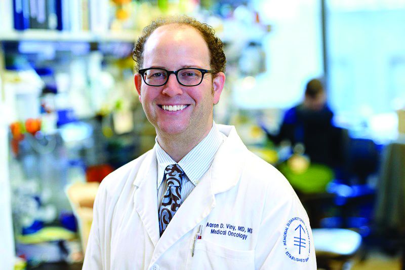
Dr. Viny is with the Memorial Sloan-Kettering Cancer Center, N.Y., where he is an assistant attending physician on the leukemia service and is a clinical researcher in the Ross Levine Lab. Follow him on Twitter @TheDoctorIsVin.
I just hung up with a friend I haven’t seen in decades. Her father has advanced cancer, and while she does not have formal medical training, a passerby wouldn’t know it. Her questions are spot on, her resources are peer reviewed and validated, and her questions I’d more likely expect from trainees in a formal oncology training program than from the director of an elementary level tutoring service.
Her father is fortunately doing well, but she’s searching for the next plan for when the standard drugs ultimately fail. We know they will fail. She’s connected to patient advocacy groups, emailing physicians across the country, and looking into clinical trials with their exhaustive lists of exclusion criteria. She sees the logistic difficulties with trials far from home. She’s hit the key issues we face every day in clinical research, and she’s never stepped foot in a medical school lecture hall.
Amazingly her story is not unique. When cancer hits close to home is when these problems become very clear. This same story could easily have been retold as the narrative of former Vice President Joe Biden and his care for his son. Both my friend and Mr. Biden, in fact, asked me the same question: How do we get the cutting-edge science from major research centers out to the rest of the country?
The Cancer Moonshot initiative has done much to promote collaboration, but one major success has been in the Count Me In initiative, a partnership between the Biden Cancer Initiative, Emerson Collective, the Broad Institute, and the Dana-Farber Cancer Institute. Their goal is to gain access to thousands of patients, collect data on treatment and outcomes, and collect biological specimens. They are not alone, the MSK-IMPACT initiative – led by David B. Solit, MD, at my institution – aims to sequence rare cancers. Both programs have heavily leveraged social media to access and engage patients.
There are of course concerns. Coming from hundreds or thousands of different sites will mean the data will likely be heterogeneous in formatting and quality. How do we ensure the security of patient data? Can we rely on patients and family members to report accurately and without bias? We know there are challenges and upside to crowdsourced patient recruitment.
David Ginsburg, MD, Karl Desch, MD, and colleagues enrolled more than 1,000 students from the University of Michigan to participate in a study on blood clotting factors. This led to many important findings on the genetic basis for coagulopathies, but also was instructive in uncovering a worrisome aspect of online patient registration. The group recorded the time taken for registrants to read the consent form – including whether the participant clicked a hyperlink that was embedded. Nearly a quarter of participants accepted the terms of the 2,833-word document in less than 10 seconds, and less than 3% clicked the hyperlink (Ann Intern Med. 2011 Sep 6;155[5]:316-22).
Are these patients, who we are asking for their partnership and trust, really understanding to what they are agreeing?
Surely there is tremendous altruism on the part of these patients. Their hopes of helping the future of cancer care does have a real track record. Crowdsourcing efforts that were less far reaching in scope made substantial impact in discovering the genetic basis for polycythemia vera. The patients, contacted largely through printed newspaper ads, have helped millions of others. What will happen when we add in the power of social media will be exciting to see – and there is something else that comes with great power, but since I can’t seem to remember what that is, I’ll just search online.
Dr. Viny is with the Memorial Sloan-Kettering Cancer Center, N.Y., where he is an assistant attending physician on the leukemia service and is a clinical researcher in the Ross Levine Lab. Follow him on Twitter @TheDoctorIsVin.
HBV, HCV, HIV testing of new cancer patients advised
Oncologists should consider testing all patients with newly diagnosed cancers for infection with the hepatitis B and C viruses, a multicenter team has recommended.
A prospective study of hepatitis B virus (HBV), hepatitis C virus (HCV), and HIV infections among 3,051 patients with newly diagnosed cancers showed that 6.5% of patients tested positive for previous HBV and 0.6% had chronic HBV infection. In addition, 2.4% of patients were positive for HCV, and 1.1% for HIV infections, reported Scott D. Ramsey, MD, PhD, from the Fred Hutchinson Cancer Research Center in Seattle, and colleagues.
“Many patients had no known risk factors for infection, suggesting that current risk-based models for screening may be insufficient. Thus, we believe our results warrant consideration of universal testing of patients with newly diagnosed cancer for HBV and HCV infection, particularly if such an approach is shown to be cost effective,” they wrote in JAMA Oncology.
The investigators noted that patients with undiagnosed hepatitis and/or HIV infections could transmit them to unsuspecting caregivers, adding that “with effective treatments available, not screening for these viruses misses an opportunity to reduce future morbidity associated with these infections and to avoid viral reactivation during treatment, with resulting morbidity and mortality.”
To estimate the prevalence of the infections in patients with newly diagnosed cancers, investigators looked at a cohort of 3,051 patients with a cancer diagnosis made within the previous 120 days at nine academic medical centers and nine community oncology centers representing a total of 41 cancer clinics affiliated with the SWOG Cancer Research Network (formerly the Southwest Oncology Group).
The median patient age was 60.6 years. Female patients constitute 60.4% of the sample; 18.1% were black, and 18.3% were of Hispanic heritage.
Of 3,050 patients for whom HBV testing results were available, 6.5% (197) were positive for previous HBV infection, compared with an estimated U.S. population prevalence of 4.7%. In addition, 0.6% (19 patients) were found to have chronic HBV, compared with an estimated 0.3% US population prevalence.
HCV infections were detected in 2.4% (71 of 2990 patients), compared with an estimated population prevalence of 1.3%, and HIV infections were detected in 1.1%, compared with a background estimated population prevalence of 0.3%.
In all, 32 patients were diagnosed with viral infections by testing performed for the study, including 8 patients with chronic HBV, 22 with HCV, and 2 with HIV.
Additionally, the authors found that 4 patients with chronic HBV, 23 with HCV, and 7 with HIV had no identifiable risk factors.
The highest prevalence of infections occurred among patients with liver cancer, nonliver and noncolorectal cancers of the gastrointestinal tract, head and neck cancers, lung cancers, and prostate cancer. A finding of viral positivity changed the treatment plan in only 8% of all infected patients, however.
“Given that most HIV-infected patients in our study knew their viral status, the yield of universal HIV testing among patients with newly diagnosed cancer may likely be low. Although age-directed screening is recommended for HIV and HCV, uptake rates in primary care are variable and low overall,” Dr. Ramsey and his colleagues wrote.
The study was supported by grants from the National Cancer Institute. Dr. Ramsey and several co-authors reported receiving NCI grants, and multiple co-authors reported grants and/or consulting fees from various companies.
SOURCE: Ramsey SD et al. JAMA Oncol. 2019 Jan 17. doi: 10.1001/jamaoncol.2018.6437.
FROM JAMA ONCOLOGY
Key clinical point: Patients with newly diagnosed cancers should be screened for viral infections that may pose a transmission risk or could be reactivated by cancer therapies.
Major finding: Infection rates of HBV, HCV, and HIV in patients with newly diagnosed cancers were 6.5%, 2.4%, and 1.1%, respectively.
Study details: Prospective study of viral infections in 3,051 patients with a diagnosis of cancer within the previous 120 days.
Disclosures: The study was supported by grants from the National Cancer Institute. Dr. Ramsey and several coauthors reported receiving NCI grants, and multiple coauthors reported grants and/or consulting fees from various companies.
Source: Ramsey SD et al. JAMA Oncology. 2019 Jan 17. doi: 10.1001/jamaoncol.2018.6437.
Checkpoint inhibitors linked to rare, but serious immune-related side effects
Checkpoint inhibitors can cause rare, but serious, hematological immune-related adverse events (hem-irAEs), which require early detection and intervention, according to a recent French study.
Immune thrombocytopenia, hemolytic anemia, and neutropenia were the most common hem-irAEs in the population, reported lead author, Nicolas Delanoy, MD, of Gustave Roussy, Université Paris-Saclay, Villejuif, France, and his colleagues.
“About 71% of patients treated have any-grade irAEs and 10% have grade 3-4 irAEs after anti-PD-1 immunotherapy,” the investigators wrote. The report is in The Lancet Haematology. “In most cases, they involve the skin, gastrointestinal tract, thyroid or endocrine glands, liver, lungs, or joints. However, all organs can potentially be affected, including the hemopoietic system.”
Despite this possibility, few reports detail the frequency or character of hematological toxicities from immunotherapy.
The present study involved 948 patients who entered into three French registries between 2014 and 2018. The first registry, consisting of 745 patients, was observed prospectively during checkpoint inhibitor therapy. The other two registries provided retrospective data on confirmed irAEs or hem-irAEs.
Among 745 patients followed during checkpoint inhibitor therapy, four developed hem-irAEs, providing an incidence rate of 0.5%. The other two databases added 31 patients with confirmed hem-irAEs, allowing for characterization of 35 total cases.
The group of 35 patients had a median age of 65 years, with more men (n = 21) than women (n = 14). Melanoma was the most common type of malignancy (43%), followed by non–small-cell lung cancer (34%), lymphoma (11%), and others. The majority of patients received nivolumab (57%), slightly fewer received pembrolizumab (40%), and a small minority received atezolizumab (3%).
Immune thrombocytopenia, hemolytic anemia, and neutropenia were the most common hem-irAEs, each occurring in nine patients (26%). Five patients (14%) had aplastic anemia or pancytopenia, two patients had bicytopenia (6%; neutropenia and anemia or thrombocytopenia and anemia), and one patient had pure red cell aplasia (3%).
Hem-irAEs resolved in 60% of patients, but two patients (6%) died due to febrile neutropenia. Overall, 71% of hem-irAEs were grade 4.
These findings suggest that hem-irAEs are rare, but they are often serious, and potentially life-threatening, the researchers noted.
In 7 of 35 patients (20%) who were rechallenged with checkpoint inhibitor therapy, 3 (43%) had recurrence of hem-irAEs. This finding should elicit caution and close monitoring if rechallenge is elected.
“This observational study encourages further, in-depth investigations of hematological immune toxicities, to search for biomarkers that can be helpful for earlier detection,” the investigators concluded.
This study was funded by Gustave Roussy and the Gustave Roussy Immunotherapy Program. Dr. Delanoy reported nonfinancial support from Sanofi and other authors reported financial relationships with pharmaceutical companies.
SOURCE: Delanoy N et al. Lancet Haematol. 2018 Dec 4. doi: 10.1016/S2352-3026(18)30175-3.
FROM THE LANCET HAEMATOLOGY
Key clinical point:
Major finding: Checkpoint inhibitor therapy led to hematological toxicity in 0.5% of patients.
Study details: A study of 948 patients in French registries who were observed prospectively or retrospectively, including a case series of 35 patients treated with checkpoint inhibitor therapy who developed hematologic, immune-related adverse events.
Disclosures: This study was funded by Gustave Roussy and the Gustave Roussy Immunotherapy Program. Dr. Delanoy reported nonfinancial support from Sanofi and other authors reported financial relationships with pharmaceutical companies.
Source: Delanoy N et al. Lancet Haematol. 2018 Dec 4. doi: 10.1016/S2352-3026(18)30175-3.
Soy didn’t up all-cause mortality in breast cancer survivors
A cohort of Chinese women who are breast cancer survivors had no increased mortality from soy intake, according to a new study.

The work adds to the existing body of evidence that women with breast cancer, or risk for breast cancer, don’t need to modify their soy intake to mitigate risk, said the study’s first author, Suzanne C. Ho, PhD.
Speaking at the annual meeting of the North American Menopause Society, Dr. Ho noted that the combination of increasing breast cancer incidence and improved outcome has resulted in larger numbers of breast cancer survivors in Hong Kong, where she is professor emerita at the Chinese University of Hong Kong.
The prospective, ongoing study examines the association between soy intake pre- and postdiagnosis and total mortality for Chinese women who are breast cancer survivors. Dr. Ho said that she and her colleagues hypothesized that they would not see higher mortality among women who had higher soy intake – and this was the case.
Of 1,497 breast cancer survivors drawn from two facilities in Hong Kong, those who consumed higher quantities of dietary soy did not have increased risk of all-cause mortality, compared with those in the lowest tertile of soy consumption.
There are theoretical underpinnings for thinking that soy could be a player in cancer risk, but the biochemistry and epidemiology behind the studies are complicated. Estrogen plays a role in human breast cancer, and many modern breast cancer treatments actually dampen endogenous estrogens.
However, epidemiologic data have shown that consumption of soy-based foods – which contain phytoestrogens, primarily in the form of isoflavones – is inversely associated with developing breast cancer.
This is all part of why soy-based foods have been thought of as a mixed bag with regard to breast cancer: Soy isoflavones are, said Dr. Ho, “Natural estrogen receptor modulators that possess both estrogenlike and antiestrogenic properties.”
Other chemicals contained in soy may fight cancer, with effects that are antioxidative and strengthen immune response. Soy constituents also inhibit DNA topoisomerase I and II, proteases, tyrosine kinases, and inositol phosphate, effects that can slow tumor growth. Still, one soy isoflavone, genistein, actually can promote growth of estrogen-dependent tumors in rats, said Dr. Ho
Dr. Ho and her colleagues enrolled Hong Kong residents for the study of mortality among breast cancer survivors. Participants were included if they were Chinese, female, aged 24-77 years, and had their first primary breast cancer histologically confirmed within 12 months of entering the study. Cancer had to be graded below stage III.
Using a 109-item validated food questionnaire, investigators gathered information about participants’ soy intake and general diet for the year prior to breast cancer diagnosis. Other patient characteristics, relevant prognostic information from medical records, and anthropometric data were collected at baseline, and repeated at 18, 36, and 60 months.
The primary outcome measure – all-cause mortality during the follow-up period – was tracked for a mean 50.9 months, with a 78% retention rate for study participants, said Dr. Ho. In total, 96 patients died during follow-up, making up 5.9% of the premenopausal and 7% of the postmenopausal participants.
Statistical analysis corrected for potential confounders, including patient and disease characteristics and treatment modalities, as well as overall energy consumption.
Patients were evenly divided into tertiles of soy isoflavone intake, with cutpoints of 3.77 mg/1,000 kcal and 10.05 mg/1,000 kcal for the lower limit of the two higher tertiles. For the highest tertile, though, mean isoflavone intake was actually 20.87 mg/1,000 kcal.
Patient, disease, and treatment characteristics did not differ significantly among the tertiles.
An adjusted statistical analysis looked at pre- and postmenopausal women separately by tertile of soy isoflavone consumption, setting the hazard ratio for all-cause mortality at 1.00 for women in the lowest tertile of soy consumption.
For premenopausal women in the middle tertile, the HR was 0.45 (95% confidence interval, 0.20-1.00), and 0.86 for those in the highest tertile (95% CI, 0.43-1.72); 782 participants, in all, were premenopausal.
For the 715 postmenopausal women, the HR for those in the middle tertile of soy consumption was 0.94 (95% CI, 0.43-2.05), and 1.11 in the highest (95% CI, 0.54-2.29).
Taking all pre- and postmenopausal participants together, those in the middle tertile of soy isoflavone intake had an all-cause mortality HR of 0.63 (95% CI, 0.37-1.09). For the highest tertile of the full cohort, the HR was 0.95 (95% CI, 0.58-1.55).
Confidence intervals were wide in these findings, but Dr. Ho noted that “moderate soy food intake might be associated with better survival.”
“Prediagnosis soy intake did not increase the risk of all-cause mortality in breast cancer survivors,” said Dr. Ho, findings she called “consistent with the literature that soy consumption does not adversely effect breast cancer survival.”
The study is ongoing, she explained, and “longer follow-up will provide further evidence on the effect of pre- and postdiagnosis soy intake on breast cancer outcomes.”
The study had a homogeneous population of southern Chinese women, with fairly good retention and robust statistical adjustment for confounders. However, it wasn’t possible to assess bioavailability of isoflavones and their metabolites, which can vary according to individual microbiota. Also, researchers did not track whether patients used traditional Chinese medicine.
The World Cancer Research Fund International supported the study. Dr. Ho reported no conflicts of interest.
SOURCE: Ho S et al. NAMS 2018, Abstract S-23.
A cohort of Chinese women who are breast cancer survivors had no increased mortality from soy intake, according to a new study.
The work adds to the existing body of evidence that women with breast cancer, or risk for breast cancer, don’t need to modify their soy intake to mitigate risk, said the study’s first author, Suzanne C. Ho, PhD.
Speaking at the annual meeting of the North American Menopause Society, Dr. Ho noted that the combination of increasing breast cancer incidence and improved outcome has resulted in larger numbers of breast cancer survivors in Hong Kong, where she is professor emerita at the Chinese University of Hong Kong.
The prospective, ongoing study examines the association between soy intake pre- and postdiagnosis and total mortality for Chinese women who are breast cancer survivors. Dr. Ho said that she and her colleagues hypothesized that they would not see higher mortality among women who had higher soy intake – and this was the case.
Of 1,497 breast cancer survivors drawn from two facilities in Hong Kong, those who consumed higher quantities of dietary soy did not have increased risk of all-cause mortality, compared with those in the lowest tertile of soy consumption.
There are theoretical underpinnings for thinking that soy could be a player in cancer risk, but the biochemistry and epidemiology behind the studies are complicated. Estrogen plays a role in human breast cancer, and many modern breast cancer treatments actually dampen endogenous estrogens.
However, epidemiologic data have shown that consumption of soy-based foods – which contain phytoestrogens, primarily in the form of isoflavones – is inversely associated with developing breast cancer.
This is all part of why soy-based foods have been thought of as a mixed bag with regard to breast cancer: Soy isoflavones are, said Dr. Ho, “Natural estrogen receptor modulators that possess both estrogenlike and antiestrogenic properties.”
Other chemicals contained in soy may fight cancer, with effects that are antioxidative and strengthen immune response. Soy constituents also inhibit DNA topoisomerase I and II, proteases, tyrosine kinases, and inositol phosphate, effects that can slow tumor growth. Still, one soy isoflavone, genistein, actually can promote growth of estrogen-dependent tumors in rats, said Dr. Ho
Dr. Ho and her colleagues enrolled Hong Kong residents for the study of mortality among breast cancer survivors. Participants were included if they were Chinese, female, aged 24-77 years, and had their first primary breast cancer histologically confirmed within 12 months of entering the study. Cancer had to be graded below stage III.
Using a 109-item validated food questionnaire, investigators gathered information about participants’ soy intake and general diet for the year prior to breast cancer diagnosis. Other patient characteristics, relevant prognostic information from medical records, and anthropometric data were collected at baseline, and repeated at 18, 36, and 60 months.
The primary outcome measure – all-cause mortality during the follow-up period – was tracked for a mean 50.9 months, with a 78% retention rate for study participants, said Dr. Ho. In total, 96 patients died during follow-up, making up 5.9% of the premenopausal and 7% of the postmenopausal participants.
Statistical analysis corrected for potential confounders, including patient and disease characteristics and treatment modalities, as well as overall energy consumption.
Patients were evenly divided into tertiles of soy isoflavone intake, with cutpoints of 3.77 mg/1,000 kcal and 10.05 mg/1,000 kcal for the lower limit of the two higher tertiles. For the highest tertile, though, mean isoflavone intake was actually 20.87 mg/1,000 kcal.
Patient, disease, and treatment characteristics did not differ significantly among the tertiles.
An adjusted statistical analysis looked at pre- and postmenopausal women separately by tertile of soy isoflavone consumption, setting the hazard ratio for all-cause mortality at 1.00 for women in the lowest tertile of soy consumption.
For premenopausal women in the middle tertile, the HR was 0.45 (95% confidence interval, 0.20-1.00), and 0.86 for those in the highest tertile (95% CI, 0.43-1.72); 782 participants, in all, were premenopausal.
For the 715 postmenopausal women, the HR for those in the middle tertile of soy consumption was 0.94 (95% CI, 0.43-2.05), and 1.11 in the highest (95% CI, 0.54-2.29).
Taking all pre- and postmenopausal participants together, those in the middle tertile of soy isoflavone intake had an all-cause mortality HR of 0.63 (95% CI, 0.37-1.09). For the highest tertile of the full cohort, the HR was 0.95 (95% CI, 0.58-1.55).
Confidence intervals were wide in these findings, but Dr. Ho noted that “moderate soy food intake might be associated with better survival.”
“Prediagnosis soy intake did not increase the risk of all-cause mortality in breast cancer survivors,” said Dr. Ho, findings she called “consistent with the literature that soy consumption does not adversely effect breast cancer survival.”
The study is ongoing, she explained, and “longer follow-up will provide further evidence on the effect of pre- and postdiagnosis soy intake on breast cancer outcomes.”
The study had a homogeneous population of southern Chinese women, with fairly good retention and robust statistical adjustment for confounders. However, it wasn’t possible to assess bioavailability of isoflavones and their metabolites, which can vary according to individual microbiota. Also, researchers did not track whether patients used traditional Chinese medicine.
The World Cancer Research Fund International supported the study. Dr. Ho reported no conflicts of interest.
SOURCE: Ho S et al. NAMS 2018, Abstract S-23.
REPORTING FROM NAMS 2018
Key clinical point: Soy consumption did not increase mortality risk in breast cancer survivors.
Major finding: The hazard ratios for all-cause mortality were 0.63 and 0.95 for the two highest tertiles of soy consumption.
Study details: An ongoing prospective cohort study of 1,497 female breast cancer survivors in Hong Kong.
Disclosures: The World Cancer Research Fund International supported the study. Dr. Ho reported no conflicts of interest.
Source: Ho S et al. NAMS 2018, Abstract S-23.
Daily News Special: SABCS
Stories include: uUing low-dose tamoxifen, the latest findings from the KATHERINE trial, results of a meta-analysis of neoadjuvant chemotherapy, and capecitabine in early stage triple negative breast cancer.
Amazon Alexa
Apple Podcasts
Google Podcasts
Spotify
Stories include: uUing low-dose tamoxifen, the latest findings from the KATHERINE trial, results of a meta-analysis of neoadjuvant chemotherapy, and capecitabine in early stage triple negative breast cancer.
Amazon Alexa
Apple Podcasts
Google Podcasts
Spotify
Stories include: uUing low-dose tamoxifen, the latest findings from the KATHERINE trial, results of a meta-analysis of neoadjuvant chemotherapy, and capecitabine in early stage triple negative breast cancer.
Amazon Alexa
Apple Podcasts
Google Podcasts
Spotify

Poor-prognosis cancers linked to highest suicide risk in first year
Suicide risk significantly increases within the first year of a cancer diagnosis, with risk varying by type of cancer, according to investigators who conducted a retrospective analysis representing nearly 4.7 million patients.
Risk of suicide in that first year after diagnosis was especially high in pancreatic and lung cancers, while by contrast, breast and prostate cancer did not increase suicide risk, reported the researchers, led by Hesham Hamoda, MD, MPH, of Boston Children’s Hospital/Harvard Medical School, and Ahmad Alfaar, MBBCh, MSc, of Charité–Universitätsmedizin Berlin.
That variation in suicide risk by cancer type suggests that prognosis and 5-year relative survival play a role in increasing suicide rates, according to Dr. Hamoda, Dr. Alfaar, and their coauthors.
“After the diagnosis, it is important that health care providers be vigilant in screening for suicide and ensuring that patients have access to social and emotional support,” they wrote in a report published in Cancer. Their analysis was based on 4,671,989 patients with a diagnosis of cancer in the Surveillance, Epidemiology, and End Results (SEER) database between 2000 and 2014. Out of 1,005,825 of those patients who died within the first year of diagnosis, the cause of death was suicide for 1,585, or 0.16%.
Overall, the risk of suicide increased significantly among cancer patients versus the general population, with an observed-to-expected (O/E) ratio of 2.51 per 10,000 person-years, the investigators found. The risk was highest in the first 6 months, with an O/E mortality of 3.13 versus 1.8 in the latter 6 months.
The highest ratios were seen for pancreatic cancer, with an O/E ratio of 8.01, and lung cancer, with a ratio of 6.05, the researchers found in further analysis.
Significant increases in suicide risk were also seen for colorectal cancer (2.08) and melanoma (1.45), though rates were not significantly different versus the general population for breast (1.23) and prostate (0.99), according to the reported data.
Suicide risk was relatively high for any cancer with distant metastases (5.63), though still significantly higher at 1.65 in persons with localized/regional disease, the data show.
The increased suicide risk persisted more than 1 year after the cancer diagnosis, though not to the degree observed within that first year, they added.
Most patients with suicide as a cause of death were white (90.2%) and male (87%). Nearly 60% were between the ages of 65 and 84 at the time of suicide.
Social support plays an integral role in suicide prevention among cancer patients, the researchers noted.
Previous studies suggest that support programs may decrease suicide risk by making patients better aware of their prognosis, receptive to decreased social stigma, or less likely to have stress related to cost of care, they said.
“Discussing the quality of life after diagnosis, the effectiveness of therapy, and the prognosis of the disease and maintaining a trusting relationship with health care professionals all decrease the likelihood of suicide immediately after a diagnosis of cancer,” they said.
Dr. Hamoda, Dr. Alfaar, and their coauthors reported no conflicts of interest. Funding for the study came in part from the German Academic Exchange Service (Dr. Alfaar).
SOURCE: Saad AM, et al. Cancer 2019 Jan 7. doi: 10.1002/cncr.31876.
FROM CANCER
Key clinical point: A cancer diagnosis significantly increases risk of suicide in comparison to the general population, particularly for poorer-prognosis cancers.
Major finding: The observed-to-expected mortality ratio was substantially higher for pancreatic cancer (8.01), and lung cancer (6.05), but not significantly increased for breast (1.23) and prostate (0.99).
Study details: A retrospective population-based study of 4,671,989 cancer patients.
Disclosures: The authors reported no conflicts of interest. Funding for the study came in part from the German Academic Exchange Service.
Source: Saad AM et al. Cancer. 2019 Jan 7. doi: 10.1002/cncr.31876.
Higher AML, MDS risk linked to solid tumor chemotherapy
There is an increased risk for therapy-related myelodysplastic syndrome or acute myeloid leukemia (tMDS/AML) following chemotherapy for the majority of solid tumor types, according to an analysis of cancer registry data.
These findings suggest a substantial expansion in the patients at risk for tMDS/AML because, in the past, excess risks were established only after chemotherapy for cancers of the lung, ovary, breast, soft tissue, testis, and brain or central nervous system,” Lindsay M. Morton, PhD, of the National Institutes of Health, and her colleagues wrote in JAMA Oncology.
The researchers retrospectively analyzed data from 1,619 patients with tMDS/AML who were diagnosed with an initial primary solid tumor from 2000 to 2013. Data came from the National Cancer Institute’s Surveillance, Epidemiology, and End Results (SEER) Program and Medicare claims.
Study participants were given initial chemotherapy and lived for at least 1 year after treatment. Subsequently, Dr. Morton and her colleagues linked patient database records with Medicare insurance claim information to confirm the accuracy of chemotherapy data.
“Because registry data [does] not include treatment details, we used an alternative database to provide descriptive information on population-based patterns of chemotherapeutic drug use,” the researchers wrote in JAMA Oncology.
After statistical analysis, the researchers found that the risk of developing tMDS/AML was significantly elevated following chemotherapy administration for 22 of 23 solid tumor types, excluding colon cancer. They reported a 1.5-fold to more than 10-fold increased relative risk for tMDS/AML in those patients who received chemotherapy for those 22 solid cancer types, compared with the general population.
The relative risks were highest after chemotherapy for bone, soft-tissue, and testis cancers.
The researchers found that the absolute risk of developing tMDS/AML was low. Excess absolute risks ranged from 1.4 to greater than 15 cases per 10,000 person-years, compared with the general population, in those 22 solid cancer types. The greatest absolute risks were for peritoneum, small-cell lung, bone, soft-tissue, and fallopian tube cancers.
“For patients treated with chemotherapy at the present time, approximately three-quarters of tMDS/AML cases expected to occur within the next 5 years will be attributable to chemotherapy,” they added.
The researchers acknowledged a key limitation of the study was the limited data on dosing and patient-specific chemotherapy. As a result, Dr. Morton and her colleagues called for a cautious interpretation of the magnitude of the risk.
The study was supported by the Intramural Research Program of the National Institutes of Health, National Cancer Institute, and the California Department of Public Health. The authors reported having no conflicts of interest.
SOURCE: Morton LM et al. JAMA Oncol. 2018 Dec 20. doi: 10.1001/jamaoncol.2018.5625.
Possibly the most clinical relevant finding of the study by Lindsay M. Morton, PhD, and her colleagues is that patients who received chemotherapy for solid tumor treatment at a younger age were at the highest relative risk for tMDS/AML.
The incidence of tMDS/AML was highest among patients treated with chemotherapy for bone, soft-tissue, and testicular cancers, where the median age of onset is often by 30 years, and the mean onset occurs before age 50.
The researchers also noted an increased risk for tMDS/AML associated with prolonged survival from primary tumors.
Going forward, research should consider those patients at highest risk for tMDS/AML and risk-assessment models for these therapy-related myeloid neoplasms should take into account the clonal evolution of subclinical mutations into overt disease.
The study findings point to the unanswered question of how best to perform risk assessment of chemotherapy in solid tumors. That risk stratification could include the probability of the specific chemotherapy agent initiating disease, the benefit of tumor regression from chemotherapy, and the potential consequences of tumor progression if chemotherapy is not administered.
Shyam A. Patel, MD, PhD, is with the department of medicine at Stanford (Calif.) University. Dr. Patel reported having no financial disclosures. These comments are adapted from his accompanying editorial (JAMA Oncol. 2018 Dec 20. doi: 10.1001/jamaoncol.2018.5617 ).
There is an increased risk for therapy-related myelodysplastic syndrome or acute myeloid leukemia (tMDS/AML) following chemotherapy for the majority of solid tumor types, according to an analysis of cancer registry data.
These findings suggest a substantial expansion in the patients at risk for tMDS/AML because, in the past, excess risks were established only after chemotherapy for cancers of the lung, ovary, breast, soft tissue, testis, and brain or central nervous system,” Lindsay M. Morton, PhD, of the National Institutes of Health, and her colleagues wrote in JAMA Oncology.
The researchers retrospectively analyzed data from 1,619 patients with tMDS/AML who were diagnosed with an initial primary solid tumor from 2000 to 2013. Data came from the National Cancer Institute’s Surveillance, Epidemiology, and End Results (SEER) Program and Medicare claims.
Study participants were given initial chemotherapy and lived for at least 1 year after treatment. Subsequently, Dr. Morton and her colleagues linked patient database records with Medicare insurance claim information to confirm the accuracy of chemotherapy data.
“Because registry data [does] not include treatment details, we used an alternative database to provide descriptive information on population-based patterns of chemotherapeutic drug use,” the researchers wrote in JAMA Oncology.
After statistical analysis, the researchers found that the risk of developing tMDS/AML was significantly elevated following chemotherapy administration for 22 of 23 solid tumor types, excluding colon cancer. They reported a 1.5-fold to more than 10-fold increased relative risk for tMDS/AML in those patients who received chemotherapy for those 22 solid cancer types, compared with the general population.
The relative risks were highest after chemotherapy for bone, soft-tissue, and testis cancers.
The researchers found that the absolute risk of developing tMDS/AML was low. Excess absolute risks ranged from 1.4 to greater than 15 cases per 10,000 person-years, compared with the general population, in those 22 solid cancer types. The greatest absolute risks were for peritoneum, small-cell lung, bone, soft-tissue, and fallopian tube cancers.
“For patients treated with chemotherapy at the present time, approximately three-quarters of tMDS/AML cases expected to occur within the next 5 years will be attributable to chemotherapy,” they added.
The researchers acknowledged a key limitation of the study was the limited data on dosing and patient-specific chemotherapy. As a result, Dr. Morton and her colleagues called for a cautious interpretation of the magnitude of the risk.
The study was supported by the Intramural Research Program of the National Institutes of Health, National Cancer Institute, and the California Department of Public Health. The authors reported having no conflicts of interest.
SOURCE: Morton LM et al. JAMA Oncol. 2018 Dec 20. doi: 10.1001/jamaoncol.2018.5625.
FROM JAMA ONCOLOGY
Key clinical point:
Major finding: Treatment with chemotherapy was linked with a 1.5-fold to more than 10-fold increased risk for tMDS/AML.
Study details: A retrospective analysis of 1,619 patients with tMDS/AML who were diagnosed with an initial primary solid tumor from 2000 to 2013.
Disclosures: The study was supported by the Intramural Research Program of the National Institutes of Health, National Cancer Institute, and the California Department of Public Health. The authors reported having no conflicts of interest.
Source: Morton LM et al. JAMA Oncol. 2018 Dec 20. doi: 10.1001/jamaoncol.2018.5625.
Should doctors disclose preliminary results?
Outside the clinic room, I paced the hallway and pressed the phone to my ear, waiting for the resident to pick up.
“I have patient SB in clinic for her appointment now. I’m hoping to get preliminary results of her bone marrow biopsy.”
I had known SB well from her month-long inpatient stay on our leukemia service. She had come in with a white blood cell count through the roof – a relapse of her leukemia, 4 years out from her bone marrow transplant. It was devastating. After a few cycles of chemotherapy and a bone marrow biopsy yesterday to see if it had worked, she was here now to get her results and decide next steps.
“Hello!” I said and we hugged. Her mother and father accompanied her, sitting still with their hands folded nervously. SB had multiple complications during her hospitalization, and we went through how each was doing. Did she get her new heart medication? Did she do okay on the antibiotics? Was the rash improving? With each question, she and her parents seemed more nervous.
There was an elephant in that exam room. Asking a cancer patient in limbo if she refilled her heart medications becomes as trivial as asking her about the weather. SB and her parents were here for one thing, from which everything else was a distraction. The only question that mattered was the one splitting their world in two: Is their daughter in remission or not?
“She’s here with her parents now,” I said outside the door. “What do you think?” The resident told me he had looked at the case this morning, and it looked like 3% blasts. I smiled – anything under 5% is good, considered a remission. But the pathology resident still hadn’t reviewed the sample with his attending.
Inside the room, after exhausting all other conversation, I hesitated. Should I tell SB the preliminary results? Or should I wait for the final diagnosis?
I’d been burned before. Once, I told a patient with a new diagnosis of esophageal cancer that it was early stage. It was not. Upon additional radiology review, the surrounding lymph nodes were enlarged, and they were ultimately found to be metastases. That initial conversation – and the subsequent one, in which I had to walk back my reassurance that the cancer was contained – was seared into my mind.
I learned from it. Giving preliminary results can be dangerous. What if we say all clear and then learn days later it isn’t so? Or what if we reveal the cancer is progressing, causing despair and re-evaluation of life’s priorities, only to find out it was a false alarm? False alarms are terrifying, and false reassurance is cruel, yet all the while, excessive waiting can feel excruciating for the person whose very existence is suspended.
As hematologists and oncologists, we scroll through CT scans, and we look at slides ourselves. But we also value and depend on the expertise of our colleagues in other departments like pathology and radiology who have their own workflow. It’s a process; it’s for quality assurance that we don’t get immediate results, and that’s a good thing.
It depends on the patient, but often I find the most straightforward solution is to say exactly what is true. For some, the combination of incredibly high stakes coupled with extended wait times becomes agonizing. They might incorrectly read into unrelated, benign actions – if my pager goes off or I look at the computer screen a moment too long – as clues into something I know and am not sharing. They might be so distracted we cannot address anything else.
So I’ve walked back from my initial “do not share anything” reaction to “it’s sometimes okay” – as long as the patient understands the nuances of what preliminary results do and do not mean. The problem with my esophageal cancer patient was not that I had shared preliminary results; it was that I hadn’t framed them as such. I had simply stated the findings, portraying them as certain.
Now, I tend to break the fourth wall. I explain that it’s the resident’s read, that it isn’t final, and that it can be amended. Do you still want to know?
Most people say yes.
SB and her parents were in that boat. They had driven 3 hours to make this appointment. They didn’t want to drive home empty handed.
“It’s preliminary,” I carefully qualified.
“Okay.”
“The final results may be different.”
“Okay, yes. We understand.”
The three of them held hands. They were holding their breath.
“It looks like remission.”
SB cried. Her mother threw her arms around my neck. “You know, she broke down when you stepped out,” her father whispered to me. “She was sure it meant bad news.”
I tried to be happy for them and with them, but now I was the one holding my breath. I hoped I wouldn’t have to take it all away.
For the next 24 hours, I compulsively checked SB’s chart, hoping final results would populate that would be consistent with what I had shared.
The next day, the pathologist called me, and I called SB.
“I have the final results,” I said, followed by my favorite phrase in hematology and oncology. “I have good news.”
Dr. Yurkiewicz is a fellow in hematology and oncology at Stanford (Calif.) University. Follow her on Twitter @ilanayurkiewicz.
Lack of gut diversity hurts survival after HCT
SAN DIEGO – The success or failure of hematopoietic cell transplants may be related to the diversity of species in the gut microbiome, investigators contend.

A study of fecal samples from patients scheduled to undergo hematopoietic cell transplant (HCT) in the United States, Europe, and Japan showed that intestinal microbial diversity was significantly associated with overall survival, reported Jonathan U. Peled, MD, PhD, from the bone marrow transplantation service at Memorial Sloan Kettering Cancer Center in New York City.
“Patients with low diversity pretransplant have a poorer overall survival than patients with a higher diversity after transplantation,” he said at the annual meeting of the American Society of Hematology. “The implication is, if we could come up with a way to remediate microbiome injury, there might be time to implement it before the transplant.”
There are about 100 trillion symbiotic microbes in and on the human body, and 95% of them live in the gastrointestinal tract, which has a surface area that if stretched out would equal the size of two tennis courts, Dr. Peled said.
Previous studies by his group and others have shown that the composition of intestinal microbiota is associated with survival, relapses, graft-versus-host disease, and infections in patients in the early weeks after allogeneic HCT.
The international team previously reported that intestinal diversity measured around the time of neutrophil engraftment was predictive of overall survival, and they hypothesized that the same might be true of the pretransplant microbiota composition.
To test this idea, they collected 1,922 stools samples approximately once weekly for 3 weeks pretransplant from 991 adult patients scheduled for allo-HCT for various diagnoses in two U.S. cohorts and one cohort each in Europe and Japan.
They found that on average patients in all four cohorts had microbiota diversity values ranging from 70% to 150% lower than those of healthy volunteers whose samples were sequenced by the investigators, as well as those in a publicly available database.
The investigators also asked whether one or more bacterial species accounted for 30% or more of a microbiome, a phenomenon known as “monodomination.”
“A third, or in some cases 95+%, of bacteria in an individual’s intestine are all the exact same strain,” Dr. Peled said.
They found that the incidence and prevalence of the monodomination phenotype was “strikingly similar” at all four transplant sites.
The patterns of microbiota injury were similar despite differences in antibiotic strategies among the four centers and different dietary patterns of patients in the regions studied.
Studies in animal models suggest that T cells responsible for graft-versus-host disease migrate to the gut as early as 2 or 3 days post transplant, and by that time more than half of transplant patients have a monodomination event in their guts, Dr. Peled said.
Some strategies to remediate or prevent microbiome damage – and potentially improve HCT outcomes – may include the use of over-the-counter or custom-made probiotics, fecal transplants, a “prebiotic” approach in which the patient is asked to follow a specific diet that promotes the growth of beneficial bacteria, or a “postbiotic” approach in which patients receive metabolites of agents or foods thought to have a beneficial effect.
“Finally, we can think about different antibiotic strategies, to use or not use different types of antibiotics at different times in a rational way,” Dr. Peled said.
Dr. Peled reported current or prior financial relationships with Seres Therapeutics, the Parker Institute for Cancer Immunotherapy, and Merck/Society for Immunotherapy of Cancer.
SOURCE: Peled JU et al. ASH 2018, Abstract 811.
SAN DIEGO – The success or failure of hematopoietic cell transplants may be related to the diversity of species in the gut microbiome, investigators contend.
A study of fecal samples from patients scheduled to undergo hematopoietic cell transplant (HCT) in the United States, Europe, and Japan showed that intestinal microbial diversity was significantly associated with overall survival, reported Jonathan U. Peled, MD, PhD, from the bone marrow transplantation service at Memorial Sloan Kettering Cancer Center in New York City.
“Patients with low diversity pretransplant have a poorer overall survival than patients with a higher diversity after transplantation,” he said at the annual meeting of the American Society of Hematology. “The implication is, if we could come up with a way to remediate microbiome injury, there might be time to implement it before the transplant.”
There are about 100 trillion symbiotic microbes in and on the human body, and 95% of them live in the gastrointestinal tract, which has a surface area that if stretched out would equal the size of two tennis courts, Dr. Peled said.
Previous studies by his group and others have shown that the composition of intestinal microbiota is associated with survival, relapses, graft-versus-host disease, and infections in patients in the early weeks after allogeneic HCT.
The international team previously reported that intestinal diversity measured around the time of neutrophil engraftment was predictive of overall survival, and they hypothesized that the same might be true of the pretransplant microbiota composition.
To test this idea, they collected 1,922 stools samples approximately once weekly for 3 weeks pretransplant from 991 adult patients scheduled for allo-HCT for various diagnoses in two U.S. cohorts and one cohort each in Europe and Japan.
They found that on average patients in all four cohorts had microbiota diversity values ranging from 70% to 150% lower than those of healthy volunteers whose samples were sequenced by the investigators, as well as those in a publicly available database.
The investigators also asked whether one or more bacterial species accounted for 30% or more of a microbiome, a phenomenon known as “monodomination.”
“A third, or in some cases 95+%, of bacteria in an individual’s intestine are all the exact same strain,” Dr. Peled said.
They found that the incidence and prevalence of the monodomination phenotype was “strikingly similar” at all four transplant sites.
The patterns of microbiota injury were similar despite differences in antibiotic strategies among the four centers and different dietary patterns of patients in the regions studied.
Studies in animal models suggest that T cells responsible for graft-versus-host disease migrate to the gut as early as 2 or 3 days post transplant, and by that time more than half of transplant patients have a monodomination event in their guts, Dr. Peled said.
Some strategies to remediate or prevent microbiome damage – and potentially improve HCT outcomes – may include the use of over-the-counter or custom-made probiotics, fecal transplants, a “prebiotic” approach in which the patient is asked to follow a specific diet that promotes the growth of beneficial bacteria, or a “postbiotic” approach in which patients receive metabolites of agents or foods thought to have a beneficial effect.
“Finally, we can think about different antibiotic strategies, to use or not use different types of antibiotics at different times in a rational way,” Dr. Peled said.
Dr. Peled reported current or prior financial relationships with Seres Therapeutics, the Parker Institute for Cancer Immunotherapy, and Merck/Society for Immunotherapy of Cancer.
SOURCE: Peled JU et al. ASH 2018, Abstract 811.
REPORTING FROM ASH 2018
Key clinical point:
Major finding: Hematopoietic cell transplant candidates in the United States, Europe, and Japan all had similar patterns of low intestinal microbiota diversity and predomination of a single taxonomic species.
Study details: An analysis of stool samples from 991 adults scheduled for hematopoietic cell transplant at four transplant centers in the United States, Germany, and Japan.
Disclosures: Dr. Peled reported current or prior financial relationships with Seres Therapeutics, the Parker Institute for Cancer Immunotherapy, and Merck/Society for Immunotherapy of Cancer.
Source: Peled JU et al. ASH 2018, Abstract 811.