User login
The Cause of All That Stress: Tonsillectomy?
This transcript has been edited for clarity.
You know those times in your life when you’re just feeling ... stressed? You’re on the edge; you have no chill; everything just sort of gets to you. If you can step away from the anxiety for a moment, you might ask yourself where it’s all coming from. Is it really the stuff in your inbox at work or is it money issues at home? Is it something with your relationship, or maybe it’s your sleep quality or your diet? One thing you probably won’t blame for those acute stress reactions is the tonsillectomy you had as a kid. But according to new research, maybe you should.
Tonsillectomy and adenoidectomy are among the most common surgical procedures young people in the United States undergo, with about 300,000 cases a year, according to recent numbers. That’s down a bit from numbers a decade or so ago, but suffice it to say, a good chunk of the population is walking around right now without their tonsils.
The data supporting tonsillectomy have never been great. The two big indications for the surgery are recurrent sore throat — data show that tonsillectomy reduces this by about 0.7 sore throats per year— and obstructive sleep apnea (OSA). The data for improvement of OSA are a bit better than the data for sore throats.
Also, tonsillectomy is a relatively quick, relatively well-reimbursed surgery with indications that are — let’s be honest — somewhat subjective, and so variation is high. One study found that in a single Vermont town, nearly 60% of the population had had their tonsils removed by the time they turned 18. A few towns over, the rate was 20%.
A few factors have led to the decline of tonsillectomy in recent years. Reimbursement rates have gone down a bit. Additionally, better data collection and statistical analysis have shown that the benefits of the procedure are relatively modest.
And then there is a body of medical literature that at first struck me as surprising and almost bizarre: data linking tonsillectomy to subsequent physical and psychiatric disorders.
I teach a course on interpretation of the medical literature, and one of the first things I teach my students is to check their gut when they see the conclusion of a study.
Basically, even before you read the data, have a sense in your own mind if the hypothesis seems reasonable. If a paper is going to conclude that smoking leads to increased risk for bone cancer, I’d say that seems like a reasonable thing to study. If a paper purports to show a link between eating poultry and bone cancer, I’m going to be reading it with quite a bit more skepticism.
The technical term for that process is assessing “biologic plausibility.” If we’re talking tonsils, we have to ask ourselves: Is it plausible that removing someone’s tonsils when they are young should lead to major problems in the future?
At first blush, it didn’t seem very plausible to me.
But the truth is, there are quite a few studies out there demonstrating links like this: links between tonsillectomy and irritable bowel syndrome; links between tonsillectomy and cancer; links between tonsillectomy and depression.
And this week, appearing in JAMA Network Open, is a study linking tonsillectomy with stress disorders.
Researchers leveraged Sweden’s health database, which contains longitudinal data on basically every person who has lived in Sweden since 1981. This database let them know who had a tonsillectomy or adenoidectomy, and when, and what happened to them later in life.
I think the best way to present these data is to show you what they found, and then challenge that finding, and then show you what they did in anticipation of the challenges we would have to their findings. It’s a pretty thorough study.
So, topline results here. The researchers first identified 83,957 individuals who had their tonsils removed. They matched each of them with 10 controls who did not have their tonsils removed but were the same age and sex.
Over around 30 years of follow-up, those people who had their tonsils removed were 43% more likely to develop a stress-related disorder. Among the specific disorders, the risk for PTSD was substantially higher: 55% higher in the tonsillectomy group.
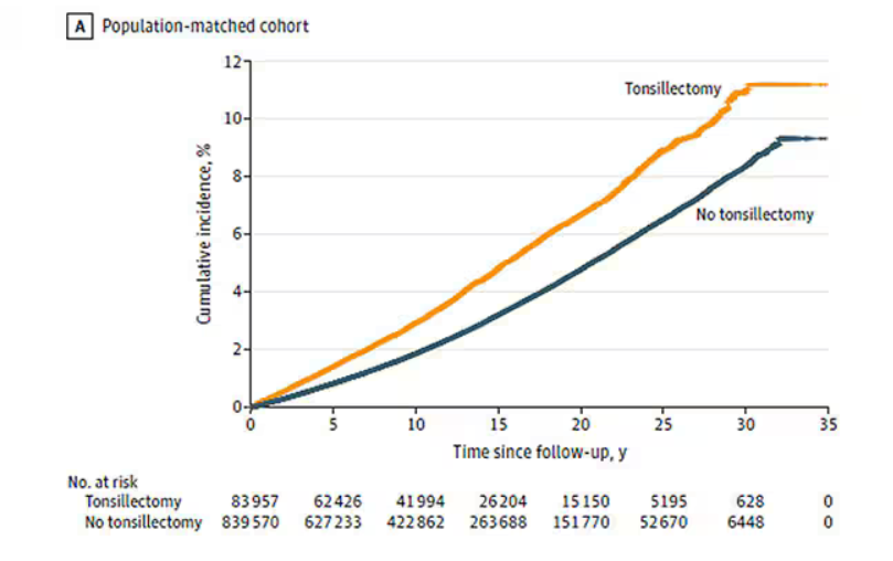
That’s pretty surprising, but I bet you already want to push back against this. Sure, the control group was the same age and sex, but other factors might be different between the two groups. You’d be right to think so. People who got their tonsils out were more likely to have parents with a history of stress-related disorders and who had lower educational attainment. But the primary results were adjusted for those factors.
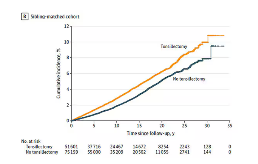
There’s more to a family than parental educational attainment, of course. To account for household factors that might be harder to measure, the researchers created a second control group, this one comprising the siblings of people who had their tonsils removed but who hadn’t themselves had their tonsils removed.
The relationship between tonsillectomy and stress disorders in this population was not quite as robust but still present: a 34% increase in any stress disorder and a 41% increase in the risk for PTSD.
Maybe kids who get their tonsils out are just followed more closely thereafter, so doctors might notice a stress disorder and document it in the medical record; whereas with other kids it might go unnoticed. This is known as ascertainment bias. The researchers addressed this in a sensitivity analysis where they excluded new diagnoses of stress disorders that occurred in the first 3 years after tonsillectomy. The results were largely unchanged.
So how do we explain these data? We observe a correlation between tonsillectomy in youth and stress disorders in later life. But correlation is not causation. One possibility, perhaps even the most likely possibility, is that tonsillectomy is a marker of some other problem. Maybe these kids are more prone to infections and are therefore more likely to need their tonsils removed. Then, after a lifetime of more infections than average, their stress responses are higher. Or maybe kids with a higher BMI are more likely to have their tonsils removed due to sleep apnea concerns, and it’s that elevated BMI that leads to higher stress in later life.
Or maybe this is causal. Maybe there actually is biological plausibility here. The authors suggest that removal of tonsils might lead to broader changes in the immune system; after all, tonsillar tissue is on the front line of our defense against pathogens that might enter our bodies through our mouths or noses. Immunologic changes lead to greater inflammation over time, and there is decent evidence to link chronic inflammation to a variety of physical and psychological disorders.
In support of this, the authors show that the kids with tonsillectomy were more likely to be hospitalized for an infectious disease in the future as well, in magnitudes similar to the increased risk for stress. But they don’t actually show that the relationship between tonsillectomy and stress is mediated by that increased risk for infectious disease.
In the end, I find these data really intriguing. Before I dug into the literature, it seemed highly unlikely that removal of these small lumps of tissue would have much of an effect on anything. Now I’m not so sure. A few things can be removed from the human body without any consequences, but it can be hard to know exactly what those consequences are.
That said, given the rather marginal benefits of tonsillectomy and the growing number of studies expanding on the risks, I expect that we’ll see the rates of the surgery decline even further in the future.
F. Perry Wilson, MD, MSCE, is an associate professor of medicine and public health and director of Yale’s Clinical and Translational Research Accelerator in New Haven, Connecticut. He reported no relevant conflicts of interest.
A version of this article first appeared on Medscape.com.
This transcript has been edited for clarity.
You know those times in your life when you’re just feeling ... stressed? You’re on the edge; you have no chill; everything just sort of gets to you. If you can step away from the anxiety for a moment, you might ask yourself where it’s all coming from. Is it really the stuff in your inbox at work or is it money issues at home? Is it something with your relationship, or maybe it’s your sleep quality or your diet? One thing you probably won’t blame for those acute stress reactions is the tonsillectomy you had as a kid. But according to new research, maybe you should.
Tonsillectomy and adenoidectomy are among the most common surgical procedures young people in the United States undergo, with about 300,000 cases a year, according to recent numbers. That’s down a bit from numbers a decade or so ago, but suffice it to say, a good chunk of the population is walking around right now without their tonsils.
The data supporting tonsillectomy have never been great. The two big indications for the surgery are recurrent sore throat — data show that tonsillectomy reduces this by about 0.7 sore throats per year— and obstructive sleep apnea (OSA). The data for improvement of OSA are a bit better than the data for sore throats.
Also, tonsillectomy is a relatively quick, relatively well-reimbursed surgery with indications that are — let’s be honest — somewhat subjective, and so variation is high. One study found that in a single Vermont town, nearly 60% of the population had had their tonsils removed by the time they turned 18. A few towns over, the rate was 20%.
A few factors have led to the decline of tonsillectomy in recent years. Reimbursement rates have gone down a bit. Additionally, better data collection and statistical analysis have shown that the benefits of the procedure are relatively modest.
And then there is a body of medical literature that at first struck me as surprising and almost bizarre: data linking tonsillectomy to subsequent physical and psychiatric disorders.
I teach a course on interpretation of the medical literature, and one of the first things I teach my students is to check their gut when they see the conclusion of a study.
Basically, even before you read the data, have a sense in your own mind if the hypothesis seems reasonable. If a paper is going to conclude that smoking leads to increased risk for bone cancer, I’d say that seems like a reasonable thing to study. If a paper purports to show a link between eating poultry and bone cancer, I’m going to be reading it with quite a bit more skepticism.
The technical term for that process is assessing “biologic plausibility.” If we’re talking tonsils, we have to ask ourselves: Is it plausible that removing someone’s tonsils when they are young should lead to major problems in the future?
At first blush, it didn’t seem very plausible to me.
But the truth is, there are quite a few studies out there demonstrating links like this: links between tonsillectomy and irritable bowel syndrome; links between tonsillectomy and cancer; links between tonsillectomy and depression.
And this week, appearing in JAMA Network Open, is a study linking tonsillectomy with stress disorders.
Researchers leveraged Sweden’s health database, which contains longitudinal data on basically every person who has lived in Sweden since 1981. This database let them know who had a tonsillectomy or adenoidectomy, and when, and what happened to them later in life.
I think the best way to present these data is to show you what they found, and then challenge that finding, and then show you what they did in anticipation of the challenges we would have to their findings. It’s a pretty thorough study.
So, topline results here. The researchers first identified 83,957 individuals who had their tonsils removed. They matched each of them with 10 controls who did not have their tonsils removed but were the same age and sex.
Over around 30 years of follow-up, those people who had their tonsils removed were 43% more likely to develop a stress-related disorder. Among the specific disorders, the risk for PTSD was substantially higher: 55% higher in the tonsillectomy group.
That’s pretty surprising, but I bet you already want to push back against this. Sure, the control group was the same age and sex, but other factors might be different between the two groups. You’d be right to think so. People who got their tonsils out were more likely to have parents with a history of stress-related disorders and who had lower educational attainment. But the primary results were adjusted for those factors.
There’s more to a family than parental educational attainment, of course. To account for household factors that might be harder to measure, the researchers created a second control group, this one comprising the siblings of people who had their tonsils removed but who hadn’t themselves had their tonsils removed.
The relationship between tonsillectomy and stress disorders in this population was not quite as robust but still present: a 34% increase in any stress disorder and a 41% increase in the risk for PTSD.
Maybe kids who get their tonsils out are just followed more closely thereafter, so doctors might notice a stress disorder and document it in the medical record; whereas with other kids it might go unnoticed. This is known as ascertainment bias. The researchers addressed this in a sensitivity analysis where they excluded new diagnoses of stress disorders that occurred in the first 3 years after tonsillectomy. The results were largely unchanged.
So how do we explain these data? We observe a correlation between tonsillectomy in youth and stress disorders in later life. But correlation is not causation. One possibility, perhaps even the most likely possibility, is that tonsillectomy is a marker of some other problem. Maybe these kids are more prone to infections and are therefore more likely to need their tonsils removed. Then, after a lifetime of more infections than average, their stress responses are higher. Or maybe kids with a higher BMI are more likely to have their tonsils removed due to sleep apnea concerns, and it’s that elevated BMI that leads to higher stress in later life.
Or maybe this is causal. Maybe there actually is biological plausibility here. The authors suggest that removal of tonsils might lead to broader changes in the immune system; after all, tonsillar tissue is on the front line of our defense against pathogens that might enter our bodies through our mouths or noses. Immunologic changes lead to greater inflammation over time, and there is decent evidence to link chronic inflammation to a variety of physical and psychological disorders.
In support of this, the authors show that the kids with tonsillectomy were more likely to be hospitalized for an infectious disease in the future as well, in magnitudes similar to the increased risk for stress. But they don’t actually show that the relationship between tonsillectomy and stress is mediated by that increased risk for infectious disease.
In the end, I find these data really intriguing. Before I dug into the literature, it seemed highly unlikely that removal of these small lumps of tissue would have much of an effect on anything. Now I’m not so sure. A few things can be removed from the human body without any consequences, but it can be hard to know exactly what those consequences are.
That said, given the rather marginal benefits of tonsillectomy and the growing number of studies expanding on the risks, I expect that we’ll see the rates of the surgery decline even further in the future.
F. Perry Wilson, MD, MSCE, is an associate professor of medicine and public health and director of Yale’s Clinical and Translational Research Accelerator in New Haven, Connecticut. He reported no relevant conflicts of interest.
A version of this article first appeared on Medscape.com.
Anaphylaxis Treatment Uncertainty Persists for Patients and Professionals
Misinformation and outdated protocols contribute to the suboptimal management of anaphylaxis by patients and healthcare professionals, based on data from two new studies presented at the American College of Allergy, Asthma and Immunology Annual Scientific Meeting.
Anaphylaxis can strike suddenly, and many patients and caregivers at risk do not know which symptoms to treat with epinephrine, said Joni Chow, DO, of Baylor College of Medicine, San Antonio, Texas, in her presentation at the meeting.
“Early identification of anaphylaxis and early intervention with epinephrine are critical for improving patient outcomes,” Chow said in an interview.
“Many allergic reactions occur in community settings, where written action plans serve to instruct patients and caregivers on how to recognize and respond to these emergencies,” she said. “Currently, anaphylaxis action plans are developed based on the consensus of healthcare professionals, with limited information available on the preferences of patients and caregivers,” she noted. However, even with action plans, many patients and families struggle to recognize and manage severe allergic reactions effectively, she added.
In response to this issue, Chow and colleagues created a survey designed to assess the understanding of anaphylaxis recognition and management by patients and caregivers and to identify their preferences regarding the elements included in the action plans.
In the study, Chow and colleagues surveyed 96 patients and caregivers in an allergy clinic waiting room. The majority (95%) of the patients were prescribed epinephrine. Although 73% said they were comfortable identifying signs of anaphylaxis, only 14% said they were likely to use epinephrine as a first-line treatment.
The most common reason given for avoiding epinephrine was uncertainty over which symptoms to treat (40.6%), followed by hesitancy to visit an emergency department (24%), hesitancy to call 911 (17.7%), uncertainty about how to use epinephrine auto-injectors (11.5%), and fear of needles (5.2%).
Although 85% of the respondents understood that antihistamine use does not prevent the need for epinephrine in cases of anaphylactic reactions, 23.7% said they would use an antihistamine as the first treatment in these cases.
For patients with rash and wheezing after a suspected allergen exposure, approximately two thirds (64.5%) of the respondents said they would inject epinephrine and 10.8% would drive to the emergency room before taking any action, Chow said in her presentation.
The relatively low impact of fear of needles was unexpected, as fear of needles is considered a significant deterrent to epinephrine use, Chow told this news organization. “However, our respondents were more inclined to acknowledge a reluctance to escalate to emergency response as the major barrier to treatment,” she said.
The survey also asked patients what features of an anaphylaxis action plan would be most helpful. A majority of respondents (93%) rated a section for the management of mild (non-anaphylactic) allergic reaction symptoms as somewhat or very important. Visual aids for injection of epinephrine and visuals of anaphylaxis symptoms also ranked as somewhat or very important for 87.6% and 81% of respondents, respectively.
The study highlights the importance of educating allergy patients on recognizing and treating anaphylaxis and demonstrates that visuals were preferred in this survey population, Chow said. “Most patients and caregivers from our surveyed population report knowing how to treat anaphylaxis, but many would not use epinephrine as the first treatment,” she noted.
“The study focused on a single community clinic, and it would be beneficial to gather feedback from patients and caregivers representing a wider variety of educational, cultural, social, and socioeconomic backgrounds,” Chow told this news organization. “Additionally, input from other stakeholders, such as school nurses, would enhance knowledge,” she said.
Clinical Anaphylaxis Protocols Fall Short
A second study presented at the meeting showed the need to improve anaphylaxis education for clinicians.
Discrepancies in anaphylaxis management include variations in the definition and treatment of the condition, according to Carly Gunderson, DO, of Memorial Healthcare System, Pembroke Pines, Florida, who presented the study at the meeting.
“So often, we see patients in our office with a history of symptoms that meet criteria for anaphylaxis, yet when they call 911 and emergency medical services (EMS) arrive, they never receive epinephrine,” Gunderson said in an interview. “They receive antihistamines, steroids, everything except epinephrine, which is incredibly concerning given that epinephrine is always the first-line treatment for anaphylaxis,” she said.
“Because EMS providers are often the first healthcare professionals to assess patients experiencing anaphylaxis, their ability to recognize and appropriately treat anaphylaxis is essential,” Gunderson emphasized.
Gunderson and colleagues analyzed data from 30 states with mandatory Advanced Cardiac Life Support protocols to identify gaps in recognizing anaphylaxis and areas for improvement in prehospital management.
Only 15 states (50%) included gastrointestinal symptoms in the definition of anaphylaxis, 40% included neurologic manifestations, and 47% used a two-organ system definition, Gunderson noted in her presentation.
All 30 state protocols recommended diphenhydramine and epinephrine for anaphylactic reactions, 90% recommended albuterol if respiratory symptoms were present, 73% recommended intravenous fluids, and 60% recommended steroids. All but one of the state protocols listed epinephrine as the first-line recommendation for anaphylaxis; 25 states allowed epinephrine autoinjectors and 17 provided autoinjectors.
“We were shocked by how many protocols didn’t include gastrointestinal (abdominal pain, vomiting) or neurologic (lethargy, altered mental status) manifestations, when these are common presenting symptoms of anaphylaxis,” Gunderson told this news organization.
“We were also disappointed by how many protocols continue to recommend outdated interventions such as first-generation antihistamines and corticosteroids in the treatment of anaphylaxis,” she said.
Although anaphylaxis management has come a long way, the current study suggests that there is clearly room for improvement in the education of healthcare providers on how to identify and treat anaphylaxis, said Gunderson. “Most people think of anaphylaxis as the typical ‘face swelling up, throat closing’ type of reaction, which it can be, but in reality, there are so many other ways that it can present,” she said. “Healthcare providers must be aware of all of these possible manifestations so that we can treat in a timely manner to improve outcomes,” she added.
Limitations of the study included the focus only on states with mandatory or model EMS protocols, Gunderson told this news organization. As for additional research, the most important next steps are practical ones, namely, identifying ways to realistically implement necessary protocol changes, she said.
Real-World Data Support Need for Education
Real-world studies are important to identify current practice and opportunities for improvement, S. Shahzad Mustafa, MD, lead physician in allergy, immunology, and rheumatology at Rochester Regional Health and clinical associate professor of medicine at the University of Rochester School of Medicine and Dentistry, Rochester, New York, said in an interview.
“Management of anaphylaxis continues to evolve, and studies like these can help standardize evidence-based care across different medical settings, such as emergency medical services, urgent care, and emergency departments,” said Mustafa, who was not involved in either study.
The findings of the two studies were not unexpected, Mustafa said. “Heterogeneity in medical care is well recognized in numerous conditions, and anaphylaxis is no different. Patients and healthcare providers continue to have hesitation to use epinephrine and continue to overly rely on antihistamines and/or systemic steroids,” he noted.
For both studies, the takeaway message is that education is paramount to optimize anaphylaxis management, Mustafa told this news organization. “Education needs to focus on timely recognition of anaphylaxis, including atypical features such as gastrointestinal symptoms, and appropriate therapy with epinephrine,” he said.
Looking ahead, “research demonstrating differences in clinical outcomes with differing approaches to anaphylaxis may highlight the importance of early recognition and treatment with epinephrine,” said Mustafa. Management of anaphylaxis also lends itself to quality improvement studies, he added.
Neither of the studies received any outside funding. The researchers had no financial conflicts to disclose. Mustafa had no disclosures related to anaphylaxis but disclosed serving on the speakers’ bureau for Genentech, GSK, AstraZeneca, Regeneron/Sanofi, and CSL Behring and received grants from Takeda.
A version of this article first appeared on Medscape.com.
Just Call It ‘Chronic Rhinitis’ and Reach for These Treatments
This transcript has been edited for clarity.
Matthew F. Watto, MD: I’m here with my great friend and America’s primary care physician, Dr. Paul Nelson Williams. Paul, are you ready to talk about rhinitis?
Paul N. Williams, MD: I’m excited. It’s always the season to talk about rhinitis.
Watto: We had a great guest for this podcast, Rhinitis and Environmental Allergies with Dr. Olajumoke Fadugba from Penn Medicine. She’s an allergist and immunologist. One of her pet peeves is when people just call everything “allergic rhinitis” because we should be calling it “chronic rhinitis,” if it’s chronic. That’s an umbrella term, and there are many buckets underneath it that people could fall into.
When you’re taking a history, you have to figure out whether it’s perennial (meaning it happens year round) because certain things can cause that. Cat dander is around all the time, so people with cats might have sinus symptoms all year. Dust mites are another one, and it’s pretty hard to avoid those. Those are some perennial allergens.
Then there is allergic vs nonallergic rhinitis, which is something I hadn’t really put too much thought into.
Williams: I didn’t realize exactly how nuanced it got. Nonallergic rhinitis can still be seasonal because changes in temperature and humidity can trigger the rhinitis. And it matters what medications you use for what.
Watto: Here are some ways you can try to figure out if rhinitis is allergic or nonallergic. Ask the patient if they have itchy eyes and are sneezing a lot. That can be more of an allergic rhinitis, but both allergic and nonallergic rhinitis have the congestion, the rhinorrhea, so you can’t figure it out based on that alone.
Dr. Fadugba said that one clue that it might be nonallergic rhinitis is the age of onset. If the symptoms are later in onset (older age), then 30%-40% of rhinitis is nonallergic. If the patient has never had allergies and now all of a sudden they have new chronic sinus symptoms, it’s probably nonallergic rhinitis. It’s a diagnosis of exclusion.
I guess they need allergy testing?
Williams: If you want to make a definitive diagnosis, you need to rule it out. I suspect that you might be able to get away with some empirical treatment. If they get better, you can feel like a winner because getting booked in for allergy testing can be a little bit of a challenge.
Watto: The main treatment difference is that the oral antihistamines do not really seem to work for nonallergic rhinitis, but they can help with allergic rhinitis. Weirdly, the nasal antihistamines and nasal steroids do seem to work for both allergic and nonallergic rhinitis.
I don’t understand the mechanism there, but if you think someone might have nonallergic rhinitis, I wouldn’t go with the oral antihistamines as your first-line treatment. I would go with a nasal spray; you pretty much can’t go wrong with either an antihistamine or a steroid nasal spray.
Williams: We typically start with the nasal sprays. That’s kind of first-line for almost everybody, allergic or nonallergic. You’re probably going to start with an intranasal steroid, and then it’s kind of dealer’s choice what the patient can tolerate and afford. Sometimes you can get them covered by insurance, at least in my experience.
I will say that this is one of the medications — like nicotine patches and other things — where we as doctors don’t really counsel patients on how to use it appropriately. So with our expert, we revisited the idea of the patient pointing the nasal spray laterally, toward their ear basically, and not spraying toward their brain. There should not be a slurping sound afterward, because “if you taste it, you waste it,” as the allergists and immunologists say. It’s supposed to sit up there and not be swallowed immediately.
If your patient is sensitive to the floral flavor of some of the fluticasones (which I don’t mind so much as a user myself), then you can try mometasone or the other formulations. They are all roughly equivalent.
Speaking of medications, which medications can cause rhinitis? Any meds we commonly use in primary care?
Williams: Apparently the combined hormonal oral contraceptives can do it. Also the phosphodiesterase 5 (PDE-5) inhibitors. Drugs that cause vasodilation can also do it. Some of the antihypertensives. I’ve seen beta-blockers and angiotensin-converting enzyme (ACE) inhibitors listed specifically, and some of the medications for benign prostatic hyperplasia (BPH). So there are a couple of medications that you can think about as a potential cause of rhinitis, although my suspicion is not going to be as high as for some of the other causes.
Watto: We mentioned medication treatments for patients who are really bothered by rhinorrhea, and maybe they are already on a steroid or an antihistamine.
You can try nasal ipratropium for people that have really prominent rhinorrhea. Dr. Fadugba said that can work well, and it’s usually taken three or four times a day. I’ve had good success prescribing it for my patients. Another one that I have never prescribed, but that Dr. Fadugba said is available over the counter, is intranasal cromolyn — a mast cell stabilizer. She said it can be beneficial.
Let’s say I had a cat allergy and I was going to visit Paul. I could use the intranasal cromolyn ahead of time to reduce rhinitis when I’m around the cats.
Paul, what about montelukast? I never know what to do with that one.
Williams: I’ve seen it prescribed as a last-ditch attempt to fix chronic rhinitis. Dr. Fadugba said she only ever prescribes it for patients who have rhinitis symptoms and asthma and never just for chronic rhinitis because it doesn’t work. And also, there have been some new black-box warnings from the US Food and Drug Administration (FDA). So unless there’s a solid indication for it, montelukast is not something you should just prescribe to try to see if it will work. That’s probably not the right approach for this.
But if the patient has challenging control asthma, and as a component, challenging nasal symptoms as well, it might be a reasonable medication to try.
Watto: And finally, Paul, how does climate change possibly have anything to do with rhinitis?
Williams: I feel like I’m just seeing more and more of the stuff every year. I don’t know if I’m more sensitive to it or because I’m having more symptoms myself, but it turns out the prevalence actually is going up.
We’re seeing more of it in part because it’s getting hotter outside, which is in turn worsening the production of allergens and increasing the allergen exposure and the severity of the symptoms that go along with it. More people are having more severe disease because the world is changing as a result of the stuff that we do. So fix that. But also be mindful and expect to see even more of these problems as you move forward in your careers.
Watto: Dr. Fadugba gave us so many great tips. You can listen to the full podcast episode here.
Dr. Watto, Clinical Assistant Professor, Department of Medicine, Perelman School of Medicine at University of Pennsylvania; Internist, Department of Medicine, Hospital Medicine Section, Pennsylvania Hospital, Philadelphia, has disclosed no relevant financial relationships. Dr. Williams, Associate Professor of Clinical Medicine, Department of General Internal Medicine, Lewis Katz School of Medicine; Staff Physician, Department of General Internal Medicine, Temple Internal Medicine Associates, Philadelphia, disclosed ties with The Curbsiders.
A version of this article first appeared on Medscape.com.
PCPs Play a Key Role in Managing and Preventing the Atopic March in Children
Primary care physicians (PCPs) play a key role in treating young patients as they progress through the “atopic march” from atopic dermatitis through food allergy, asthma, and allergic rhinitis. They can also help prevent the process from starting.
“The PCP is usually the first clinician a family with concerns about atopic conditions sees, unless they first visit urgent care or an emergency department after an allergic reaction to food. Either way, families rely on their PCP for ongoing guidance,” said Terri F. Brown-Whitehorn, MD, attending physician in the Division of Allergy and Immunology at the Center for Pediatric Eosinophilic Disorders and the Integrative Health Program at Children’s Hospital of Philadelphia.
“The most important thing PCPs can do is know that the atopic march exists, how it progresses over time, and what signs and symptoms to look for,” she told this news organization.
The Atopic March
The atopic march describes the progression of allergic diseases in a child over time, with atopic dermatitis and food allergy in infancy tending to be followed by allergic rhinitis and asthma into later childhood and adulthood.
Although the pathophysiology of the inflammation that precedes atopic dermatitis is unclear, two main hypotheses have been proposed. The first suggests a primary immune dysfunction leads to immunoglobulin E (IgE) sensitization, allergic inflammation, and a secondary disturbance of the epithelial barrier; the second starts with a primary defect in the epithelial barrier that leads to secondary immunologic dysregulation and results in inflammation.
Genetics, infection, hygiene, extreme climate, food allergens, probiotics, aeroallergens, and tobacco smoke are thought to play roles in atopic dermatitis. An estimated 10%-12% of children and 1% of adults in the United States have been reported to have the condition, and the prevalence appears to be increasing. An estimated 85% of cases occur during the first year of life and 95% before the age of 5 years.
“Atopy often, though not always, runs in families, so PCPs should inquire about the history of atopic dermatitis, IgE-mediated food allergies, allergic rhinitis, and asthma in the patient’s siblings, parents, and grandparents,” Brown-Whitehorn said.
Key Educators
PCPs treat the full gamut of atopic conditions and are key educators on ways families can help mitigate their children’s atopic march or stop it before it begins, said Gerald Bell Lee, MD, an allergist and immunologist at Children’s Healthcare of Atlanta and an associate professor in the Division of Allergy and Immunology at Emory University School of Medicine, Atlanta.
“Most parents who bring their infants with eczema to the PCP assume their child ate something that caused their rash. But the relationship between atopic dermatitis, a type of eczema, and food allergy is more complicated,” he added.
Lee said PCPs should explain to their patients what atopic dermatitis is, how it starts and progresses, and how families can help prevent the condition by, for example, introducing allergenic foods to infants at around 4-6 months of age.
Atopic Dermatitis
PCPs should inform parents and other caregivers to wash their hands before moisturizing their child, take care not to contaminate the moisturizer, and bathe their child only when the child is dirty.
“Soap removes protective natural skin oils and increases moisture loss, and exposure to soap and bathing is a main contributor to eczema,” said Lee. “Dry skin loses its protective barrier, allowing outside agents to penetrate and be identified by the immune system.”
“According to one hypothesis, parents may eat food, not wash their hands afterwards, then moisturize their baby. This unhygienic practice spreads food proteins from the adult’s meal, and possibly from contaminants present in the moisturizer, all over the baby’s body,” he added.
Lee said he and his colleagues discourage overbathing babies to minimize the risk for skin injury that begins the atopic march: “New parents are inundated with infant skincare messaging and products. But we need to weigh societal pressures against practicality and ask, ‘Is the child’s skin actually dirty?’ ”
Atopic dermatitis tends to appear on the extensor surfaces, face, and scalp in infants and around arm and leg creases in toddlers and older children. Severe forms of the condition can be more widely distributed on the body, said Aarti P. Pandya, MD, medical director of the Food Allergy Center at Children’s Mercy Kansas City and clinical assistant professor of pediatrics at the University of Missouri-Kansas City School of Medicine, Kansas City, Missouri.
Avoid Triggers, Minimize Flares
Triggers of eczema are varied and common. To help minimize flares, PCPs can encourage caregivers to avoid products with fragrances or dyes, minimize the use of soaps, and completely rinse laundry detergent from clothing and household items. “Advise them to keep fingernails short and control dander, pollen, mold, household chemicals, and tobacco smoke, as well as the child’s stress and anxiety, which can also be a trigger,” Lee said.
“Skin infections from organisms such as staph, herpes, or coxsackie can also exacerbate symptoms,” Brown-Whitehorn added. “PCPs can educate caregivers to avoid all known triggers and give them an ‘action plan’ to carry out when skin flares.”
Food Allergies
Parents may be unaware food allergens can travel far beyond the plate, Lee said. Researchers vacuuming household bedding, carpets, furniture, and other surfaces have detected unnoticeably tiny quantities of allergenic food proteins in ordinary house dust. Touching this dust appears to provide the main exposure to those allergens.
“According to the dual exposure to allergen hypothesis, an infant’s tolerance to antigens occurs through high-dose exposure by mouth, and allergic sensitization occurs through low-dose exposure through the skin,” he said. “As young as four to six months of age, even before eating solid food, a child develops eczema, has a leaky skin barrier, comes in contact with food, and develops a food allergy.”
IgE-mediated food allergies can begin at any age. “Symptoms occur when a food is ingested and the patient develops symptoms including but not limited to urticaria, angioedema, pruritus, flushing, vomiting, diarrhea, coughing, wheezing, difficulty breathing, presyncope, or syncope,” Pandya noted.
In the case of eosinophilic esophagitis, which may also be part of the atopic march, infants and toddlers often have challenging-to-treat symptoms of reflux, while school-age children have reflux and abdominal pain, and adolescents and adults may experience difficulty swallowing and impactions of food or pills, Brown-Whitehorn said.
To differentiate between food allergy and contact dermatitis, Lee suggested providers ask, “ ’Is the rash hives? If yes, is the rash generalized or in a limited area?’ Then consider the statistical probabilities. Skin problems after milk, egg, wheat, soy, peanut, tree nut, fish, shellfish, or sesame are likely due to IgE-mediated food allergy, but after ketchup or strawberry are probably from skin contact.”
Allergic Rhinitis and Asthma
“For asthma, ask about frequency of night cough and symptoms with exercise, laughing, or crying. For allergic rhinitis, look for runny nose, itchy eyes, or sneezing,” Brown-Whitehorn said.
Testing and Monitoring
Assessing the extent of eczema with the Eczema Area and Severity Index or the SCORing Atopic Dermatitis index takes time but may be necessary to obtain insurance coverage for treatments such as biologics.
Avoid ordering IgE food panels, which can result in false positives that can lead to loss of tolerance and nutritional deficiencies; psychological harm from bullying, anxiety, and decreased quality of life; and higher food and healthcare costs, Pandya said.
Treatments
Caregivers may be wary about treatments, and all the three experts this news organization spoke with stressed the importance of educating caregivers about how treatments work and what to expect from them.
“Early and aggressive atopic dermatitis treatment could prevent sensitization to food or aeroallergens, which could help prevent additional atopic diseases, including those on the atopic march,” Pandya said. “Topical steroids are considered first line at any age. Topical phosphodiesterase inhibitors are approved at 3 months of age and above. Topical calcineurin inhibitors are approved at 2 years of age and above. Wet wrap therapy and bleach baths can be effective. Other options include biologic therapy, allergen immunotherapy, and UV therapy.”
“Epinephrine auto-injectors can counteract food reactions. For allergic rhinitis, non-sedating antihistamines, steroidal nasal sprays, and nasal antihistamines help. Asthma treatments include various inhaled medications,” Brown-Whitehorn added.
When to Refer to Specialists
Involving an allergist, dermatologist, pulmonologist, or ear nose throat specialist to the patient’s care team is advisable in more challenging cases.
If a child is younger than 3 months and has moderate to severe atopic dermatitis, an underlying immune defect may be to blame, so an allergy and immunology assessment is warranted, Brown-Whitehorn said. “An allergist can help any child who has recurrent coughing or wheezing avoid the emergency room or hospitalization.”
“In pediatrics, we always try to find the medication, regimen, and avoidance strategies that use the least treatment to provide the best care for each patient,” Brown-Whitehorn added. “Children eat, play, learn, and sleep, and every stage of the atopic march affects each of these activities. As clinicians, we need to be sure that we are helping children make the best of all these activities.”
Brown-Whitehorn reported financial relationships with DBV Technologies and Regeneron Pharmaceuticals. Lee reported financial relationships with Novartis. Pandya reported financial relationships with DBV Technologies, Thermo Fisher Scientific, and Sanofi.
A version of this article first appeared on Medscape.com.
Nobel Prize in Medicine Awarded to MicroRNA Researchers
Victor Ambros, PhD, a researcher at the University of Massachusetts Chan Medical School, Worcester, and Gary Ruvkun, PhD, professor of genetics at Harvard Medical School in Boston, Massachusetts, discovered microRNAs, a new class of RNA molecules.
“Their groundbreaking discovery in the small worm Caenorhabditis elegans revealed a completely new principle of gene regulation. This turned out to be essential for multicellular organisms, including humans,” said the Nobel Assembly in a statement.
Protein Expression
Genetic information flows from DNA during transcription to messenger RNA (mRNA) and then to protein biosynthesis. In that stage, mRNAs are translated so that proteins are produced according to the genetic instructions stored in the DNA.
Different cell types or tissues express unique sets of proteins, however. This specialized expression results from precise regulation of gene activity, so that in each cell type, only the correct set of genes is active. In this way, for example, muscle cells, intestinal cells, and various types of nerve cells can fulfill their functions.
Furthermore, gene activity must constantly be fine-tuned to adapt cell functions to changing conditions in our body and environment. When gene regulation goes awry, it can lead to serious outcomes such as cancer, diabetes, or autoimmune diseases. Therefore, understanding the regulation of gene activity has been an important goal for many decades.
In the 1960s, researchers had shown that specialized proteins called transcription factors bind to specific regions of DNA and control the flow of genetic information by determining which mRNAs are produced. Since that time, thousands of transcription factors have been identified. For a long time, scientists thought that the main principles of gene regulation were understood.
Roundworm Research
In the late 1980s, Dr. Ambros and Dr. Ruvkun were postdoctoral researchers in the laboratory of Robert Horvitz, PhD, who received the Nobel Prize in 2002 with Sydney Brenner and John Sulston. In Dr. Horvitz’s laboratory, they studied the relatively inconspicuous, 1-mm long roundworm C elegans.
Despite its small size, C elegans has many specialized cell types such as nerve and muscle cells that are also found in larger, more complex animals. These features make it a popular animal model.
Dr. Ambros and Dr. Ruvkun were interested in genes that ensure that different cell types develop at the right time. They examined two mutated worm strains, lin-4 and lin-14, that exhibited defects in the temporal activation of specific genes during development. The laureates wanted to identify mutated genes and understand their function.
Dr. Ambros had previously shown that lin-4 appeared to be a negative regulator of lin-14. But how lin-14 activity was blocked was unknown.
Collaboration Yields Breakthrough
After his postdoctoral years, Dr. Ambros analyzed the lin-4 mutant in his newly established laboratory at Harvard University. Systematic mapping allowed the cloning of the gene and led to an unexpected result: lin-4 produced an unusually short RNA molecule that lacked a code for protein synthesis. These surprising results suggested that this small RNA from lin-4 was responsible for inhibiting lin-14.
At the same time, Dr. Ruvkun, in his newly founded laboratory at Massachusetts General Hospital and Harvard Medical School, studied the regulation of lin-14. In contradiction to the current understanding of gene regulation, he showed that it was not the production of lin-14 mRNA that was inhibited by lin-4. The regulation seems to occur at a later stage in the gene expression process, namely through the shutdown of protein synthesis. In addition, a section in lin-14 mRNA was discovered to be necessary for inhibition by lin-4.
The two laureates compared their results, leading to a groundbreaking discovery. The short lin-4 sequence matched complementary sequences in the relevant section of the lin-14 mRNA. Dr. Ambros and Dr. Ruvkun conducted further experiments showing that the lin-4 microRNA silences lin-14 by binding to the complementary sequences of its mRNA, thus blocking the production of the lin-14 protein. A new principle of gene regulation, mediated by a previously unknown type of RNA, the microRNA, had been discovered.
Subdued Initial Response
The results were published in Cell in 1993 and initially received little attention. However, interest grew in 2000 when Dr. Ruvkun’s research group published the discovery of another microRNA encoded by let-7.
In contrast to lin-4, let-7 was highly conserved and present throughout the animal kingdom. The article sparked great interest. In the following years, hundreds of microRNAs were identified. Today, researchers know that there are more than 1000 genes for various microRNAs in humans and that gene regulation by microRNAs is found in all multicellular organisms.
In addition to mapping new microRNAs, experiments by several research groups have elucidated fundamental mechanisms. Their binding leads to inhibition of protein synthesis or degradation of mRNA. Interestingly, a single microRNA can regulate the expression of many genes. Conversely, a single gene can be regulated by multiple microRNAs, thus coordinating and fine-tuning entire gene networks.
The cellular machinery for producing functional microRNAs is also used to produce other small RNA molecules in plants and animals, for example, as a means of protecting plants from viral infections. Andrew Z. Fire and Craig C. Mello, who were awarded the Nobel Prize in 2006, described RNA interference, in which specific mRNA molecules are inactivated by the addition of double-stranded RNA molecules to cells.
Small RNAs, Great Importance
Gene regulation by microRNA has likely existed for hundreds of millions of years. This mechanism has enabled the evolution of increasingly complex organisms.
From genetic research, it is known that cells and tissues do not develop normally without microRNAs. Abnormal regulation can lead to cancer. Mutations in genes encoding microRNAs cause, among other things, congenital deafness and eye and skeletal diseases. And mutations in one of the proteins required for microRNA production lead to the DICER1 syndrome, a rare but severe syndrome associated with cancer in various organs and tissues.
This story was translated from the Medscape German edition using several editorial tools, including AI, as part of the process. Human editors reviewed this content before publication. A version of this article appeared on Medscape.com.
FDA Approves Ustekinumab Biosimilar Otulfi
This is the fourth ustekinumab biosimilar approved in the United States. Like the reference product, ustekinumab-aauz is indicated for:
- Patients 6 years or older with moderate to severe plaque psoriasis who are candidates for phototherapy or systemic therapy
- Patients 6 years or older with active psoriatic arthritis
- Adult patients with moderately to severely active Crohn’s disease
- Adult patients with moderately to severely active ulcerative colitis
Ustekinumab-aauz, produced by a partnership between Fresenius Kabi and Formycon, has two formulations: subcutaneous injection (45 mg/0.5 mL or 90 mg/mL solution in a single-dose prefilled syringe) or intravenous infusion (130 mg/26 mL solution in a single-dose vial).
The biosimilar will launch in the United States “no later than February 22, 2025,” according to the press release, “in accordance with the patent settlement between Fresenius Kabi, Formycon, and Johnson & Johnson.”
Ustekinumab-aauz is Fresenius Kabi’s fourth biosimilar granted US approval, behind adalimumab-aacf (Idacio), tocilizumab-aazg (Tyenne), and pegfilgrastim-fpgk (Stimufend).
A version of this article first appeared on Medscape.com.
New Biologic Tulisokibart Beats Placebo in Ulcerative Colitis Trial
In one cohort of 135 patients, the primary endpoint of clinical remission occurred in 26% of those given the novel antibody to tumor necrosis factor–like cytokine 1A (TL1A) vs 1% given placebo (95% CI, 14-37, P < .001). In a smaller cohort of 43 patients genetically pretested for likely response to the new biologic, remission after treatment was only slightly higher at 32% vs 11% (95% CI, 2-38, P = .02).
The incidence of adverse events was similar in both arms, and most events were mild.

The 12-week induction trial, conducted in 14 countries by the ARTEMIS-UC Study Group and led by Bruce E. Sands, MD, MS, AGAF, a professor of medicine at Icahn School of Medicine at Mount Sinai and system chief in the Division of Gastroenterology at Mount Sinai Health System in New York City, was published in The New England Journal of Medicine.
“Our results suggest that important clinical benefit may be achieved through TL1A blockade in patients with UC,” Dr. Sands said in an interview, adding that this is the first rigorous study of a drug class with an entirely new mechanism of action that may be beneficial in other immune-mediated and fibrotic diseases.
“And it is also the first prospective randomized controlled trial in IBD to incorporate a precision-medicine approach using a predictive biomarker for response in a drug development program,” he added.
Dr. Sands stressed the urgent need for new therapies since, despite the approval of multiple new classes of agents, both small molecules and biologics, “there is still a plateau of efficacy in that less than 50% of patients achieve remission at a year.”
He added that UC may progress over time owing to fibrosis of the bowel, a condition not directly or safely addressed by any existing therapies. “Identifying novel targets such as TL1A may allow us to address a different subpopulation of patients who may not respond to the targets addressed by existing therapies,” he said.
In agreement is Jason K. Hou, MD, MS, AGAF, an associate professor of medicine at Baylor College of Medicine and section chief of gastroenterology at Michael E. DeBakey VA Medical Center, both in Houston, Texas. “Although it’s a very exciting time with more options in the last few years for treating UC, even inhibitors with new agents such as JAK inhibitors and interleukin 23 antagonists, many patients have no or only a partial response,” he said in an interview. “Targeting molecules, which has been studied for decades, may offer more than a shot in the dark.”
Why Target TL1A?
Genome-wide studies have shown elevated TL1A, a member of the tumor necrosis factor superfamily, in patients with inflammatory bowel disease (IBD).
“The interaction of TL1A and its ligand, death domain receptor 3, contributes to the immune-mediated inflammation and fibrosis seen in IBD through the downstream production of proinflammatory cytokines by multiple different immune cells, and the elaboration of collagen by fibroblasts,” Dr. Sands explained.
With the intention of targeting TL1A, his group randomly assigned patients with moderate to severe active UC who were glucocorticoid dependent or had not responded to conventional or advanced therapies, with disease extending a minimum of 15 cm from the anal verge. Across arms, the age of the mainly White, non-Hispanic participants ranged from about 37 to about 42, 35%-53% were female, and disease duration was approximately 6-8 years.
The arms received either placebo or intravenous tulisokibart at 1000 mg on day 1 and 500 mg at weeks 2, 6, and 10. Cohort 1 included patients regardless of biomarker status for likelihood of response. Cohort 2 included only patients with a positive test for likelihood of response.

Dr. Hou was surprised that response to tulisokibart vs placebo was not greater in test-identified probable responders. “The biomarker didn’t make a huge difference, just a numerical one,” he said. “It may be that more genes are involved than the test could identify, and response is more complicated. Or perhaps the placebo response was particularly high in this small group. We need a deeper dive into why.”
Earlier Application?
“This was a phase 2 study, so it’s too soon to say if tulisokibart could be used as early therapy or in severe disease,” Sands said. “However, the excellent safety profile and efficacy suggest that these populations should be explored in later studies.
Further work is needed to validate the test to predict higher likelihood of response, he added, and recruiting for a phase 3 study is now underway.
The study was supported by Prometheus Biosciences, a subsidiary of Merck. Dr. Sands disclosed multiple ties to private companies, including research support, consulting, data safety monitoring, travel, a gift, and a stock option. Several coauthors reported, variously, research support from and/or consulting for multiple private companies. Others reported employment, variously, with Prometheus and/or Merck, Spyre Therapeutics, and Mirador Therapeutics, or patent holding for IBD drugs. Dr. Hou had no relevant competing interests to disclose but will participate in the phase 3 trial.
A version of this article appeared on Medscape.com.
In one cohort of 135 patients, the primary endpoint of clinical remission occurred in 26% of those given the novel antibody to tumor necrosis factor–like cytokine 1A (TL1A) vs 1% given placebo (95% CI, 14-37, P < .001). In a smaller cohort of 43 patients genetically pretested for likely response to the new biologic, remission after treatment was only slightly higher at 32% vs 11% (95% CI, 2-38, P = .02).
The incidence of adverse events was similar in both arms, and most events were mild.
The 12-week induction trial, conducted in 14 countries by the ARTEMIS-UC Study Group and led by Bruce E. Sands, MD, MS, AGAF, a professor of medicine at Icahn School of Medicine at Mount Sinai and system chief in the Division of Gastroenterology at Mount Sinai Health System in New York City, was published in The New England Journal of Medicine.
“Our results suggest that important clinical benefit may be achieved through TL1A blockade in patients with UC,” Dr. Sands said in an interview, adding that this is the first rigorous study of a drug class with an entirely new mechanism of action that may be beneficial in other immune-mediated and fibrotic diseases.
“And it is also the first prospective randomized controlled trial in IBD to incorporate a precision-medicine approach using a predictive biomarker for response in a drug development program,” he added.
Dr. Sands stressed the urgent need for new therapies since, despite the approval of multiple new classes of agents, both small molecules and biologics, “there is still a plateau of efficacy in that less than 50% of patients achieve remission at a year.”
He added that UC may progress over time owing to fibrosis of the bowel, a condition not directly or safely addressed by any existing therapies. “Identifying novel targets such as TL1A may allow us to address a different subpopulation of patients who may not respond to the targets addressed by existing therapies,” he said.
In agreement is Jason K. Hou, MD, MS, AGAF, an associate professor of medicine at Baylor College of Medicine and section chief of gastroenterology at Michael E. DeBakey VA Medical Center, both in Houston, Texas. “Although it’s a very exciting time with more options in the last few years for treating UC, even inhibitors with new agents such as JAK inhibitors and interleukin 23 antagonists, many patients have no or only a partial response,” he said in an interview. “Targeting molecules, which has been studied for decades, may offer more than a shot in the dark.”
Why Target TL1A?
Genome-wide studies have shown elevated TL1A, a member of the tumor necrosis factor superfamily, in patients with inflammatory bowel disease (IBD).
“The interaction of TL1A and its ligand, death domain receptor 3, contributes to the immune-mediated inflammation and fibrosis seen in IBD through the downstream production of proinflammatory cytokines by multiple different immune cells, and the elaboration of collagen by fibroblasts,” Dr. Sands explained.
With the intention of targeting TL1A, his group randomly assigned patients with moderate to severe active UC who were glucocorticoid dependent or had not responded to conventional or advanced therapies, with disease extending a minimum of 15 cm from the anal verge. Across arms, the age of the mainly White, non-Hispanic participants ranged from about 37 to about 42, 35%-53% were female, and disease duration was approximately 6-8 years.
The arms received either placebo or intravenous tulisokibart at 1000 mg on day 1 and 500 mg at weeks 2, 6, and 10. Cohort 1 included patients regardless of biomarker status for likelihood of response. Cohort 2 included only patients with a positive test for likelihood of response.
Dr. Hou was surprised that response to tulisokibart vs placebo was not greater in test-identified probable responders. “The biomarker didn’t make a huge difference, just a numerical one,” he said. “It may be that more genes are involved than the test could identify, and response is more complicated. Or perhaps the placebo response was particularly high in this small group. We need a deeper dive into why.”
Earlier Application?
“This was a phase 2 study, so it’s too soon to say if tulisokibart could be used as early therapy or in severe disease,” Sands said. “However, the excellent safety profile and efficacy suggest that these populations should be explored in later studies.
Further work is needed to validate the test to predict higher likelihood of response, he added, and recruiting for a phase 3 study is now underway.
The study was supported by Prometheus Biosciences, a subsidiary of Merck. Dr. Sands disclosed multiple ties to private companies, including research support, consulting, data safety monitoring, travel, a gift, and a stock option. Several coauthors reported, variously, research support from and/or consulting for multiple private companies. Others reported employment, variously, with Prometheus and/or Merck, Spyre Therapeutics, and Mirador Therapeutics, or patent holding for IBD drugs. Dr. Hou had no relevant competing interests to disclose but will participate in the phase 3 trial.
A version of this article appeared on Medscape.com.
FROM THE NEW ENGLAND JOURNAL OF MEDICINE
Environmental Pollutants Play a Growing Role in IBD
The culprit environmental substances include heavy and transition metals, air pollutants, pesticides, and industrial contaminants. The latter encompass synthetic chemicals such as perfluoroalkyls and polyfluoroalkyls (PFAs), which are present in many common household products.
In contrast, zinc exposure may have a protective, anti-inflammatory effect, according to a research group led by Maria Manuela Estevinho, MD, of the Department of Gastroenterology of the Unidade Local de Saúde Gaia e Espinho in Vila Nova de Gaia, Portugal.
Published in Gut , the review also found limited data suggesting adverse IBD outcomes such as hospitalizations are more prevalent with increased exposure to air contaminants in particular.
“These data carry relevance toward counseling patients and family members,” coauthor Manasi Agrawal, MD, assistant professor of medicine at the Icahn School of Medicine, Mount Sinai, and a gastroenterologist at Mount Sinai Hospital in New York City, said in an interview. “At the individual level, we can try to decrease our exposure to chemicals; for example, to minimize use of pesticides and products containing in our homes. However, at the broader community level, health policy changes are needed to help with mitigation strategies and curb production.”

The physiological mechanisms by which pollutants raise IBD risk include an exaggerated immune response leading to systemic inflammation, loss of tight junction proteins leading to increased gut permeability, and dysbiosis of the intestinal microbiota.
The review found the following effects for various pollutants:
- Heavy and transition metals such as copper, lead, and cadmium were associated with gut dysbiosis, overgrowth of undesirable species of microorganisms, and loss of tight junction proteins leading to leaky gut. In all studies, individuals with IBD showed higher concentrations of such metals than healthy control individuals. While the specific profile of heavy metals varied across studies, lead, copper, and iron, were linked to IBD risk in more than one study.
- The particulate matter present in air pollution — including agricultural and wood dust as well as volcanic ash and hydrocarbon dioxin — was linked to dysbiosis and tight junction protein loss. Air pollution has also been linked to increased incidence of irritable bowel syndrome.
- Industrial and organic pollutants such as perfluoroalkyl and polyfluoroalkyl compounds, triclocarban, and polychlorinated biphenyls were also associated with gut permeability and/or reduced microbial diversity.
- Pesticides such as PFAs, organochloride and organophosphate compounds, and pyrethroids were associated with loss of tight junction proteins.
- Zinc was linked to an increase in tight junction proteins.
Commenting on the review but not involved in it, Ashwin N. Ananthakrishnan, MBBS, MD, MPH, AGAF, director of the Crohn’s and Colitis Center at Massachusetts General Hospital, and associate professor at Harvard Medical School in Boston, called it a very important study that expands our understanding of the role of environment in IBD.

“While traditionally studies have focused on dietary and other exposures related to personal behavior and lifestyle such as smoking, this expands consideration to exposures at the environmental level, where an individual may have less control,” he said in an interview.
“This shift could be critically important from a policy standpoint as modifying these risk factors may require more societal than individual efforts,” he added. He did offer a caveat, however. “While the review highlights several plausible associations, all of which merit further study, importantly, one should also avoid overinterpreting the results as there are very few high-quality studies that provide robust evidence of an association. So more work is needed.”
Recent research has suggested that environmental exposures affect IBD risk more than genetic predisposition.
As background to this review, the growing industrialization and consumerism of the developing world has seen the global number of IBD cases rise from 3.3 million in 1990 to an estimated 4.9 million in 2019, a jump of 47.5%. In the United States, IBD accounts for more than $25 billion in direct healthcare costs.
In terms of the near future, Dr. Agrawal said, “Next steps would be to measure various chemicals in pre-disease biological samples for objective assessment of the impact of chemicals on IBD risk, and such studies are already underway.”
That would mean using exposure biomarkers with high temporal resolution in preclinical samples, as well as advanced measurement techniques and machine-based composite data analysis to explain the IBD-pollutant relationship. “This approach may also provide insight into the role of different environmental insults in different stages of life and clarify whether the timing of exposure may be more critical than the duration,” the authors wrote.
Dr. Agrawal was supported by the National Institute of Diabetes and Digestive and Kidney Diseases, the International Organization For the Study of Inflammatory Bowel Disease, and the Crohn’s and Colitis Foundation. She reported consulting for Douglas Pharmaceuticals. Other authors reported lecture/consulting fees from multiple pharmaceutical/biomedical companies. Dr. Ananthakrishnan had no relevant conflicts of interest.
A version of this article appeared on Medscape.com.
The culprit environmental substances include heavy and transition metals, air pollutants, pesticides, and industrial contaminants. The latter encompass synthetic chemicals such as perfluoroalkyls and polyfluoroalkyls (PFAs), which are present in many common household products.
In contrast, zinc exposure may have a protective, anti-inflammatory effect, according to a research group led by Maria Manuela Estevinho, MD, of the Department of Gastroenterology of the Unidade Local de Saúde Gaia e Espinho in Vila Nova de Gaia, Portugal.
Published in Gut , the review also found limited data suggesting adverse IBD outcomes such as hospitalizations are more prevalent with increased exposure to air contaminants in particular.
“These data carry relevance toward counseling patients and family members,” coauthor Manasi Agrawal, MD, assistant professor of medicine at the Icahn School of Medicine, Mount Sinai, and a gastroenterologist at Mount Sinai Hospital in New York City, said in an interview. “At the individual level, we can try to decrease our exposure to chemicals; for example, to minimize use of pesticides and products containing in our homes. However, at the broader community level, health policy changes are needed to help with mitigation strategies and curb production.”
The physiological mechanisms by which pollutants raise IBD risk include an exaggerated immune response leading to systemic inflammation, loss of tight junction proteins leading to increased gut permeability, and dysbiosis of the intestinal microbiota.
The review found the following effects for various pollutants:
- Heavy and transition metals such as copper, lead, and cadmium were associated with gut dysbiosis, overgrowth of undesirable species of microorganisms, and loss of tight junction proteins leading to leaky gut. In all studies, individuals with IBD showed higher concentrations of such metals than healthy control individuals. While the specific profile of heavy metals varied across studies, lead, copper, and iron, were linked to IBD risk in more than one study.
- The particulate matter present in air pollution — including agricultural and wood dust as well as volcanic ash and hydrocarbon dioxin — was linked to dysbiosis and tight junction protein loss. Air pollution has also been linked to increased incidence of irritable bowel syndrome.
- Industrial and organic pollutants such as perfluoroalkyl and polyfluoroalkyl compounds, triclocarban, and polychlorinated biphenyls were also associated with gut permeability and/or reduced microbial diversity.
- Pesticides such as PFAs, organochloride and organophosphate compounds, and pyrethroids were associated with loss of tight junction proteins.
- Zinc was linked to an increase in tight junction proteins.
Commenting on the review but not involved in it, Ashwin N. Ananthakrishnan, MBBS, MD, MPH, AGAF, director of the Crohn’s and Colitis Center at Massachusetts General Hospital, and associate professor at Harvard Medical School in Boston, called it a very important study that expands our understanding of the role of environment in IBD.
“While traditionally studies have focused on dietary and other exposures related to personal behavior and lifestyle such as smoking, this expands consideration to exposures at the environmental level, where an individual may have less control,” he said in an interview.
“This shift could be critically important from a policy standpoint as modifying these risk factors may require more societal than individual efforts,” he added. He did offer a caveat, however. “While the review highlights several plausible associations, all of which merit further study, importantly, one should also avoid overinterpreting the results as there are very few high-quality studies that provide robust evidence of an association. So more work is needed.”
Recent research has suggested that environmental exposures affect IBD risk more than genetic predisposition.
As background to this review, the growing industrialization and consumerism of the developing world has seen the global number of IBD cases rise from 3.3 million in 1990 to an estimated 4.9 million in 2019, a jump of 47.5%. In the United States, IBD accounts for more than $25 billion in direct healthcare costs.
In terms of the near future, Dr. Agrawal said, “Next steps would be to measure various chemicals in pre-disease biological samples for objective assessment of the impact of chemicals on IBD risk, and such studies are already underway.”
That would mean using exposure biomarkers with high temporal resolution in preclinical samples, as well as advanced measurement techniques and machine-based composite data analysis to explain the IBD-pollutant relationship. “This approach may also provide insight into the role of different environmental insults in different stages of life and clarify whether the timing of exposure may be more critical than the duration,” the authors wrote.
Dr. Agrawal was supported by the National Institute of Diabetes and Digestive and Kidney Diseases, the International Organization For the Study of Inflammatory Bowel Disease, and the Crohn’s and Colitis Foundation. She reported consulting for Douglas Pharmaceuticals. Other authors reported lecture/consulting fees from multiple pharmaceutical/biomedical companies. Dr. Ananthakrishnan had no relevant conflicts of interest.
A version of this article appeared on Medscape.com.
Myth of the Month: Vitamin C vs the Common Cold
Case: A 38-year-old presents for acute onset runny nose, cough, and fever for the last 3 days. Her children at home have a similar presentation. She believes that she has been managing her symptoms well with Tylenol and rest. The patient is up to date on her COVID and flu shots and was wondering if there was anything else she could have done to prevent her symptoms. She saw a commercial about vitamin C supplements boosting the immune system and was wondering about their efficacy. How would you respond?
Studies of Vitamin C
Linus Pauling, FRS, did a summary of four relatively small published studies of vitamin C and concluded that vitamin C supplementation helped prevent and lessen colds.1 He mentioned a placebo-controlled study of vitamin C with viral inoculation which did not show any effect. His overall conclusion of efficacy for vitamin C led to the widespread belief that vitamin C was a proven effective therapy to prevent and treat the common cold. Since then, multiple trials and studies have examined the effect of vitamin C on the prevention and treatment of colds.

The Cochrane Review conducted a meta-analysis comparing 29 placebo-controlled trials involving 11,306 participants.2 Criteria included vitamin C supplementation of 0.2 g-1 g/day to study its efficacy in preventing the common cold. The analysis showed that supplemental vitamin C did not significantly reduce the incidence of colds. However, there was a statistically significant 8% reduction in adults and 14% in children in the duration of colds. In terms of treatment, there was no evidence of vitamin C’s efficacy.
A 2001 study conducted a small double-blind, randomized control trial to evaluate large doses of vitamin C as treatment for the common cold.3 Volunteers were divided and instructed to take varying doses ranging from 1 to 3 g of vitamin C vs a placebo at the onset of cold-like symptoms. Subjects were expected to assess the duration and severity of their cold. The data showed no significant difference in the severity or duration of cold symptoms between small or large vitamin C doses or placebo.

A more recent meta-analysis by Hemilä and Chalker looked at 10 placebo-controlled trials of vitamin C for the prevention and treatment of colds.4 The analysis showed a small 15% reduction in more severe cold symptoms.
Summary
While vitamin C is safe, there is no evidence for its ability to prevent the common cold. Although the Cochrane review and more a recent meta-analysis by Hemilä and Chalker demonstrated statistical significance in shortening the duration of symptoms, it was a minimal reduction with little clinical significance. .
Ms. Ibabao is a fourth year medical student at the University of Washington School of Medicine; Dr. Paauw is Professor of Medicine, Rathmann Family Foundation Endowed Chair Patient-centered Clinical Education, at the University of Washington School of Medicine, Seattle. They have no conflicts of interest.
References
1. Pauling L. The significance of the evidence about ascorbic acid and the common cold. Proc Natl Acad Sci USA. 1971;68:2678-2671.
2. Hemilä H, Chalker E. Vitamin C for preventing and treating the common cold. Cochrane Database of Systematic Reviews. 2013;1(1).
3. Audera C et al. Mega‐dose vitamin C in treatment of the common cold: a randomised controlled trial. Med J Australia. 2001;175(7):359-362.
4. Hemilä H, Chalker E. Vitamin C reduces the severity of common colds: a meta-analysis. BMC Public Health. 2023;23:2468.
Case: A 38-year-old presents for acute onset runny nose, cough, and fever for the last 3 days. Her children at home have a similar presentation. She believes that she has been managing her symptoms well with Tylenol and rest. The patient is up to date on her COVID and flu shots and was wondering if there was anything else she could have done to prevent her symptoms. She saw a commercial about vitamin C supplements boosting the immune system and was wondering about their efficacy. How would you respond?
Studies of Vitamin C
Linus Pauling, FRS, did a summary of four relatively small published studies of vitamin C and concluded that vitamin C supplementation helped prevent and lessen colds.1 He mentioned a placebo-controlled study of vitamin C with viral inoculation which did not show any effect. His overall conclusion of efficacy for vitamin C led to the widespread belief that vitamin C was a proven effective therapy to prevent and treat the common cold. Since then, multiple trials and studies have examined the effect of vitamin C on the prevention and treatment of colds.
The Cochrane Review conducted a meta-analysis comparing 29 placebo-controlled trials involving 11,306 participants.2 Criteria included vitamin C supplementation of 0.2 g-1 g/day to study its efficacy in preventing the common cold. The analysis showed that supplemental vitamin C did not significantly reduce the incidence of colds. However, there was a statistically significant 8% reduction in adults and 14% in children in the duration of colds. In terms of treatment, there was no evidence of vitamin C’s efficacy.
A 2001 study conducted a small double-blind, randomized control trial to evaluate large doses of vitamin C as treatment for the common cold.3 Volunteers were divided and instructed to take varying doses ranging from 1 to 3 g of vitamin C vs a placebo at the onset of cold-like symptoms. Subjects were expected to assess the duration and severity of their cold. The data showed no significant difference in the severity or duration of cold symptoms between small or large vitamin C doses or placebo.
A more recent meta-analysis by Hemilä and Chalker looked at 10 placebo-controlled trials of vitamin C for the prevention and treatment of colds.4 The analysis showed a small 15% reduction in more severe cold symptoms.
Summary
While vitamin C is safe, there is no evidence for its ability to prevent the common cold. Although the Cochrane review and more a recent meta-analysis by Hemilä and Chalker demonstrated statistical significance in shortening the duration of symptoms, it was a minimal reduction with little clinical significance. .
Ms. Ibabao is a fourth year medical student at the University of Washington School of Medicine; Dr. Paauw is Professor of Medicine, Rathmann Family Foundation Endowed Chair Patient-centered Clinical Education, at the University of Washington School of Medicine, Seattle. They have no conflicts of interest.
References
1. Pauling L. The significance of the evidence about ascorbic acid and the common cold. Proc Natl Acad Sci USA. 1971;68:2678-2671.
2. Hemilä H, Chalker E. Vitamin C for preventing and treating the common cold. Cochrane Database of Systematic Reviews. 2013;1(1).
3. Audera C et al. Mega‐dose vitamin C in treatment of the common cold: a randomised controlled trial. Med J Australia. 2001;175(7):359-362.
4. Hemilä H, Chalker E. Vitamin C reduces the severity of common colds: a meta-analysis. BMC Public Health. 2023;23:2468.
The New Formula for Stronger, Longer-Lasting Vaccines
Vaccines work pretty well. But with a little help, they could work better.
Stanford researchers have developed a new vaccine helper that combines two kinds of adjuvants, ingredients that improve a vaccine’s efficacy, in a novel, customizable system.
In lab tests, the experimental additive improved the effectiveness of COVID-19 and HIV vaccine candidates, though it could be adapted to stimulate immune responses to a variety of pathogens, the researchers said. It could also be used one day to fine-tune vaccines for vulnerable groups like young children, older adults, and those with compromised immune systems.
“Current vaccines are not perfect,” said lead study author Ben Ou, a PhD candidate and researcher in the lab of Eric Appel, PhD, an associate professor of materials science and engineering, at Stanford University in California. “Many fail to generate long-lasting immunity or immunity against closely related strains [such as] flu or COVID vaccines. One way to improve them is to design more potent vaccine adjuvants.”
The study marks an advance in an area of growing scientific interest: Combining different adjuvants to enhance the immune-stimulating effect.
The Stanford scientists developed sphere-shaped nanoparticles, like tiny round cages, made of saponins, immune-stimulating molecules common in adjuvant development. To these nanoparticles, they attached Toll-like receptor (TLR) agonists, molecules that have become a focus in vaccine research because they stimulate a variety of immune responses.
Dr. Ou and the team tested the new adjuvant platform in COVID and HIV vaccines, comparing it to vaccines containing alum, a widely used adjuvant. (Alum is not used in COVID vaccines available in the United States.)
The nanoparticle-adjuvanted vaccines triggered stronger, longer-lasting effects.
Notably, the combination of the new adjuvant system with a SARS-CoV-2 virus vaccine was effective in mice against the original SARS-CoV-2 virus and against Delta, Omicron, and other variants that emerged in the months and years after the initial outbreak.
“Since our nanoparticle adjuvant platform is more potent than traditional/clinical vaccine adjuvants,” Dr. Ou said, “we expected mice to produce broadly neutralizing antibodies and better breadth responses.”
100 Years of Adjuvants
The first vaccine adjuvants were aluminum salts mixed into shots against pertussis, diphtheria, and tetanus in the 1920s. Today, alum is still used in many vaccines, including shots for diphtheria, tetanus, and pertussis; hepatitis A and B; human papillomavirus; and pneumococcal disease.
But since the 1990s, new adjuvants have come on the scene. Saponin-based compounds, harvested from the soapbark tree, are used in the Novavax COVID-19 Vaccine, Adjuvanted; a synthetic DNA adjuvant in the Heplisav-B vaccine against hepatitis B; and oil in water adjuvants using squalene in the Fluad and Fluad Quadrivalent influenza vaccines. Other vaccines, including those for chickenpox, cholera, measles, mumps, rubella, and mRNA-based COVID vaccines from Pfizer-BioNTech and Moderna, don’t contain adjuvants.
TLR agonists have recently become research hotspots in vaccine science.
“TLR agonists activate the innate immune system, putting it on a heightened alert state that can result in a higher antibody production and longer-lasting protection,” said David Burkhart, PhD, a research professor in biomedical and pharmaceutical sciences at the University of Montana in Missoula. He is also the chief operating officer of Inimmune, a biotech company developing vaccines and immunotherapies.
Dr. Burkhart studies TLR agonists in vaccines and other applications. “Different combinations activate different parts of the immune system,” he said. “TLR4 might activate the army, while TLR7 might activate the air force. You might need both in one vaccine.”
TLR agonists have also shown promise against Alzheimer’s disease, allergies, cancer, and even addiction. In immune’s experimental immunotherapy using TLR agonists for advanced solid tumors has just entered human trials, and the company is looking at a TLR agonist therapy for allergic rhinitis.
Combining Forces
In the new study, researchers tested five different combinations of TLR agonists hooked to the saponin nanoparticle framework. Each elicited a slightly different response from the immune cells.
“Our immune systems generate different downstream immune responses based on which TLRs are activated,” Dr. Ou said.
Ultimately, the advance could spur the development of vaccines tuned for stronger immune protection.
“We need different immune responses to fight different types of pathogens,” Dr. Ou said. “Depending on what specific virus/disease the vaccine is formulated for, activation of one specific TLR may confer better protection than another TLR.”
According to Dr. Burkhart, combining a saponin with a TLR agonist has found success before.
Biopharma company GSK (formerly GlaxoSmithKline) used the combination in its AS01 adjuvant, in the vaccine Shingrix against herpes zoster. The live-attenuated yellow fever vaccine, given to more than 600 million people around the world and considered one of the most powerful vaccines ever developed, uses several TLR agonists.
The Stanford paper, Dr. Burkhart said, “is a nice demonstration of the enhanced efficacy [that] adjuvants can provide to vaccines by exploiting the synergy different adjuvants and TLR agonists can provide when used in combination.”
Tailoring Vaccines
The customizable aspect of TLR agonists is important too, Dr. Burkhart said.
“The human immune system changes dramatically from birth to childhood into adulthood into older maturity,” he said. “It’s not a one-size-fits-all. Vaccines need to be tailored to these populations for maximum effectiveness and safety. TLRAs [TLR agonists] are a highly valuable tool in the vaccine toolbox. I think it’s inevitable we’ll have more in the future.”
That’s what the Stanford researchers hope for.
They noted in the study that the nanoparticle platform could easily be used to test different TLR agonist adjuvant combinations in vaccines.
But human studies are still a ways off. Tests in larger animals would likely come next, Dr. Ou said.
“We now have a single nanoparticle adjuvant platform with formulations containing different TLRs,” Dr. Ou said. “Scientists can pick which specific formulation is the most suitable for their needs.”
A version of this article first appeared on Medscape.com.
FROM SCIENCE ADVANCES