User login
ATA’s risk assessment guidelines for thyroid nodules using sonography patterns validated
DENVER – The malignancy risk of thyroid nodules can be assessed with reassuring accuracy using ultrasound and the guidelines developed by the American Thyroid Association.
Ultrasound assessment is the first step of the evaluation of any patient with one or more thyroid nodules. “Maybe it shouldn’t be, but, for now, it is,” noted David L. Steward, MD, at the annual meeting of the American Thyroid Association.

The ATA guidelines categorize thyroid nodules on the basis of their ultrasound patterns, with the high risk of malignancy being in nodules that are taller than they are wide and /or have microcalcifications, irregular margins, hypoechoic areas, extrathyroidal extension, interrupted rim calcification with soft tissue extrusion, and suspicious lymph nodes. Between 70% and 90% of thyroids with such patterns will contain malignancy, according to the ATA guidelines. Lesions with an intermediate risk of malignancy have such sonographic findings as hypoechoic solid tissue and regular margins; between 10% and 20% of these are malignant. The third category in the ATA’s guidelines are those that are of low suspicion, with hyperechoic solid tissue, isoechoic solid tissue, partially cystic with eccentric solid area, and regular margins; 5%-10% of these are malignant. Thyroid nodules with a very-low risk of malignancy (less than 3%) are spongiform or partially cystic with no suspicious findings. Finally, benign nodules, of which less than 1% contain malignancy, are cysts, he said.
“We found that the size of the nodule on ultrasound that underwent fine needle aspiration was inversely correlated with malignancy risk: The lower risk nodules were larger,” he said.
Using the ATA’s system, 9 (4%) of the nodules were high risk, 64 (31%) were intermediate risk, 79 (38%) were low risk, 54 (26%) were very-low risk, and none were benign. Five of the nodules were not included in the results presented.
There was good correlation between the Bethesda and ATA classification systems. Of the lesions that were malignant or suspicious for malignancy in the Bethesda system, 77% were very-high risk for malignancy on ultrasound according to the ATA. Of the lesions that were atypia of undetermined significance (AUS)/follicular lesion of undetermined significance (FLUS), 22% were very high risk according to the ATA. Neither of the systems classified as malignant any of the lesions as follicular/Hurthle cell cancer, benign, or nondiagnostic.
The AUS/FLUS nodules “tend to be all over the map,” he noted. Looking at just the AUS/FLUS nodules, malignancy was found on pathology in 100% classified by the ATA system as being high risk; in 21% of those called intermediate risk; in 17% of those called low risk; and in 12% of the very-low risk group.
The study was funded by the University of Cincinnati. Dr. Steward said his only disclosure is that he was a member of the ATA committee that wrote the guidelines under evaluation in this study.
DENVER – The malignancy risk of thyroid nodules can be assessed with reassuring accuracy using ultrasound and the guidelines developed by the American Thyroid Association.
Ultrasound assessment is the first step of the evaluation of any patient with one or more thyroid nodules. “Maybe it shouldn’t be, but, for now, it is,” noted David L. Steward, MD, at the annual meeting of the American Thyroid Association.
The ATA guidelines categorize thyroid nodules on the basis of their ultrasound patterns, with the high risk of malignancy being in nodules that are taller than they are wide and /or have microcalcifications, irregular margins, hypoechoic areas, extrathyroidal extension, interrupted rim calcification with soft tissue extrusion, and suspicious lymph nodes. Between 70% and 90% of thyroids with such patterns will contain malignancy, according to the ATA guidelines. Lesions with an intermediate risk of malignancy have such sonographic findings as hypoechoic solid tissue and regular margins; between 10% and 20% of these are malignant. The third category in the ATA’s guidelines are those that are of low suspicion, with hyperechoic solid tissue, isoechoic solid tissue, partially cystic with eccentric solid area, and regular margins; 5%-10% of these are malignant. Thyroid nodules with a very-low risk of malignancy (less than 3%) are spongiform or partially cystic with no suspicious findings. Finally, benign nodules, of which less than 1% contain malignancy, are cysts, he said.
“We found that the size of the nodule on ultrasound that underwent fine needle aspiration was inversely correlated with malignancy risk: The lower risk nodules were larger,” he said.
Using the ATA’s system, 9 (4%) of the nodules were high risk, 64 (31%) were intermediate risk, 79 (38%) were low risk, 54 (26%) were very-low risk, and none were benign. Five of the nodules were not included in the results presented.
There was good correlation between the Bethesda and ATA classification systems. Of the lesions that were malignant or suspicious for malignancy in the Bethesda system, 77% were very-high risk for malignancy on ultrasound according to the ATA. Of the lesions that were atypia of undetermined significance (AUS)/follicular lesion of undetermined significance (FLUS), 22% were very high risk according to the ATA. Neither of the systems classified as malignant any of the lesions as follicular/Hurthle cell cancer, benign, or nondiagnostic.
The AUS/FLUS nodules “tend to be all over the map,” he noted. Looking at just the AUS/FLUS nodules, malignancy was found on pathology in 100% classified by the ATA system as being high risk; in 21% of those called intermediate risk; in 17% of those called low risk; and in 12% of the very-low risk group.
The study was funded by the University of Cincinnati. Dr. Steward said his only disclosure is that he was a member of the ATA committee that wrote the guidelines under evaluation in this study.
Key clinical point:
Major finding: Of the lesions that were malignant or suspicious for malignancy in the Bethesda system, 77% were very-high risk for malignancy on ultrasound, according to the ATA.
Data source: Prospective validation of the ATA’s ultrasound risk assessment guidelines on 211 thyroid nodules excised from 199 patients.
Disclosures: The study was funded by the University of Cincinnati. Dr. Steward said his only disclosure is that he was a member of the ATA committee that wrote the guidelines under evaluation in this study.
Endoscopic, laparoscopic pseudocyst drainage comparable if necrotic debris minimal
SAN DIEGO – Endoscopic and laparoscopic drainage worked about equally well for pancreatic pseudocysts and walled off necrosis in a small randomized trial from India, the first to compare the two options.
Both are in common use, but until now it wasn’t clear if one was better than the other. The findings mean that “in general, one could do either; the choice of treatment depends [largely] on the expertise available. As an endoscopist, I prefer endoscopic drainage,” said gastroenterologist Pramod Garg, of the All India Institute of Medical Sciences, New Delhi.

Laparoscopic drainage was a technical success in 23 of the 30 patients (76.6%) randomized to it, six of whom (20%) had symptomatic pseudocysts larger than 6 cm for more than 6 weeks; the rest had walled off necrosis (WON) containing less than 30% necrotic debris. Five of the other patients were converted to open surgery, and two underwent percutaneous drainage. One of the 30 patients required endoscopic lavage and necrosectomy for secondary infection.
Endoscopic drainage, meanwhile, was technically successful in 22 of 30 patients (73.3%) with similar distributions of pseudocysts and WON. Most of the other patients needed subsequent endoscopic lavage and necrosectomy for secondary infection.
Clinical success – defined as resolution by week 4 – was 100% in the laparoscopic and 97% (29/30) in the endoscopic groups; one endoscopic patient had a splenic artery pseudoaneurysm that required further surgery. The differences in technical and clinical success rates were not statistically significant. There were no recurrences and no deaths in either group after an average follow-up of 22 months.
Although it seems okay to opt for either approach, “it’s very important for us to assess the amount of necrotic debris. If the amount is sizable, say 50% or more of the volume, one should hesitate before doing purely endoscopic drainage.” As seen in the study, “the chances of developing an infection are pretty high, especially if,” like the investigators, “you place only a plastic stent,” Dr. Garg said at the annual Digestive Disease Week.
Laparoscopic drainage would probably be better when there’s a lot of necrotic tissue, and certainly so if patients need their gallbladder removed, because it can be taken out at the same time. If endoscopy is still the choice, “you should be prepared to do repeat procedures for endoscopic lavage and necrosectomy. The chance of infection may be less if you use a metal stent with a wide diameter,” Dr. Garg said. Before tackling WON with endoscopy, he suggested getting input from a radiologist and surgeon.
Laparoscopic cystogastrostomy was done in the usual manner, with an endostapler to create a wide cystogastrostomy, necrotic debris suction, and concomitant cholecystectomies as needed.
Endoscopic drainage was performed under endosonographic guidance in the 13 patients without bulging cysts, and directly in the 17 patients whose cysts bulged. A balloon was used to dilate the cystogastrostomy tract to 12-15 mm, and a 10 F double pigtail plastic stent placed to keep it open.
Patients in both groups received perioperative antibiotics. The demographic, clinical, and laboratory parameters and etiology of acute pancreatitis were comparable between the two groups. Patients tended to be in their mid-30s, and about 75% in both groups were women. Over a third in each group had gallstone disease. The median hospital stay in both groups was about a week. Fever was more common following endoscopic drainage, probably because of the higher incidence of secondary infection.
Patients with complicated pseudocysts, coagulopathies, or organ failure were excluded from the investigation, as well as those otherwise unfit for surgery.
There was no industry funding for the work, and Dr. Garg had no disclosures.
SAN DIEGO – Endoscopic and laparoscopic drainage worked about equally well for pancreatic pseudocysts and walled off necrosis in a small randomized trial from India, the first to compare the two options.
Both are in common use, but until now it wasn’t clear if one was better than the other. The findings mean that “in general, one could do either; the choice of treatment depends [largely] on the expertise available. As an endoscopist, I prefer endoscopic drainage,” said gastroenterologist Pramod Garg, of the All India Institute of Medical Sciences, New Delhi.
Laparoscopic drainage was a technical success in 23 of the 30 patients (76.6%) randomized to it, six of whom (20%) had symptomatic pseudocysts larger than 6 cm for more than 6 weeks; the rest had walled off necrosis (WON) containing less than 30% necrotic debris. Five of the other patients were converted to open surgery, and two underwent percutaneous drainage. One of the 30 patients required endoscopic lavage and necrosectomy for secondary infection.
Endoscopic drainage, meanwhile, was technically successful in 22 of 30 patients (73.3%) with similar distributions of pseudocysts and WON. Most of the other patients needed subsequent endoscopic lavage and necrosectomy for secondary infection.
Clinical success – defined as resolution by week 4 – was 100% in the laparoscopic and 97% (29/30) in the endoscopic groups; one endoscopic patient had a splenic artery pseudoaneurysm that required further surgery. The differences in technical and clinical success rates were not statistically significant. There were no recurrences and no deaths in either group after an average follow-up of 22 months.
Although it seems okay to opt for either approach, “it’s very important for us to assess the amount of necrotic debris. If the amount is sizable, say 50% or more of the volume, one should hesitate before doing purely endoscopic drainage.” As seen in the study, “the chances of developing an infection are pretty high, especially if,” like the investigators, “you place only a plastic stent,” Dr. Garg said at the annual Digestive Disease Week.
Laparoscopic drainage would probably be better when there’s a lot of necrotic tissue, and certainly so if patients need their gallbladder removed, because it can be taken out at the same time. If endoscopy is still the choice, “you should be prepared to do repeat procedures for endoscopic lavage and necrosectomy. The chance of infection may be less if you use a metal stent with a wide diameter,” Dr. Garg said. Before tackling WON with endoscopy, he suggested getting input from a radiologist and surgeon.
Laparoscopic cystogastrostomy was done in the usual manner, with an endostapler to create a wide cystogastrostomy, necrotic debris suction, and concomitant cholecystectomies as needed.
Endoscopic drainage was performed under endosonographic guidance in the 13 patients without bulging cysts, and directly in the 17 patients whose cysts bulged. A balloon was used to dilate the cystogastrostomy tract to 12-15 mm, and a 10 F double pigtail plastic stent placed to keep it open.
Patients in both groups received perioperative antibiotics. The demographic, clinical, and laboratory parameters and etiology of acute pancreatitis were comparable between the two groups. Patients tended to be in their mid-30s, and about 75% in both groups were women. Over a third in each group had gallstone disease. The median hospital stay in both groups was about a week. Fever was more common following endoscopic drainage, probably because of the higher incidence of secondary infection.
Patients with complicated pseudocysts, coagulopathies, or organ failure were excluded from the investigation, as well as those otherwise unfit for surgery.
There was no industry funding for the work, and Dr. Garg had no disclosures.
AT DDW 2016
Key clinical point: Choosing between endoscopic and laparoscopic drainage for pancreatic pseudocysts comes down to local expertise and the amount of necrotic tissue that needs to be removed.
Major finding: Clinical success – defined as resolution by week 4 – was 100% in the laparoscopic and 97% (29/30) in the endoscopic groups.
Data source: Randomized trial with 60 patients.
Disclosures: There was no industry funding for the work, and the presenter had no disclosures.

VIDEO: Asymptomatic pancreatic cysts rarely became malignant
Only 1% of adults with asymptomatic neoplastic pancreatic cysts developed invasive pancreatic adenocarcinoma after more than 5 years of follow-up, according to a multicenter retrospective study reported in the June issue of Clinical Gastroenterology and Hepatology.
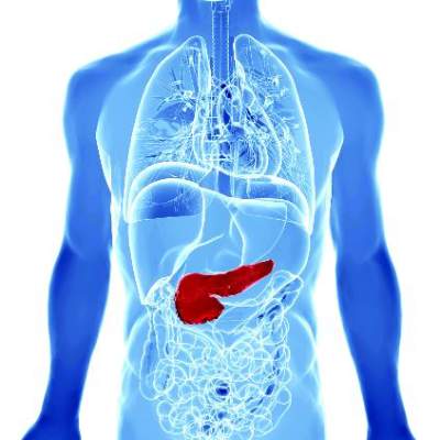
Furthermore, there were no malignant conversions among patients lacking American Gastroenterological Association high-risk features – that is, mural nodules, dilated pancreatic ducts, or cysts measuring more than 3 cm, said Dr. Wilson Kwong at the University of California San Diego Health Sciences in La Jolla. “There is a very low risk of malignant transformation of asymptomatic neoplastic pancreatic cysts after 5 years,” he and his associates wrote.
Up to 20% of cross-sectional imaging studies reveal incidental pancreatic cysts, the researchers noted. Cysts with neoplastic features are recommended for indefinite surveillance, even though there is little or no data on their natural history and malignant potential beyond 5- 10 years, they added. Therefore, they studied 310 patients who underwent endoscopic ultrasound of pancreatic cysts at an academic medical center, a Veterans’ Affairs hospital, and two community health care systems in California between 2002 and 2010. The most common age at enrollment was 66 years, 60% of patients were women, and the median follow-up period was 87 months (range, 60 to 189 months). A total of 90% of patients were followed for 5-10 years, while 10% were followed for more than 10 years (Clin Gastroenterol Hepatol. 2016 Feb 10. doi: 10.1016/j.cgh.2015.11.013).
Source: American Gastroenterological Association
In all, three patients developed invasive pancreatic malignancies after 6, 8, and 11 years of follow-up, for an overall conversion rate of 1%. Conversion rates by subgroup were 0% for patients with no high-risk AGA features, 1% (one case) for patients with one high-risk feature, and 15% (two cases) for patients with two high-risk features. “Because the risk of malignant transformation beyond 5 years is lower than the 1.4% mortality risk of pancreatic resection at high-volume centers, the argument can be made that discontinuing surveillance after 5 years is justified,” the researchers said. Specifically, surveillance could be discontinued after 5 years for neoplastic pancreatic cysts with up to one high-risk feature, particularly if patients have significant comorbidities that increase their risk of imminent death from other causes, they added. In contrast, healthy patients in their 60s and 70s might benefit from long-term surveillance given their longer life expectancy, they said. “Among patients with two high-risk features who remain surgically fit, discussion of surgery or surveillance beyond 5 years should be considered,” they emphasized.
A total of two patients developed high-grade dysplasia – a risk factor for invasive pancreatic cancer – but even so, the aggregate rate of cancer and high-grade dysplasia was 1.6%, only slightly higher than the fatality rate associated with pancreatic resection, the researchers noted. By excluding patients with recent acute pancreatitis (because of the likelihood of pseudocysts), they might have inadvertently excluded “a small number” of patients with pancreatic intraductal papillary mucinous neoplasms, they added.
The University of California San Diego Health Care System supported the study. The investigators had no disclosures.
Kwong et al. present important data demonstrating a low risk of malignant transformation for pancreas cysts followed for more than 5 years, which is similar to the risk of surgical resection. Mortality from nonpancreatic causes was found to be eightfold higher than mortality from pancreatic cancer. The goal of pancreas cyst surveillance is to prevent death from pancreatic cancer, currently accomplished by identifying high-risk cysts for surgical resection. When evaluating the utility of surveillance, patient and cyst characteristics can be considered.
Elderly patients with multiple comorbidities are unlikely to benefit from long-term surveillance as they may be poor surgical candidates and are unlikely to die from the malignant progression of a pancreas cyst. Healthy patients with a family history of pancreatic cancer and/or identifiable genetic risk factors, however, may benefit from long-term surveillance. Although demonstrated to be infrequent, cysts that have been stable for 5-10 years rarely may progress to cancer. The presence of more than one high-risk cyst feature increased the risk of progression from approximately 1% to 15%. The study of larger groups of cysts with morphologic high-risk features is required. The addition of molecular and genetic cyst and patient features has the potential to assist in risk stratification.
Clarifying which cysts and patients are likely to benefit from surveillance and resection is of increasing importance as high-resolution, cross-sectional imaging identifies greater numbers of pancreas cysts.
Dr. Harry R. Aslanian, AGAF, is director, Advanced Endoscopy Fellowship, and associate professor, Yale University, New Haven, Conn. He is a consultant for Boston Scientific and Olympus.
Only 1% of adults with asymptomatic neoplastic pancreatic cysts developed invasive pancreatic adenocarcinoma after more than 5 years of follow-up, according to a multicenter retrospective study reported in the June issue of Clinical Gastroenterology and Hepatology.
Furthermore, there were no malignant conversions among patients lacking American Gastroenterological Association high-risk features – that is, mural nodules, dilated pancreatic ducts, or cysts measuring more than 3 cm, said Dr. Wilson Kwong at the University of California San Diego Health Sciences in La Jolla. “There is a very low risk of malignant transformation of asymptomatic neoplastic pancreatic cysts after 5 years,” he and his associates wrote.
Up to 20% of cross-sectional imaging studies reveal incidental pancreatic cysts, the researchers noted. Cysts with neoplastic features are recommended for indefinite surveillance, even though there is little or no data on their natural history and malignant potential beyond 5- 10 years, they added. Therefore, they studied 310 patients who underwent endoscopic ultrasound of pancreatic cysts at an academic medical center, a Veterans’ Affairs hospital, and two community health care systems in California between 2002 and 2010. The most common age at enrollment was 66 years, 60% of patients were women, and the median follow-up period was 87 months (range, 60 to 189 months). A total of 90% of patients were followed for 5-10 years, while 10% were followed for more than 10 years (Clin Gastroenterol Hepatol. 2016 Feb 10. doi: 10.1016/j.cgh.2015.11.013).
Source: American Gastroenterological Association
In all, three patients developed invasive pancreatic malignancies after 6, 8, and 11 years of follow-up, for an overall conversion rate of 1%. Conversion rates by subgroup were 0% for patients with no high-risk AGA features, 1% (one case) for patients with one high-risk feature, and 15% (two cases) for patients with two high-risk features. “Because the risk of malignant transformation beyond 5 years is lower than the 1.4% mortality risk of pancreatic resection at high-volume centers, the argument can be made that discontinuing surveillance after 5 years is justified,” the researchers said. Specifically, surveillance could be discontinued after 5 years for neoplastic pancreatic cysts with up to one high-risk feature, particularly if patients have significant comorbidities that increase their risk of imminent death from other causes, they added. In contrast, healthy patients in their 60s and 70s might benefit from long-term surveillance given their longer life expectancy, they said. “Among patients with two high-risk features who remain surgically fit, discussion of surgery or surveillance beyond 5 years should be considered,” they emphasized.
A total of two patients developed high-grade dysplasia – a risk factor for invasive pancreatic cancer – but even so, the aggregate rate of cancer and high-grade dysplasia was 1.6%, only slightly higher than the fatality rate associated with pancreatic resection, the researchers noted. By excluding patients with recent acute pancreatitis (because of the likelihood of pseudocysts), they might have inadvertently excluded “a small number” of patients with pancreatic intraductal papillary mucinous neoplasms, they added.
The University of California San Diego Health Care System supported the study. The investigators had no disclosures.
FROM CLINICAL GASTROENTEROLOGY AND HEPATOLOGY
Key clinical point: Asymptomatic neoplastic pancreatic cysts rarely become malignant, especially in the absence of multiple American Gastroenterological Association high-risk features.
Major finding: Only 1% of patients developed invasive pancreatic adenocarcinoma after more than 5 years of surveillance.
Data source: A multicenter retrospective study of 310 patients who underwent endoscopic ultrasound evaluations of pancreatic cysts.
Disclosures: The University of California San Diego Health Care System supported the study. The investigators had no disclosures.

Metabolic factors associated with pancreatic NETs
SAN DIEGO – Diabetes (not recent onset), obesity, and gallstone disease were associated with the occurrence of sporadic pancreatic neuroendocrine tumors (PNET) in the prospective, case-control European EpiNet study.
Cigarette smoking, first-degree family history of cancer, and alcohol consumption were not significantly associated with PNET in this study, which was reported at the annual Digestive Disease Week.
“This is the first prospective multicenter study investigating risk factors for the occurrence of PNET. We confirmed that diabetes, obesity and gallstone disease are significantly associated with PNET. The role of obesity is confirmed for the first time. However, the study failed to confirm an association with family history of cancer, smoking cigarettes, alcohol consumption, hormonal reproductive factors, or allergies. This is different from pancreatic cancer,” said lead author Dr. Roberto Valente, Sapienza University of Rome, Italy.
PNET is a rare tumor, comprising 1% of all pancreatic tumors and 10% of all pancreatic neoplasms.
“The incidence of PNET is rising over time and we don’t know why. Thus far, risk factors for PNET have been poorly investigated. Many factors associated with pancreatic cancer have not been previously studied in PNET, including allergies, use of aspirin, and reproductive/gynecologic factors in women,” he said.
Previous case-control studies in small samples identified diabetes, smoking, alcohol consumption, and a family history of cancer as potential risk factors. Dr. Valente and coinvestigators sought to evaluate risk and protective factors for PNET in a more rigorous study.
Participants were drawn from five European countries. The study included 201 cases of sporadic, histologically proven PNET diagnosed within 24 months of the initiation of the study, and 603 controls matched for age and sex. For both cases and controls, about 50% were male, mean age was 59 years, and 97% were Caucasian. Among cases, 76.6% had nonfunctioning PNET that were equally distributed anatomically.
Participants responded to standardized questionnaires about demographics, and environmental, and familial risk factors using a predefined standard questionnaire.
Obesity (defined as body mass index [BMI] greater than 30) was significantly more prevalent in cases than controls (26.5% versus 19.26%, respectively, P = .03). Diabetes was also significantly more prevalent in cases than controls (18.5% versus 12.5%, respectively P = .003).
No differences were found between cases and controls in family history of any cancer, alcohol consumption, coffee drinking, or cigarette smoking.
Looking at previous medical history, only gallstone disease was strongly associated with PNET, but this was of borderline statistical significance (19.2% versus 13.2%, respectively, P = .06).
The investigators looked at previously uninvestigated potentially protective factors such as allergies (asthma, eczema, hay fever), and use of aspirin. None of these was significantly more prevalent in cases compared with controls.
Reproductive/hormonal factors were analyzed in 98 cases and 294 controls. None of these factors was significantly different in cases or controls.
Univariate regression analysis adjusted for age found that diabetes (P = .02), diabetes for more than 1 year (P = .002), obesity (P = .02), and previous gallstone disease (P = .04) were significant risk factors for PNET.
Multivariate logistic regression analysis confirmed diabetes as a significant risk factor (P = .009), and obesity and gallstone disease were of borderline significance (P = .09 and P = .07, respectively).
“The reported risk factors partially overlap with those reported for pancreatic adenocarcinoma, thus suggesting a possible organ-specific carcinogenic effect,” Dr Valente noted.
AT DDW® 2016

Surgery has edge over surveillance for micropapillary thyroid cancer
BALTIMORE – Hemithyroidectomy for low-risk micropapillary thyroid cancer can have advantages over active surveillance, according to findings from a study that examined outcomes by cost and quality of life data.
Endocrinologists and surgeons need to have in-depth conversations with their patients to determine their level of anxiety about cancer, surgery, and about their quality of life, to determine the best course of treatment, researchers at the University of California, San Francisco (UCSF) reported at the annual meeting of the American Association of Endocrine Surgeons.

“Our study found that hemithyroidectomy is cost effective in the majority of scenarios,” presenter Shriya Venkatesh said. “However, patient perception of micropapillary thyroid cancer as well as [the patient’s] life expectancy can play a major role in deciding which therapeutic option to choose.”
The study involved a cost-effectiveness analysis of the surgery vs. active surveillance, “which is especially relevant in our current times,” Ms. Venkatesh said in an interview. “What we wanted to do is give physicians information for when they approach their patients, not only in assessing the tumor from the medical aspect but also when looking at it from quality-of-life and cost-benefit perspectives.”
Both courses of management were modeled over a 20-year period with Medicare data and literature review to calculate costs and health utilities. The UCSF researchers used Markov statistical models for both approaches in which the reference case was a 40-year-old, otherwise healthy patient with a recent diagnosis of micropapillary thyroid cancer without high-risk factors. Either hemithyroidectomy or surveillance would be reasonable treatment options.
“We found that hemithyroidectomy was about $8,000 more costly than active surveillance, but it also afforded an increase in about 1.09 quality-adjusted life years,” Ms. Venkatesh said. Hemithyroidectomy is most cost effective for patients with a life expectancy of 3 years or more and who perceive that living with low-grade thyroid cancer would have even a modest detriment on their quality of life, she said.
“Unfortunately there is no current published quality-of-life assessment of active surveillance for thyroid cancer,” Ms. Venkatesh said. “We believe that estimating active surveillance to the equivalent of surgery underestimates the anxiety some patients may feel upon receiving their diagnosis.”
The paucity of literature on active surveillance for thyroid cancer prompted the UCSF researchers to turn to the prostate cancer literature, which has more data on active surveillance, to try to determine the disutility of active surveillance for micropapillary thyroid cancer. “Our extrapolation from the literature yields a mean disutility of 0.11,” she said.
However, the utility estimates the researchers came up with were variable, Ms. Venkatesh said. “This really pushes physicians to have that conversation with their patients, not only about the physical aspects of how they’re doing but also the mental aspects,” she said.

But quality of life is difficult to quantify, senior author Dr. Insoo Suh said in an interview. “What we found is that no matter how one measures quality of life, the qualitative degree of quality of life decrease that people associate with ‘living with cancer’ need not be that significant in order for surgery to be a potentially cost-effective treatment for them,” said Dr. Suh, an endocrine surgeon at UCSF and an ACS Fellow.
During the discussion, Dr. Peter Angelos of the University of Chicago and an ACS Fellow, said, “I’m curious how this information should impact the individual decision-making and informed consent for a specific patient, because I’m not sure that an individual patient would care if active surveillance is more cost effective or not.”
“When speaking to your patients, obviously discussing the rates of progression of the disease is important and then [so is] talking to them about different therapeutic options,” Ms. Venkatesh said. “The physician should also make an assessment about the patient’s quality of life to see if there are likely to be any changes due to the diagnosis.”
The limitations of the study include the extrapolation of data from the prostate cancer literature to define a utility scale and also the reference case used in the Markov model. Other utility measures showed variability as well.
Ms. Venkatesh, Dr. Suh and their coauthors had no financial relationships to disclose.
BALTIMORE – Hemithyroidectomy for low-risk micropapillary thyroid cancer can have advantages over active surveillance, according to findings from a study that examined outcomes by cost and quality of life data.
Endocrinologists and surgeons need to have in-depth conversations with their patients to determine their level of anxiety about cancer, surgery, and about their quality of life, to determine the best course of treatment, researchers at the University of California, San Francisco (UCSF) reported at the annual meeting of the American Association of Endocrine Surgeons.
“Our study found that hemithyroidectomy is cost effective in the majority of scenarios,” presenter Shriya Venkatesh said. “However, patient perception of micropapillary thyroid cancer as well as [the patient’s] life expectancy can play a major role in deciding which therapeutic option to choose.”
The study involved a cost-effectiveness analysis of the surgery vs. active surveillance, “which is especially relevant in our current times,” Ms. Venkatesh said in an interview. “What we wanted to do is give physicians information for when they approach their patients, not only in assessing the tumor from the medical aspect but also when looking at it from quality-of-life and cost-benefit perspectives.”
Both courses of management were modeled over a 20-year period with Medicare data and literature review to calculate costs and health utilities. The UCSF researchers used Markov statistical models for both approaches in which the reference case was a 40-year-old, otherwise healthy patient with a recent diagnosis of micropapillary thyroid cancer without high-risk factors. Either hemithyroidectomy or surveillance would be reasonable treatment options.
“We found that hemithyroidectomy was about $8,000 more costly than active surveillance, but it also afforded an increase in about 1.09 quality-adjusted life years,” Ms. Venkatesh said. Hemithyroidectomy is most cost effective for patients with a life expectancy of 3 years or more and who perceive that living with low-grade thyroid cancer would have even a modest detriment on their quality of life, she said.
“Unfortunately there is no current published quality-of-life assessment of active surveillance for thyroid cancer,” Ms. Venkatesh said. “We believe that estimating active surveillance to the equivalent of surgery underestimates the anxiety some patients may feel upon receiving their diagnosis.”
The paucity of literature on active surveillance for thyroid cancer prompted the UCSF researchers to turn to the prostate cancer literature, which has more data on active surveillance, to try to determine the disutility of active surveillance for micropapillary thyroid cancer. “Our extrapolation from the literature yields a mean disutility of 0.11,” she said.
However, the utility estimates the researchers came up with were variable, Ms. Venkatesh said. “This really pushes physicians to have that conversation with their patients, not only about the physical aspects of how they’re doing but also the mental aspects,” she said.
But quality of life is difficult to quantify, senior author Dr. Insoo Suh said in an interview. “What we found is that no matter how one measures quality of life, the qualitative degree of quality of life decrease that people associate with ‘living with cancer’ need not be that significant in order for surgery to be a potentially cost-effective treatment for them,” said Dr. Suh, an endocrine surgeon at UCSF and an ACS Fellow.
During the discussion, Dr. Peter Angelos of the University of Chicago and an ACS Fellow, said, “I’m curious how this information should impact the individual decision-making and informed consent for a specific patient, because I’m not sure that an individual patient would care if active surveillance is more cost effective or not.”
“When speaking to your patients, obviously discussing the rates of progression of the disease is important and then [so is] talking to them about different therapeutic options,” Ms. Venkatesh said. “The physician should also make an assessment about the patient’s quality of life to see if there are likely to be any changes due to the diagnosis.”
The limitations of the study include the extrapolation of data from the prostate cancer literature to define a utility scale and also the reference case used in the Markov model. Other utility measures showed variability as well.
Ms. Venkatesh, Dr. Suh and their coauthors had no financial relationships to disclose.
AT AAES 2016
Key clinical point: Patient psychological factors are key determinants in choosing a course of management for low-risk micropapillary thyroid cancer.
Major finding: Hemithyroidectomy typically costs about $8,000 more than active surveillance but also accounts for improved quality of life in these patients.
Data source: Markov models for both courses of management over a 20-year period with Medicare data and literature review to calculate costs and health utilities.
Disclosures: Ms. Venkatesh and her coauthors reported having no financial disclosures.

VIDEO: Proposed revision of medullary thyroid cancer staging improves risk-stratification analysis
BOSTON – An analysis of data from medullary thyroid cancer patients that partitioned the patients into groups with similar overall survival has spurred a rethink of the current American Joint Committee on Cancer (AJCC) staging system.
The results from researchers at Duke University, Durham, N.C., presented at the annual meeting of the Endocrine Society by Dr. Mohamed Abdelgadir Adam, are timely, as the AJCC has embarked on a reconsideration of the staging of cancers, including medullary thyroid cancer (MTC), as part revisions for the eighth edition of the staging system.
“The existing AJCC staging system for MTC appears to be less than optimal in discriminating the risk of mortality among disease stage groups,” said Dr. Adam, who discussed the findings in a video interview.
MTC, a neuroendocrine tumor that affects C cells of the thyroid, comprises 3%-5% of all cases of thyroid cancer and it can be a more aggressive disease than differentiated thyroid cancer. Yet the current AJCC MTC staging system has been extrapolated from differentiated thyroid cancer data.
“We sought to evaluate how well the current AJCC seventh edition stage groupings predict survival for patients with MTC, to suggest a possible staging revision to sharpen estimates of prognosis,” said Dr. Adam.
The researchers utilized the National Cancer Data Base, representing over 70% of incident cancer cases in the United States.
MTC patients who underwent thyroid surgery from 1998 to 2012 were identified. Patients with missing values for pathologic T, N, or M were excluded. The primary outcome in the 3,315 patients was survival.
The researchers used a form of decision-tree analysis called recursive partitioning. In general, recursive partitioning is able to classify a population by splitting subjects into subgroups, each of which is homogeneous based on the particular outcome. In this study, the subgroup allocations were based on T, N, and M stages, with the outcome being overall survival. Kaplan-Meier and adjusted survival analyses enabled survival differences among the four subgroups (groups I, II, III and IV) to be explored.
The four groups were distinct in terms of survival time and allowed more accurate risk stratification. In particular, groups I and II were markedly better distinguished from one another than is the case with the current staging system. Survival differences across the stages were more distinct with the newly created T, N, and M groupings, compared with the current AJCC staging system.
After adjustment, survival differences across TNM groups were more distinct with the newly created TNM groupings (compared to subgroup I, hazard ratio of 3.06 for subgroup II; HR, 6.79 for III; and HR, 17.03 for IV), compared with the current AJCC staging (compared to stage I, HR, 1.45 for stage II; HR, 2.17 for III; and HR, 5.33 for IV).
“The AJCC is reevaluating all staging schemas, including MTC. The current AJCC staging system could be improved with the newly identified TNM groupings suggested here for more accurate patient risk stratification and possibly treatment selection,” said Dr. Adam.
Dr. Adam had no disclosures.
The video associated with this article is no longer available on this site. Please view all of our videos on the MDedge YouTube channel
Key clinical point: A proposed revision of the AJCC thyroid cancer staging system improves risk stratification analysis.
Major finding: In the proposed staging system, compared to subgroup I, hazard ratio for survival was 3.06 for subgroup II; HR, 6.79 for III; and HR, 17.03 for IV, compared with the current AJCC staging of HR, 1.45 for stage II; HR, 2.17 for III; and HR, 5.33 for IV.
Data source: Data from 3,315 patients with medullary thyroid cancer was drawn from the National Cancer Database.
Disclosures: Dr. Adam had no disclosures.

Adjuvant endocrine therapy for premenopausal breast cancer patients should be individualized
Oncologists should take an individualized approach when making decisions about adjuvant endocrine therapies for premenopausal hormone receptor–positive, HER2-negative early breast cancer, suggests an analysis of a pair of randomized phase III trials published online in the Journal of Clinical Oncology.
Investigators led by Meredith M. Regan, Sc.D., of Dana-Farber Cancer Institute in Boston, analyzed data from the TEXT (Tamoxifen and Exemestane Trial) and SOFT (Suppression of Ovarian Function Trial) trials of adjuvant endocrine therapies, comprising a total of nearly 5,000 women.
Results suggested that the absolute improvement in the 5-year breast cancer–free interval rate with exemestane plus ovarian function suppression (OFS) versus tamoxifen with or without OFS ranged from less than 1% in women with a lowest recurrence risk based on clinicopathologic factors to 10%-15% in women with a highest risk.
“TEXT and SOFT demonstrated that premenopausal women with hormone receptor–positive disease benefit, on average, from exemestane plus OFS versus tamoxifen with or without OFS. However, individualized treatment decisions should weigh the benefits against the adverse effects and costs of these therapy options,” the investigators wrote.
“In the absence of predictive biomarkers, consideration of a patient’s prognosis, as illustrated by STEPP [Subpopulation Treatment Effect Pattern Plot] analysis of a composite measure of recurrence risk in the TEXT and SOFT populations, is integral to this decision making,” they added.
In the SOFT trial, women were randomized to 5 years of tamoxifen alone (as an active comparator), tamoxifen plus OFS, or exemestane (Aromasin) plus OFS. In the TEXT trial, women were randomized to 5 years of exemestane plus OFS or of tamoxifen plus OFS.
Dr. Regan and colleagues based their analyses on a total of 4,891 women. They assessed each patient’s composite recurrence risk from a Cox model that included a set of conventional clinicopathologic factors: age, nodal status, tumor size and grade, and estrogen receptor, progesterone receptor, and Ki-67 expression levels. And they used STEPP methodology to assess the impact of endocrine therapy across groups having different risk.
The median duration of follow-up was 5.6 years in the SOFT trial and 6 years in the TEXT trial. Results showed that the 5-year breast cancer–free interval rate was 90.8% for the study cohort as a whole. But it ranged considerably from 98.6% for patients with composite risk in the lowest quartile to 77.5% for patients with composite risk in the highest quartiles, the investigators reported (J Clin Oncol. 2016. doi: 10.1200/JCO.2015.64.3171).
In the SOFT population, patients who remained premenopausal after neoadjuvant or adjuvant chemotherapy had an absolute improvement of 5% or more in the 5-year breast cancer–free interval rate with exemestane plus OFS, compared with tamoxifen plus OFS or tamoxifen alone. The difference was 10%-15% for the subset at intermediate to high risk for recurrence.
In addition, a benefit of tamoxifen plus OFS over tamoxifen alone was evident in patients having the highest composite risk.
Among patients who were not given chemotherapy, who on average had the lowest composite recurrence risk, the 5-year breast cancer–free interval rate was excellent regardless of the endocrine therapy received.
In the TEXT trial population, the benefit of exemestane plus OFS over tamoxifen plus OFS in 5-year breast cancer–free interval rate ranged from 5% to 15%. Again, the patients who were not given chemotherapy, who had the lowest composite recurrence risk, fared well regardless of which endocrine therapy they received.
These findings should help guide clinical decisions in premenopausal women with hormone receptor–positive, HER2-negative breast cancer, both at the extremes of risk and in the scenario of intermediate risk, where factors such as patient preference, tolerance, and cost play a greater role, according to the investigators.
“Further follow-up of TEXT and SOFT patients is essential to guide patient care,” they concluded.
FROM THE JOURNAL OF CLINICAL ONCOLOGY
Key clinical point: Decisions about adjuvant endocrine therapies in premenopausal women with HR–positive, HER2-negative early breast cancer should be individualized.
Major finding: The absolute improvement in 5-year breast cancer–free interval rate with exemestane plus OFS versus tamoxifen with or without OFS ranged from less than 1% for those with lowest recurrence risk to 15% for those with highest recurrence risk.
Data source: An analysis of data from 4,891 women treated in the TEXT and SOFT trials of adjuvant endocrine therapies for premenopausal HR–positive, HER2-negative breast cancer.
Disclosures: Dr. Reagan disclosed that her institution receives research funding from Veridex, OncoGenex, Pfizer, Ipsen, Novartis, Merck, Ferring Pharmaceuticals, Celgene, and AstraZeneca.
Early biopsy predicts levonorgestrel IUD response in endometrial cancer
SAN DIEGO – Endometrial pathology findings at 3 months predicted response to levonorgestrel-releasing IUD treatment for complex atypical hyperplasia or grade 1 endometrial cancer at the MD Anderson Cancer Center in Houston.
Twenty-nine of 32 women (91%) who responded by 12 months showed stromal, glandular, or other endometrial changes indicating an effect at 3 months, vs. only 3 of 9 nonresponders (33%) (P less than .001). There were no differences in responders versus nonresponders in median age (47 vs. 56 years, P = .2) or body mass index (45 vs. 55 kg/m2, P = .16).

The finding addresses an “unmet need” for markers of response to levonorgestrel-releasing IUD therapy. “You can look at [early] pathology” and have an idea how patients will do, Dr. Shannon Westin, a study investigator who is with the department of gynecologic oncology at MD Anderson, said at the annual meeting of the Society of Gynecologic Oncology.
Twenty-seven of 29 women (93%) with complex atypical hyperplasia (CAH) responded completely to the IUD, meaning they had normal endometrium or hyperplasia without atypia at 12 months. The response rate for endometrial cancer was 67%; 7 of 12 women had a complete response, and an 8th was diagnosed at 12 months with CAH, indicating a partial response. The rest of the patients remained stable or progressed.
Endometrial biopsies were performed every 3 months; the team also did molecular testing on tumors from 20 patients. Baseline protein Ki67 – a marker of proliferation – was significantly higher in nonresponders. Expression of several estrogen-induced genes was higher in responders.
Patients opted for the IUD to retain fertility or because obesity or comorbidities precluded surgery. Exclusion criteria included prior treatment for CAH or endometrial cancer, evidence of extrauterine spread, or levonorgestrel IUD contraindications, such as uterine infection.
Adverse events – primarily irregular bleeding and cramping – were mild and tended to resolve by 12 months. Treatment had little effect on measures of social, mental, and physical function. About half of the patients were white, a third were Hispanic, and most of the remaining patients were black.
There was no external funding for the work. Dr. Westin is a consultant for AstraZeneca, Medivation, Roche, Ovation, and Vermillion, and reported receiving research funding from AstraZeneca, Critical Outcomes Technologies, and Novartis.
SAN DIEGO – Endometrial pathology findings at 3 months predicted response to levonorgestrel-releasing IUD treatment for complex atypical hyperplasia or grade 1 endometrial cancer at the MD Anderson Cancer Center in Houston.
Twenty-nine of 32 women (91%) who responded by 12 months showed stromal, glandular, or other endometrial changes indicating an effect at 3 months, vs. only 3 of 9 nonresponders (33%) (P less than .001). There were no differences in responders versus nonresponders in median age (47 vs. 56 years, P = .2) or body mass index (45 vs. 55 kg/m2, P = .16).
The finding addresses an “unmet need” for markers of response to levonorgestrel-releasing IUD therapy. “You can look at [early] pathology” and have an idea how patients will do, Dr. Shannon Westin, a study investigator who is with the department of gynecologic oncology at MD Anderson, said at the annual meeting of the Society of Gynecologic Oncology.
Twenty-seven of 29 women (93%) with complex atypical hyperplasia (CAH) responded completely to the IUD, meaning they had normal endometrium or hyperplasia without atypia at 12 months. The response rate for endometrial cancer was 67%; 7 of 12 women had a complete response, and an 8th was diagnosed at 12 months with CAH, indicating a partial response. The rest of the patients remained stable or progressed.
Endometrial biopsies were performed every 3 months; the team also did molecular testing on tumors from 20 patients. Baseline protein Ki67 – a marker of proliferation – was significantly higher in nonresponders. Expression of several estrogen-induced genes was higher in responders.
Patients opted for the IUD to retain fertility or because obesity or comorbidities precluded surgery. Exclusion criteria included prior treatment for CAH or endometrial cancer, evidence of extrauterine spread, or levonorgestrel IUD contraindications, such as uterine infection.
Adverse events – primarily irregular bleeding and cramping – were mild and tended to resolve by 12 months. Treatment had little effect on measures of social, mental, and physical function. About half of the patients were white, a third were Hispanic, and most of the remaining patients were black.
There was no external funding for the work. Dr. Westin is a consultant for AstraZeneca, Medivation, Roche, Ovation, and Vermillion, and reported receiving research funding from AstraZeneca, Critical Outcomes Technologies, and Novartis.
AT THE ANNUAL MEETING ON WOMEN’S CANCER
Key clinical point: Pathology and molecular findings identify good candidates for levonorgestrel IUD therapy.
Major finding: Twenty-nine of 32 women (91%) who responded by 12 months showed stromal, glandular, or other endometrial changes indicating an effect at 3 months, versus only 3 of 9 nonresponders (33%) (P less than .001).
Data source: A prospective investigation of 41 women.
Disclosures: There was no external funding for the work. Dr. Westin is a consultant for and receives research funding from AstraZeneca and several other companies.

A call for definitive trial of statins in breast cancer
SAN ANTONIO – A definitive phase III randomized controlled trial of statin therapy during adjuvant endocrine therapy for breast cancer is warranted in light of recent encouraging evidence pointing to a protective effect against disease recurrence, Melissa L. Bondy, Ph.D., said at the San Antonio Breast Cancer Symposium.
“The next phase of research in this area is to learn whether we can improve survival in breast cancer patients by having them take cholesterol-lowering medication. We really need to have a phase III trial in the adjuvant setting that includes these cholesterol-lowering drugs,” said Dr. Bondy, professor of cancer prevention and population sciences at Baylor Medical College, Houston.
Her call for a formal, randomized trial came while serving as discussant for two positive reports of an apparent beneficial effect of statins on breast cancer recurrence-free survival: one from the BIG 1-98 trial of adjuvant endocrine therapy for early-stage breast cancer, the other a meta-analysis of 12 published studies totaling 72,774 breast cancer patients.

Dr. Bondy found particularly compelling the Breast International Group (BIG) 1-98 study results presented by Dr. Signe Borgquist, an oncologist at Lund (Sweden) University. This secondary analysis of the impact of cholesterol-lowering medication included 7,963 postmenopausal women with early-stage, estrogen receptor–positive invasive breast cancer who were randomized to 5 years of tamoxifen, the aromatase inhibitor letrozole (Femara), or the two drugs in sequence. Serum total cholesterol levels and the use of cholesterol-lowering medications – a decision left up to individual physicians and patients – were assessed at baseline and again every 6 months for 5.5 years.
With a median 8 years of prospective follow-up, during which 1,432 patients experienced breast cancer recurrence, the disease-free survival rate was 18% higher in the 637 patients already on cholesterol-lowering medication – mainly statins – at enrollment, compared with those who were not. The distant recurrence-free interval was 19% better as well, with both analyses adjusted for their form of endocrine therapy, tumor size and grade, nodal status, local therapy, peritumoral vascular invasion, and other potential confounders.
Another 697 BIG 1-98 participants initiated cholesterol-lowering medication during their adjuvant endocrine therapy. In multivariate adjusted analyses, their disease-free survival rate was 21% greater and distant recurrence-free interval was 26% better than in participants not on a statin or other cholesterol-lowering medication during the trial.
As a possible mechanism by which statins might exert anticancer effects, Dr. Borgquist proposed that lowering cholesterol levels may deprive tumor cells of the ability to satisfy their increased demand for cholesterol uptake. This is a result of attenuated signaling through the estrogen receptor by the cholesterol metabolite 27-hydroxycholesterol, levels of which correlate with systemic total cholesterol.
Dr. Bondy offered another possible explanation for the reduced risk of breast cancer recurrence seen in women on cholesterol-lowering medication in BIG 1-98: “Many groups have shown that LDL affects the immune system by binding and inactivating all kinds of microorganisms and their toxic products. In other words, statins appear to affect the microbiome.”
She commented that, although the use of statins was nonrandomized in BIG 1-98, she was favorably impressed by the 8-year follow-up and careful monitoring of total cholesterol levels at 6-month intervals throughout the 5 years of adjuvant endocrine therapy.
Dr. Bondy noted that other studies – again, nonrandomized – suggest statins also interrupt the growth of colorectal cancer and other malignancies.

Dr. Sashidhar Manthravadi presented a meta-analysis of the 12 published studies that have reported data on the association of statins with recurrence-free and/or overall survival in breast cancer patients. Statin use was associated with a significant 34% improvement in recurrence-free survival.
Moreover, this benefit was confined to breast cancer patients on the lipophilic statins atorvastatin and simvastatin; the use of hydrophilic statins was not associated with a significant improvement in recurrence-free survival, according to Dr. Manthravadi, an internal medicine resident at the University of Missouri, Kansas City.
The use of statins also was associated with a 27% improvement in breast cancer–specific survival and a 33% improvement in overall survival, compared with statin nonusers; however, neither of these results achieved statistical significance, he added.
Dr. Bondy cautioned against making too much of the apparent distinction between lipophilic and hydrophilic statin in terms of protection against breast cancer recurrence, given the inherent limitations of a meta-analysis.
“There’s always a lot of missing information, as well as a failure to adjust for comorbidities. And follow-up times in these 12 studies ranged from 2.5 to 11.5 years,” she noted.
The ongoing BIG 1-98 trial is sponsored by the International Breast Cancer Study Group. Dr. Borgquist, Dr. Bondy, and Dr. Manthravadi reported having no financial conflicts of interest.
SAN ANTONIO – A definitive phase III randomized controlled trial of statin therapy during adjuvant endocrine therapy for breast cancer is warranted in light of recent encouraging evidence pointing to a protective effect against disease recurrence, Melissa L. Bondy, Ph.D., said at the San Antonio Breast Cancer Symposium.
“The next phase of research in this area is to learn whether we can improve survival in breast cancer patients by having them take cholesterol-lowering medication. We really need to have a phase III trial in the adjuvant setting that includes these cholesterol-lowering drugs,” said Dr. Bondy, professor of cancer prevention and population sciences at Baylor Medical College, Houston.
Her call for a formal, randomized trial came while serving as discussant for two positive reports of an apparent beneficial effect of statins on breast cancer recurrence-free survival: one from the BIG 1-98 trial of adjuvant endocrine therapy for early-stage breast cancer, the other a meta-analysis of 12 published studies totaling 72,774 breast cancer patients.
Dr. Bondy found particularly compelling the Breast International Group (BIG) 1-98 study results presented by Dr. Signe Borgquist, an oncologist at Lund (Sweden) University. This secondary analysis of the impact of cholesterol-lowering medication included 7,963 postmenopausal women with early-stage, estrogen receptor–positive invasive breast cancer who were randomized to 5 years of tamoxifen, the aromatase inhibitor letrozole (Femara), or the two drugs in sequence. Serum total cholesterol levels and the use of cholesterol-lowering medications – a decision left up to individual physicians and patients – were assessed at baseline and again every 6 months for 5.5 years.
With a median 8 years of prospective follow-up, during which 1,432 patients experienced breast cancer recurrence, the disease-free survival rate was 18% higher in the 637 patients already on cholesterol-lowering medication – mainly statins – at enrollment, compared with those who were not. The distant recurrence-free interval was 19% better as well, with both analyses adjusted for their form of endocrine therapy, tumor size and grade, nodal status, local therapy, peritumoral vascular invasion, and other potential confounders.
Another 697 BIG 1-98 participants initiated cholesterol-lowering medication during their adjuvant endocrine therapy. In multivariate adjusted analyses, their disease-free survival rate was 21% greater and distant recurrence-free interval was 26% better than in participants not on a statin or other cholesterol-lowering medication during the trial.
As a possible mechanism by which statins might exert anticancer effects, Dr. Borgquist proposed that lowering cholesterol levels may deprive tumor cells of the ability to satisfy their increased demand for cholesterol uptake. This is a result of attenuated signaling through the estrogen receptor by the cholesterol metabolite 27-hydroxycholesterol, levels of which correlate with systemic total cholesterol.
Dr. Bondy offered another possible explanation for the reduced risk of breast cancer recurrence seen in women on cholesterol-lowering medication in BIG 1-98: “Many groups have shown that LDL affects the immune system by binding and inactivating all kinds of microorganisms and their toxic products. In other words, statins appear to affect the microbiome.”
She commented that, although the use of statins was nonrandomized in BIG 1-98, she was favorably impressed by the 8-year follow-up and careful monitoring of total cholesterol levels at 6-month intervals throughout the 5 years of adjuvant endocrine therapy.
Dr. Bondy noted that other studies – again, nonrandomized – suggest statins also interrupt the growth of colorectal cancer and other malignancies.
Dr. Sashidhar Manthravadi presented a meta-analysis of the 12 published studies that have reported data on the association of statins with recurrence-free and/or overall survival in breast cancer patients. Statin use was associated with a significant 34% improvement in recurrence-free survival.
Moreover, this benefit was confined to breast cancer patients on the lipophilic statins atorvastatin and simvastatin; the use of hydrophilic statins was not associated with a significant improvement in recurrence-free survival, according to Dr. Manthravadi, an internal medicine resident at the University of Missouri, Kansas City.
The use of statins also was associated with a 27% improvement in breast cancer–specific survival and a 33% improvement in overall survival, compared with statin nonusers; however, neither of these results achieved statistical significance, he added.
Dr. Bondy cautioned against making too much of the apparent distinction between lipophilic and hydrophilic statin in terms of protection against breast cancer recurrence, given the inherent limitations of a meta-analysis.
“There’s always a lot of missing information, as well as a failure to adjust for comorbidities. And follow-up times in these 12 studies ranged from 2.5 to 11.5 years,” she noted.
The ongoing BIG 1-98 trial is sponsored by the International Breast Cancer Study Group. Dr. Borgquist, Dr. Bondy, and Dr. Manthravadi reported having no financial conflicts of interest.
EXPERT ANALYSIS FROM SABCS 2015

ITC: Study provides first evidence of paclitaxel benefit for anaplastic thyroid cancer
LAKE BUENA VISTA, FLA. – Weekly infusions of paclitaxel delayed progression in some patients with the very aggressive anaplastic thyroid cancer, a small prospective study determined.
The drug was most effective as adjuvant therapy for patients who had already undergone chemotherapy plus resection of the primary tumor, Dr. Naoyoshi Onoda reported in a poster session at the International Thyroid Conference. They survived for a median of 1 year (112-788 days) – an impressive feat considering that most patients with anaplastic thyroid cancer die within 6 months of diagnosis.
This finding suggests a place for paclitaxel as a standardized therapy for such patients, said Dr. Onoda of Osaka (Japan) City University. “We have objective data supporting standardized chemotherapy for the first time in the world.”
Anaplastic thyroid cancer is a very rare – but very aggressive – disease; there is no standardized treatment option. Dr. Onoda and his colleagues conducted a national prospective open-label study of weekly paclitaxel infusions in 56 patients with the malignancy.
The cohort was a median of 71 years old. All had stage IV disease: 10 were grade A, 18 grade B, 24 grade C, and four grade X. They received 80 mg/m2 infusions once a week. The median number of cycles was 2, although it ranged from 0-23 cycles.
Almost everyone (98%) experienced adverse events; the most common was anemia (77%). About a quarter (28%) experienced adverse events of at least grade 3, but there were no serious events and no deaths related to the study drug.
The objective response rate and the clinical benefit rate were 23% and 79%. The agent was not curative; at the last follow-up, no patient had achieved a complete response, and 43 of 56 in the study had died of their disease. “Overall, the median time to progression was only 47 days, and median overall survival just 227 days. That is so very short. But it’s a little bit longer than we had been seeing rates from reported cases,” he said at the meeting held by the American Thyroid Association, Asia-Oceania Thyroid Association, European Thyroid Association, and Latin American Thyroid Society.
The study was sponsored by the Prospective Clinical Study Committee of the Anaplastic Thyroid Carcinoma Research Consortium of Japan (ATCCJ). Dr. Onoda had no financial disclosures.
AT ITC 2015
Key clinical point: Paclitaxel may have use as a standardized treatment in anaplastic thyroid cancer.
Major finding: Weekly paclitaxel extended survival to a median of 1 year as adjuvant therapy for patients with resected anaplastic thyroid cancer.
Data source: The prospective open-label study involved 56 patients.
Disclosures: The study was sponsored by the Prospective Clinical Study Committee of the Anaplastic Thyroid Carcinoma Research Consortium of Japan (ATCCJ). Dr. Onoda had no financial disclosures.