User login
Pancreatic Panniculitis
Drug Hypersensitivity Reactions Presenting as a Morbilliform Eruption With Islands of Sparing [letter]
Phaeohyphomycosis
What Is Your Diagnosis? Desmoplastic Spitz Nevus
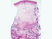
Syringocystadenoma Papilliferum
Lichtenberg Figures and Lightning: Case Reports and Review of the Literature
Thyroid Dermopathy: An Underrecognized Cause of Leg Edema
Current and Emerging Therapeutic Modalities for Hyperhidrosis, Part 1: Conservative and Noninvasive Treatments
Rosacea in the Pediatric Population
Epidemiology
Rosacea in childhood is most likely underreported because of its clinical similarity to other erythematous facial disorders.1 Rosacea is generally thought of as a disease of fair-skinned, young to middle-aged adults, though it has been noted to affect people of other complexions and ages.2 Most full-blown cases in the pediatric population have been in light-skinned children ranging from infants to adolescents.
Etiology
The etiology of rosacea is unknown, though certain exacerbating factors undoubtedly have a role in predisposed individuals.3,4 Emotions such as anger, anxiety, and embarrassment can lead to flushing. Environmental conditions such as wind, cold, humidity, or heat from any source (eg, sun, sauna, whirlpool, vigorous exercise) can do the same. Vasodilators such as alcohol or vasodilatory medications can lead to flushing, though these are not likely causes in children. Spicy foods such as chili, curry, and peppers, as well as hot foods and beverages including coffee, tea, and hot chocolate, may contribute to symptoms. Irritants such as alcohol-based cleansers, astringents, perfume, shaving lotion, certain soaps, sunscreen, and facecloths may aggravate rosacea.3,4 Saprophytic mites (Demodex folliculorum and Demodex brevis) may cause an inflammatory or allergic reaction by either blocking hair follicles or acting as vectors for microorganisms that some believe may be responsible for or may trigger rosacea.5 Immunodeficiency, as in patients with human immunodeficiency virus, also may contribute to the development of rosacea.6
Genetics plays an uncertain role in the development of blushing and ultimately rosacea. If vasodilator substances or mediators are implicated in the development of rosacea as postulated, the disease may have a genetic basis because such mediators are often under the control of single genes.7 In one study, 20% of children with rosacea were found to have a history of rosacea in their immediate families, though this number may be an underestimate because only one parent of each patient was examined and half of the parents clinically diagnosed with rosacea reported no familial involvement.8 A family history of perioral dermatitis also may be important, as this condition may be related to rosacea.9
Pathophysiology
Chronic transient vasodilation, as occurs with blushing, is the earliest representation of rosacea. The warmth and redness associated with flushing is caused by vasodilation, allowing excess blood flow, and by engorgement of the subpapillary venous plexus.2 Flushing without sweating is typically seen in children and is likely due to circulating vasodilator substances or mediators such as bradykinin, catecholamines, cytokines, endorphins, gastrin, histamine, neuropeptides, serotonin, substance P, and vascular endothelial growth factor.10 A flaw in the autonomic innervation of the cutaneous vasculature also is a likely mechanism.10
Clinical Description
The first stage of rosacea consists of blushing, in which the face becomes bright red in response to certain stimuli (Figure). Episodes of erythema are recurrent and last longer than normal physiologic flushing, which typically subsides within minutes.11 Telangiectasias can become apparent over time. Children in this early stage of the condition may complain of burning or irritation.
Please refer to the PDF to view the figure
In the second, or intermediate, stage of rosacea, the rash consists of papules and pustules on a background of erythema with telangiectasias confined to the child's face. Although peripheral involvement of the back, upper chest, and scalp may be seen in adults, these areas seem to be spared in the pediatric population.12
The third, or late, stage of rosacea involves coarse skin, inflammatory nodules, or gross enlargement of facial features.11 Such chronic changes do not occur in children as they do in adults, presumably because the disease process takes more time to evolve.13
Eye involvement can occur in children.14 It may include manifestations such as blepharoconjunctivitis, episcleritis, keratitis, meibomianitis, chalazia, hordeola, and hyperemic conjunctivae.15,16 Although any of these eye conditions can potentially occur in children, meibomian gland inflammation and keratitis are the common findings noted.17 Peripheral vascularization followed by subepithelial infiltrates can lead to scarring or perforation in the lower two thirds of the cornea. The disease may be unilateral, but most commonly it affects both eyes.17
Steroid-induced rosacea also has been termed iatrosacea because of its mode of acquisition.18 Topical fluorinated and low-dose corticosteroids can cause a rosacealike dermatitis of the face consisting of persistent erythema with papules, pustules, telangiectasias, and sometimes atrophy.19-22 Corticosteroids may be an exacerbating factor leading to classic rosacea rather than the cause of an independent disease.2 The distribution of steroid rosacea to the eyelids and lateral face may help distinguish it from the centrally located typical rosacea.23 A case of pediatric rosacea associated with the use of topical fluorinated glucocorticosteroids was identified in a 9-month-old boy and 16-year-old girl, both with erythematous patches and papules on the cheeks, paranasal areas, and chin.18,23 Forty-six boys and 60 girls, ranging in age of onset from 6 months to 13 years (average, 7 years), were diagnosed with steroid rosacea. Nearly all of the children had perinasal and perioral involvement of erythematous skin interspersed with papules and/or pustules. The lower eyelids were affected in roughly half of the patients.8
Diagnosis
Consistent flushing in children may be a sign of vasomotor instability and early rosacea. These children may blush more frequently and with greater intensity for longer periods than their peers exposed to the same stimuli.24 Thus, blushing in the early stage of rosacea may be an accentuation of the body's normal physiologic response system. A diagnosis of pediatric rosacea beyond the initial stage should be considered when a healthy child has acuminate papules and small pustules of the face, especially if there also exists flushing, telangiectasias, or a family history of rosacea.14
Differential Diagnosis
The earliest form of rosacea, facial blushing, may be difficult to distinguish from flushing due to other causes. Blushing due to emotions such as embarrassment or anger and to exercise-induced flushing are both appropriate reactions to such stimuli, whereas blushing in the first stage of rosacea may be an exaggeration of this phenomenon.2 The main pathway for thermoregulatory and emotional flushing is the cervical sympathetic outflow tract.25 Gustatory blushing, as occurs with consumption of spicy foods, is mediated by autonomic neurons via a branch of the trigeminal nerve.2 Sweating often occurs in conjunction with the aforementioned causes of flushing, but it is rarely associated with rosacea flushing. Thus, sweating may be helpful in reaching a diagnosis; however, exceptions exist.2 Frey syndrome (auriculotemporal syndrome), which is characterized by warmth and sweating in the malar region caused by aberrant autonomic fiber connections after damage in the parotid region, may mimic the early stage of rosacea.
The intermediate stage of pediatric rosacea may be confused with other papulopustular disorders such as acne vulgaris, perioral dermatitis, and lupus erythematosus (Table). Careful attention to symptoms, distribution of facial lesions, and potential biopsy results are warranted to distinguish between the conditions. Steroid rosacea and perioral dermatitis may be variants of rosacea or completely separate conditions.9,26
Please refer to the PDF to view the table
Perioral dermatitis is a rosacealike dermatitis characterized by erythematous papules and pustules usually confined to the perioral region, though the perinasal and periocular areas may be involved.27 A granulomatous perioral dermatitis with tiny, closely spaced, flesh-colored papules in the perioral, perinasal, and periorbital areas was described in children aged 3 to 11 years.28 All cases had spontaneous resolution of symptoms regardless of treatment. The patients did not exhibit flushing or telangiectasias.28
The classic butterfly rash, consisting of erythema and telangiectasia of the malar region and associated with systemic lupus erythematosus, also can be confused with rosacea. Histopathologic examination and direct immunofluorescence of the lesion may help to differentiate lupus from rosacea.14
Laboratory Diagnosis
There is no specific histologic change unique to rosacea.12 The most common findings are telangiectasia, edema, elastosis, a variable amount of superficial and deep perivascular lymphohistiocytic inflammatory infiltrate loosely arranged around the hair follicles, and especially architectural disruption of the upper dermis.12,28 Depending on the variant of rosacea, there may be an exaggeration of one or more histopathologic signs.12 For example, granulomatous rosacea may contain collections of granulomas with multinucleated giant cells.28
Treatment
Treatment is gradual and largely determined by the clinical type of rosacea. Children with early or intermediate stages of rosacea are encouraged to avoid their individual local triggers to prevent flares. Topical corticosteroids, especially fluorinated medications, should be discouraged, because even low-potency steroids, including over-the-counter preparations and hydrocortisone 1%, have been shown to cause worsening of the condition.8
Traditional therapy for rosacea includes topical and systemic antibiotics, topical metronidazole, and topical retinoids. Oral tetracycline can be used for adolescents in doses similar to those prescribed for adults. It should not be used in children younger than approximately 9 years17 because it is known to cause dental staining and to be deposited in the skeletal system where it can cause temporary depression in bone growth.29,30 Azithromycin or a low dose of doxycycline can be used with good results.31,32 For younger children, oral erythromycin is safe and effective to eliminate the erythema, papules, and pustules of rosacea.32 Topical erythromycin and clindamycin have been used with varying results. Azelaic acid and isotretinoin also may be effective.33 Topical metronidazole 0.75% gel has proven effective in clinical trials.34,35 A combination of systemic antibiotics and topical treatment may lead to a substantial reduction in inflammatory lesions, erythema, and the size and diameter of telangiectatic vessels.32
Eyelid hygiene and erythromycin or bacitracin ointment, to improve meibomian gland function, are appropriate initial treatments for the ocular manifestations of childhood rosacea.17,36 A low dose of steroid drops can manage significant irritation when needed. Systemic therapy with tetracycline or other oral pharmaceuticals used to treat the face also may work for ocular symptoms.17
The treatment of steroid rosacea is a slow process often involving antiacne agents such as benzoyl peroxide and oral or topical antibiotics.23 Abrupt discontinuation of topical steroids followed by administration of antibiotics is a suitable treatment option. Prior recommendation has been to taper all topical steroids to prevent rebound flare; however, one study found clearing of symptoms by week 3 in 22% of patients, by week 4 in 86% of patients, and by week 8 in 100% of patients following abrupt cessation of topical steroids and a regimen of oral erythromycin stearate or topical clindamycin phosphate in children with erythromycin allergy or intolerance.8 Thus, a gradual withdrawal of topical nonfluorinated steroids may not be necessary. However, it is more common than not for children to experience an initial flare of their condition upon withdrawal from topical fluorinated steroids. This is followed by a slow and steady fading.23
- Buxton PK. Acne and rosacea. BMJ. 1988;296:43-45.
- Greaves MW. Flushing and flushing syndromes, rosacea and perioral dermatitis. In: Champion RH, Burton JL, Burns DA, et al, eds. Textbook of Dermatology. Vol 3. Oxford, England: Blackwell Science; 1998:2099-2111.
- McDonnell JK, Tomecki KJ. Rosacea: an update. Cleve Clin J Med. 2000;67:587-590.
- Zuber TJ. Rosacea. Prim Care. 2000;27:309-318.
- Roihu T, Kariniemi A-L. Demodex mites in acne rosacea. J Cutan Pathol. 1998;25:550-552.
- Vin-Christian K, Maurer TA, Berger TG. Acne rosacea as a cutaneous manifestation of HIV infection. J Am Acad Dermatol. 1994;30:139-140.
- Bamford JT. Rosacea: current thoughts on origin. Semin Cutan Med Surg. 2001;20:199-206.
- Weston WL, Morelli JG. Steroid rosacea in prepubertal children. Arch Pediatr Adolesc Med. 2000;154:62-64.
- Weston WL, Morelli JG. Identical twins with perioral dermatitis. Pediatr Dermatol. 1998;15:144.
- Landow K. Unraveling the mystery of rosacea. Postgrad Med. 2002;112:51-58.
- Jansen T, Plewig G. Rosacea. In: DJ Demis, ed. Clinical Dermatology. Philadelphia, Pa: Lippincott Williams and Wilkins; 1997:1-11.
- Marks R, Harcourt-Webster JN. Histopathology of rosacea. Arch Dermatol. 1969;100:683-691.
- Savin JA, Alexander S, Marks R. A rosacea-like eruption of children. Br J Derm. 1972;87:425-429.
- Drolet B, Paller AS. Childhood rosacea. Pediatr Dermatol. 1992;9:22-26.
- Browning DJ, Proia AD. Ocular rosacea. Surv Ophthalmol. 1986;31:145-158.
- Jenkins MS, Brown SI, Lempert SL, et al. Ocular rosacea. Am J Ophthalmol. 1979;88:618-622.
- Erzurum SA, Feder RS, Greenwald MJ. Acne rosacea with keratitis in childhood. Arch Ophthalmol. 1993;111:228-230.
- Litt JZ. Steroid-induced rosacea. Am Fam Physician. 1993;48:67-71.
- Leyden JJ, Thew M, Kligman AM. Steroid rosacea. Arch Dermatol. 1974;110:619-622.
- Hogan DJ, Epstein JD, Lane PR. Perioral dermatitis: an uncommon condition? Can Med Assoc J. 1986;134:1025-1028.
- Hogan DJ, Rooney ME. Facial telangiectasia associated with long-term application of a topical corticosteroid to the scalp. J Am Acad Dermatol. 1989;20:1129-1130.
- Coskey RJ. Perioral dermatitis. Cutis. 1984;34:55-56, 58.
- Franco HL, Weson WL. Steroid rosacea in children. Pediatrics. 1979;64:36-38.
- Wilkin JK. Flushing reactions: consequences and mechanisms. Ann Intern Med. 1981;95:468-476.
- Drummond PD, Lance JW. Facial flushing mediated by the sympathetic nervous system. Brain. 1987;110:793-803.
- Manders SM, Lucky AW. Perioral dermatitis in childhood.J Am Acad Dermatol. 1992;27:688-692.
- Boeck K, Abeck D, Werfel S, et al. Perioral dermatitis in children—clinical presentation, pathogenesis-related factors and response to topical metronidazole. Dermatology. 1997;195:235-238.
- Frieden IJ, Prose NS, Fletcher V, et al. Granulomatous perioral dermatitis in children. Arch Dermatol. 1989;125:369-373.
- Howard R, Tsuchiya A. Adult skin disease in the pediatric patient. Dermatol Clin. 1998;16:593-608.
- Gruber GG, Callen JP. Systemic complications of commonly used dermatologic drugs. Cutis. 1978;21:825-829.
- Caputo R, Barbareschi M, Veraldi S. Azithromycin: a new drug for systemic treatment of inflammatory acneic lesions. G Ital Dermatol Venereol.2003;138:327-331.
- Bikowski JB. Subantimicrobial dose for acne and rosacea. SKINmed. 2003;2:234-245.
- Szepietowski J. Azelaic acid gel: use in the treatment of rosacea. Dermatol Klin (Wroclaw). 2003;3:150.
- Anonymous. Topical metronidazole for rosacea. Med Lett Drugs Ther. 1989;31:75-76.
- Micali G, Licastro R, Lembo D. Treatment of rosacea with metronidazole gel 0.75%. G Ital Dermatol Venereol. 1992;127:247-250.
- McCully JP, Dougherty JM, Deneau DG. Classification of chronic blepharitis.
Ophthalmology. 1982;89:1173-1180.
Epidemiology
Rosacea in childhood is most likely underreported because of its clinical similarity to other erythematous facial disorders.1 Rosacea is generally thought of as a disease of fair-skinned, young to middle-aged adults, though it has been noted to affect people of other complexions and ages.2 Most full-blown cases in the pediatric population have been in light-skinned children ranging from infants to adolescents.
Etiology
The etiology of rosacea is unknown, though certain exacerbating factors undoubtedly have a role in predisposed individuals.3,4 Emotions such as anger, anxiety, and embarrassment can lead to flushing. Environmental conditions such as wind, cold, humidity, or heat from any source (eg, sun, sauna, whirlpool, vigorous exercise) can do the same. Vasodilators such as alcohol or vasodilatory medications can lead to flushing, though these are not likely causes in children. Spicy foods such as chili, curry, and peppers, as well as hot foods and beverages including coffee, tea, and hot chocolate, may contribute to symptoms. Irritants such as alcohol-based cleansers, astringents, perfume, shaving lotion, certain soaps, sunscreen, and facecloths may aggravate rosacea.3,4 Saprophytic mites (Demodex folliculorum and Demodex brevis) may cause an inflammatory or allergic reaction by either blocking hair follicles or acting as vectors for microorganisms that some believe may be responsible for or may trigger rosacea.5 Immunodeficiency, as in patients with human immunodeficiency virus, also may contribute to the development of rosacea.6
Genetics plays an uncertain role in the development of blushing and ultimately rosacea. If vasodilator substances or mediators are implicated in the development of rosacea as postulated, the disease may have a genetic basis because such mediators are often under the control of single genes.7 In one study, 20% of children with rosacea were found to have a history of rosacea in their immediate families, though this number may be an underestimate because only one parent of each patient was examined and half of the parents clinically diagnosed with rosacea reported no familial involvement.8 A family history of perioral dermatitis also may be important, as this condition may be related to rosacea.9
Pathophysiology
Chronic transient vasodilation, as occurs with blushing, is the earliest representation of rosacea. The warmth and redness associated with flushing is caused by vasodilation, allowing excess blood flow, and by engorgement of the subpapillary venous plexus.2 Flushing without sweating is typically seen in children and is likely due to circulating vasodilator substances or mediators such as bradykinin, catecholamines, cytokines, endorphins, gastrin, histamine, neuropeptides, serotonin, substance P, and vascular endothelial growth factor.10 A flaw in the autonomic innervation of the cutaneous vasculature also is a likely mechanism.10
Clinical Description
The first stage of rosacea consists of blushing, in which the face becomes bright red in response to certain stimuli (Figure). Episodes of erythema are recurrent and last longer than normal physiologic flushing, which typically subsides within minutes.11 Telangiectasias can become apparent over time. Children in this early stage of the condition may complain of burning or irritation.
Please refer to the PDF to view the figure
In the second, or intermediate, stage of rosacea, the rash consists of papules and pustules on a background of erythema with telangiectasias confined to the child's face. Although peripheral involvement of the back, upper chest, and scalp may be seen in adults, these areas seem to be spared in the pediatric population.12
The third, or late, stage of rosacea involves coarse skin, inflammatory nodules, or gross enlargement of facial features.11 Such chronic changes do not occur in children as they do in adults, presumably because the disease process takes more time to evolve.13
Eye involvement can occur in children.14 It may include manifestations such as blepharoconjunctivitis, episcleritis, keratitis, meibomianitis, chalazia, hordeola, and hyperemic conjunctivae.15,16 Although any of these eye conditions can potentially occur in children, meibomian gland inflammation and keratitis are the common findings noted.17 Peripheral vascularization followed by subepithelial infiltrates can lead to scarring or perforation in the lower two thirds of the cornea. The disease may be unilateral, but most commonly it affects both eyes.17
Steroid-induced rosacea also has been termed iatrosacea because of its mode of acquisition.18 Topical fluorinated and low-dose corticosteroids can cause a rosacealike dermatitis of the face consisting of persistent erythema with papules, pustules, telangiectasias, and sometimes atrophy.19-22 Corticosteroids may be an exacerbating factor leading to classic rosacea rather than the cause of an independent disease.2 The distribution of steroid rosacea to the eyelids and lateral face may help distinguish it from the centrally located typical rosacea.23 A case of pediatric rosacea associated with the use of topical fluorinated glucocorticosteroids was identified in a 9-month-old boy and 16-year-old girl, both with erythematous patches and papules on the cheeks, paranasal areas, and chin.18,23 Forty-six boys and 60 girls, ranging in age of onset from 6 months to 13 years (average, 7 years), were diagnosed with steroid rosacea. Nearly all of the children had perinasal and perioral involvement of erythematous skin interspersed with papules and/or pustules. The lower eyelids were affected in roughly half of the patients.8
Diagnosis
Consistent flushing in children may be a sign of vasomotor instability and early rosacea. These children may blush more frequently and with greater intensity for longer periods than their peers exposed to the same stimuli.24 Thus, blushing in the early stage of rosacea may be an accentuation of the body's normal physiologic response system. A diagnosis of pediatric rosacea beyond the initial stage should be considered when a healthy child has acuminate papules and small pustules of the face, especially if there also exists flushing, telangiectasias, or a family history of rosacea.14
Differential Diagnosis
The earliest form of rosacea, facial blushing, may be difficult to distinguish from flushing due to other causes. Blushing due to emotions such as embarrassment or anger and to exercise-induced flushing are both appropriate reactions to such stimuli, whereas blushing in the first stage of rosacea may be an exaggeration of this phenomenon.2 The main pathway for thermoregulatory and emotional flushing is the cervical sympathetic outflow tract.25 Gustatory blushing, as occurs with consumption of spicy foods, is mediated by autonomic neurons via a branch of the trigeminal nerve.2 Sweating often occurs in conjunction with the aforementioned causes of flushing, but it is rarely associated with rosacea flushing. Thus, sweating may be helpful in reaching a diagnosis; however, exceptions exist.2 Frey syndrome (auriculotemporal syndrome), which is characterized by warmth and sweating in the malar region caused by aberrant autonomic fiber connections after damage in the parotid region, may mimic the early stage of rosacea.
The intermediate stage of pediatric rosacea may be confused with other papulopustular disorders such as acne vulgaris, perioral dermatitis, and lupus erythematosus (Table). Careful attention to symptoms, distribution of facial lesions, and potential biopsy results are warranted to distinguish between the conditions. Steroid rosacea and perioral dermatitis may be variants of rosacea or completely separate conditions.9,26
Please refer to the PDF to view the table
Perioral dermatitis is a rosacealike dermatitis characterized by erythematous papules and pustules usually confined to the perioral region, though the perinasal and periocular areas may be involved.27 A granulomatous perioral dermatitis with tiny, closely spaced, flesh-colored papules in the perioral, perinasal, and periorbital areas was described in children aged 3 to 11 years.28 All cases had spontaneous resolution of symptoms regardless of treatment. The patients did not exhibit flushing or telangiectasias.28
The classic butterfly rash, consisting of erythema and telangiectasia of the malar region and associated with systemic lupus erythematosus, also can be confused with rosacea. Histopathologic examination and direct immunofluorescence of the lesion may help to differentiate lupus from rosacea.14
Laboratory Diagnosis
There is no specific histologic change unique to rosacea.12 The most common findings are telangiectasia, edema, elastosis, a variable amount of superficial and deep perivascular lymphohistiocytic inflammatory infiltrate loosely arranged around the hair follicles, and especially architectural disruption of the upper dermis.12,28 Depending on the variant of rosacea, there may be an exaggeration of one or more histopathologic signs.12 For example, granulomatous rosacea may contain collections of granulomas with multinucleated giant cells.28
Treatment
Treatment is gradual and largely determined by the clinical type of rosacea. Children with early or intermediate stages of rosacea are encouraged to avoid their individual local triggers to prevent flares. Topical corticosteroids, especially fluorinated medications, should be discouraged, because even low-potency steroids, including over-the-counter preparations and hydrocortisone 1%, have been shown to cause worsening of the condition.8
Traditional therapy for rosacea includes topical and systemic antibiotics, topical metronidazole, and topical retinoids. Oral tetracycline can be used for adolescents in doses similar to those prescribed for adults. It should not be used in children younger than approximately 9 years17 because it is known to cause dental staining and to be deposited in the skeletal system where it can cause temporary depression in bone growth.29,30 Azithromycin or a low dose of doxycycline can be used with good results.31,32 For younger children, oral erythromycin is safe and effective to eliminate the erythema, papules, and pustules of rosacea.32 Topical erythromycin and clindamycin have been used with varying results. Azelaic acid and isotretinoin also may be effective.33 Topical metronidazole 0.75% gel has proven effective in clinical trials.34,35 A combination of systemic antibiotics and topical treatment may lead to a substantial reduction in inflammatory lesions, erythema, and the size and diameter of telangiectatic vessels.32
Eyelid hygiene and erythromycin or bacitracin ointment, to improve meibomian gland function, are appropriate initial treatments for the ocular manifestations of childhood rosacea.17,36 A low dose of steroid drops can manage significant irritation when needed. Systemic therapy with tetracycline or other oral pharmaceuticals used to treat the face also may work for ocular symptoms.17
The treatment of steroid rosacea is a slow process often involving antiacne agents such as benzoyl peroxide and oral or topical antibiotics.23 Abrupt discontinuation of topical steroids followed by administration of antibiotics is a suitable treatment option. Prior recommendation has been to taper all topical steroids to prevent rebound flare; however, one study found clearing of symptoms by week 3 in 22% of patients, by week 4 in 86% of patients, and by week 8 in 100% of patients following abrupt cessation of topical steroids and a regimen of oral erythromycin stearate or topical clindamycin phosphate in children with erythromycin allergy or intolerance.8 Thus, a gradual withdrawal of topical nonfluorinated steroids may not be necessary. However, it is more common than not for children to experience an initial flare of their condition upon withdrawal from topical fluorinated steroids. This is followed by a slow and steady fading.23
- Buxton PK. Acne and rosacea. BMJ. 1988;296:43-45.
- Greaves MW. Flushing and flushing syndromes, rosacea and perioral dermatitis. In: Champion RH, Burton JL, Burns DA, et al, eds. Textbook of Dermatology. Vol 3. Oxford, England: Blackwell Science; 1998:2099-2111.
- McDonnell JK, Tomecki KJ. Rosacea: an update. Cleve Clin J Med. 2000;67:587-590.
- Zuber TJ. Rosacea. Prim Care. 2000;27:309-318.
- Roihu T, Kariniemi A-L. Demodex mites in acne rosacea. J Cutan Pathol. 1998;25:550-552.
- Vin-Christian K, Maurer TA, Berger TG. Acne rosacea as a cutaneous manifestation of HIV infection. J Am Acad Dermatol. 1994;30:139-140.
- Bamford JT. Rosacea: current thoughts on origin. Semin Cutan Med Surg. 2001;20:199-206.
- Weston WL, Morelli JG. Steroid rosacea in prepubertal children. Arch Pediatr Adolesc Med. 2000;154:62-64.
- Weston WL, Morelli JG. Identical twins with perioral dermatitis. Pediatr Dermatol. 1998;15:144.
- Landow K. Unraveling the mystery of rosacea. Postgrad Med. 2002;112:51-58.
- Jansen T, Plewig G. Rosacea. In: DJ Demis, ed. Clinical Dermatology. Philadelphia, Pa: Lippincott Williams and Wilkins; 1997:1-11.
- Marks R, Harcourt-Webster JN. Histopathology of rosacea. Arch Dermatol. 1969;100:683-691.
- Savin JA, Alexander S, Marks R. A rosacea-like eruption of children. Br J Derm. 1972;87:425-429.
- Drolet B, Paller AS. Childhood rosacea. Pediatr Dermatol. 1992;9:22-26.
- Browning DJ, Proia AD. Ocular rosacea. Surv Ophthalmol. 1986;31:145-158.
- Jenkins MS, Brown SI, Lempert SL, et al. Ocular rosacea. Am J Ophthalmol. 1979;88:618-622.
- Erzurum SA, Feder RS, Greenwald MJ. Acne rosacea with keratitis in childhood. Arch Ophthalmol. 1993;111:228-230.
- Litt JZ. Steroid-induced rosacea. Am Fam Physician. 1993;48:67-71.
- Leyden JJ, Thew M, Kligman AM. Steroid rosacea. Arch Dermatol. 1974;110:619-622.
- Hogan DJ, Epstein JD, Lane PR. Perioral dermatitis: an uncommon condition? Can Med Assoc J. 1986;134:1025-1028.
- Hogan DJ, Rooney ME. Facial telangiectasia associated with long-term application of a topical corticosteroid to the scalp. J Am Acad Dermatol. 1989;20:1129-1130.
- Coskey RJ. Perioral dermatitis. Cutis. 1984;34:55-56, 58.
- Franco HL, Weson WL. Steroid rosacea in children. Pediatrics. 1979;64:36-38.
- Wilkin JK. Flushing reactions: consequences and mechanisms. Ann Intern Med. 1981;95:468-476.
- Drummond PD, Lance JW. Facial flushing mediated by the sympathetic nervous system. Brain. 1987;110:793-803.
- Manders SM, Lucky AW. Perioral dermatitis in childhood.J Am Acad Dermatol. 1992;27:688-692.
- Boeck K, Abeck D, Werfel S, et al. Perioral dermatitis in children—clinical presentation, pathogenesis-related factors and response to topical metronidazole. Dermatology. 1997;195:235-238.
- Frieden IJ, Prose NS, Fletcher V, et al. Granulomatous perioral dermatitis in children. Arch Dermatol. 1989;125:369-373.
- Howard R, Tsuchiya A. Adult skin disease in the pediatric patient. Dermatol Clin. 1998;16:593-608.
- Gruber GG, Callen JP. Systemic complications of commonly used dermatologic drugs. Cutis. 1978;21:825-829.
- Caputo R, Barbareschi M, Veraldi S. Azithromycin: a new drug for systemic treatment of inflammatory acneic lesions. G Ital Dermatol Venereol.2003;138:327-331.
- Bikowski JB. Subantimicrobial dose for acne and rosacea. SKINmed. 2003;2:234-245.
- Szepietowski J. Azelaic acid gel: use in the treatment of rosacea. Dermatol Klin (Wroclaw). 2003;3:150.
- Anonymous. Topical metronidazole for rosacea. Med Lett Drugs Ther. 1989;31:75-76.
- Micali G, Licastro R, Lembo D. Treatment of rosacea with metronidazole gel 0.75%. G Ital Dermatol Venereol. 1992;127:247-250.
- McCully JP, Dougherty JM, Deneau DG. Classification of chronic blepharitis.
Ophthalmology. 1982;89:1173-1180.
Trachyonychia: A Case Report and Review of Manifestations, Associations, and Treatments
Trachyonychia (“rough nails”) is best considered a reaction or morphologic pattern with a variety of clinical presentations and etiologies. It may involve only 1 or as many as 20 nails (20-nail dystrophy). It can be a manifestation of lichen planus, psoriasis, alopecia areata, immunoglobulin A deficiency, atopic dermatitis, and ichthyosis vulgaris. Nail matrix biopsy results and physical examination findings help in establishing the cause of this condition, though often trachyonychia is an isolated finding. When trachyonychia occurs in childhood as a manifestation of lichen planus, it tends to resolve with time. We review a case of trachyonychia, its association, its diagnostic evaluation, and treatment options.
Trachyonychia means "rough nails." This condition may involve only 1 or as many as 20 nails. It is best considered a reaction or morphologic pattern with a variety of clinical presentations and etiologies. Clinical presentations are rough nails with a sandpapered appearance and numerous small superficial pits that make the nails shiny1; onychorrhexis, onychoschizia, distal chipping, and yellow onychauxis of the great toenail; and closely arranged longitudinal ridges, distal notching, and layered splitting.2,3 Nail matrix biopsy results combined with clinical findings have linked trachyonychia with lichen planus generally,4 lichen planus in children,5 psoriasis,6 alopecia areata,7 IgA deficiency,8 atopic dermatitis,9 and ichthyosis vulgaris.10 The term 20-nail dystrophy of childhood11 refers to a trachyonychia variant likely caused by lichen planus. Some who consider the term a misnomer—in part because not all nails are necessarily involved—think that perhaps it should be abandoned.12 back to top
Case Report A 10-year-old girl presented with a 1-year history of worsening nail dystrophy. The patient had no history of psoriasis, atopic dermatitis, alopecia, or other skin disease, and family history was unremarkable. Except for dystrophy and hyperkeratosis identified on nails of both hands and both feet (Figure), physical examination findings were normal. Results of a fungal nail culture were negative, and the nail matrix biopsy specimen showed a bandlike lymphocytic infiltrate in the superficial dermis, with vacuolar alteration of the basal level. The diagnosis was trachyonychia secondary to lichen planus. Daily use of flurandrenolone tape and monthly intralesional injections of triamcinolone 2.5 mg/mL did not improve this patient's condition. After 4 months of injections in the distal nail folds, she was lost to follow-up.
Comment Often, the onset of trachyonychia is insidious. The condition usually develops on all nails simultaneously. Trachyonychia also can occur on individual nails over many months. Peak age of onset is 3 to 12 years. Trachyonychia occurs, however, in multigenerational families,13 in all age groups, in twins in the United States14 and Europe,15 in both sexes, and in all ethnic groups. This condition has been associated with ichthyosis vulgaris combined with alopecia universalis,16 ungual lichen planus and alopecia areata,17 koilonychia,18 primary biliary cirrhosis,19 and vitiligo.20 In chronic graft versus host (GVH) disease, trachyonychia can be an isolated finding21 or part of a constellation of cutaneous symptoms.22 It may be associated with dystrophy, atrophy, and, often, ulceration of the lunula.23 In the proper setting, the nail findings and clinical presentation of chronic GVH disease can resemble those of dyskeratosis congentia.24 A mother and her 7-year-old daughter with chronic GVH disease had balanced translocation 46, XX, t(6q13;10p13).25 A 15-year-old white boy with chronic GVH disease had recurrent episodes of immune thrombocytopenic purpura, autoimmune hemolytic anemia, and mild depression of immunoglobulin levels.26
Nail matrix biopsy results and physical examination findings help in establishing the cause of trachyonychia, though this condition often is an isolated finding.27 In the case of lichen planus,28 some patients also have flat polished purple papules on the body and white lacy or reticulated plaques in the mouth.29 Nail biopsy specimens can show hyperkeratosis, hypergranulosis, and acanthosis in the ventral portion of the proximal nail fold and in the nail matrix; a bandlike lymphocytic infiltrate in the superficial dermis; and vacuolar alterations in the basal layer. Nail abnormalities can develop in 1% to 10% of patients with lichen planus.30 In the case of psoriasis, psoriasiform plaques sometimes develop on other body areas, and nail biopsy specimens can show psoriasis evidence such as psoriasiform hyperplasia and neutrophils. In the case of atopic dermatitis, spongiosis31 (intercellular edema of the epidermis) also can occur in nail matrix biopsy specimens.32 In the case of alopecia areata, lymphocytes can be present in the nail matrix, patches of nonscarring alopecia can develop on the scalp, and nail pits can develop in a gridlike pattern (giving a pounded brass appearance) on the nail plates. Evaluation of trachyonychia should include a check for fungus—a fungal culture or periodic acid–Schiff staining of a nail clipping. Some authors have suggested that longitudinal nail biopsy may be a useful diagnostic tool in certain cases of acquired nail dystrophy.33
Hazelrigg et al11 stated that trachyonychia is self-limited and self-resolving in children. Specifically, trachyonychia tends to resolve with time when it occurs in childhood as a manifestation of lichen planus. Rarely, there is nail destruction in 20-nail dystrophy. If destruction occurs, the diagnosis is lichen planus—a form not restricted to the proximal nail fold but extended to the matrix. If the matrix is involved in lichen planus, a pterygium can develop—a manifestation rarely seen in 20-nail dystrophy.
Treatments for trachyonychia include intralesional injections of triamcinolone 2.5 to 3 mg/mL into the proximal nail folds.2,34 Injections are painful and thus difficult in children. Medications for systemic treatment include prednisolone,35 antimalarials,36 and etretinate.37 Seven-month therapy with topical psoralen and UVA light is reported effective.38 In treating psoriatic nail disease, topical 5-fluorouracil39 and cyclosporine40 are useful. Clear nail hardeners can be applied to nails to improve their appearance.
In a study of 15 children, intramuscularly injected triamcinolone acetonide 0.5 to 1 mg/kg per month was prescribed for children with typical nail lichen planus.41 Therapy duration was increased from 3 to 6 months, until the proximal half of the nail was normal. No treatment was prescribed for patients with 20-nail dystrophy or idiopathic atrophy of the nails. Treatment with systemic corticosteroids was effective in curing typical nail lichen planus. For 2 children, the disease recurred during follow-up. Recurrences were always responsive to therapy. Two children with 20-nail dystrophy improved without any therapy. Nail lesions caused by idiopathic atrophy of the nails remained unchanged during follow-up.
Trachyonychia and 20-nail dystrophy continue to present difficulties in classification, diagnosis, and treatment. With the advent of new immunomodulators, it is hoped that more effective treatments will be developed. Prompt diagnosis of these conditions aids in patient education and therapy. back to top
- Tosti A, Bardazzi F, Paraccini BM. Idiopathic trachyonychia (twenty-nail dystrophy): a pathological study of 23 patients. Br J Dermatol. 1994;131:866-872.
- Samman PD. Trachyonychia (rough nails). Br J Dermatol. 1979;101:701-705.
- Kechijian P. Twenty-nail dystrophy of childhood: a reappraisal. Cutis. 1985;35:38-41.
- Scher RK, Fischbein R, Ackerman AB. Twenty-nail dystrophy: a variant of lichen planus. Arch Dermatol. 1978;114:612-613.
- Silverman RA, Rhodes AR. Twenty-nail dystrophy of childhood: a sign of localized lichen planus. Pediatr Dermatol. 1984;1:207-210.
- Schissel DJ, Elston DM. Topical 5-fluorouracil treatment for psoriatic trachyonychia. Cutis. 1998:62:27-28.
- Horn RT Jr, Odom RB. Twenty-nail dystrophy of alopecia areata. Arch Dermatol. 1980;116;573-574.
- Leong AB, Gange RW, O'Connor RD. Twenty-nail dystrophy (trachyonychia) associated with selective IgA deficiency. J Pediatr. 1982;100:418-420.
- Braun-Falco O, Dorn M, Neubert U, et al. Trachyonychia: 20-nail dystrophy. Hautarzt. 1981;32:17-22.
- James WD, Odom RB, Horn RT. Twenty-nail dystrophy and ichthyosis vulgaris. Arch Dermatol. 1981;117:316.
- Hazelrigg DE, Duncan WC, Jarratt M. Twenty-nail dystrophy of childhood. Arch Dermatol. 1977;113:73-75.
- Baran R, Dawber R. Twenty-nail dystrophy of childhood: a misnamed syndrome. Cutis. 1987;39:481-482.
- Arias AM, Yung CW, Rendler S, et al. Familial severe twenty-nail dystrophy in identical twins. Pediatr Dermatol. 1988;5:117-119.
- Commens CA. Twenty nail dystrophy in identical twins. Pediatr Dermatol. 1988;5:117-119.
- Crosby DL, Swanson SL, Fleischer AB. Twenty-nail dystrophy of childhood with koilonychia. Clin Pediatr (Phila). 1991;30:117-119.
- Karakayali G, Lenk N, Gungor E, et al. Twenty-nail dystrophy in monozygotic twins. J Eur Acad Dermatol Venereol. 1995;33:903-905.
- Taniguchi S, Kutsuna H, Tani Y, et al. Twenty-nail dystrophy (trachyonychia) caused by lichen planus in a patient with alopecia universalis and ichthyosis vulgaris. J Am Acad Dermatol. 1995;33(5 pt 2):903-905.
- Kanwar AJ, Ghosh S, Thami GP, et al. Twenty-nail dystrophy due to lichen planus in a patient with alopecia areata. Clin Exp Dermatol. 1993;18:293-294.
- Jeanmougin M, Civatte J. Sandy nails and twenty-nail dystrophy of childhood: apropos of 2 cases. Dermatologica. 1984;168:242-246.
- Sowden JM, Cartwright PH, Green JR, et al. Isolated lichen planus of the nails associated with primary biliary cirrhosis. Br J Dermatol. 1989;121:659-662.
- Khandpur S, Reddy BS. An association of twenty-nail dystrophy with vitiligo. J Dermatol. 2001;28:38-42.
- Palencia SI, Rodriguez-Peralto JL, Castano E, et al. Lichenoid nail changes as sole external manifestation of graft vs. host dise
Trachyonychia (“rough nails”) is best considered a reaction or morphologic pattern with a variety of clinical presentations and etiologies. It may involve only 1 or as many as 20 nails (20-nail dystrophy). It can be a manifestation of lichen planus, psoriasis, alopecia areata, immunoglobulin A deficiency, atopic dermatitis, and ichthyosis vulgaris. Nail matrix biopsy results and physical examination findings help in establishing the cause of this condition, though often trachyonychia is an isolated finding. When trachyonychia occurs in childhood as a manifestation of lichen planus, it tends to resolve with time. We review a case of trachyonychia, its association, its diagnostic evaluation, and treatment options.
Trachyonychia means "rough nails." This condition may involve only 1 or as many as 20 nails. It is best considered a reaction or morphologic pattern with a variety of clinical presentations and etiologies. Clinical presentations are rough nails with a sandpapered appearance and numerous small superficial pits that make the nails shiny1; onychorrhexis, onychoschizia, distal chipping, and yellow onychauxis of the great toenail; and closely arranged longitudinal ridges, distal notching, and layered splitting.2,3 Nail matrix biopsy results combined with clinical findings have linked trachyonychia with lichen planus generally,4 lichen planus in children,5 psoriasis,6 alopecia areata,7 IgA deficiency,8 atopic dermatitis,9 and ichthyosis vulgaris.10 The term 20-nail dystrophy of childhood11 refers to a trachyonychia variant likely caused by lichen planus. Some who consider the term a misnomer—in part because not all nails are necessarily involved—think that perhaps it should be abandoned.12 back to top
Case Report A 10-year-old girl presented with a 1-year history of worsening nail dystrophy. The patient had no history of psoriasis, atopic dermatitis, alopecia, or other skin disease, and family history was unremarkable. Except for dystrophy and hyperkeratosis identified on nails of both hands and both feet (Figure), physical examination findings were normal. Results of a fungal nail culture were negative, and the nail matrix biopsy specimen showed a bandlike lymphocytic infiltrate in the superficial dermis, with vacuolar alteration of the basal level. The diagnosis was trachyonychia secondary to lichen planus. Daily use of flurandrenolone tape and monthly intralesional injections of triamcinolone 2.5 mg/mL did not improve this patient's condition. After 4 months of injections in the distal nail folds, she was lost to follow-up.
Comment Often, the onset of trachyonychia is insidious. The condition usually develops on all nails simultaneously. Trachyonychia also can occur on individual nails over many months. Peak age of onset is 3 to 12 years. Trachyonychia occurs, however, in multigenerational families,13 in all age groups, in twins in the United States14 and Europe,15 in both sexes, and in all ethnic groups. This condition has been associated with ichthyosis vulgaris combined with alopecia universalis,16 ungual lichen planus and alopecia areata,17 koilonychia,18 primary biliary cirrhosis,19 and vitiligo.20 In chronic graft versus host (GVH) disease, trachyonychia can be an isolated finding21 or part of a constellation of cutaneous symptoms.22 It may be associated with dystrophy, atrophy, and, often, ulceration of the lunula.23 In the proper setting, the nail findings and clinical presentation of chronic GVH disease can resemble those of dyskeratosis congentia.24 A mother and her 7-year-old daughter with chronic GVH disease had balanced translocation 46, XX, t(6q13;10p13).25 A 15-year-old white boy with chronic GVH disease had recurrent episodes of immune thrombocytopenic purpura, autoimmune hemolytic anemia, and mild depression of immunoglobulin levels.26
Nail matrix biopsy results and physical examination findings help in establishing the cause of trachyonychia, though this condition often is an isolated finding.27 In the case of lichen planus,28 some patients also have flat polished purple papules on the body and white lacy or reticulated plaques in the mouth.29 Nail biopsy specimens can show hyperkeratosis, hypergranulosis, and acanthosis in the ventral portion of the proximal nail fold and in the nail matrix; a bandlike lymphocytic infiltrate in the superficial dermis; and vacuolar alterations in the basal layer. Nail abnormalities can develop in 1% to 10% of patients with lichen planus.30 In the case of psoriasis, psoriasiform plaques sometimes develop on other body areas, and nail biopsy specimens can show psoriasis evidence such as psoriasiform hyperplasia and neutrophils. In the case of atopic dermatitis, spongiosis31 (intercellular edema of the epidermis) also can occur in nail matrix biopsy specimens.32 In the case of alopecia areata, lymphocytes can be present in the nail matrix, patches of nonscarring alopecia can develop on the scalp, and nail pits can develop in a gridlike pattern (giving a pounded brass appearance) on the nail plates. Evaluation of trachyonychia should include a check for fungus—a fungal culture or periodic acid–Schiff staining of a nail clipping. Some authors have suggested that longitudinal nail biopsy may be a useful diagnostic tool in certain cases of acquired nail dystrophy.33
Hazelrigg et al11 stated that trachyonychia is self-limited and self-resolving in children. Specifically, trachyonychia tends to resolve with time when it occurs in childhood as a manifestation of lichen planus. Rarely, there is nail destruction in 20-nail dystrophy. If destruction occurs, the diagnosis is lichen planus—a form not restricted to the proximal nail fold but extended to the matrix. If the matrix is involved in lichen planus, a pterygium can develop—a manifestation rarely seen in 20-nail dystrophy.
Treatments for trachyonychia include intralesional injections of triamcinolone 2.5 to 3 mg/mL into the proximal nail folds.2,34 Injections are painful and thus difficult in children. Medications for systemic treatment include prednisolone,35 antimalarials,36 and etretinate.37 Seven-month therapy with topical psoralen and UVA light is reported effective.38 In treating psoriatic nail disease, topical 5-fluorouracil39 and cyclosporine40 are useful. Clear nail hardeners can be applied to nails to improve their appearance.
In a study of 15 children, intramuscularly injected triamcinolone acetonide 0.5 to 1 mg/kg per month was prescribed for children with typical nail lichen planus.41 Therapy duration was increased from 3 to 6 months, until the proximal half of the nail was normal. No treatment was prescribed for patients with 20-nail dystrophy or idiopathic atrophy of the nails. Treatment with systemic corticosteroids was effective in curing typical nail lichen planus. For 2 children, the disease recurred during follow-up. Recurrences were always responsive to therapy. Two children with 20-nail dystrophy improved without any therapy. Nail lesions caused by idiopathic atrophy of the nails remained unchanged during follow-up.
Trachyonychia and 20-nail dystrophy continue to present difficulties in classification, diagnosis, and treatment. With the advent of new immunomodulators, it is hoped that more effective treatments will be developed. Prompt diagnosis of these conditions aids in patient education and therapy. back to top
- Tosti A, Bardazzi F, Paraccini BM. Idiopathic trachyonychia (twenty-nail dystrophy): a pathological study of 23 patients. Br J Dermatol. 1994;131:866-872.
- Samman PD. Trachyonychia (rough nails). Br J Dermatol. 1979;101:701-705.
- Kechijian P. Twenty-nail dystrophy of childhood: a reappraisal. Cutis. 1985;35:38-41.
- Scher RK, Fischbein R, Ackerman AB. Twenty-nail dystrophy: a variant of lichen planus. Arch Dermatol. 1978;114:612-613.
- Silverman RA, Rhodes AR. Twenty-nail dystrophy of childhood: a sign of localized lichen planus. Pediatr Dermatol. 1984;1:207-210.
- Schissel DJ, Elston DM. Topical 5-fluorouracil treatment for psoriatic trachyonychia. Cutis. 1998:62:27-28.
- Horn RT Jr, Odom RB. Twenty-nail dystrophy of alopecia areata. Arch Dermatol. 1980;116;573-574.
- Leong AB, Gange RW, O'Connor RD. Twenty-nail dystrophy (trachyonychia) associated with selective IgA deficiency. J Pediatr. 1982;100:418-420.
- Braun-Falco O, Dorn M, Neubert U, et al. Trachyonychia: 20-nail dystrophy. Hautarzt. 1981;32:17-22.
- James WD, Odom RB, Horn RT. Twenty-nail dystrophy and ichthyosis vulgaris. Arch Dermatol. 1981;117:316.
- Hazelrigg DE, Duncan WC, Jarratt M. Twenty-nail dystrophy of childhood. Arch Dermatol. 1977;113:73-75.
- Baran R, Dawber R. Twenty-nail dystrophy of childhood: a misnamed syndrome. Cutis. 1987;39:481-482.
- Arias AM, Yung CW, Rendler S, et al. Familial severe twenty-nail dystrophy in identical twins. Pediatr Dermatol. 1988;5:117-119.
- Commens CA. Twenty nail dystrophy in identical twins. Pediatr Dermatol. 1988;5:117-119.
- Crosby DL, Swanson SL, Fleischer AB. Twenty-nail dystrophy of childhood with koilonychia. Clin Pediatr (Phila). 1991;30:117-119.
- Karakayali G, Lenk N, Gungor E, et al. Twenty-nail dystrophy in monozygotic twins. J Eur Acad Dermatol Venereol. 1995;33:903-905.
- Taniguchi S, Kutsuna H, Tani Y, et al. Twenty-nail dystrophy (trachyonychia) caused by lichen planus in a patient with alopecia universalis and ichthyosis vulgaris. J Am Acad Dermatol. 1995;33(5 pt 2):903-905.
- Kanwar AJ, Ghosh S, Thami GP, et al. Twenty-nail dystrophy due to lichen planus in a patient with alopecia areata. Clin Exp Dermatol. 1993;18:293-294.
- Jeanmougin M, Civatte J. Sandy nails and twenty-nail dystrophy of childhood: apropos of 2 cases. Dermatologica. 1984;168:242-246.
- Sowden JM, Cartwright PH, Green JR, et al. Isolated lichen planus of the nails associated with primary biliary cirrhosis. Br J Dermatol. 1989;121:659-662.
- Khandpur S, Reddy BS. An association of twenty-nail dystrophy with vitiligo. J Dermatol. 2001;28:38-42.
- Palencia SI, Rodriguez-Peralto JL, Castano E, et al. Lichenoid nail changes as sole external manifestation of graft vs. host dise