User login
Light-headedness and a petechial rash
A Good-quality patient-oriented evidence
B Inconsistent or limited-quality patient-oriented evidence
C Consensus, usual practice, opinion, disease-oriented evidence, case series
A 31-YEAR-OLD MAN with a history of depression, alcohol abuse, and rosacea sought care at our clinic for worsening light-headedness, rash, and bruising. Two weeks earlier, he had been to the emergency department (ED) for a syncopal event. The work-up at that time included:
- electrocardiogram showing sinus tachycardia
- lower extremity duplex ultrasound that was negative for deep vein thrombosis or fluid collection
- computed tomography angiogram of the chest that was negative for pulmonary embolus
- lab work that showed normal platelets and international normalized ratio.
The patient did, however, have a notable decrease in hemoglobin from a baseline of 15 g/dL to 12 g/dL, low serum albumin, and mildly elevated aspartate aminotransferase (AST) and total bilirubin.
The patient received 3 liters of intravenous normal saline and was discharged home to follow up as an outpatient. During his visit to our clinic, we noted an extensive purpuric rash on the posterior aspect of his thighs, although his right thigh was worse than his left (FIGURE 1). The patient also had a diffuse follicular petechial rash on his legs. The circumference of his right thigh was 61 cm and his left thigh was 51.5 cm.
FIGURE 1
Diffuse ecchymosis on the right thigh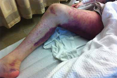
Repeat labs in the clinic showed a further decrease in hemoglobin to 9.2 g/dL. The patient was advised to go to the ED, where a work-up revealed that he had normocytic-normochromic anemia; his hemoglobin was 8.9 g/dL. Liver function tests revealed elevated AST and alanine aminotransferase (AST:ALT=3.66).
The patient was admitted to the family medicine inpatient service, where his hemoglobin continued to decline. He received a total of 4 units of packed red blood cells.
WHAT IS YOUR DIAGNOSIS?
HOW WOULD YOU TREAT THIS PATIENT?
Diagnosis: Scurvy
Upon further questioning, the patient revealed that his diet consisted solely of alcohol and meat. A serum vitamin C was ordered and returned at 0.1 mg/dL (normal, 0.4-2.0 mg/dL).
Alcoholism is a big tip-off. The diagnosis of scurvy is usually clinical, based on risk factors that suggest a diet deficient in vitamin C. The most common risk factor is alcoholism.1 Other risk factors include adults living alone (notably men), poverty, poor access to groceries, reclusiveness, dementia, nutritional ignorance, avoidance of “acid” foods because of purported allergy, gastrointestinal disorders (eg, colitis, inflammatory disease), poor dentition, food fads or food avoidances, cancer, schizophrenia, and depression.1
Men more than women. Men between the ages of 20 and 39 and those older than 60 years have a higher prevalence of vitamin C deficiency than similarly aged women, according to a National Health and Nutrition Examination Survey of 7277 children and adults.2 Overall, 8.2% of the men and 6% of the women surveyed were deficient in vitamin C.2
Henoch-Schönlein purpura, leukemia included in differential Dx
The differential diagnosis of scurvy includes autoimmune diseases, coagulopathies, hematologic malignancies, adverse effects of medications, meningococcemia, necrotizing gingivitis, senile purpura, thrombophlebitis, other vitamin deficiencies (vitamins K, D, B12, folate, and zinc), and physical abuse or trauma.
Autoimmune diseases such as systemic lupus erythematosus, Henoch-Schönlein purpura, Sjögren syndrome, and rheumatoid arthritis often involve cutaneous manifestations of purpuric and petechial rash. These diseases tend to have systemic involvement that can include the airways, kidneys, and nervous system—as well as rheumatologic findings of joint involvement.3 Making the diagnosis hinges on autoantibody serologies and inflammatory markers.
Coagulopathies such as clotting factor deficiencies, platelet dysfunction, and disseminated intravascular coagulation can also present with skin rash and anemia. Laboratory analysis shows abnormalities of blood cell counts (anemia, throm-bocytopenia), prothrombin time (PT), activated partial thromboplastin time (aPTT), and fibrinogen and clotting factor deficiencies.
Hematologic malignancies such as leukemia or lymphoma can cause petechial and purpuric rashes. Fever, fatigue, weight loss, bone pain, easy bleeding or bruising, lymphadenopathy, and hepatosplenomegaly highlight the systemic manifestations of these malignancies. Positive findings on complete blood count (CBC), coagulation profile, erythrocyte sedimentation rate, and bone marrow biopsy help confirm this diagnosis.
Petechial or purpuric rash can be an adverse effect of anticoagulants, antiplatelet agents, and nonsteroidal anti-inflammatory drugs. Symptoms range in severity and generally resolve with the discontinuation of the offending agent.
Meningococcemia involves a maculopapular rash with petechia and ecchymosis resulting from the spread of bacterial infection from cerebral spinal fluid (CSF) to the systemic circulation. Infection-related effects on clotting factors can be identified with a CBC, PT/aPTT times, and testing for fibrinogen and fibrin degradation products. CSF analysis and culture along with a history and exam findings of fever, malaise, hypotension, and meningeal signs point to meningitis.
Scurvy: What you’ll see and what the lab work will show
Follicular hyperkeratosis and perifollicular hemorrhagic rash with corkscrew hairs are pathognomonic for scurvy and occur early in the disease. Ecchymosis (FIGURE 2) and splinter hemorrhages are also present.
FIGURE 2
Ecchymosis and petechial rash seen in scurvy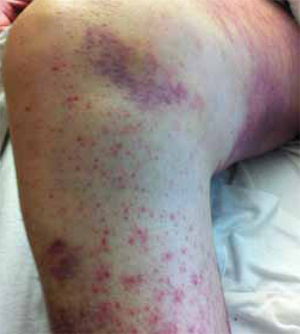
Patients may experience impaired wound healing; abnormal bleeding; muscle, joint, and bone pain; and pathologic fractures.4 There may also be a history of swollen, bleeding gums, tooth loss, and conjunctival varicosities.1,4 Patients may complain of fatigue, exercise intolerance, and depressed mood.
A history of alcoholism is, of course, a red flag. But it’s also important to take note when patients describe a severe food aversion. They may, for instance, tell you that they only eat protein, breads, or other carbohydrates, or that they avoid acidic foods because they have an allergy or because these foods upset their stomach.
Lab work. Serum concentration of ascorbic acid can be checked, although this tends to reflect recent dietary intake rather than actual tissue stores of vitamin C.5 It is estimated that clinical symptoms of scurvy begin to appear when the total body pool of vitamin C has decreased to approximately 300 mg, which reflects a serum level of <0.19 mg/dL.4 Other dietary deficiencies are likely to be present, as well.
There is a leukocyte ascorbate level that more accurately reflects tissue stores of vitamin C, although this test is difficult to perform and not routinely available. (A more practical diagnostic approach is to observe whether clinical abnormalities disappear with repletion therapy.)
Mild anemia is common and its degree correlates with the severity and duration of the scurvy.1 A normocytic-normochromic anemia is usually present and the reticulocyte count is elevated.
Macrocytic anemia may be present due to inadequate folate levels. (Ascorbic acid prevents oxidation of folate to other forms that are excreted in the urine, thus vitamin C deficiency may deplete folate stores by increasing urinary excretion.1)
Iron deficiency anemia is rare in scurvy, even though vitamin C is involved with the absorption of iron.1 Leukopenia is uncommon and platelet counts are usually normal.
Treat with ascorbic acid
Patients with scurvy will need to be treated with a repletion dose of ascorbic acid 100 mg 3 times a day1 (strength of recommendation: A). Manifestations of scurvy recede quickly and usually disappear within a few weeks. Subjective improvement commonly occurs within 24 hours.1
Educating our patient
Given the severity of our patient’s case, he was told that he needed to take 1500 mg ascorbic acid daily. He also received information on alcohol abuse treatment and was counseled on proper nutrition. He was advised to maintain a balanced diet that included the recommended daily amount of all essential vitamins.
Within 48 hours, his hemoglobin stabilized.
CORRESPONDENCE
Sean Robinson, MD, Oregon Health and Science University, 3181 SW Sam Jackson Park Road, FM, Portland, OR 97239; [email protected]
1. Hirschmann JV, Raugi GJ. Adult scurvy. J Am Acad Dermatol. 1999;41:895-906.
2. Hampl JS, Taylor CA, Johnston CS. Vitamin C deficiency and depletion in the United States: the Third National Health and Nutrition Examination Survey, 1988 to 1994. Am J Public Health. 2004;94:870-875.
3. Reamy BV, Williams PM, Lindsay TJ. Henoch-Schönlein purpura. Am Fam Physician. 2009;80:697-704.
4. Nguyen RT, Cowley DM, Muir JB. Scurvy: a cutaneous clinical diagnosis. Australas J Dermatol. 2003;44:48-51.
5. Olmedo JM, Yiannias JA, Windgassen EB, et al. Scurvy: a disease almost forgotten. Int J Dermatol. 2006;45:909-913.
A Good-quality patient-oriented evidence
B Inconsistent or limited-quality patient-oriented evidence
C Consensus, usual practice, opinion, disease-oriented evidence, case series
A 31-YEAR-OLD MAN with a history of depression, alcohol abuse, and rosacea sought care at our clinic for worsening light-headedness, rash, and bruising. Two weeks earlier, he had been to the emergency department (ED) for a syncopal event. The work-up at that time included:
- electrocardiogram showing sinus tachycardia
- lower extremity duplex ultrasound that was negative for deep vein thrombosis or fluid collection
- computed tomography angiogram of the chest that was negative for pulmonary embolus
- lab work that showed normal platelets and international normalized ratio.
The patient did, however, have a notable decrease in hemoglobin from a baseline of 15 g/dL to 12 g/dL, low serum albumin, and mildly elevated aspartate aminotransferase (AST) and total bilirubin.
The patient received 3 liters of intravenous normal saline and was discharged home to follow up as an outpatient. During his visit to our clinic, we noted an extensive purpuric rash on the posterior aspect of his thighs, although his right thigh was worse than his left (FIGURE 1). The patient also had a diffuse follicular petechial rash on his legs. The circumference of his right thigh was 61 cm and his left thigh was 51.5 cm.
FIGURE 1
Diffuse ecchymosis on the right thigh
Repeat labs in the clinic showed a further decrease in hemoglobin to 9.2 g/dL. The patient was advised to go to the ED, where a work-up revealed that he had normocytic-normochromic anemia; his hemoglobin was 8.9 g/dL. Liver function tests revealed elevated AST and alanine aminotransferase (AST:ALT=3.66).
The patient was admitted to the family medicine inpatient service, where his hemoglobin continued to decline. He received a total of 4 units of packed red blood cells.
WHAT IS YOUR DIAGNOSIS?
HOW WOULD YOU TREAT THIS PATIENT?
Diagnosis: Scurvy
Upon further questioning, the patient revealed that his diet consisted solely of alcohol and meat. A serum vitamin C was ordered and returned at 0.1 mg/dL (normal, 0.4-2.0 mg/dL).
Alcoholism is a big tip-off. The diagnosis of scurvy is usually clinical, based on risk factors that suggest a diet deficient in vitamin C. The most common risk factor is alcoholism.1 Other risk factors include adults living alone (notably men), poverty, poor access to groceries, reclusiveness, dementia, nutritional ignorance, avoidance of “acid” foods because of purported allergy, gastrointestinal disorders (eg, colitis, inflammatory disease), poor dentition, food fads or food avoidances, cancer, schizophrenia, and depression.1
Men more than women. Men between the ages of 20 and 39 and those older than 60 years have a higher prevalence of vitamin C deficiency than similarly aged women, according to a National Health and Nutrition Examination Survey of 7277 children and adults.2 Overall, 8.2% of the men and 6% of the women surveyed were deficient in vitamin C.2
Henoch-Schönlein purpura, leukemia included in differential Dx
The differential diagnosis of scurvy includes autoimmune diseases, coagulopathies, hematologic malignancies, adverse effects of medications, meningococcemia, necrotizing gingivitis, senile purpura, thrombophlebitis, other vitamin deficiencies (vitamins K, D, B12, folate, and zinc), and physical abuse or trauma.
Autoimmune diseases such as systemic lupus erythematosus, Henoch-Schönlein purpura, Sjögren syndrome, and rheumatoid arthritis often involve cutaneous manifestations of purpuric and petechial rash. These diseases tend to have systemic involvement that can include the airways, kidneys, and nervous system—as well as rheumatologic findings of joint involvement.3 Making the diagnosis hinges on autoantibody serologies and inflammatory markers.
Coagulopathies such as clotting factor deficiencies, platelet dysfunction, and disseminated intravascular coagulation can also present with skin rash and anemia. Laboratory analysis shows abnormalities of blood cell counts (anemia, throm-bocytopenia), prothrombin time (PT), activated partial thromboplastin time (aPTT), and fibrinogen and clotting factor deficiencies.
Hematologic malignancies such as leukemia or lymphoma can cause petechial and purpuric rashes. Fever, fatigue, weight loss, bone pain, easy bleeding or bruising, lymphadenopathy, and hepatosplenomegaly highlight the systemic manifestations of these malignancies. Positive findings on complete blood count (CBC), coagulation profile, erythrocyte sedimentation rate, and bone marrow biopsy help confirm this diagnosis.
Petechial or purpuric rash can be an adverse effect of anticoagulants, antiplatelet agents, and nonsteroidal anti-inflammatory drugs. Symptoms range in severity and generally resolve with the discontinuation of the offending agent.
Meningococcemia involves a maculopapular rash with petechia and ecchymosis resulting from the spread of bacterial infection from cerebral spinal fluid (CSF) to the systemic circulation. Infection-related effects on clotting factors can be identified with a CBC, PT/aPTT times, and testing for fibrinogen and fibrin degradation products. CSF analysis and culture along with a history and exam findings of fever, malaise, hypotension, and meningeal signs point to meningitis.
Scurvy: What you’ll see and what the lab work will show
Follicular hyperkeratosis and perifollicular hemorrhagic rash with corkscrew hairs are pathognomonic for scurvy and occur early in the disease. Ecchymosis (FIGURE 2) and splinter hemorrhages are also present.
FIGURE 2
Ecchymosis and petechial rash seen in scurvy
Patients may experience impaired wound healing; abnormal bleeding; muscle, joint, and bone pain; and pathologic fractures.4 There may also be a history of swollen, bleeding gums, tooth loss, and conjunctival varicosities.1,4 Patients may complain of fatigue, exercise intolerance, and depressed mood.
A history of alcoholism is, of course, a red flag. But it’s also important to take note when patients describe a severe food aversion. They may, for instance, tell you that they only eat protein, breads, or other carbohydrates, or that they avoid acidic foods because they have an allergy or because these foods upset their stomach.
Lab work. Serum concentration of ascorbic acid can be checked, although this tends to reflect recent dietary intake rather than actual tissue stores of vitamin C.5 It is estimated that clinical symptoms of scurvy begin to appear when the total body pool of vitamin C has decreased to approximately 300 mg, which reflects a serum level of <0.19 mg/dL.4 Other dietary deficiencies are likely to be present, as well.
There is a leukocyte ascorbate level that more accurately reflects tissue stores of vitamin C, although this test is difficult to perform and not routinely available. (A more practical diagnostic approach is to observe whether clinical abnormalities disappear with repletion therapy.)
Mild anemia is common and its degree correlates with the severity and duration of the scurvy.1 A normocytic-normochromic anemia is usually present and the reticulocyte count is elevated.
Macrocytic anemia may be present due to inadequate folate levels. (Ascorbic acid prevents oxidation of folate to other forms that are excreted in the urine, thus vitamin C deficiency may deplete folate stores by increasing urinary excretion.1)
Iron deficiency anemia is rare in scurvy, even though vitamin C is involved with the absorption of iron.1 Leukopenia is uncommon and platelet counts are usually normal.
Treat with ascorbic acid
Patients with scurvy will need to be treated with a repletion dose of ascorbic acid 100 mg 3 times a day1 (strength of recommendation: A). Manifestations of scurvy recede quickly and usually disappear within a few weeks. Subjective improvement commonly occurs within 24 hours.1
Educating our patient
Given the severity of our patient’s case, he was told that he needed to take 1500 mg ascorbic acid daily. He also received information on alcohol abuse treatment and was counseled on proper nutrition. He was advised to maintain a balanced diet that included the recommended daily amount of all essential vitamins.
Within 48 hours, his hemoglobin stabilized.
CORRESPONDENCE
Sean Robinson, MD, Oregon Health and Science University, 3181 SW Sam Jackson Park Road, FM, Portland, OR 97239; [email protected]
1. Hirschmann JV, Raugi GJ. Adult scurvy. J Am Acad Dermatol. 1999;41:895-906.
2. Hampl JS, Taylor CA, Johnston CS. Vitamin C deficiency and depletion in the United States: the Third National Health and Nutrition Examination Survey, 1988 to 1994. Am J Public Health. 2004;94:870-875.
3. Reamy BV, Williams PM, Lindsay TJ. Henoch-Schönlein purpura. Am Fam Physician. 2009;80:697-704.
4. Nguyen RT, Cowley DM, Muir JB. Scurvy: a cutaneous clinical diagnosis. Australas J Dermatol. 2003;44:48-51.
5. Olmedo JM, Yiannias JA, Windgassen EB, et al. Scurvy: a disease almost forgotten. Int J Dermatol. 2006;45:909-913.
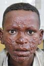
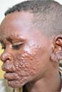
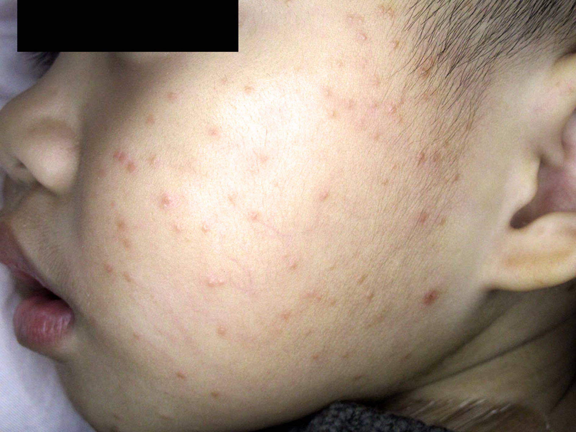
Facial changes
| 
|
| FIGURE 1 | FIGURE 2 |
The physician suspected that the boy had lepromatous leprosy (FIGURES 1 and 2). His father was also examined and he exhibited more subtle signs of leprosy (several patches of hypopigmented skin with diminished sensation). The laboratory performed a slit skin exam on the boy’s ear lobe and many acid-fast bacilli--characteristic of Mycobacterium leprae--were found. The boy was started on the World Health Organization’s standard multidrug therapy using rifampin, clofazimine, and dapsone. The father was also treated.
Leprosy (Hansen’s disease) is caused by M leprae and is still endemic in many parts of the developing world where there is poverty and poor access to clean water. At one time, people with leprosy were isolated to leper colonies because the disease was disfiguring and the community was afraid that it was highly contagious. Current science and epidemiology tell us that leprosy is transmitted via droplets from the nose and mouth during close and frequent contact over a period of years, and not by casual contact. Thus doctors working with patients who have leprosy are at no real risk of becoming infected. In the United States, eating or handling armadillos is a risk factor for leprosy, as armadillos are natural hosts for M leprae.
Photos and text for Photo Rounds Friday courtesy of Richard P. Usatine, MD. This case was adapted from: Berggren R, Usatine R. In: Usatine R, Smith M, Mayeaux EJ, et al. Color Atlas of Family Medicine. 2nd ed. New York, NY: McGraw-Hill; 2013:53-79.
To learn more about the Color Atlas of Family Medicine, see:
• http://www.amazon.com/Color-Atlas-Family-Medicine/dp/0071474641
You can now get the Color Atlas of Family Medicine as an app for mobile devices by clicking this link:
|
|
|
| FIGURE 1 | FIGURE 2 |
The physician suspected that the boy had lepromatous leprosy (FIGURES 1 and 2). His father was also examined and he exhibited more subtle signs of leprosy (several patches of hypopigmented skin with diminished sensation). The laboratory performed a slit skin exam on the boy’s ear lobe and many acid-fast bacilli--characteristic of Mycobacterium leprae--were found. The boy was started on the World Health Organization’s standard multidrug therapy using rifampin, clofazimine, and dapsone. The father was also treated.
Leprosy (Hansen’s disease) is caused by M leprae and is still endemic in many parts of the developing world where there is poverty and poor access to clean water. At one time, people with leprosy were isolated to leper colonies because the disease was disfiguring and the community was afraid that it was highly contagious. Current science and epidemiology tell us that leprosy is transmitted via droplets from the nose and mouth during close and frequent contact over a period of years, and not by casual contact. Thus doctors working with patients who have leprosy are at no real risk of becoming infected. In the United States, eating or handling armadillos is a risk factor for leprosy, as armadillos are natural hosts for M leprae.
Photos and text for Photo Rounds Friday courtesy of Richard P. Usatine, MD. This case was adapted from: Berggren R, Usatine R. In: Usatine R, Smith M, Mayeaux EJ, et al. Color Atlas of Family Medicine. 2nd ed. New York, NY: McGraw-Hill; 2013:53-79.
To learn more about the Color Atlas of Family Medicine, see:
• http://www.amazon.com/Color-Atlas-Family-Medicine/dp/0071474641
You can now get the Color Atlas of Family Medicine as an app for mobile devices by clicking this link:
Acral papular rash
Gianotti-Crosti syndrome (GCS) was diagnosed in this young patient. GCS is a pediatric disease that is believed to involve a cutaneous reaction pattern related to viral and bacterial infections or to vaccination. It is associated with the hepatitis B virus, Epstein-Barr virus, enteroviruses, parainfluenza viruses, and other viral infections. The eruption has also occurred following vaccination (hepatitis A, others).
Most patients with GCS do not need treatment because it is a self-limited benign disease. Although the course is variable and the skin lesions may persist for up to 60 days, the lesions will heal without scarring.
In patients who have severe pruritus, topical antipruritic lotions or oral histamines can provide relief. Medium-potency topical steroids may have some benefit, but patients should be closely monitored because there have been reports of exacerbations of lesions with steroid use.
In the case of this patient, the lesions resolved one week after applying 0.1% mometasone furoate cream once a day.
Adapted from: Liaw FY, Huang CF, Wu LW, et al. Photo Rounds: Acral papular rash in a 2-year-old boy. J Fam Pract. 2012;61:157-159.
To learn more about The Color Atlas of Family Medicine, see:
http://www.amazon.com/Color-Atlas-Family-Medicine/dp/0071474641
You can now get The Color Atlas of Family Medicine as an app for mobile devices including the iPhone and iPad by clicking this link:
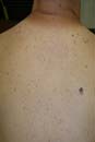
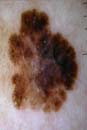
Mole on back
| 
|
| FIGURE 1 | FIGURE 2 |
A closer look at the lesion (FIGURE 1) using the dermatoscope (FIGURE 2) revealed an atypical network, negative network, streaming pigment at the periphery, and a blue/white veil—suggestive of melanoma.
The FP performed a saucerization (scoop shave biopsy) and sent the full lesion to pathology. The report came back as a superficial spreading melanoma with a Breslow depth of 0.35 mm. The lesion site was then re-excised with 1 cm margins; no sentinel node biopsies were required.
In this case, listening to the lungs with the patient’s shirt off and noticing the "ugly duckling" lesion led to a lifesaving intervention.
Photos and text for Photo Rounds Friday courtesy of Richard P. Usatine, MD. This case was adapted from: Marghoob A. Dermoscopy. In: Usatine R, Smith M, Mayeaux EJ, et al, eds. The Color Atlas of Family Medicine. New York, NY: McGraw-Hill; 2009:1019-1029.
To learn more about The Color Atlas of Family Medicine, see:
http://www.amazon.com/Color-Atlas-Family-Medicine/dp/0071474641
You can now get The Color Atlas of Family Medicine as an app for mobile devices including the iPhone and iPad by clicking this link:
|
|
|
| FIGURE 1 | FIGURE 2 |
A closer look at the lesion (FIGURE 1) using the dermatoscope (FIGURE 2) revealed an atypical network, negative network, streaming pigment at the periphery, and a blue/white veil—suggestive of melanoma.
The FP performed a saucerization (scoop shave biopsy) and sent the full lesion to pathology. The report came back as a superficial spreading melanoma with a Breslow depth of 0.35 mm. The lesion site was then re-excised with 1 cm margins; no sentinel node biopsies were required.
In this case, listening to the lungs with the patient’s shirt off and noticing the "ugly duckling" lesion led to a lifesaving intervention.
Photos and text for Photo Rounds Friday courtesy of Richard P. Usatine, MD. This case was adapted from: Marghoob A. Dermoscopy. In: Usatine R, Smith M, Mayeaux EJ, et al, eds. The Color Atlas of Family Medicine. New York, NY: McGraw-Hill; 2009:1019-1029.
To learn more about The Color Atlas of Family Medicine, see:
http://www.amazon.com/Color-Atlas-Family-Medicine/dp/0071474641
You can now get The Color Atlas of Family Medicine as an app for mobile devices including the iPhone and iPad by clicking this link:
Marks on forearms
The scars on the patient’s arms were highly suggestive of skin popping. She subsequently admitted to skin popping cocaine, which leaves round, depressed scars. The patient indicated that she had given up trying to find veins for injecting cocaine and had begun injecting directly into her skin. The local skin necrosis caused round scars with skin atrophy.
The unusual pattern of skin scars provided the FP with an opportunity to discuss the patient’s cocaine use and its relationship to the palpitations she had been experiencing.
Photos and text for Photo Rounds Friday courtesy of Richard P. Usatine, MD. This case was adapted from: Usatine, R. Injection drug use. In: Usatine R, Smith M, Mayeaux EJ, et al, eds. The Color Atlas of Family Medicine. New York, NY: McGraw-Hill; 2009:1006-1009.
To learn more about The Color Atlas of Family Medicine, see:
• http://www.amazon.com/Color-Atlas-Family-Medicine/dp/0071474641
You can now get The Color Atlas of Family Medicine as an app for mobile devices including the iPhone, iPad, and all Android devices by clicking this link:
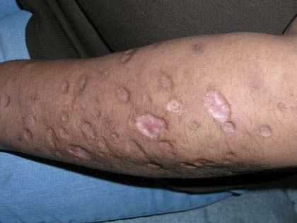
Blistering rash in an older man
A Good-quality patient-oriented evidence
B Inconsistent or limited-quality patient-oriented evidence
C Consensus, usual practice, opinion, disease-oriented evidence, case series
A 76-YEAR-OLD MAN sought care for a rash that had gotten progressively worse over the previous 3 weeks. He indicated that the rash was initially red, itchy, and located over his abdomen, but as time went by, new blisters developed in the axillae and groin, and they were painful. The patient did not have any arthralgias or systemic symptoms. The medications he was taking included simvastatin, albuterol, and finasteride.
On physical examination, the patient was in mild distress due to the pain and anxiety, and his temperature was 36.5°C (97.7°F). He had confluent areas of erythematous, denuded skin spanning his trunk, back, and proximal upper and lower extremities (FIGURE 1). Tense, fluid-filled blisters were most prominent in the groin and in the axillae, bilaterally.
FIGURE 1
Diffuse rash on the trunk and in the axillae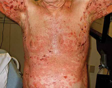
WHAT IS YOUR DIAGNOSIS?
HOW WOULD YOU TREAT THIS PATIENT?
Diagnosis: Bullous pemphigoid
This patient had a severe and refractory case of bullous pemphigoid (BP), which was confirmed with a biopsy of the lesions.
BP is a rare autoimmune, blistering skin disease that typically occurs after age 60.1 The incidence rises with age, and is higher among women than men.1 The pathogenesis of BP involves development of autoantibodies against the subepidermal basement membrane. Deposition of immunoglobulin G (IgG) occurs, leading to immune-mediated destruction and subepidermal blistering.2
Patients will present with new-onset, widespread eruptions of bullous lesions and urticarial plaques (FIGURE 2).2 Bullae are frequent on flexural surfaces such as the groin and axillae. Urticarial plaques are often pruritic. Oral involvement occurs in a minority of cases.2 Nikolsky’s sign—exfoliation of the outermost layer of skin upon slight rubbing—is absent in BP.
FIGURE 2
Fluid-filled vesicles and bullae on right anterior thigh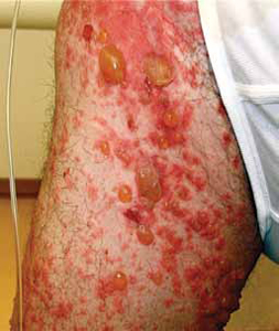
Differential: Other autoimmune and blistering skin conditions
Two additional pemphigoid subtypes are part of the differential when a patient presents with a blistering skin condition: pemphigoid gestationis and mucous membrane pemphigoid.2
Pemphigoid gestationis occurs exclusively during pregnancy and the puerperium, and is self-limited.
Mucous membrane pemphigoid is pathophysiologically similar to BP, but distributes preferentially on mucosal surfaces.
Pemphigus vulgaris, another autoimmune blistering skin disease, is characterized by sparse intact bullae. The mucous membrane is frequently involved, and there is a positive Nikolsky’s sign.
Additional conditions to keep in mind include epidermolysis bullosa acquisita, dermatitis herpetiformis, bullous erythema multiforme, and bullous lupus erythematosus.
Biopsy confirms the Dx
A biopsy of a lesion confirms the diagnosis of BP and will help differentiate it from the conditions mentioned above.
Light microscopy shows eosinophil-rich subepidermal inflammatory infiltrate.2 Direct immunofluorescence displays the characteristic linear deposits of IgG and complement C3 along the basement membrane. Immunofluorescent testing on human salt-split skin may also be performed.
Drug induced? There is a subset of BP, called drug-induced BP, in which the onset of the disease is associated with the initiation of a medication. Furosemide is the most common culprit,3 although many additional medications have been described.
The pathophysiology of drug-induced BP is poorly understood.3 In some cases, discontinuation of the offending medication may halt progression and prevent recurrence. In other cases, the disease will progress to a chronic form regardless of medication discontinuation. It is reasonable to attempt medication discontinuation trials in cases where drug-induced BP is suspected.
Treat with corticosteroids
The traditional treatment of BP is high-dose oral corticosteroids. However, long-term use of systemic corticosteroids can cause significant morbidity and has been linked to an increased mortality rate in the elderly population.4 A potent topical corticosteroid, such as clobetasol propionate cream 10 to 30 g/d tapering over 4 months, or 40 g/d tapering over 12 months,5,6 is an effective alternative (strength of recommendation [SOR]: A).
Other options include methotrexate, mycophenolate, azathioprine, niacinamide, doxycycline, intravenous (IV) immunoglobulin, and plasma exchange. These therapies are typically used in combination with corticosteroids, or after initial treatment failure. Evidence regarding their effectiveness is limited7 (SOR: B).
Although the disease is occasionally self-limited after the initial episode, most patients with BP will achieve clinical remission with medical intervention. Patients often experience recurrent outbreaks and require chronic use of immunosuppressive agents.
Our patient required ongoing care
Our patient was prescribed prednisone 80 mg/d PO in combination with topical clobetasol cream. Despite these treatments, the disease progressed. One week later, approximately 80% of his body surface was involved. He was admitted for fluid replacement and monitoring for infection.
Subsequent initiation of methotrexate, niacinamide, doxycycline, and topical clobetasol led to clinical remission. Unfortunately, the patient relapsed approximately 3 months later and required a second hospital stay.
In the ensuing months, the patient’s course was marked by frequent relapses and significant morbidity. Further treatment trials have included IV immunoglobulin, mycophenolate, and azathioprine.
CORRESPONDENCE
Casey Z. MacVane, MD, MPH, Department of Emergency Medicine, Maine Medical Center, 47 Bramhall Street, Portland, ME 04102; [email protected]
1. Langan SM, Smeeth L, Hubbard R, et al. Bullous pemphigoid and pemphigus vulgaris—incidence and mortality in the UK: population based cohort study. BMJ. 2008;337:a180.-
2. Yancey KB, Egan CA. Pemphigoid: clinical, histological, immunopathologic, and therapeutic considerations. JAMA. 2000;248:350-356.
3. Lee JJ, Downham TF, 2nd. Furosemide-induced bullous pemphigoid: case report and review of literature. J Drugs Dermatol. 2006;5:562-564.
4. Rzany B, Partscht K, Jung M, et al. Risk factors for lethal outcome in patients with bullous pemphigoid. Arch Dermatol. 2002;138:903-908.
5. Joly P, Roujeau JC, Benichou J, et al. A comparison of oral and topical corticosteroids in patients with bullous pemphigoid. N Engl J Med. 2002;346:321-327.
6. Joly P, Roujeau JC, Benichou J, et al. A comparison of two regimens of topical corticosteroids in the treatment of patients with bullous pemphigoid: a multicenter randomized study. J Invest Dermatol. 2009;129:1681-1687.
7. Kirtschig G, Middleton P, Bennett C, et al. Interventions for bullous pemphigoid. Cochrane Database Syst Rev. 2010;(10):CD002292.
A Good-quality patient-oriented evidence
B Inconsistent or limited-quality patient-oriented evidence
C Consensus, usual practice, opinion, disease-oriented evidence, case series
A 76-YEAR-OLD MAN sought care for a rash that had gotten progressively worse over the previous 3 weeks. He indicated that the rash was initially red, itchy, and located over his abdomen, but as time went by, new blisters developed in the axillae and groin, and they were painful. The patient did not have any arthralgias or systemic symptoms. The medications he was taking included simvastatin, albuterol, and finasteride.
On physical examination, the patient was in mild distress due to the pain and anxiety, and his temperature was 36.5°C (97.7°F). He had confluent areas of erythematous, denuded skin spanning his trunk, back, and proximal upper and lower extremities (FIGURE 1). Tense, fluid-filled blisters were most prominent in the groin and in the axillae, bilaterally.
FIGURE 1
Diffuse rash on the trunk and in the axillae
WHAT IS YOUR DIAGNOSIS?
HOW WOULD YOU TREAT THIS PATIENT?
Diagnosis: Bullous pemphigoid
This patient had a severe and refractory case of bullous pemphigoid (BP), which was confirmed with a biopsy of the lesions.
BP is a rare autoimmune, blistering skin disease that typically occurs after age 60.1 The incidence rises with age, and is higher among women than men.1 The pathogenesis of BP involves development of autoantibodies against the subepidermal basement membrane. Deposition of immunoglobulin G (IgG) occurs, leading to immune-mediated destruction and subepidermal blistering.2
Patients will present with new-onset, widespread eruptions of bullous lesions and urticarial plaques (FIGURE 2).2 Bullae are frequent on flexural surfaces such as the groin and axillae. Urticarial plaques are often pruritic. Oral involvement occurs in a minority of cases.2 Nikolsky’s sign—exfoliation of the outermost layer of skin upon slight rubbing—is absent in BP.
FIGURE 2
Fluid-filled vesicles and bullae on right anterior thigh
Differential: Other autoimmune and blistering skin conditions
Two additional pemphigoid subtypes are part of the differential when a patient presents with a blistering skin condition: pemphigoid gestationis and mucous membrane pemphigoid.2
Pemphigoid gestationis occurs exclusively during pregnancy and the puerperium, and is self-limited.
Mucous membrane pemphigoid is pathophysiologically similar to BP, but distributes preferentially on mucosal surfaces.
Pemphigus vulgaris, another autoimmune blistering skin disease, is characterized by sparse intact bullae. The mucous membrane is frequently involved, and there is a positive Nikolsky’s sign.
Additional conditions to keep in mind include epidermolysis bullosa acquisita, dermatitis herpetiformis, bullous erythema multiforme, and bullous lupus erythematosus.
Biopsy confirms the Dx
A biopsy of a lesion confirms the diagnosis of BP and will help differentiate it from the conditions mentioned above.
Light microscopy shows eosinophil-rich subepidermal inflammatory infiltrate.2 Direct immunofluorescence displays the characteristic linear deposits of IgG and complement C3 along the basement membrane. Immunofluorescent testing on human salt-split skin may also be performed.
Drug induced? There is a subset of BP, called drug-induced BP, in which the onset of the disease is associated with the initiation of a medication. Furosemide is the most common culprit,3 although many additional medications have been described.
The pathophysiology of drug-induced BP is poorly understood.3 In some cases, discontinuation of the offending medication may halt progression and prevent recurrence. In other cases, the disease will progress to a chronic form regardless of medication discontinuation. It is reasonable to attempt medication discontinuation trials in cases where drug-induced BP is suspected.
Treat with corticosteroids
The traditional treatment of BP is high-dose oral corticosteroids. However, long-term use of systemic corticosteroids can cause significant morbidity and has been linked to an increased mortality rate in the elderly population.4 A potent topical corticosteroid, such as clobetasol propionate cream 10 to 30 g/d tapering over 4 months, or 40 g/d tapering over 12 months,5,6 is an effective alternative (strength of recommendation [SOR]: A).
Other options include methotrexate, mycophenolate, azathioprine, niacinamide, doxycycline, intravenous (IV) immunoglobulin, and plasma exchange. These therapies are typically used in combination with corticosteroids, or after initial treatment failure. Evidence regarding their effectiveness is limited7 (SOR: B).
Although the disease is occasionally self-limited after the initial episode, most patients with BP will achieve clinical remission with medical intervention. Patients often experience recurrent outbreaks and require chronic use of immunosuppressive agents.
Our patient required ongoing care
Our patient was prescribed prednisone 80 mg/d PO in combination with topical clobetasol cream. Despite these treatments, the disease progressed. One week later, approximately 80% of his body surface was involved. He was admitted for fluid replacement and monitoring for infection.
Subsequent initiation of methotrexate, niacinamide, doxycycline, and topical clobetasol led to clinical remission. Unfortunately, the patient relapsed approximately 3 months later and required a second hospital stay.
In the ensuing months, the patient’s course was marked by frequent relapses and significant morbidity. Further treatment trials have included IV immunoglobulin, mycophenolate, and azathioprine.
CORRESPONDENCE
Casey Z. MacVane, MD, MPH, Department of Emergency Medicine, Maine Medical Center, 47 Bramhall Street, Portland, ME 04102; [email protected]
1. Langan SM, Smeeth L, Hubbard R, et al. Bullous pemphigoid and pemphigus vulgaris—incidence and mortality in the UK: population based cohort study. BMJ. 2008;337:a180.-
2. Yancey KB, Egan CA. Pemphigoid: clinical, histological, immunopathologic, and therapeutic considerations. JAMA. 2000;248:350-356.
3. Lee JJ, Downham TF, 2nd. Furosemide-induced bullous pemphigoid: case report and review of literature. J Drugs Dermatol. 2006;5:562-564.
4. Rzany B, Partscht K, Jung M, et al. Risk factors for lethal outcome in patients with bullous pemphigoid. Arch Dermatol. 2002;138:903-908.
5. Joly P, Roujeau JC, Benichou J, et al. A comparison of oral and topical corticosteroids in patients with bullous pemphigoid. N Engl J Med. 2002;346:321-327.
6. Joly P, Roujeau JC, Benichou J, et al. A comparison of two regimens of topical corticosteroids in the treatment of patients with bullous pemphigoid: a multicenter randomized study. J Invest Dermatol. 2009;129:1681-1687.
7. Kirtschig G, Middleton P, Bennett C, et al. Interventions for bullous pemphigoid. Cochrane Database Syst Rev. 2010;(10):CD002292.
Abscess on buttocks
Upon questioning, the patient admitted to using injectable anabolic steroids for body-building. He said that he couldn’t be sure that the needles he'd used had always been sterile.
The patient’s abscess was drained and the site healed by second intention. The family physician (FP) talked to the patient about the detrimental effects of anabolic steroids, the risks of sharing needles, and the fact that he could be a successful body-builder with the right weight-lifting program and a high-protein diet.
Because of the patient’s needle-sharing history, the FP ordered tests for hepatitis C and human immunodeficiency virus (HIV). The patient was fully immunized for hepatitis B as a child so the FP did not order any hepatitis B serologies.
Fortunately, the patient was negative for hepatitis C and HIV.
Text for Photo Rounds Friday courtesy of Richard P. Usatine, MD. Photo courtesy of Bill Rodney, MD. This case was adapted from: Usatine, R. Injection drug use. In: Usatine R, Smith M, Mayeaux EJ, et al, eds. The Color Atlas of Family Medicine. New York, NY: McGraw-Hill; 2009:1006-1009.
To learn more about The Color Atlas of Family Medicine, see:
• http://www.amazon.com/Color-Atlas-Family-Medicine/dp/0071474641
You can now get The Color Atlas of Family Medicine as an app for mobile devices including the iPhone, iPad, and all Android devices by clicking this link:
Papular nodular eruption
Secondary syphilis was confirmed when the rapid plasma reagin came back with a titer of 1:512. The treponemal blood test was also positive and the HIV test was negative. The biopsy showed Treponema pallidum in the tissue.
The patient was called to come in for treatment. He admitted to being involved in unsafe sex with many partners while addicted to cocaine. Syphilis is the great imitator and while this particular pattern is not the most common pattern of secondary syphilis, it is helpful to be aware of the great variability in which secondary syphilis may present.
The patient was treated with a single IM injection of benzathine penicillin. Within one week, the eruption was gone and the patient continued with his drug treatment program. At a 6-month follow-up, he was sober and his RPR titer was down 4-fold.
Photos and text for Photo Rounds Friday courtesy of Richard P. Usatine, MD. This case was adapted from: Smith M. Cocaine. In: Usatine R, Smith M, Mayeaux EJ, et al, eds. The Color Atlas of Family Medicine. New York, NY: McGraw-Hill; 2009:1000-1003.
To learn more about The Color Atlas of Family Medicine, see:
• http://www.amazon.com/Color-Atlas-Family-Medicine/dp/0071474641
You can now get The Color Atlas of Family Medicine as an app for mobile devices including the iPhone, iPad, and all Android devices by clicking this link:
Poor dentition
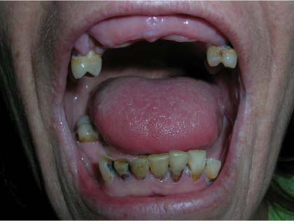
The patient admitted to a 20-year history of methamphetamine use. The rampant dental caries and gingivitis commonly seen in methamphetamine users is known as “meth mouth.”
There are several causes of meth mouth:
- Vasoconstriction leads to decreased saliva production and dry mouth, which often result in consumption of large quantities of sugary beverages.
- Preoccupation with obtaining and using drugs often leads methamphetamine users to neglect their oral hygiene.
- Methamphetamine-induced bruxism also damages the teeth.
- Neglect of early symptoms and lack of access to (or failure to seek) dental care often lead to unsalvageable teeth that can only be extracted.
Referral for dental care is indicated for patients with gingivitis and dental caries caused by chronic methamphetamine use. On a daily basis, patients should brush their teeth with a soft-bristled toothbrush and use dental floss to treat and prevent oral pathology. Rinsing with a chlorhexidine-containing mouthwash may be a reasonable alternative for patients who find it too painful to floss. This patient, however, was lost to follow-up.
Photos and text for Photo Rounds Friday courtesy of Richard P. Usatine, MD. This case was adapted from: Rowe, M, Schechtman A. Methamphetamine. In: Usatine R, Smith M, Mayeaux EJ, et al, eds. The Color Atlas of Family Medicine. New York, NY: McGraw-Hill; 2009:994-999.
To learn more about The Color Atlas of Family Medicine, see:
• http://www.amazon.com/Color-Atlas-Family-Medicine/dp/0071474641
You can now get The Color Atlas of Family Medicine as an app for mobile devices including the iPhone, iPad, and all Android devices by clicking this link:
Sores on arms
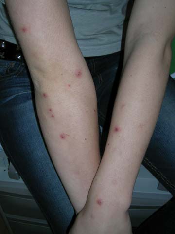
This patient was given a diagnosis of methamphetamine-induced self-excoriations, also known as meth mites.
The FP in this case suspected methamphetamine use and obtained a drug use history. The young woman admitted to smoking methamphetamine in a pipe, and said that when she did, her skin became itchy and she compulsively picked at it. Methamphetamine users may experience formication, the hallucination that bugs are crawling under the skin. The skin excoriations resulting from picking at the imagined bugs are known as meth mites.
Methamphetamine is known on the street as meth, crank, ice, and crystal. It is abused by smoking, injecting, snorting, and oral ingestion. Smoking or injecting the drug gives an intense, short-lived “flash” or rush. Snorting or oral ingestion creates euphoria, but no rush. Effects of methamphetamine use—euphoria, increased libido, and impaired judgment—may lead to increased high-risk sexual behaviors such as unprotected sexual intercourse and contact with multiple sexual partners. As a result, methamphetamine users are at increased risk of contracting sexually transmitted infections including human immunodeficiency virus.
Treatment involves referral to a 12-step program. Crystal Meth Anonymous is a 12-step program modeled on the 12 steps of Alcoholics Anonymous. If Crystal Meth Anonymous meetings are not available, any 12-step program can help in recovery and maintaining sobriety. While there are currently no Food and Drug Administration-approved medications to help treat methamphetamine dependence, several medications under study have shown favorable early results, including modafinil, bupropion, and some antiepileptic drugs.
In this case, the patient was given a prescription for a moderate strength topical steroid (0.1% triamcinolone cream) and encouraged to seek treatment for her drug addiction.
Photos and text for Photo Rounds Friday courtesy of Richard P. Usatine, MD. This case was adapted from: Rowe, M, Schechtman A. Methamphetamine. In: Usatine R, Smith M, Mayeaux EJ, et al, eds. The Color Atlas of Family Medicine. New York, NY: McGraw-Hill; 2009:994-999.
To learn more about The Color Atlas of Family Medicine, see:
• http://www.amazon.com/Color-Atlas-Family-Medicine/dp/0071474641
You can now get The Color Atlas of Family Medicine as an app for mobile devices including the iPhone, iPad, and all Android devices by clicking this link: