User login
| 
|
| FIGURE 1 | FIGURE 2 |
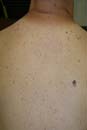
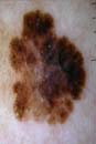
A closer look at the lesion (FIGURE 1) using the dermatoscope (FIGURE 2) revealed an atypical network, negative network, streaming pigment at the periphery, and a blue/white veil—suggestive of melanoma.
The FP performed a saucerization (scoop shave biopsy) and sent the full lesion to pathology. The report came back as a superficial spreading melanoma with a Breslow depth of 0.35 mm. The lesion site was then re-excised with 1 cm margins; no sentinel node biopsies were required.
In this case, listening to the lungs with the patient’s shirt off and noticing the "ugly duckling" lesion led to a lifesaving intervention.
Photos and text for Photo Rounds Friday courtesy of Richard P. Usatine, MD. This case was adapted from: Marghoob A. Dermoscopy. In: Usatine R, Smith M, Mayeaux EJ, et al, eds. The Color Atlas of Family Medicine. New York, NY: McGraw-Hill; 2009:1019-1029.
To learn more about The Color Atlas of Family Medicine, see:
http://www.amazon.com/Color-Atlas-Family-Medicine/dp/0071474641
You can now get The Color Atlas of Family Medicine as an app for mobile devices including the iPhone and iPad by clicking this link:
|
|
|
| FIGURE 1 | FIGURE 2 |
A closer look at the lesion (FIGURE 1) using the dermatoscope (FIGURE 2) revealed an atypical network, negative network, streaming pigment at the periphery, and a blue/white veil—suggestive of melanoma.
The FP performed a saucerization (scoop shave biopsy) and sent the full lesion to pathology. The report came back as a superficial spreading melanoma with a Breslow depth of 0.35 mm. The lesion site was then re-excised with 1 cm margins; no sentinel node biopsies were required.
In this case, listening to the lungs with the patient’s shirt off and noticing the "ugly duckling" lesion led to a lifesaving intervention.
Photos and text for Photo Rounds Friday courtesy of Richard P. Usatine, MD. This case was adapted from: Marghoob A. Dermoscopy. In: Usatine R, Smith M, Mayeaux EJ, et al, eds. The Color Atlas of Family Medicine. New York, NY: McGraw-Hill; 2009:1019-1029.
To learn more about The Color Atlas of Family Medicine, see:
http://www.amazon.com/Color-Atlas-Family-Medicine/dp/0071474641
You can now get The Color Atlas of Family Medicine as an app for mobile devices including the iPhone and iPad by clicking this link: