User login
Light-headedness and a petechial rash
A Good-quality patient-oriented evidence
B Inconsistent or limited-quality patient-oriented evidence
C Consensus, usual practice, opinion, disease-oriented evidence, case series
A 31-YEAR-OLD MAN with a history of depression, alcohol abuse, and rosacea sought care at our clinic for worsening light-headedness, rash, and bruising. Two weeks earlier, he had been to the emergency department (ED) for a syncopal event. The work-up at that time included:
- electrocardiogram showing sinus tachycardia
- lower extremity duplex ultrasound that was negative for deep vein thrombosis or fluid collection
- computed tomography angiogram of the chest that was negative for pulmonary embolus
- lab work that showed normal platelets and international normalized ratio.
The patient did, however, have a notable decrease in hemoglobin from a baseline of 15 g/dL to 12 g/dL, low serum albumin, and mildly elevated aspartate aminotransferase (AST) and total bilirubin.
The patient received 3 liters of intravenous normal saline and was discharged home to follow up as an outpatient. During his visit to our clinic, we noted an extensive purpuric rash on the posterior aspect of his thighs, although his right thigh was worse than his left (FIGURE 1). The patient also had a diffuse follicular petechial rash on his legs. The circumference of his right thigh was 61 cm and his left thigh was 51.5 cm.
FIGURE 1
Diffuse ecchymosis on the right thigh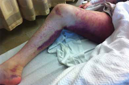
Repeat labs in the clinic showed a further decrease in hemoglobin to 9.2 g/dL. The patient was advised to go to the ED, where a work-up revealed that he had normocytic-normochromic anemia; his hemoglobin was 8.9 g/dL. Liver function tests revealed elevated AST and alanine aminotransferase (AST:ALT=3.66).
The patient was admitted to the family medicine inpatient service, where his hemoglobin continued to decline. He received a total of 4 units of packed red blood cells.
WHAT IS YOUR DIAGNOSIS?
HOW WOULD YOU TREAT THIS PATIENT?
Diagnosis: Scurvy
Upon further questioning, the patient revealed that his diet consisted solely of alcohol and meat. A serum vitamin C was ordered and returned at 0.1 mg/dL (normal, 0.4-2.0 mg/dL).
Alcoholism is a big tip-off. The diagnosis of scurvy is usually clinical, based on risk factors that suggest a diet deficient in vitamin C. The most common risk factor is alcoholism.1 Other risk factors include adults living alone (notably men), poverty, poor access to groceries, reclusiveness, dementia, nutritional ignorance, avoidance of “acid” foods because of purported allergy, gastrointestinal disorders (eg, colitis, inflammatory disease), poor dentition, food fads or food avoidances, cancer, schizophrenia, and depression.1
Men more than women. Men between the ages of 20 and 39 and those older than 60 years have a higher prevalence of vitamin C deficiency than similarly aged women, according to a National Health and Nutrition Examination Survey of 7277 children and adults.2 Overall, 8.2% of the men and 6% of the women surveyed were deficient in vitamin C.2
Henoch-Schönlein purpura, leukemia included in differential Dx
The differential diagnosis of scurvy includes autoimmune diseases, coagulopathies, hematologic malignancies, adverse effects of medications, meningococcemia, necrotizing gingivitis, senile purpura, thrombophlebitis, other vitamin deficiencies (vitamins K, D, B12, folate, and zinc), and physical abuse or trauma.
Autoimmune diseases such as systemic lupus erythematosus, Henoch-Schönlein purpura, Sjögren syndrome, and rheumatoid arthritis often involve cutaneous manifestations of purpuric and petechial rash. These diseases tend to have systemic involvement that can include the airways, kidneys, and nervous system—as well as rheumatologic findings of joint involvement.3 Making the diagnosis hinges on autoantibody serologies and inflammatory markers.
Coagulopathies such as clotting factor deficiencies, platelet dysfunction, and disseminated intravascular coagulation can also present with skin rash and anemia. Laboratory analysis shows abnormalities of blood cell counts (anemia, throm-bocytopenia), prothrombin time (PT), activated partial thromboplastin time (aPTT), and fibrinogen and clotting factor deficiencies.
Hematologic malignancies such as leukemia or lymphoma can cause petechial and purpuric rashes. Fever, fatigue, weight loss, bone pain, easy bleeding or bruising, lymphadenopathy, and hepatosplenomegaly highlight the systemic manifestations of these malignancies. Positive findings on complete blood count (CBC), coagulation profile, erythrocyte sedimentation rate, and bone marrow biopsy help confirm this diagnosis.
Petechial or purpuric rash can be an adverse effect of anticoagulants, antiplatelet agents, and nonsteroidal anti-inflammatory drugs. Symptoms range in severity and generally resolve with the discontinuation of the offending agent.
Meningococcemia involves a maculopapular rash with petechia and ecchymosis resulting from the spread of bacterial infection from cerebral spinal fluid (CSF) to the systemic circulation. Infection-related effects on clotting factors can be identified with a CBC, PT/aPTT times, and testing for fibrinogen and fibrin degradation products. CSF analysis and culture along with a history and exam findings of fever, malaise, hypotension, and meningeal signs point to meningitis.
Scurvy: What you’ll see and what the lab work will show
Follicular hyperkeratosis and perifollicular hemorrhagic rash with corkscrew hairs are pathognomonic for scurvy and occur early in the disease. Ecchymosis (FIGURE 2) and splinter hemorrhages are also present.
FIGURE 2
Ecchymosis and petechial rash seen in scurvy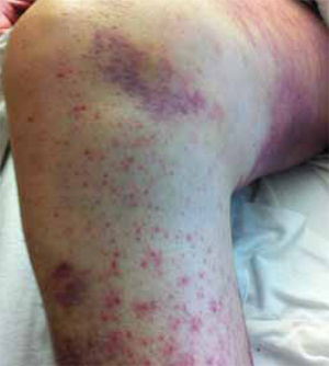
Patients may experience impaired wound healing; abnormal bleeding; muscle, joint, and bone pain; and pathologic fractures.4 There may also be a history of swollen, bleeding gums, tooth loss, and conjunctival varicosities.1,4 Patients may complain of fatigue, exercise intolerance, and depressed mood.
A history of alcoholism is, of course, a red flag. But it’s also important to take note when patients describe a severe food aversion. They may, for instance, tell you that they only eat protein, breads, or other carbohydrates, or that they avoid acidic foods because they have an allergy or because these foods upset their stomach.
Lab work. Serum concentration of ascorbic acid can be checked, although this tends to reflect recent dietary intake rather than actual tissue stores of vitamin C.5 It is estimated that clinical symptoms of scurvy begin to appear when the total body pool of vitamin C has decreased to approximately 300 mg, which reflects a serum level of <0.19 mg/dL.4 Other dietary deficiencies are likely to be present, as well.
There is a leukocyte ascorbate level that more accurately reflects tissue stores of vitamin C, although this test is difficult to perform and not routinely available. (A more practical diagnostic approach is to observe whether clinical abnormalities disappear with repletion therapy.)
Mild anemia is common and its degree correlates with the severity and duration of the scurvy.1 A normocytic-normochromic anemia is usually present and the reticulocyte count is elevated.
Macrocytic anemia may be present due to inadequate folate levels. (Ascorbic acid prevents oxidation of folate to other forms that are excreted in the urine, thus vitamin C deficiency may deplete folate stores by increasing urinary excretion.1)
Iron deficiency anemia is rare in scurvy, even though vitamin C is involved with the absorption of iron.1 Leukopenia is uncommon and platelet counts are usually normal.
Treat with ascorbic acid
Patients with scurvy will need to be treated with a repletion dose of ascorbic acid 100 mg 3 times a day1 (strength of recommendation: A). Manifestations of scurvy recede quickly and usually disappear within a few weeks. Subjective improvement commonly occurs within 24 hours.1
Educating our patient
Given the severity of our patient’s case, he was told that he needed to take 1500 mg ascorbic acid daily. He also received information on alcohol abuse treatment and was counseled on proper nutrition. He was advised to maintain a balanced diet that included the recommended daily amount of all essential vitamins.
Within 48 hours, his hemoglobin stabilized.
CORRESPONDENCE
Sean Robinson, MD, Oregon Health and Science University, 3181 SW Sam Jackson Park Road, FM, Portland, OR 97239; [email protected]
1. Hirschmann JV, Raugi GJ. Adult scurvy. J Am Acad Dermatol. 1999;41:895-906.
2. Hampl JS, Taylor CA, Johnston CS. Vitamin C deficiency and depletion in the United States: the Third National Health and Nutrition Examination Survey, 1988 to 1994. Am J Public Health. 2004;94:870-875.
3. Reamy BV, Williams PM, Lindsay TJ. Henoch-Schönlein purpura. Am Fam Physician. 2009;80:697-704.
4. Nguyen RT, Cowley DM, Muir JB. Scurvy: a cutaneous clinical diagnosis. Australas J Dermatol. 2003;44:48-51.
5. Olmedo JM, Yiannias JA, Windgassen EB, et al. Scurvy: a disease almost forgotten. Int J Dermatol. 2006;45:909-913.
A Good-quality patient-oriented evidence
B Inconsistent or limited-quality patient-oriented evidence
C Consensus, usual practice, opinion, disease-oriented evidence, case series
A 31-YEAR-OLD MAN with a history of depression, alcohol abuse, and rosacea sought care at our clinic for worsening light-headedness, rash, and bruising. Two weeks earlier, he had been to the emergency department (ED) for a syncopal event. The work-up at that time included:
- electrocardiogram showing sinus tachycardia
- lower extremity duplex ultrasound that was negative for deep vein thrombosis or fluid collection
- computed tomography angiogram of the chest that was negative for pulmonary embolus
- lab work that showed normal platelets and international normalized ratio.
The patient did, however, have a notable decrease in hemoglobin from a baseline of 15 g/dL to 12 g/dL, low serum albumin, and mildly elevated aspartate aminotransferase (AST) and total bilirubin.
The patient received 3 liters of intravenous normal saline and was discharged home to follow up as an outpatient. During his visit to our clinic, we noted an extensive purpuric rash on the posterior aspect of his thighs, although his right thigh was worse than his left (FIGURE 1). The patient also had a diffuse follicular petechial rash on his legs. The circumference of his right thigh was 61 cm and his left thigh was 51.5 cm.
FIGURE 1
Diffuse ecchymosis on the right thigh
Repeat labs in the clinic showed a further decrease in hemoglobin to 9.2 g/dL. The patient was advised to go to the ED, where a work-up revealed that he had normocytic-normochromic anemia; his hemoglobin was 8.9 g/dL. Liver function tests revealed elevated AST and alanine aminotransferase (AST:ALT=3.66).
The patient was admitted to the family medicine inpatient service, where his hemoglobin continued to decline. He received a total of 4 units of packed red blood cells.
WHAT IS YOUR DIAGNOSIS?
HOW WOULD YOU TREAT THIS PATIENT?
Diagnosis: Scurvy
Upon further questioning, the patient revealed that his diet consisted solely of alcohol and meat. A serum vitamin C was ordered and returned at 0.1 mg/dL (normal, 0.4-2.0 mg/dL).
Alcoholism is a big tip-off. The diagnosis of scurvy is usually clinical, based on risk factors that suggest a diet deficient in vitamin C. The most common risk factor is alcoholism.1 Other risk factors include adults living alone (notably men), poverty, poor access to groceries, reclusiveness, dementia, nutritional ignorance, avoidance of “acid” foods because of purported allergy, gastrointestinal disorders (eg, colitis, inflammatory disease), poor dentition, food fads or food avoidances, cancer, schizophrenia, and depression.1
Men more than women. Men between the ages of 20 and 39 and those older than 60 years have a higher prevalence of vitamin C deficiency than similarly aged women, according to a National Health and Nutrition Examination Survey of 7277 children and adults.2 Overall, 8.2% of the men and 6% of the women surveyed were deficient in vitamin C.2
Henoch-Schönlein purpura, leukemia included in differential Dx
The differential diagnosis of scurvy includes autoimmune diseases, coagulopathies, hematologic malignancies, adverse effects of medications, meningococcemia, necrotizing gingivitis, senile purpura, thrombophlebitis, other vitamin deficiencies (vitamins K, D, B12, folate, and zinc), and physical abuse or trauma.
Autoimmune diseases such as systemic lupus erythematosus, Henoch-Schönlein purpura, Sjögren syndrome, and rheumatoid arthritis often involve cutaneous manifestations of purpuric and petechial rash. These diseases tend to have systemic involvement that can include the airways, kidneys, and nervous system—as well as rheumatologic findings of joint involvement.3 Making the diagnosis hinges on autoantibody serologies and inflammatory markers.
Coagulopathies such as clotting factor deficiencies, platelet dysfunction, and disseminated intravascular coagulation can also present with skin rash and anemia. Laboratory analysis shows abnormalities of blood cell counts (anemia, throm-bocytopenia), prothrombin time (PT), activated partial thromboplastin time (aPTT), and fibrinogen and clotting factor deficiencies.
Hematologic malignancies such as leukemia or lymphoma can cause petechial and purpuric rashes. Fever, fatigue, weight loss, bone pain, easy bleeding or bruising, lymphadenopathy, and hepatosplenomegaly highlight the systemic manifestations of these malignancies. Positive findings on complete blood count (CBC), coagulation profile, erythrocyte sedimentation rate, and bone marrow biopsy help confirm this diagnosis.
Petechial or purpuric rash can be an adverse effect of anticoagulants, antiplatelet agents, and nonsteroidal anti-inflammatory drugs. Symptoms range in severity and generally resolve with the discontinuation of the offending agent.
Meningococcemia involves a maculopapular rash with petechia and ecchymosis resulting from the spread of bacterial infection from cerebral spinal fluid (CSF) to the systemic circulation. Infection-related effects on clotting factors can be identified with a CBC, PT/aPTT times, and testing for fibrinogen and fibrin degradation products. CSF analysis and culture along with a history and exam findings of fever, malaise, hypotension, and meningeal signs point to meningitis.
Scurvy: What you’ll see and what the lab work will show
Follicular hyperkeratosis and perifollicular hemorrhagic rash with corkscrew hairs are pathognomonic for scurvy and occur early in the disease. Ecchymosis (FIGURE 2) and splinter hemorrhages are also present.
FIGURE 2
Ecchymosis and petechial rash seen in scurvy
Patients may experience impaired wound healing; abnormal bleeding; muscle, joint, and bone pain; and pathologic fractures.4 There may also be a history of swollen, bleeding gums, tooth loss, and conjunctival varicosities.1,4 Patients may complain of fatigue, exercise intolerance, and depressed mood.
A history of alcoholism is, of course, a red flag. But it’s also important to take note when patients describe a severe food aversion. They may, for instance, tell you that they only eat protein, breads, or other carbohydrates, or that they avoid acidic foods because they have an allergy or because these foods upset their stomach.
Lab work. Serum concentration of ascorbic acid can be checked, although this tends to reflect recent dietary intake rather than actual tissue stores of vitamin C.5 It is estimated that clinical symptoms of scurvy begin to appear when the total body pool of vitamin C has decreased to approximately 300 mg, which reflects a serum level of <0.19 mg/dL.4 Other dietary deficiencies are likely to be present, as well.
There is a leukocyte ascorbate level that more accurately reflects tissue stores of vitamin C, although this test is difficult to perform and not routinely available. (A more practical diagnostic approach is to observe whether clinical abnormalities disappear with repletion therapy.)
Mild anemia is common and its degree correlates with the severity and duration of the scurvy.1 A normocytic-normochromic anemia is usually present and the reticulocyte count is elevated.
Macrocytic anemia may be present due to inadequate folate levels. (Ascorbic acid prevents oxidation of folate to other forms that are excreted in the urine, thus vitamin C deficiency may deplete folate stores by increasing urinary excretion.1)
Iron deficiency anemia is rare in scurvy, even though vitamin C is involved with the absorption of iron.1 Leukopenia is uncommon and platelet counts are usually normal.
Treat with ascorbic acid
Patients with scurvy will need to be treated with a repletion dose of ascorbic acid 100 mg 3 times a day1 (strength of recommendation: A). Manifestations of scurvy recede quickly and usually disappear within a few weeks. Subjective improvement commonly occurs within 24 hours.1
Educating our patient
Given the severity of our patient’s case, he was told that he needed to take 1500 mg ascorbic acid daily. He also received information on alcohol abuse treatment and was counseled on proper nutrition. He was advised to maintain a balanced diet that included the recommended daily amount of all essential vitamins.
Within 48 hours, his hemoglobin stabilized.
CORRESPONDENCE
Sean Robinson, MD, Oregon Health and Science University, 3181 SW Sam Jackson Park Road, FM, Portland, OR 97239; [email protected]
1. Hirschmann JV, Raugi GJ. Adult scurvy. J Am Acad Dermatol. 1999;41:895-906.
2. Hampl JS, Taylor CA, Johnston CS. Vitamin C deficiency and depletion in the United States: the Third National Health and Nutrition Examination Survey, 1988 to 1994. Am J Public Health. 2004;94:870-875.
3. Reamy BV, Williams PM, Lindsay TJ. Henoch-Schönlein purpura. Am Fam Physician. 2009;80:697-704.
4. Nguyen RT, Cowley DM, Muir JB. Scurvy: a cutaneous clinical diagnosis. Australas J Dermatol. 2003;44:48-51.
5. Olmedo JM, Yiannias JA, Windgassen EB, et al. Scurvy: a disease almost forgotten. Int J Dermatol. 2006;45:909-913.