User login
EHR alerts boosted MRA prescribing to patients with HFrEF
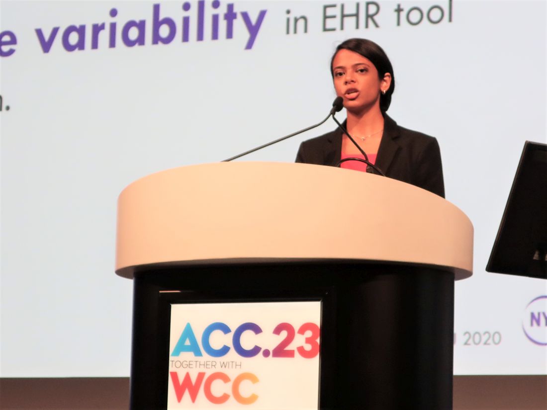
NEW ORLEANS – EHR-embedded alerts that a patient with heart failure with reduced ejection fraction (HFrEF) is a great candidate for treatment with a mineralocorticoid receptor antagonist (MRA) more than doubled prescribing of this “pillar” class for HFrEF, compared with control practices that used usual care and no alerts.
That’s according to results of BETTER CARE-HF, a single-center, randomized trial with more than 2,000 patients and involving 180 cardiologists.
“EHR-embedded tools cans be a rapid, low-cost, and high-impact method to increase prescription of life-saving therapies across large populations,” said Amrita Mukhopadhyay, MD, at the joint scientific sessions of the American College of Cardiology and the World Heart Federation.
Her study targeted underprescribing of an MRA – spironolactone or eplerenone (Inspra) – because of its “vastly underprescribed” status in U.S. practice, where roughly two-thirds of patients with HFrEF do not receive an MRA despite clear recommendations from several medical groups that it is an essential part of treatment for most patients with HFrEF. Dr. Mukhopadhyay estimated that more comprehensive prescribing of MRAs to U.S. patients with HFrEF could prevent more than 20,000 deaths annually.
She also explained that the EHR-embedded alert was carefully devised, through interviews with cardiologists and pilot testing, to optimize the nudge so that it was less intrusive but effective for capturing attention and initiating action.
‘Clinically relevant, impressive results’
“This is a really important study, because despite overwhelming evidence for more than a decade favoring MRA use for patients with HFrEF there is an incredibly large treatment gap. MRAs can reduce all-cause death in people with HFrEF by 25%-30%, as well as reduce hospitalizations for heart failure, at a cost of less than $50 a year,” commented Gregg C. Fonarow, MD, interim chief of cardiology at the University of California, Los Angeles. The study showed “very clinically relevant, impressive results” for individualized, patient-specific alerts to prescribe an MRA and order the laboratory tests, particularly for serum potassium levels, needed to safely start the treatment, Dr. Fonarow said in an interview.
The BETTER CARE-HF study ran at more than 60 practices in the New York City region operated by the NYU Langone Health system, which sponsored the study. The trial randomized 180 cardiologists from these practices in a cluster format to one of three study arms: Sixty cardiologists received the EHR-embedded alerts for their relevant patients (755 patients) when the patient was in the physician’s office; another 60 cardiologists received a less tailored, monthly message that flagged all patients with HFrEF in a cardiologist’s practice who remained untreated candidates for MRA intervention (812 patients); and a third arm of 60 cardiologists and their HFrEF patients served as controls where the clinicians received no alert or message (644 patients).
The study included 2,211 patients with HFrEF and not on MRA treatment at baseline who were all identified as good candidates for starting treatment with the class, with no contraindications, no preexisting hyperkalemia, and no advanced-stage renal dysfunction.
The study’s primary outcome was the percentage of patients in each subgroup who received a new prescription for an MRA. This occurred in 29.6% of the patients whose physicians received an alert, in 15.6% of the patients whose physicians received a monthly message, and in 11.7% of patients in the control practices. Statistical analyses showed that the alerts led to a significant 2.53-fold increase in MRA prescribing, while the messages linked with a significant 67% increase in prescribing, compared with the control practices, reported Dr. Mukhopadhyay, a health services researcher at NYU Langone Health in New York. Simultaneously with her report, the results also appeared in the Journal of the American College of Cardiology.
The findings also showed that the alert and message had no significant impact on the prescribing of any other medication classes for HFrEF, compared with the controls. And the alert intervention had minimal adverse effects. While patients in the alert arm showed a significant, 45% relative increase in the incidence of hyperkalemia episodes, compared with control patients (because of a 4.5% absolute increase in hyperkalemia events), the rate of “significant” hyperkalemia with a value of at least 5.5 mmol/L, occurred in 5.0% of patients in the alert group and 5.1% of patients in the control arm.
Potassium testing poses another barrier
Even though the alerts substantially improved MRA prescribing, 70% of patients deemed MRA eligible in the alert subgroup still failed to receive a prescription. One additional barrier specific to MRA prescribing is the need it triggers for serial laboratory testing to monitor serum potassium levels. “Potassium testing generates additional work outside the index visit, which along with the risk for hyperkalemia exists as a barrier,” commented Lee R. Goldberg, MD, a heart failure specialist and professor at the University of Pennsylvania in Philadelphia. “This may be the next aspect to focus on to improve MRA uptake,” he said as a designated discussant for the report.

“It’s not enough to just prompt medication treatment. We also need to prompt appropriate laboratory testing,” noted Dr. Fonarow.
He also said that the approach tested by Dr. Mukhopadhyay could now be expanded to outpatient cardiologists. “The onus is on everyone involved in caring for patients with HFrEF failure to explain why maximum effort is not being made to deploy” all of the guideline-directed medical therapies for the disorder.
EHR alerts “are one way to bridge the prescribing gap, but we need multiple approaches so that all eligible patients receive guideline-directed medical therapy,” Dr. Fonarow said.
BETTER CARE-HF received no commercial funding, and Dr. Mukhopadhyay had no disclosures. Dr. Fonarow has been a consultant to AstraZeneca, Amgen, Cytokinetics, Lilly, Merck, Novartis, and Pfizer. Dr. Goldberg has received personal fees from Abbott, VisCardia, and Zoll/Respircardia.
NEW ORLEANS – EHR-embedded alerts that a patient with heart failure with reduced ejection fraction (HFrEF) is a great candidate for treatment with a mineralocorticoid receptor antagonist (MRA) more than doubled prescribing of this “pillar” class for HFrEF, compared with control practices that used usual care and no alerts.
That’s according to results of BETTER CARE-HF, a single-center, randomized trial with more than 2,000 patients and involving 180 cardiologists.
“EHR-embedded tools cans be a rapid, low-cost, and high-impact method to increase prescription of life-saving therapies across large populations,” said Amrita Mukhopadhyay, MD, at the joint scientific sessions of the American College of Cardiology and the World Heart Federation.
Her study targeted underprescribing of an MRA – spironolactone or eplerenone (Inspra) – because of its “vastly underprescribed” status in U.S. practice, where roughly two-thirds of patients with HFrEF do not receive an MRA despite clear recommendations from several medical groups that it is an essential part of treatment for most patients with HFrEF. Dr. Mukhopadhyay estimated that more comprehensive prescribing of MRAs to U.S. patients with HFrEF could prevent more than 20,000 deaths annually.
She also explained that the EHR-embedded alert was carefully devised, through interviews with cardiologists and pilot testing, to optimize the nudge so that it was less intrusive but effective for capturing attention and initiating action.
‘Clinically relevant, impressive results’
“This is a really important study, because despite overwhelming evidence for more than a decade favoring MRA use for patients with HFrEF there is an incredibly large treatment gap. MRAs can reduce all-cause death in people with HFrEF by 25%-30%, as well as reduce hospitalizations for heart failure, at a cost of less than $50 a year,” commented Gregg C. Fonarow, MD, interim chief of cardiology at the University of California, Los Angeles. The study showed “very clinically relevant, impressive results” for individualized, patient-specific alerts to prescribe an MRA and order the laboratory tests, particularly for serum potassium levels, needed to safely start the treatment, Dr. Fonarow said in an interview.
The BETTER CARE-HF study ran at more than 60 practices in the New York City region operated by the NYU Langone Health system, which sponsored the study. The trial randomized 180 cardiologists from these practices in a cluster format to one of three study arms: Sixty cardiologists received the EHR-embedded alerts for their relevant patients (755 patients) when the patient was in the physician’s office; another 60 cardiologists received a less tailored, monthly message that flagged all patients with HFrEF in a cardiologist’s practice who remained untreated candidates for MRA intervention (812 patients); and a third arm of 60 cardiologists and their HFrEF patients served as controls where the clinicians received no alert or message (644 patients).
The study included 2,211 patients with HFrEF and not on MRA treatment at baseline who were all identified as good candidates for starting treatment with the class, with no contraindications, no preexisting hyperkalemia, and no advanced-stage renal dysfunction.
The study’s primary outcome was the percentage of patients in each subgroup who received a new prescription for an MRA. This occurred in 29.6% of the patients whose physicians received an alert, in 15.6% of the patients whose physicians received a monthly message, and in 11.7% of patients in the control practices. Statistical analyses showed that the alerts led to a significant 2.53-fold increase in MRA prescribing, while the messages linked with a significant 67% increase in prescribing, compared with the control practices, reported Dr. Mukhopadhyay, a health services researcher at NYU Langone Health in New York. Simultaneously with her report, the results also appeared in the Journal of the American College of Cardiology.
The findings also showed that the alert and message had no significant impact on the prescribing of any other medication classes for HFrEF, compared with the controls. And the alert intervention had minimal adverse effects. While patients in the alert arm showed a significant, 45% relative increase in the incidence of hyperkalemia episodes, compared with control patients (because of a 4.5% absolute increase in hyperkalemia events), the rate of “significant” hyperkalemia with a value of at least 5.5 mmol/L, occurred in 5.0% of patients in the alert group and 5.1% of patients in the control arm.
Potassium testing poses another barrier
Even though the alerts substantially improved MRA prescribing, 70% of patients deemed MRA eligible in the alert subgroup still failed to receive a prescription. One additional barrier specific to MRA prescribing is the need it triggers for serial laboratory testing to monitor serum potassium levels. “Potassium testing generates additional work outside the index visit, which along with the risk for hyperkalemia exists as a barrier,” commented Lee R. Goldberg, MD, a heart failure specialist and professor at the University of Pennsylvania in Philadelphia. “This may be the next aspect to focus on to improve MRA uptake,” he said as a designated discussant for the report.
“It’s not enough to just prompt medication treatment. We also need to prompt appropriate laboratory testing,” noted Dr. Fonarow.
He also said that the approach tested by Dr. Mukhopadhyay could now be expanded to outpatient cardiologists. “The onus is on everyone involved in caring for patients with HFrEF failure to explain why maximum effort is not being made to deploy” all of the guideline-directed medical therapies for the disorder.
EHR alerts “are one way to bridge the prescribing gap, but we need multiple approaches so that all eligible patients receive guideline-directed medical therapy,” Dr. Fonarow said.
BETTER CARE-HF received no commercial funding, and Dr. Mukhopadhyay had no disclosures. Dr. Fonarow has been a consultant to AstraZeneca, Amgen, Cytokinetics, Lilly, Merck, Novartis, and Pfizer. Dr. Goldberg has received personal fees from Abbott, VisCardia, and Zoll/Respircardia.
AT ACC 2023
Atorvastatin cut anthracycline cardiac dysfunction in lymphoma
NEW ORLEANS – Atorvastatin treatment of patients with lymphoma undergoing treatment with an anthracycline significantly cut the incidence of incident cardiac dysfunction by about two-thirds during 12 months of treatment, in a multicenter, randomized trial with 300 enrolled patients.
“These data support the use of atorvastatin among patients with lymphoma being treated with anthracyclines where prevention of cardiac systolic dysfunction is important,” concluded Tomas G. Neilan, MD, at the joint scientific sessions of the American College of Cardiology and the World Heart Federation.

He highlighted that an important difference between the new study, STOP-CA, and a major prior study with a neutral effect published in 2022, was that STOP-CA “was powered for a major change” in cardiac function as the study’s primary outcome, a decline from baseline in left ventricular ejection fraction (LVEF) of at least 10% that also reduced ejection fraction to less than 55%.
“We can consider these medications [atorvastatin] for patients at higher risk for cardiac toxicity from anthracyclines, such as patients who receive a higher dose of an anthracycline, older patients, people with obesity, and women, commented Anita Deswal, MD, professor and chair of the department of cardiology at the University of Texas MD Anderson Cancer Center, Houston, who was not involved with the study.
A basis for an ‘important discussion’ with patients
“For patients receiving higher doses of anthracyclines, the STOP-CA trial says that whether to start a statin for cardiac protection is now an important discussion” for these patients to have with their treating clinicians. ”That was not the case before today,” commented Ronald M. Witteles, MD, a cardiologist and professor who specializes in cardio-oncology at Stanford (Calif.) University.
“For a patient being treated for lymphoma or for another cancer and treated with equal or higher anthracycline doses, such as patients with a sarcoma, this trial’s results at the very least warrant a discussion between physicians and patients to make the decision,” Dr. Witteles, who was not involved in the study, said in an interview. But he also cautioned that “whether an individual patient should take a statin in this scenario is still not a no-brainer. While the trial was positive, it was for an imaging rather than for a clinical endpoint.”
Experts noted that a similar study with the clinical endpoint of heart failure would require both many more randomized patients as well as much longer follow-up. STOP-CA was not powered for this endpoint. During its 12-month duration, a total of 11 patients developed heart failure, with no between group difference.
STOP-CA enrolled adults with lymphoma (Hodgkin or non-Hodgkin) and scheduled to undergo anthracycline treatment at eight U.S. centers and one in Canada, and excluded patients already on statin treatment or those for whom a statin was already indicated. Of the 300 enrolled patients, 286 had 12-month follow-up. Randomization assigned patients to receive either 40 mg daily of atorvastatin or placebo.
Their cumulative, median anthracycline dose was 300 mg/m2, which is typical for treating lymphoma, but higher than the typical dose use for patients with breast cancer. At baseline, average LVEF was 63%, and after 12 months this had declined to 59%. Forty-six of the 286 patients assessed after 12 months fulfilled the primary outcome of at least a 10–percentage point reduction from baseline in their LVEF and a decline in LVEF to less than 55%. Researchers used cardiac MR to assess LVEF at baseline, and in most patients at follow-up, but a minority of patients had their follow-up assessments by echocardiography because of logistical issues. Greater than 90% of patients were adherent to their assigned regimen.
Tripled incidence of cardiac dysfunction in placebo patients
The incidence of this outcome was 9% among the patients who received atorvastatin, and 22% among those on placebo, a significant difference. The calculated odds of the primary outcome was 2.9-fold more likely among the patients treated with placebo, compared with those who received atorvastatin, also a significant difference.
The study’s secondary outcome was patients who had at least a 5% drop from baseline in their LVEF and with a LVEF of less than 55% after 12 months. This outcome occurred in 13% of patients treated with atorvastatin and in 29% of those who received placebo, a significant difference.
The atorvastatin and placebo arms showed no significant differences in adverse events during the study, with roughly similar incidence rates for muscle pain, elevated liver enzymes, and renal failure. None of the enrolled patients developed myositis.
Atorvastatin treatment also produced an expected average 37% decline from baseline in levels of LDL cholesterol.
“This was a well-designed and important trial,” said Dr. Witteles. “Anthracyclines remain a mainstay of cancer therapies for a number of malignancies, such as lymphoma and sarcoma, and the cardiac side effects of development of cardiac dysfunction are unequivocally real.”
The importance of a clinically meaningful effect
The results especially contrast with the findings from the PREVENT study, published in 2022, which compared a daily, 40-mg atorvastatin treatment with placebo in 279 randomized patients with breast cancer and treated for 24 months. However, patients in PREVENT had a cumulative, median anthracycline dose of 240 mg/m2, and the study’s primary outcome was the average change from baseline in LVEF after 24 months of treatment, which was a reduction of 0.08 percentage points in the placebo arm, a nonsignificant difference.
In STOP-CA, the average change in LVEF from baseline was a 1–percentage point reduction in the placebo arm, compared with the atorvastatin-treated patients, a difference that was statistically significant, but “not clinically significant,” said Dr. Neilan, director of the cardio-oncology program at Massachusetts General Hospital, Boston. He cited the good fortune of the STOP-CA investigators when they received a recommendation from reviewers early on to design their study to track a clinically meaningful change in LVEF rather than just looking at the average overall change.

Dr. Deswal also noted that it is unlikely that future studies will examine the efficacy of a statin for preventing LVEF in patients across the range of cancers that are eligible for anthracycline treatment. As a result, she predicted that “we may have to extrapolate” the results from STOP-CA to patients with other cancer types.
STOP-CA received no commercial funding. Dr. Neilan has been a consultant for and received fees from Abbvie, Amgen, Bristol-Myers Squibb, CRC Oncology, Genentech, Roche, and Sanofi, and has received grant funding from AstraZeneca and Bristol Myers Squib. Dr. Deswal and Dr. Witteles had no relevant disclosures.
NEW ORLEANS – Atorvastatin treatment of patients with lymphoma undergoing treatment with an anthracycline significantly cut the incidence of incident cardiac dysfunction by about two-thirds during 12 months of treatment, in a multicenter, randomized trial with 300 enrolled patients.
“These data support the use of atorvastatin among patients with lymphoma being treated with anthracyclines where prevention of cardiac systolic dysfunction is important,” concluded Tomas G. Neilan, MD, at the joint scientific sessions of the American College of Cardiology and the World Heart Federation.
He highlighted that an important difference between the new study, STOP-CA, and a major prior study with a neutral effect published in 2022, was that STOP-CA “was powered for a major change” in cardiac function as the study’s primary outcome, a decline from baseline in left ventricular ejection fraction (LVEF) of at least 10% that also reduced ejection fraction to less than 55%.
“We can consider these medications [atorvastatin] for patients at higher risk for cardiac toxicity from anthracyclines, such as patients who receive a higher dose of an anthracycline, older patients, people with obesity, and women, commented Anita Deswal, MD, professor and chair of the department of cardiology at the University of Texas MD Anderson Cancer Center, Houston, who was not involved with the study.
A basis for an ‘important discussion’ with patients
“For patients receiving higher doses of anthracyclines, the STOP-CA trial says that whether to start a statin for cardiac protection is now an important discussion” for these patients to have with their treating clinicians. ”That was not the case before today,” commented Ronald M. Witteles, MD, a cardiologist and professor who specializes in cardio-oncology at Stanford (Calif.) University.
“For a patient being treated for lymphoma or for another cancer and treated with equal or higher anthracycline doses, such as patients with a sarcoma, this trial’s results at the very least warrant a discussion between physicians and patients to make the decision,” Dr. Witteles, who was not involved in the study, said in an interview. But he also cautioned that “whether an individual patient should take a statin in this scenario is still not a no-brainer. While the trial was positive, it was for an imaging rather than for a clinical endpoint.”
Experts noted that a similar study with the clinical endpoint of heart failure would require both many more randomized patients as well as much longer follow-up. STOP-CA was not powered for this endpoint. During its 12-month duration, a total of 11 patients developed heart failure, with no between group difference.
STOP-CA enrolled adults with lymphoma (Hodgkin or non-Hodgkin) and scheduled to undergo anthracycline treatment at eight U.S. centers and one in Canada, and excluded patients already on statin treatment or those for whom a statin was already indicated. Of the 300 enrolled patients, 286 had 12-month follow-up. Randomization assigned patients to receive either 40 mg daily of atorvastatin or placebo.
Their cumulative, median anthracycline dose was 300 mg/m2, which is typical for treating lymphoma, but higher than the typical dose use for patients with breast cancer. At baseline, average LVEF was 63%, and after 12 months this had declined to 59%. Forty-six of the 286 patients assessed after 12 months fulfilled the primary outcome of at least a 10–percentage point reduction from baseline in their LVEF and a decline in LVEF to less than 55%. Researchers used cardiac MR to assess LVEF at baseline, and in most patients at follow-up, but a minority of patients had their follow-up assessments by echocardiography because of logistical issues. Greater than 90% of patients were adherent to their assigned regimen.
Tripled incidence of cardiac dysfunction in placebo patients
The incidence of this outcome was 9% among the patients who received atorvastatin, and 22% among those on placebo, a significant difference. The calculated odds of the primary outcome was 2.9-fold more likely among the patients treated with placebo, compared with those who received atorvastatin, also a significant difference.
The study’s secondary outcome was patients who had at least a 5% drop from baseline in their LVEF and with a LVEF of less than 55% after 12 months. This outcome occurred in 13% of patients treated with atorvastatin and in 29% of those who received placebo, a significant difference.
The atorvastatin and placebo arms showed no significant differences in adverse events during the study, with roughly similar incidence rates for muscle pain, elevated liver enzymes, and renal failure. None of the enrolled patients developed myositis.
Atorvastatin treatment also produced an expected average 37% decline from baseline in levels of LDL cholesterol.
“This was a well-designed and important trial,” said Dr. Witteles. “Anthracyclines remain a mainstay of cancer therapies for a number of malignancies, such as lymphoma and sarcoma, and the cardiac side effects of development of cardiac dysfunction are unequivocally real.”
The importance of a clinically meaningful effect
The results especially contrast with the findings from the PREVENT study, published in 2022, which compared a daily, 40-mg atorvastatin treatment with placebo in 279 randomized patients with breast cancer and treated for 24 months. However, patients in PREVENT had a cumulative, median anthracycline dose of 240 mg/m2, and the study’s primary outcome was the average change from baseline in LVEF after 24 months of treatment, which was a reduction of 0.08 percentage points in the placebo arm, a nonsignificant difference.
In STOP-CA, the average change in LVEF from baseline was a 1–percentage point reduction in the placebo arm, compared with the atorvastatin-treated patients, a difference that was statistically significant, but “not clinically significant,” said Dr. Neilan, director of the cardio-oncology program at Massachusetts General Hospital, Boston. He cited the good fortune of the STOP-CA investigators when they received a recommendation from reviewers early on to design their study to track a clinically meaningful change in LVEF rather than just looking at the average overall change.
Dr. Deswal also noted that it is unlikely that future studies will examine the efficacy of a statin for preventing LVEF in patients across the range of cancers that are eligible for anthracycline treatment. As a result, she predicted that “we may have to extrapolate” the results from STOP-CA to patients with other cancer types.
STOP-CA received no commercial funding. Dr. Neilan has been a consultant for and received fees from Abbvie, Amgen, Bristol-Myers Squibb, CRC Oncology, Genentech, Roche, and Sanofi, and has received grant funding from AstraZeneca and Bristol Myers Squib. Dr. Deswal and Dr. Witteles had no relevant disclosures.
AT ACC 2023
Transcatheter tricuspid valve repair effective and safe for regurgitation
NEW ORLEANS – In the first pivotal randomized, controlled trial of a transcatheter device for the repair of severe tricuspid regurgitation, a large reduction in valve dysfunction was associated with substantial improvement in quality of life (QOL) persisting out of 1 year of follow-up, according to results of the TRILUMINATE trial.
Based on the low procedural risks of the repair, the principal investigator, Paul Sorajja, MD, called the results “very clinically meaningful” as he presented the results at the joint scientific sessions of the American College of Cardiology and the World Heart Federation.

Conducted at 65 centers in the United States, Canada, and North America, TRILUMINATE evaluated a transcatheter end-to-end (TEER) repair performed with the TriClip G4 Delivery System (Abbott). The study included two cohorts, both of which will be followed for 5 years. One included patients with very severe tricuspid regurgitation enrolled in a single arm. Data on this cohort is expected later in 2023.
In the randomized portion of the study, 350 patients enrolled with severe tricuspid regurgitation underwent TEER with a clipping device and then were followed on the guideline-directed therapy (GDMT) for heart failure they were receiving at baseline. The control group was managed on GDMT alone.
The primary composite endpoint at 1 year was a composite of death from any cause and/or tricuspid valve surgery, hospitalization for heart failure, and quality of life as measured with the Kansas City Cardiomyopathy questionnaire (KCCQ).
Benefit driven by quality of life
For the primary endpoint, the win ratio, a statistical calculation of those who did relative to those who did not benefit, was 1.48, signifying a 48% advantage (P = .02). This was driven almost entirely by the KCCQ endpoint. There was no significant difference death and/or tricuspid valve surgery, which occurred in about 10% of both groups (P = .75) or heart failure hospitalization, which was occurred in slightly more patients randomized to repair (14.9% vs. 12.1%; P = .41).
For KCCQ, the mean increase at 1 year was 12.3 points in the repair group versus 0.6 points (P < .001) in the control group. With an increase of 5-10 points typically considered to be clinically meaningful, the advantage of repair over GDMT at the threshold of 15 points or greater was highly statistically significant (49.7% vs. 26.4%; P < .0001).
This advantage was attributed to control of regurgitation. The proportion achieving moderate or less regurgitation sustained at 1 year was 87% in the repair group versus 4.8% in the GDMT group (P < .0001).
When assessed independent of treatment, KCCQ benefits at 1 year increased in a stepwise fashion as severity of regurgitation was reduced, climbing from 2 points if there was no improvement to 6 points with one grade in improvement and then to 18 points with at least a two-grade improvement.
For regurgitation, “the repair was extremely effective,” said Dr. Sorajja of Allina Health Minneapolis Heart Institute at Abbott Northwestern Hospital, Minneapolis. He added that the degree of regurgitation control in the TRILUMINATE trial “is the highest ever reported.” With previous trials with other transcatheter devices in development, the improvement so far has been on the order of 70%-80%.
For enrollment in TRILUMINATE, patients were required to have at least an intermediate risk of morbidity or mortality from tricuspid valve surgery. Exclusion criteria included a left ventricular ejection fraction (LVEF) less than 20% and severe pulmonary hypertension.
More than 70% of patients had the highest (torrential) or second highest (massive) category of regurgitation on a five-level scale by echocardiography. Almost all the remaining were at the third level (severe).
Of those enrolled, the average age was roughly 78 years. About 55% were women. Nearly 60% were in New York Heart Association class III or IV heart failure and most had significant comorbidities, including hypertension (> 80%), atrial fibrillation (about 90%), and renal disease (35%). Patients with diabetes (16%), chronic obstructive pulmonary disease (10%), and liver disease (7.5%) were represented in lower numbers.
Surgery is not necessarily an option
All enrolled patients were considered to be at intermediate or greater risk for mortality with surgical replacement of the tricuspid valve, but Dr. Sorajja pointed out that surgery, which involves valve replacement, is not necessarily an alternative to valve repair. Even in fit patients, the high morbidity, mortality, and extended hospital stay associated with surgical valve replacement makes this procedure unattractive.
In this trial, most patients who underwent the transcatheter procedure were discharged within a day. The safety was excellent, Dr. Sorajja said. Only three patients (1.7%) had a major adverse event. This included two cases of new-onset renal failure and one cardiovascular death. There were no cases of endocarditis requiring surgery or any other type of nonelective cardiovascular surgery, including for any device-related issue.
In the sick population enrolled, Dr. Sorajja characterized the number of adverse events over 1 year as “very low.”

These results are important, according to Kendra Grubb, MD, surgical director of the Structural Heart and Valve Center, Emory University, Atlanta. While she expressed surprise that there was no signal of benefit on hard endpoints at 1 year, she emphasized that “these patients feel terrible,” and they are frustrating to manage because surgery is often contraindicated or impractical.
“Finally, we have something for this group,” she said, noting that the mortality from valve replacement surgery even among patients who are fit enough for surgery to be considered is about 10%.
Ajay Kirtane, MD, director of the Cardiac Catheterization Laboratories at Columbia University, New York, was more circumspect. He agreed that the improvement in QOL was encouraging, but cautioned that QOL is a particularly soft outcome in a nonrandomized trial in which patients may feel better just knowing that there regurgitation has been controlled. He found the lack of benefit on hard outcomes not just surprising but “disappointing.”
Still, he agreed the improvement in QOL is potentially meaningful for a procedure that appears to be relatively safe.
Dr. Sorajja reported financial relationships with Boston Scientific, Edwards Lifesciences, Foldax. 4C Medical, Gore Medtronic, Phillips, Siemens, Shifamed, Vdyne, xDot, and Abbott Structural, which provided funding for this trial. Dr. Grubb reported financial relationships with Abbott Vascular, Ancora Heart, Bioventrix, Boston Scientific, Edwards Lifesciences, 4C Medical, JenaValve, and Medtronic. Dr. Kirtane reported financial relationships with Abbott Vascular, Amgen, Boston Scientific, Chiesi, Medtronic, Opsens, Phillips, ReCor, Regeneron, and Zoll.
NEW ORLEANS – In the first pivotal randomized, controlled trial of a transcatheter device for the repair of severe tricuspid regurgitation, a large reduction in valve dysfunction was associated with substantial improvement in quality of life (QOL) persisting out of 1 year of follow-up, according to results of the TRILUMINATE trial.
Based on the low procedural risks of the repair, the principal investigator, Paul Sorajja, MD, called the results “very clinically meaningful” as he presented the results at the joint scientific sessions of the American College of Cardiology and the World Heart Federation.
Conducted at 65 centers in the United States, Canada, and North America, TRILUMINATE evaluated a transcatheter end-to-end (TEER) repair performed with the TriClip G4 Delivery System (Abbott). The study included two cohorts, both of which will be followed for 5 years. One included patients with very severe tricuspid regurgitation enrolled in a single arm. Data on this cohort is expected later in 2023.
In the randomized portion of the study, 350 patients enrolled with severe tricuspid regurgitation underwent TEER with a clipping device and then were followed on the guideline-directed therapy (GDMT) for heart failure they were receiving at baseline. The control group was managed on GDMT alone.
The primary composite endpoint at 1 year was a composite of death from any cause and/or tricuspid valve surgery, hospitalization for heart failure, and quality of life as measured with the Kansas City Cardiomyopathy questionnaire (KCCQ).
Benefit driven by quality of life
For the primary endpoint, the win ratio, a statistical calculation of those who did relative to those who did not benefit, was 1.48, signifying a 48% advantage (P = .02). This was driven almost entirely by the KCCQ endpoint. There was no significant difference death and/or tricuspid valve surgery, which occurred in about 10% of both groups (P = .75) or heart failure hospitalization, which was occurred in slightly more patients randomized to repair (14.9% vs. 12.1%; P = .41).
For KCCQ, the mean increase at 1 year was 12.3 points in the repair group versus 0.6 points (P < .001) in the control group. With an increase of 5-10 points typically considered to be clinically meaningful, the advantage of repair over GDMT at the threshold of 15 points or greater was highly statistically significant (49.7% vs. 26.4%; P < .0001).
This advantage was attributed to control of regurgitation. The proportion achieving moderate or less regurgitation sustained at 1 year was 87% in the repair group versus 4.8% in the GDMT group (P < .0001).
When assessed independent of treatment, KCCQ benefits at 1 year increased in a stepwise fashion as severity of regurgitation was reduced, climbing from 2 points if there was no improvement to 6 points with one grade in improvement and then to 18 points with at least a two-grade improvement.
For regurgitation, “the repair was extremely effective,” said Dr. Sorajja of Allina Health Minneapolis Heart Institute at Abbott Northwestern Hospital, Minneapolis. He added that the degree of regurgitation control in the TRILUMINATE trial “is the highest ever reported.” With previous trials with other transcatheter devices in development, the improvement so far has been on the order of 70%-80%.
For enrollment in TRILUMINATE, patients were required to have at least an intermediate risk of morbidity or mortality from tricuspid valve surgery. Exclusion criteria included a left ventricular ejection fraction (LVEF) less than 20% and severe pulmonary hypertension.
More than 70% of patients had the highest (torrential) or second highest (massive) category of regurgitation on a five-level scale by echocardiography. Almost all the remaining were at the third level (severe).
Of those enrolled, the average age was roughly 78 years. About 55% were women. Nearly 60% were in New York Heart Association class III or IV heart failure and most had significant comorbidities, including hypertension (> 80%), atrial fibrillation (about 90%), and renal disease (35%). Patients with diabetes (16%), chronic obstructive pulmonary disease (10%), and liver disease (7.5%) were represented in lower numbers.
Surgery is not necessarily an option
All enrolled patients were considered to be at intermediate or greater risk for mortality with surgical replacement of the tricuspid valve, but Dr. Sorajja pointed out that surgery, which involves valve replacement, is not necessarily an alternative to valve repair. Even in fit patients, the high morbidity, mortality, and extended hospital stay associated with surgical valve replacement makes this procedure unattractive.
In this trial, most patients who underwent the transcatheter procedure were discharged within a day. The safety was excellent, Dr. Sorajja said. Only three patients (1.7%) had a major adverse event. This included two cases of new-onset renal failure and one cardiovascular death. There were no cases of endocarditis requiring surgery or any other type of nonelective cardiovascular surgery, including for any device-related issue.
In the sick population enrolled, Dr. Sorajja characterized the number of adverse events over 1 year as “very low.”
These results are important, according to Kendra Grubb, MD, surgical director of the Structural Heart and Valve Center, Emory University, Atlanta. While she expressed surprise that there was no signal of benefit on hard endpoints at 1 year, she emphasized that “these patients feel terrible,” and they are frustrating to manage because surgery is often contraindicated or impractical.
“Finally, we have something for this group,” she said, noting that the mortality from valve replacement surgery even among patients who are fit enough for surgery to be considered is about 10%.
Ajay Kirtane, MD, director of the Cardiac Catheterization Laboratories at Columbia University, New York, was more circumspect. He agreed that the improvement in QOL was encouraging, but cautioned that QOL is a particularly soft outcome in a nonrandomized trial in which patients may feel better just knowing that there regurgitation has been controlled. He found the lack of benefit on hard outcomes not just surprising but “disappointing.”
Still, he agreed the improvement in QOL is potentially meaningful for a procedure that appears to be relatively safe.
Dr. Sorajja reported financial relationships with Boston Scientific, Edwards Lifesciences, Foldax. 4C Medical, Gore Medtronic, Phillips, Siemens, Shifamed, Vdyne, xDot, and Abbott Structural, which provided funding for this trial. Dr. Grubb reported financial relationships with Abbott Vascular, Ancora Heart, Bioventrix, Boston Scientific, Edwards Lifesciences, 4C Medical, JenaValve, and Medtronic. Dr. Kirtane reported financial relationships with Abbott Vascular, Amgen, Boston Scientific, Chiesi, Medtronic, Opsens, Phillips, ReCor, Regeneron, and Zoll.
AT ACC 2023
Posttransplant NASH patients fare worse with older donor livers
All-cause mortality was twice as high and death from an infectious cause was more than three times as high for patients with NASH who received liver grafts from octogenarian donors than for those who received a liver from someone younger than 50.
“The findings from this study implicate a critical need to investigate donor age as an important risk factor for poorer host and graft survival,” wrote the authors, led by David Lee, MD, of the University of Maryland, Baltimore.
“Given the possibility of infectious graft complications, post–liver transplant follow-ups may need to be more comprehensive and frequent in these individuals who receive grafts from older donors,” they add.
The study was published online in Digestive and Liver Disease.
Donor age trends
Dr. Lee and colleagues pulled data from the United Network for Organ Sharing (UNOS) Standard Transplant Analysis and Research database, a national database of deidentified donor and recipient transplant data. The analysis excluded recipients younger than 18, those with a living donor, those who had hepatocellular carcinoma prior to transplant, and those who had been diagnosed with additional liver disorders apart from NASH.
The team identified 8,88 recipients with NASH who received a liver transplant from 2005–2019. They stratified recipients by donor age. The 5,187 patients who received livers from donors who were younger than 50 served as the reference group. The remainder were placed into four cohorts – 1,842 whose donors were in their 50s, 1,290 whose donors were in their 60s, 504 whose donors were in their 70s, and 65 whose donors were in their 80s.
The researchers found that in comparison with the reference group, the average age of recipients in each donor-age cohort was progressively older. Two donor-age cohorts had significantly higher proportions of recipients with diabetes than the 46.5% in the reference group – the sexagenarian cohort (51.7%) and the octogenarian group (66.2%).
The median follow-up time ranged from 2.35–3.61 years across all age groups.
The researchers found that for all donor-age groups excluding donors in their 60s, recipients had higher risk of all-cause mortality after transplant than the reference group. Recipients with donors in their 50s had a 16% greater risk for death (P = .01), and recipients with donors in their 70s had a 20% greater risk (P = .05). For recipients with octogenarian donors, the adjusted hazard ratio for all-cause mortality was 2.01 (P < .001).
Only recipients in the octogenarian donor cohort were at increased risk of graft failure, compared with the reference group (aHR, 3.72; P = .002).
As donor age increased, the recipient’s risk of dying from sepsis and infectious causes rose, compared with the reference group. Recipients’ likelihood of sepsis death increased by 71% (P = .001) with donors in their 50s, 73% (P = .003) with donors in their 60s, and 76% (P = .03) with donors in their 70s. For recipients with octogenarian donors, the risk more than tripled (aHR, 3.58; P = .007). Likewise, recipients with donors in their 70s were 73% more likely to die from infectious causes. That risk nearly quadrupled among those with donors in their 80s.
Recipient factors at play?
While the study found a relationship between liver donor age and recipient outcomes, it is not clear whether any other recipient factors may have contributed to the higher risk of all-cause mortality, sad Nancy Reau, MD, chief of the hepatology section at Rush University Medical Center, Chicago. The researchers did not parse out whether younger recipients did better with older organs than older recipients or whether older recipients fared worse with younger organs, she said in an interview.
“I wasn’t convinced that they had demonstrated that the recipient may not have played a role in that,” said Dr. Reau, who wasn’t involved with the study.
The analysis only a found an increased risk of graft failure among recipients who received organs from octogenarian donors, so factors other than liver transplant may have contributed to all-cause mortality, she noted.
The UNOS database has some limitations, noted Timothy Pruett, MD, who directs the liver transplant program at the University of Minnesota, Minneapolis. Because the database pulls information from transplantation centers across the country, it can be difficult to standardize specific patient variables in the data.
While it’s clear that a patient died, it’s less certain whether an infection was the cause of death and whether that infection was somehow associated with the liver, noted Dr. Pruett, who wasn’t involved in the research. For example, a patient could have had broken a hip, gone to the hospital, and contracted pneumonia, which led to their death.
“There’s just not much granularity in the database, and we can’t overextrapolate what we see,” Dr. Pruett said in an interview.
Knowledge gaps
Dr. Lee agreed that more research is needed to understand what may be driving higher mortality rates among patients who receive older organs. “There are still a lot of gaps in knowledge with respect to why.”
Dr. Reau said she is curious as to whether certain comorbidities, such as previous infection, diabetes, or obesity, could predict worse outcomes for recipients with older organs.
“We would love to give all of our patients younger organs, but if that leads to even more disparity in need [compared] to availability and the alternative is not surviving, I think you have to place [this work] into context,” she said.
The study findings shouldn’t be used to deter patients with NASH from considering older organs, Dr. Reau said. More insight as to which populations might want to be choosier owing to an elevated risk would be beneficial, she added.
Dr. Lee, Dr. Pruett, and Dr. Reau reported no relevant financial relationships.
A version of this article first appeared on Medscape.com.
FROM DIGESTIVE AND LIVER DISEASE
Ovarian cancer risk lower with daily aspirin, despite genetics
new research suggests.
The study found that daily or almost daily aspirin use was associated with a 13% reduction in ovarian cancer risk, which was not modified by an individual’s polygenic score (PGS).
“Our findings suggest that frequent use of aspirin is associated with reduced ovarian cancer risk, regardless of whether a woman has lower or higher genetic susceptibility to ovarian cancer, as predicted by a set of known, common risk variants,” said lead author Lauren M. Hurwitz, PhD, MHS, division of cancer epidemiology and genetics at the National Cancer Institute, Rockville, Md.
The study was published online in JAMA Network Open.
Patients diagnosed with ovarian cancer face difficult survival odds, which make preventive strategies especially important. Evidence suggests that frequent aspirin use can reduce the risk for ovarian cancer by about 13%, but it’s unclear whether genetic factors change those odds.
Although promising for chemoprevention, aspirin use can also come with downsides, including gastric ulcer and hemorrhagic stroke, which is why identifying and targeting individuals at higher risk for ovarian cancer who may benefit from frequent aspirin use is important.
In the current analysis, Dr. Hurwitz and colleagues used a PGS to determine whether the protective effects of daily or near-daily aspirin use for 6 months or more could be modified by genetics.
The study was a pooled analysis of eight case-controlled studies from the Ovarian Cancer Association Consortium conducted in the United Kingdom, the United States, and Australia over a 14-year period. The researchers looked at genetic data and data on frequent aspirin use among 4,476 case patients with nonmucinous ovarian cancer (average age, 57) and 6,659 control participants (average age, 58). Overall, 575 patients (13%) and 1,030 controls (15%) reported frequent aspirin use.
The authors used a PGS previously developed using 22 single-nucleotide variants. Because this PGS was developed for nonmucinous epithelial ovarian cancer, only these patients were included in the analysis.
Consistent with previous evidence, the authors found that frequent aspirin use was associated with a 13% lower risk for nonmucinous ovarian cancer (odds ratio, 0.87). And, notably, this association did not differ by PGS. Risk reductions were greatest for high-grade serous (OR, 0.83) and endometrioid tumors (OR, 0.73), with no evidence that PGS modified this association.
Overall, “we observed consistent protective associations between frequent aspirin use and nonmucinous ovarian cancer across strata of genetic susceptibility to ovarian cancer,” the authors conclude. “This work expands on the evidence base to suggest that chemoprevention programs could target individuals at higher risk of ovarian cancer.”
However, the authors noted that they were unable to test for effect modifications by specific pathogenic variants, such as BRCA1 or BRCA2.
“Our study did not address whether aspirin use is associated with reduced ovarian cancer risk among BRCA or other pathogenic variant carriers, and so our findings should not be used to inform discussions around aspirin use for these specific high-risk groups,” Dr. Hurwitz said. “Women with higher genetic susceptibility to ovarian cancer based on these common risk variants should discuss with their doctor the benefits and harms of taking aspirin for disease prevention.”
This study was supported by a grant from the DoD Ovarian Cancer Research Program. Dr. Hurwitz reports no relevant financial relationships, but several coauthors did report funding and support.
A version of this article first appeared on Medscape.com.
FROM JAMA NETWORK OPEN
Docs struggle to keep up with the flood of new medical knowledge. Here’s advice
making it much tougher for physicians to identify innovative findings and newer guidelines for helping patients. Yet not keeping up with the latest information can put doctors at risk.
“Most doctors are feeling lost about keeping up to date,” said John P.A. Ioannidis, MD, professor of medicine at Stanford (Calif.) University School of Medicine. “The vast majority of new studies are either wrong or not useful, but physicians cannot sort out which are those studies.”
The sheer number of new studies may even force some doctors to retreat from areas where they have not kept up, said Stephen A. Martin, MD, professor of family medicine and community health at the University of Massachusetts, Worcester. “When doctors don’t feel they can stay current, they may refer more cases to specialists or narrow their focus,” he said.
Some specialties have a greater challenge than others
Dr. Martin said the deluge of studies heavily impacts generalists because they have a wider field of information to keep up with. However, certain specialties like oncology are particularly flooded with new findings.
Specialties with the greatest number of published studies are reportedly oncology, cardiology, and neurology. A 2021 study found that the number of articles with the word “stroke” in them increased five times from 2000 to 2020. And investigative treatments targeting cancer nearly quadrupled just between 2010 and 2020.
What’s more, physicians spend a great deal of time sifting through studies that are ultimately useless. In a survey of internists by Univadis, which is part of WebMD/Medscape, 82% said that fewer than half of the studies they read actually had an impact on how they practice medicine.
“You often have to dig into an article and learn more about a finding before you now whether it’s useful,” Dr. Martin said. “And in the end, relatively few new findings are truly novel ones that are useful for patient care.”
So what can a physician do? First, find out what you don’t know
Looking for new findings needs to be carried out systematically, according to William B. Cutrer, MD, MEd, a pediatric intensivist who is associate dean for undergraduate medical education at Vanderbilt University School of Medicine, Nashville, Tenn.
“Before you start, you have to know what you don’t know, and that’s often not so easy,” he said. “You may get a spark about what you don’t know in an encounter with a patient or colleague or through patient outcomes data,” he said.
Dr. Martin, on the other hand, advocates a broad approach that involves finding out at least a little about everything in one’s field. “If you have a good base, you’re not starting from zero when you encounter a new clinical situation,” he said.
“The idea is that you don’t need to memorize most things, but you do need to know how to access them,” Dr. Martin said. “I memorize the things I do all the time, such as dosing or indicated testing, but I look up things that I don’t see that often and ones that have some complexity.”
Updating the old ways
For generations, doctors have stayed current by going to meetings, conversing with colleagues, and reading journals, but many physicians have updated these methods through various resources on the internet.
For example, meetings went virtual during the pandemic, and now that face-to-face meetings are back, many of them retain a virtual option, said Kevin Campbell, MD, a cardiologist at Health First Medical Group, Melbourne, Fla. “I typically go to one or two conferences a year, but I also learn a lot digitally,” he said.
As to journal reading, “assessing an article is an essential skill,” Dr. Cutrer said. “It’s important to quickly decide whether a journal article is worth reading or not. One answer to this problem is to consult summaries of important articles. But summaries are sometimes unhelpful, and it is hard to know which articles are significant. Therefore, doctors have been reaching out to others who can research the articles for them.”
For many years, some physicians have pooled their resources in journal clubs. “You get a chance to cross-cultivate your skills with others,” Dr. Ioannidis said. “But you need someone who is well informed and dedicated to run the journal club, using evidence-based principles.”
Dr. Cutrer said physicians like to cast their net wide because they are understandably wary of changing their practice based on one study. “Unless there is one large study that is really well designed, doctors will need two or more findings to be convinced,” he said. This requires having the ability to match studies across many journals.
Using research summaries
In the past two decades, physicians have gained access to countless summaries of journal articles prepared by armies of clinical experts working for review services such as the New England Journal of Medicine’s “Journal Watch,” Annals of Internal Medicine’s “In the Clinic,” and BMJ’s “State of the Arts.”
In addition to summarizing findings from a wide variety of journals in plain language, reviewers may compare them to similar studies and assess the validity of the finding by assigning a level of evidence.
Some commercial ventures provide similar services. Betsy Jones, executive vice president of clinical decisions at EBSCO, said the DynaMed service is now available through an app on the physician’s smartphone or through the electronic health record.
Physicians like this approach. Many specialists have noted that reading full-length articles was not an efficient use of their time, while even more said that reviews are efficient.
Exchanging information online
Physicians are increasingly keeping current by using the internet, especially on social media, Dr. Cutrer said. “Young doctors in particular are more likely to keep up digitally,” he said.
Internet-based information has become so widespread that disparities in health care from region to region have somewhat abated, according to Stuart J. Fischer, MD, an orthopedic surgeon at Summit Orthopaedics and Sports Medicine, New Jersey. “One positive outcome of this plethora of information today is that geographic disparities in clinical practice are not as great as they used to be,” he said.
Rather than chatting up colleagues in the hallway, many physicians have come to rely on internet-based discussion boards.
Blogs, podcasts, and Twitter
Blogs and podcasts, often focused on a specialty, can be a great way for physicians to keep up, said UMass Chan professor Dr. Martin. “Podcasts in particular have enhanced the ability to stay current,” he said. “You want to find someone you trust.”
Internal medicine podcasts include Annals on Call, where doctors discuss articles in the Annals of Internal Medicine, and the Curbsiders, where two internists interview a guest expert.
Orthopedic surgeons can visit podcasts like Nailed it, Orthobullets, the Ortho Show, and Inside Orthopedics. Neurologists can consult Brainwaves, Neurology Podcast, Practical Neurology Podcast, and Clinical Neurology with KD. And pediatricians can drop in on Talking Pediatrics, The Cribsiders, and PedsCases.
Meanwhile, Twitter has become a particularly effective way to broadcast new findings, speeding up the transition from the bench to the bedside, said Dr. Campbell, the Florida cardiologist.
“I visit cardio-specific resources on Twitter,” he said. “They can be real-time video chats or posted messages. They spur discussion like a journal club. Colleagues present cases and drop in and out of the discussion.”
Others are not as enthusiastic. Although Stanford’s Dr. Ioannidis is in the heart of the Silicon Valley, he is leery of some of the new digital methods. “I don’t use Twitter,” he says. “You just add more people to the process, which could only make things more confusing. I want to be able to think a lot about it.”
Cutting-edge knowledge at the point of care
Consulting the literature often takes place at the point of care, when a particular patient requires treatment. This can be done by using clinical decision support (CDS) and by using clinical practice guidelines (CPGs), which are typically developed by panels of doctors at specialty societies.
“It used to be that the doctor was expected to know everything,” said Ms. Jones at DynaMed. “Today there is no way to keep up with it all. Doctors often need a quick memory jog.”
Ms. Jones said the CDS result always requires the doctor’s interpretation. “It is up to the doctor to decide whether a new finding is the best choice for his or her patient,” she said.
Dr. Martin recommends going easy on point-of-care resources. “They can be used for showing a patient a differential diagnosis list or checking the cost of a procedure, but they are harder to use for novel developments that require time and context to evaluate their impact,” he said.
CPGs, meanwhile, have a high profile in the research world. In a 2018 study, Dr. Ioannidis found that 8 of the 15 most-cited articles were CPGs, disease definitions, or disease statistics.
Dr. Fischer said CPGs are typically based on thorough reviews of the literature, but they do involve experts’ interpretation of the science. “It can be difficult to obtain specific answers to some medical questions, especially for problems with complex treatments or variations,” he said.
As a result, Dr. Fischer said doctors have to use their judgment in applying CPGs to a specific patient. “For example, the orthopedic surgeon would normally recommend a total hip replacement for patients with a bad hip, but it might not be appropriate for an overweight patient.”
Stay skeptical
There are many novel ways for physicians to keep current, including summaries of articles, discussion boards, blogs, podcasts, Twitter, clinical decision support, and clinical practice guidelines.
Even with all these new services, though, doctors need to retain a healthy amount of skepticism about new research findings, Dr. Ioannidis said. “Ask yourself questions such as: Does it deal with a real problem? Am I getting the real information? Is it relevant to real patients? Is it offering good value for money?”
A version of this article first appeared on Medscape.com.
A better MS measure?
“When you measure disability, what you really want to know is how things are changing in the patient’s life and not your perception of how they’re changing,” said Mark Gudesblatt, MD, who presented a study comparing the technique, called quantitative gait analysis, to other measures at a poster session during the annual meeting held by the Americas Committee for Treatment and Research in Multiple Sclerosis (ACTRIMS).
The device, called Protokinetics, has been used in clinical studies for Alzheimer’s disease, Parkinson’s disease, Huntington’s disease, stroke, Friederich’s ataxia, and other conditions. The device is a digitized carpet that senses weight change and pressure as the individual walks.
“We can actually measure performance, and the performance is not just how fast you walk 25 feet. We’re measuring things that underlie how you walk: step length, step length variability, velocity, weight shift, how much time you spend on one leg. So it’s like listening to a symphony. We’re not measuring just the trumpets or the violins, we’re measuring everything,” said Dr. Gudesblatt, who is medical director of the Comprehensive MS Center at South Shore Neurologic Associates, Patchogue, N.Y.
Commonly used measures include the Expanded Disability Status Scale (EDSS), the 25-foot time walk (25’TW), and the Timed Up and Go (TUG).
Those measures are useful but don’t really measure up to clinical need, Dr. Gudesblatt said. “What you want is no evidence of disease activity, whether that’s multiple dimensions of thinking or multiple dimensions of walking, or changes on an MRI that are not the radiologist’s impression. Patients always say: ‘Doc, I’m worse.’ And we say: ‘Well, your exam is unchanged, your MRI has not changed. But they are worse for reasons – either their perception or their performance. So you can measure this very granularly, and you can relate it to their fear of falling, their balance confidence. This ups the game,” said Dr. Gudesblatt.
“And here’s where it gets even more interesting. You can use this for signatures of disease,” he added. The data can, for example, suggest that instead of Parkinson’s disease, a patient may have a Parkinson’s variant. “What we’re doing is showing how the 25-foot timed walk and Timed Up and Go are very traditional, conservative measures. They’re equivalent to the Pony Express. They’re good, but not where you want to be.”
Technology provides more sensitive, but more complex data
Digital tools to measure a variety of functions, including gait, cognition, and upper limb function are becoming increasingly common in MS, according to Catherine Larochelle, MD, PhD, who was asked for comment. “They are easily providing measures that are likely more sensitive and diverse and probably more meaningful about the daily functional status of a person than our usual EDSS,” said Dr. Larochelle, who is an associate professor at Université de Montréal.
The next step is to determine how best to use the complex data that such devices generate. “Lots of research is being done to better understand how to use the rich but complex data obtained with these tools to provide useful information to people with MS and their clinical team, to help guide shared clinical decisions, and likely accelerate and improve outcomes in clinical trials. So this is a very exciting new era in terms of clinical neurological assessment,” said Dr. Larochelle.
Granular gait analysis
Dr. Gudesblatt and colleagues analyzed retrospective data from 105 people with MS (69% female; average age, 53.7 years). Participants underwent all tests on the same day. The digital gait analysis captured velocity, double support, cadence, functional ambulation profile, gait variability index, and walk ratio over three trials conducted at preferred walking speed (PWS) and during dual task walking.
There were statistically significant relationships (P ≤ .01) between TUG and 25’TW (R2 = 0.62). There were also significant relationships between 25’TW and digital parameters measured at PWS: velocity (R2 = 0.63); double support (R2 = 0.74); cadence (R2 = 0.56); and gait variability index (R2 = 0.54). During dual task walking, there were relationships between 25’TW and velocity (R2 = 0.53); double support (R2 = 0.30); cadence (R2 = 0.43); and gait variability index (R2 = 0.46).
TUG values were significantly associated with gait parameters during PWS: velocity (R2 = 0.71); double support (R2 = 0.75); cadence (R2 = 0.43); gait variability index (R2 = 0.45); and walk ratio (R2 = 0.06). During dual task walking, TUG values were significantly associated with velocity (R2 = 0.55), double support (R2 = 0.21), cadence (R2 = 0.45), and gait variability index (R2 = 0.39).
“With the availability multiple effective disease modifying therapies and the future potential of restorative or reparative treatments, more granular, validated standardized outcome measures are urgently needed,” said Dr. Gudesblatt. Analysis of gait cycle can provide clinically useful information not adequately captured by the current, more traditional approaches of measuring outcomes in MS.
Dr. Gudesblatt and Dr. Larochelle have no relevant financial disclosures.
FROM ACTRIMS FORUM 2023
Childhood trauma tied to increased Parkinson’s disease severity
new research shows.
Results of the first study to evaluate the relationship between childhood trauma and PD investigators found that the relationship appears to be dose dependent. Patients with PD who reported more than one ACE all experienced a statistically significant decrease in QOL, and for each additional ACE, there was significant worsening of motor symptoms.
This study supports a recent-call to-action paper in JAMA Neurology encouraging adoption of “trauma-informed neurology,” study investigator Indu Subramanian, MD, clinical professor, department of neurology, University of California, Los Angeles, said in an interview.
“We need to start asking about ACEs in everyone. It should be part of our medical intake,” said Dr. Subramanian, who is also the director of the Southwest Parkinson’s Disease Research, Education, and Clinical Center, West Los Angeles Veterans Affairs Medical Center.
The study was published online in Neurology: Clinical Practice.
Hard on the mind and body
A robust body of literature has clearly established a connection between ACEs, which include physical and emotional abuse, neglect, and household dysfunction, and negative physical health outcomes across the lifespan. These include stroke, dementia, diabetes, cancer, cardiovascular disease, autoimmune disorders, hypertension, and premature death as well as psychosocial health outcomes such as anxiety, depression, substance use, and suicide.
However, until now, the effects of childhood trauma have not been evaluated in a PD population.
As part of the MVP study, 712 adults with PD responded to an online survey asking about childhood trauma.
As anticipated, patients with the least reported childhood trauma reported the highest current QOL and lowest patient-reported motor and nonmotor symptom burden compared with peers with higher reported childhood trauma, the researchers reported.
PD symptom burden increased and QOL decreased as the number of ACEs increased.
Patients with ACE scores of 4 or higher reported greater PD symptom severity for 45% of the variables assessed, including apathy, muscle pain, daytime sleepiness, restless leg syndrome, depression, fatigue, comprehension, and anxiety (P < .05), compared with peers with trauma scores of 0.
Limitations of the study included the cross-sectional nature, which prevents making any causal determinations. Also, the ACE questionnaire, because it is self-reported and a retrospective collection of data, introduces the risk for recall bias. In addition, 65% of respondents were women, and racial and ethnic minority groups were not well represented.
Looking ahead, Dr. Subramanian and coauthors believe future research should “attempt to include more diverse populations, attempt improve the response rate of these sensitive questions and, most importantly, determine whether the adverse outcomes associated with childhood trauma can be mitigated with lifestyle modification, psychosocial support, and intervention in adulthood.”
“As a trauma-informed approach, something sorely lacking yet needed in the field of movement disorders, clinicians can proactively screen for ACEs while being mindful to avoid retraumatization,” they suggested. “They can begin to identify how ACEs may physiologically contribute to PD symptom and focus on targeting appropriate interventions that may improve outcomes.”
Life experiences matter
In a comment, Michael S. Okun, MD, medical advisor, Parkinson’s Foundation, and director of the Norman Fixel Institute for Neurological Diseases, University of Florida Health, Gainesville, said that “the idea that childhood trauma could be associated with a mild increase in severity of Parkinson’s symptoms such as apathy, pain, sleepiness and depression is fascinating.”
“We should however temper our enthusiasm for the results of this study because they were obtained through a direct patient survey, and not collected from large well characterized medical database,” Dr. Okun said.
He added” “If the data on childhood trauma and Parkinson’s can be replicated, we must ask why this could be?
“For Parkinson clinicians this as a reminder of how important obtaining a complete life history can be when strategizing on a plan to reduce motor and nonmotor Parkinson symptoms. Life experiences matter and can impact symptoms,” Dr. Okun said.
The MVP study was initiated with support of the National Center for Complementary and Integrative Health. The ongoing data collection has been supported by a donation from Sondra and Bill Fondren. Dr. Subramanian and Dr. Okun disclosed no potential conflicts of interest.
A version of this article first appeared on Medscape.com.
FROM NEUROLOGY: CLINICAL PRACTICE
Does CRC risk in IBD extend to close family members?
new research suggests.
In a large Swedish study, a history of IBD among first-degree relatives was not associated with an increased risk of CRC, even when considering various characteristics of IBD and CRC history.
The findings suggest that extra screening for CRC may not be needed for children, siblings, or parents of those with IBD, say the study authors, led by Kai Wang, MD, PhD, with Harvard School of Public Health, Boston. The findings strengthen the theory that it’s inflammation or atypism of the colon of people with IBD that confers the increased CRC risk.
“There is nothing in this study that changes our existing practice,” said Ashwin Ananthakrishnan, MD, MPH, with Massachusetts General Hospital and Harvard Medical School in Boston, who was not involved in the research. “It is already the thought that inflammation in IBD increases risk of cancer,” which would not increase CRC risk among family members.
The study appeared in the International Journal of Cancer.
Patients with IBD are known to be at increased risk for CRC. However, the association between family history of IBD and CRC risk remains less clear. Current CRC screening recommendations are the same for patients who have family members with IBD and for those who do not.
The Swedish nationwide case-control study included 69,659 individuals with CRC, of whom 1,599 (2.3%) had IBD, and 343,032 matched control persons who did not have CRC, of whom 1,477 (0.4%) had IBD.
Overall, 2.2% of CRC case patients and control patients had at least one first-degree relative who had a history of IBD.
After adjusting for family history of CRC, the authors did not find an increase in risk for CRC among first-degree relatives of people with IBD (odds ratio, 0.96; 95% confidence interval, 0.91-1.02).
The null association was consistently observed regardless of IBD subtype (Crohn’s disease or ulcerative colitis), the number of first-degree relatives with IBD, age at first IBD diagnosis, maximum location or extent of IBD, or type of relative (parent, sibling, or offspring). The null association remained for early-onset CRC diagnosed before age 50.
Overall, these findings suggest that IBD and CRC may not have substantial familial clustering or shared genetic susceptibility and provide “robust evidence that a family history of IBD did not increase the risk of CRC, supporting use of the same routine CRC screening strategy in offspring, siblings, and parents of IBD patients as in the general population,” Dr. Wang and colleagues conclude.
This “well-done” study is one of the largest to date to evaluate first-degree relatives of IBD patients and their risk of CRC, said Shannon Chang, MD, with NYU Langone Health Inflammatory Bowel Disease Center, who wasn’t involved in the research.
The findings are reassuring, as the authors assessed several factors and found that family members of patients with IBD are not at higher risk for CRC, compared with the general population, Dr. Chang added.
Support for the study was provided by the National Institutes of Health, the American Cancer Society, ALF funding, the Swedish Research Council, and the Swedish Cancer Foundation. Dr. Wang, Dr. Chang, and Dr. Ananthakrishnan have disclosed no relevant financial relationships.
A version of this article first appeared on Medscape.com.
FROM THE INTERNATIONAL JOURNAL OF CANCER
Which nonopioid meds are best for easing acute low back pain?
based on data from more than 3,000 individuals.
Acute low back pain (LBP) remains a common cause of disability worldwide, with a high socioeconomic burden, write Alice Baroncini, MD, of RWTH University Hospital, Aachen, Germany, and colleagues.
In an analysis published in the Journal of Orthopaedic Research, a team of investigators from Germany examined which nonopioid drugs are best for treating LBP.
The researchers identified 18 studies totaling 3,478 patients with acute low back pain of less than 12 weeks’ duration. They selected studies that only investigated the lumbar spine, and studies involving opioids were excluded. The mean age of the patients across all the studies was 42.5 years, and 54% were women. The mean duration of symptoms before treatment was 15.1 days.
Overall, muscle relaxants and NSAIDs demonstrated effectiveness in reducing pain and disability for acute LBP patients after about 1 week of use.
In addition, studies of a combination of NSAIDs and paracetamol (also known as acetaminophen) showed a greater improvement than NSAIDs alone, but paracetamol/acetaminophen alone had no significant impact on LBP.
Most patients with acute LBP experience spontaneous recovery and reduction of symptoms, thus the real impact of most medications is uncertain, the researchers write in their discussion. The lack of a placebo effect in the selected studies reinforces the hypothesis that nonopioid medications improve LBP symptoms, they say.
However, “while this work only focuses on the pharmacological management of acute LBP, it is fundamental to highlight that the use of drugs should always be a second-line strategy once other nonpharmacological, noninvasive therapies have proved to be insufficient,” the researchers write.
The study findings were limited by several factors, including the inability to distinguish among different NSAID classes, the inability to conduct a subanalysis of the best drug or treatment protocol for a given drug class, and the short follow-up period for the included studies, the researchers note.
More research is needed to address the effects of different drugs on LBP recurrence, they add.
However, the results support the current opinion that NSAIDs can be effectively used for LBP, strengthened by the large number of studies and relatively low risk of bias, the researchers conclude.
The current study addresses a common cause of morbidity among patients and highlights alternatives to opioid analgesics for its management, Suman Pal, MBBS, a specialist in hospital medicine at the University of New Mexico, Albuquerque, said in an interview.
Dr. Pal said he was not surprised by the results. “The findings of the study mirror prior studies,” he said. “However, the lack of benefit of paracetamol alone needs to be highlighted as important to clinical practice.”
A key message for clinicians is the role of NSAIDs in LBP, Dr. Pal said. “NSAIDs, either alone or in combination with paracetamol or myorelaxants, can be effective therapy for select patients with acute LBP.” However, “further research is needed to better identify which patients would derive most benefit from this approach,” he said.
Other research needs include more evidence to better understand the appropriate duration of therapy, given the potential for adverse effects with chronic NSAID use, Dr. Pal said.
The study received no outside funding. The researchers and Dr. Pal have disclosed no relevant financial relationships.
A version of this article first appeared on Medscape.com.
FROM THE JOURNAL OF ORTHOPAEDIC RESEARCH