User login
Bringing you the latest news, research and reviews, exclusive interviews, podcasts, quizzes, and more.
Powered by CHEST Physician, Clinician Reviews, MDedge Family Medicine, Internal Medicine News, and The Journal of Clinical Outcomes Management.
Depression drops COPD medication adherence
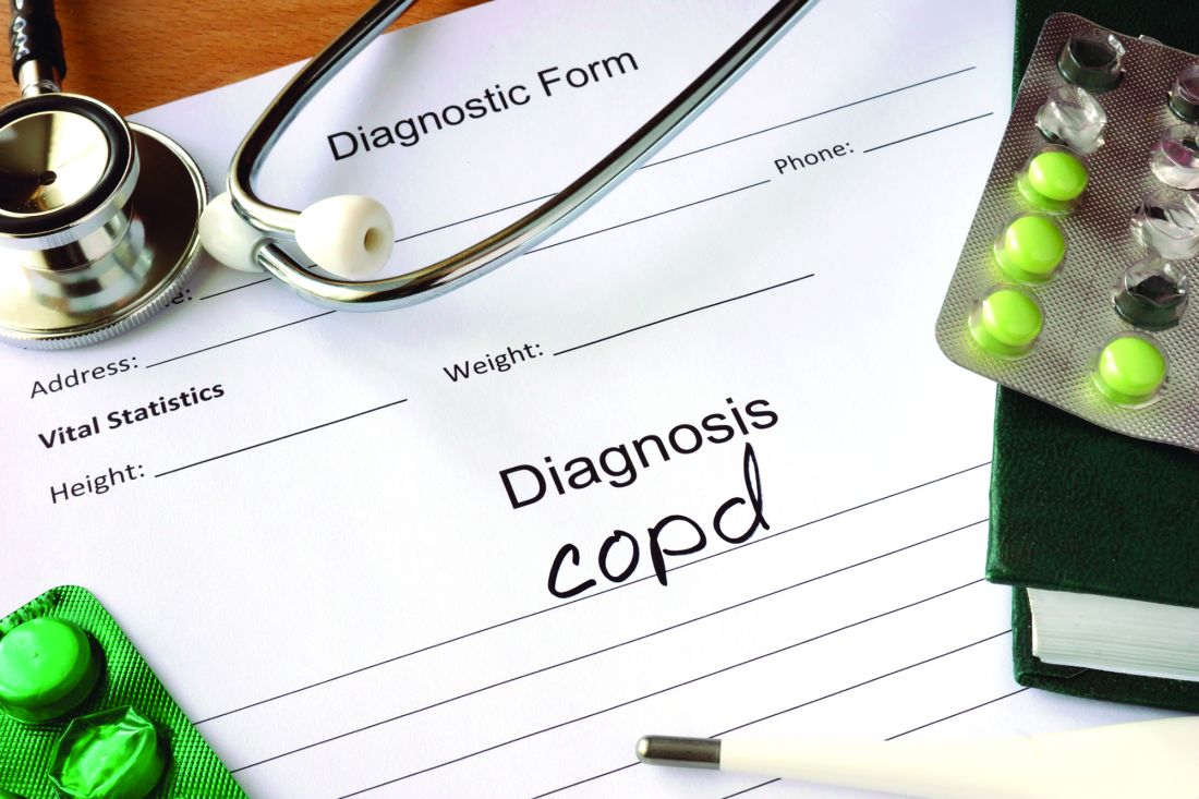
Patients with chronic obstructive pulmonary disease (COPD) who also suffer from depression are less likely to take their COPD maintenance medications, according to a review of Medicare claims by researchers at the University of Maryland, Baltimore. Researchers found that patients with newly diagnosed depression were about 7% less likely to have good adherence to their medications. For more on this research, see the article in CHEST Physician, available at http://www.mdedge.com/chestphysician/article/115659/depression/depression-drops-copd-medication-adherence.
Patients with chronic obstructive pulmonary disease (COPD) who also suffer from depression are less likely to take their COPD maintenance medications, according to a review of Medicare claims by researchers at the University of Maryland, Baltimore. Researchers found that patients with newly diagnosed depression were about 7% less likely to have good adherence to their medications. For more on this research, see the article in CHEST Physician, available at http://www.mdedge.com/chestphysician/article/115659/depression/depression-drops-copd-medication-adherence.
Patients with chronic obstructive pulmonary disease (COPD) who also suffer from depression are less likely to take their COPD maintenance medications, according to a review of Medicare claims by researchers at the University of Maryland, Baltimore. Researchers found that patients with newly diagnosed depression were about 7% less likely to have good adherence to their medications. For more on this research, see the article in CHEST Physician, available at http://www.mdedge.com/chestphysician/article/115659/depression/depression-drops-copd-medication-adherence.
Absolute humidity most important environmental factor in global influenza
Absolute humidity and temperature are the most important environmental drivers of global influenza, despite differences in outbreak patterns between tropical and temperate countries, according to a new analysis by U.S.-based researchers.
Using convergent cross-mapping and an empirical dynamic modeling approach on data collected by the World Health Organization, investigators led by George Sugihara, PhD, of the Scripps Institution of Oceanography at the University of California, San Diego, confirmed a hypothetical U-shaped relationship between influenza outbreaks and absolute humidity. At low latitudes in the tropics, absolute humidity has a positive effect, increasing the likelihood of influenza as humidity rises but at higher latitudes in temperate countries, absolute humidity has a negative effect, making influenza more likely when absolute humidity is low.
While absolute humidity was the most important factor in the likelihood of influenza outbreaks, the U-shaped relationship was dictated by average temperature. An average temperature below 70 °F had little effect on the negative relationship between absolute humidity and influenza at that range of temperatures, but if the temperature was between 75 °F and 85 °F, the effect was positive. Above 85 °F, aerosol transmission of influenza is blocked, the investigators noted.
“Augmented with further laboratory testing, these population-level results could help set the stage for public health initiatives such as placing humidifiers in schools and hospitals during cold, dry, temperate winter, and in the tropics, perhaps using dehumidifiers or air conditioners set above 75 °F to dry air in public buildings,” Dr. Sugihara and his colleagues wrote.
Find the full study in Proceedings of the National Academy of Sciences of the United States of America (doi: 10.1073/pnas.1607747113).
Pulmonary Function Tests
The video associated with this article is no longer available on this site. Please view all of our videos on the MDedge YouTube channel
The video associated with this article is no longer available on this site. Please view all of our videos on the MDedge YouTube channel
The video associated with this article is no longer available on this site. Please view all of our videos on the MDedge YouTube channel
2014-2015 influenza vaccine ineffective against predominant strain
The 2014-2015 influenza vaccines offered little protection against the predominant influenza A/H3N2 virus, but were effective against influenza B, according to the vaccine effectiveness estimates provided by the U.S. Flu Vaccine Effectiveness Network.
Preferential use of the live attenuated influenza vaccine (LAIV) among young children, a recommendation previously published by the Advisory Committee on Immunization Practices, was not supported.
During the 2014-2015 influenza season, a total of 9,710 patients seeking outpatient medical treatment for acute respiratory infection with cough were enrolled into the U.S. Flu Vaccine Effectiveness study, reported Richard Zimmerman, MD, of the University of Pittsburgh, and his colleagues (Clin Infect Dis. 2016 Oct 4. doi: 10.1093/cid/ciw635).
Of these, 9,311 participants had complete data, and 7,078 (76%) tested negative for influenza. A total of 1,840 participants tested positive for influenza A – 99% of these cases were strain A/H3N2 – and 395 participants tested positive for influenza B.
Of the 4,360 vaccinated participants with known vaccine type, 39.7% received standard dose trivalent, 1.6% received high dose trivalent, 46.8% received standard dose quadrivalent, and 11.9% received quadrivalent live-attenuated vaccines.
For influenza A and B combined, the overall adjusted vaccine effectiveness was 19% (95% Confidence Interval, 10-27%) against all medically attended influenza and was statistically significant in all age groups except 18-49 years.
Across all vaccine types, the vaccine effectiveness for the A/H3N2 strain was 6% (95% CI, -5-17%), estimates were similar across all age groups, and all vaccine types were similarly ineffective. These estimates were “consistent with a mismatch between the vaccine and circulating viruses,” the researchers noted.
Overall vaccine effectiveness for influenza B/Yamagata was 55% (95% CI, 43% to 65%) and was similarly significant in all age strata except 50-64 year olds. Trivalent vaccines were more effective at preventing influenza B and, of note, no cases of influenza B occurred among those who received a high dose trivalent flu vaccine.
The study was supported by the Centers for Disease Control and Prevention and the National Institutes of Health. Dr. Zimmerman and four other investigators reported receiving research funding from several pharmaceutical companies.
[email protected]
On Twitter @jessnicolecraig
Key clinical point:
Major finding: Across all vaccine types, the vaccine effectiveness for the A/H3N2 strain was 6%.
Data source: Retrospective analysis of 9,710 patients who sought outpatient medical treatment during the 2014-2015 influenza season.
Disclosures: The study was supported by the Centers for Disease Control and Prevention and the National Institutes of Health. Dr. Zimmerman and four other investigators reported receiving research funding from several pharmaceutical companies.
Pediatric OSA improved with oral montelukast
The majority of children with obstructive sleep apnea (OSA) who took oral montelukast showed reductions in their apnea-hypopnea index (AHI) scores, in a randomized, double-blind placebo-controlled study.
Typically, OSA in children is treated by adenotonsillectomy, according to Leila Kheirandish-Gozal, MD, director of clinical sleep research at the University of Chicago, and her colleagues. Prior to this study, only one randomized controlled trial had showed that children with mild OSA “responded favorably” to the leukotriene modifier montelukast (Pediatrics. 2012 Aug 31. doi: 10.1542/peds.2012-0310).
Twenty (71%) of the children who received montelukast had fewer AHI events per hour of total sleep time at the end of the study. The average number of such events for these patients was 4.2 plus or minus 2.8 after taking the drug, compared with 9.2 plus or minus 4.1 at the beginning of the study (P less than .0001). Only two (6.9%) of the patients who took the placebo had lower AHI scores at the end of the study, with the average AHI score for the placebo group having been 8.7 plus or minus 4.9 events per hour of total sleep time. At baseline, the average score for patients in the placebo group was 8.2 plus or minus 5.0 AHI events per hour of total sleep time at baseline.
Another improvement seen by patients who received the drug was a decrease in the number of 3% reductions in arterial oxygen saturation per hour of sleep. At the beginning of the study, these patients had 7.2 plus or minus 3.1 of these events; by the end of the study, the number of these events was down to 2.8 plus or minus 1.8 (P less than .001). No significant decrease in the number of these events was seen among patients in the placebo group.
In this study, “montelukast emerges as favorably reducing the severity of OSA short term in children 2-10 years of age. These findings add to the existing evidence supporting a therapeutic role for anti-inflammatory approaches in the management of this highly prevalent condition in children, and clearly justify future studies targeting the long-term benefits of these approaches in children with OSA,” the researchers wrote.
All patients participated in overnight sleep studies following a referral to one of two sleep clinics by their primary care pediatrician or pediatric otolaryngologist, at the beginning of the study. Children who had been diagnosed with symptomatic snoring and had an AHI score of greater than 2 events per hour of total sleep time, and for whom adenotonsillectomy was contemplated, were included in the study.
Central, obstructive, mixed apneic events were counted and hypopneas were assessed. OSA was defined “as the absence of airflow with continued chest wall and abdominal movement for a duration of at least two breaths,” the investigators said. Hypopneas were defined “as a decrease in oronasal flow greater than 50% on either the thermistor or nasal pressure transducer signal. with a corresponding decrease in arterial oxygen saturation greater than 3% or arousal,” Dr. Kheirandish-Gozal and her coauthors said.
Patients were excluded from the study for a variety of reasons, including having severe OSA requiring early surgical intervention.
Adverse events included headache in two children, one from the experimental group and one from the placebo group, and nausea in two subjects from the placebo group and in one from the montelukast group.
Merck provided tablets used in this study. Dr. Kheirandish-Gozal reported grants from Merck and the National Institutes of Health during the conduct of the study. David Gozal, MD, is supported by the Herbert T. Abelson Chair in Pediatrics at the University of Chicago.
Key clinical point:
Major finding: 71% of patients who took montelukast had a significant reduction in AHI events per hour of total sleep time (P less than .0001).
Data source: A prospective, randomized, double-blind placebo-controlled study of 57 children with obstructive sleep apnea.
Disclosures: Merck provided tablets used in this study. Dr. Kheirandish-Gozal reported grants from Merck and the National Institutes of Health during the conduct of the study. David Gozal, MD, is supported by the Herbert T. Abelson Chair in Pediatrics at the University of Chicago.
Oxygen therapy no advantage in stable COPD with moderate desaturation
Longterm supplemental oxygen had no benefit on multiple outcome measures in patients with stable chronic obstructive pulmonary disease (COPD) and resting or exercise-induced moderate desaturation, Robert Wise, MD, and his colleagues in The Long-Term Oxygen Treatment Trial (LOTT) Research Group reported.
Recommendations that supplemental oxygen be administered to patients with severe desaturation – an oxyhemoglobin saturation of less than 89% on pulse oximetry (SpO2) – date to two trials performed in the 1970s. Since that time, subsequent studies have been performed in patients with COPD and mild-to-moderate daytime hypoxemia, but the studies were underpowered to assess mortality and the impact of oxygen therapy on hospitalization, exercise performance, and quality of life were unclear.
Dr. Wise, professor of medicine and director of research, in the division of pulmonary and critical care medicine, Johns Hopkins University, Baltimore, and his fellow LOTT researchers examined whether longterm treatment with supplemental oxygen would extend life and avoid hospitalization among patients who had stable COPD with moderate resting desaturation – defined as an SpO2 of 89% to 93% – and patients who had stable COPD with moderate exercise-induced desaturation during the 6-minute walk test – defined as an SpO2 of at least 80% for at least 5 minutes and less than 90% for 10 seconds or more.
The 738 study participants, about 75% of whom were men, were randomly assigned at one of 42 centers either to receive (368) or not to receive (370) longterm supplemental oxygen. In the supplemental oxygen group, patients with resting desaturation were prescribed 24-hour oxygen, and those with desaturation only during exercise were prescribed oxygen during exercise and sleep (N Engl J Med. 2016;375:1617-27. DOI: 10.1056/NEJMoa1604344).
The groups were balanced for oxygen-desaturation type: 60 (16%) and 73 (20%) had oxygen desaturation only at rest, 171 (46%) and 148 (40%) had oxygen desaturation only upon exercise, and 139 (38%) and 147 (40%) had oxygen desaturation at rest and upon exercise. Patients were followed for 1 to 6 years.
Supplemental oxygen, regardless of prescription type or adherence, failed to benefit patients overall or any subgroup of patients with stable COPD and moderate desaturation. The results were similar for all groups based on measures of time to death or first hospitalization (hazard ratio, 0.94; 95% confidence interval [CI], 0.79 to 1.12; P = .52), hospitalization for a COPD-related hospitalizations (rate ratio, 0.99; 95% CI, 0.83 to 1.17), non–COPD-related hospitalizations (rate ratio, 1.03; 95% CI, 0.90 to 1.18), the rate of all hospitalizations (rate ratio, 1.01; 95% CI, 0.91 to 1.13), and the rate of all COPD exacerbations (rate ratio, 1.08; 95% CI, 0.98 to 1.19). Additionally, patients who did and did not receive oxygen treatment did not differ based on changes on measures of quality of life, depression, anxiety, or functional status.
Oxygen treatment also was not without risk. Among the 51 adverse events attributed to the use of supplemental oxygen were 23 reports of tripping over equipment, including two cases that necessitated hospitalization. There were five patients who reported six cases of fires or burns, including one who had to be hospitalized.
The researchers acknowledged that some patients may not have enrolled in the trial because they were too ill or felt that oxygen was beneficial. “Highly symptomatic patients who declined enrollment might have had a different response to oxygen than what we observed in the enrolled patients,” they noted.
Additionally, uniform devices weren’t used for oxygen delivery, so the amount of oxygen delivered may have varied, and the study did not evaluate the immediate effects of oxygen on symptoms or exercise performance. Nocturnal oxygen saturation was not measured, and “some patients with COPD and severe nocturnal desaturation might benefit from nocturnal oxygen supplementation,” they pointed out. Moreover, “patients’ self-reported adherence may have been an overestimate of their actual oxygen use,” they added, noting, however, that there was good agreement with use “as measured by means of serial meter readings on the concentrator.”
Based on the results, the authors concluded, “the consistency of the null findings strengthens the overall conclusion that long-term supplemental oxygen in patients with stable COPD and resting or exercise-induced moderate desaturation has no benefit with regard to the multiple outcomes measured.”
LOTT was funded by the National Heart, Lung, and Blood Institute and the Centers for Medicare and Medicaid Services. LOTT researchers reported relationships with a wide variety of drug companies.
[email protected]
On Twitter @maryjodales
This landmark study is the largest to date to provide quality evidence about clinically relevant outcomes of longterm oxygen therapy for COPD patients with moderate hypoxemia, a prescribing decision that is relatively costly and potentially places a burden on patients. For patients with moderate hypoxemia, longterm oxygen therapy consistently did not affect outcomes, and the results were not modified by the type of oxygen prescription, desaturation profile, oxygen use, sex, smoking status, or lung function.
Given the available current data, longterm oxygen therapy should be prescribed to prolong survival among patients with COPD who have more than 3 weeks of severe resting hypoxemia (PaO2 of no more than 55 mm Hg or SaO2 of less than 88%) while they are breathing ambient air. Oxygen therapy might still be appropriate in selected patients with moderate exertional hypoxemia and intractable breathlessness despite appropriate evidence-based treatment.
Ambient air or oxygen can be used to evaluate the potential benefit. Oxygen therapy can be discontinued if the patient perceives no benefit within a day or two. Selected patients who benefit should be prescribed oxygen, and I think that this treatment that should be covered by insurance payers. However, longterm oxygen therapy should not be routinely prescribed in patients with mild or moderate hypoxemia at rest or during exercise.
Magnus Ekström, MD, PhD , is with the Division of Respiratory Medicine and Allergology, Lund (Sweden) University, and the department of medicine, Blekinge Hospital, Karlskrona, Sweden. He had no relevant financial disclosures and made these remarks in an editorial that accompanied the published study ( N Engl J. Med. 2016;375: 1683-4 ).
Longterm supplemental oxygen had no benefit on multiple outcome measures in patients with stable chronic obstructive pulmonary disease (COPD) and resting or exercise-induced moderate desaturation, Robert Wise, MD, and his colleagues in The Long-Term Oxygen Treatment Trial (LOTT) Research Group reported.
Recommendations that supplemental oxygen be administered to patients with severe desaturation – an oxyhemoglobin saturation of less than 89% on pulse oximetry (SpO2) – date to two trials performed in the 1970s. Since that time, subsequent studies have been performed in patients with COPD and mild-to-moderate daytime hypoxemia, but the studies were underpowered to assess mortality and the impact of oxygen therapy on hospitalization, exercise performance, and quality of life were unclear.
Dr. Wise, professor of medicine and director of research, in the division of pulmonary and critical care medicine, Johns Hopkins University, Baltimore, and his fellow LOTT researchers examined whether longterm treatment with supplemental oxygen would extend life and avoid hospitalization among patients who had stable COPD with moderate resting desaturation – defined as an SpO2 of 89% to 93% – and patients who had stable COPD with moderate exercise-induced desaturation during the 6-minute walk test – defined as an SpO2 of at least 80% for at least 5 minutes and less than 90% for 10 seconds or more.
The 738 study participants, about 75% of whom were men, were randomly assigned at one of 42 centers either to receive (368) or not to receive (370) longterm supplemental oxygen. In the supplemental oxygen group, patients with resting desaturation were prescribed 24-hour oxygen, and those with desaturation only during exercise were prescribed oxygen during exercise and sleep (N Engl J Med. 2016;375:1617-27. DOI: 10.1056/NEJMoa1604344).
The groups were balanced for oxygen-desaturation type: 60 (16%) and 73 (20%) had oxygen desaturation only at rest, 171 (46%) and 148 (40%) had oxygen desaturation only upon exercise, and 139 (38%) and 147 (40%) had oxygen desaturation at rest and upon exercise. Patients were followed for 1 to 6 years.
Supplemental oxygen, regardless of prescription type or adherence, failed to benefit patients overall or any subgroup of patients with stable COPD and moderate desaturation. The results were similar for all groups based on measures of time to death or first hospitalization (hazard ratio, 0.94; 95% confidence interval [CI], 0.79 to 1.12; P = .52), hospitalization for a COPD-related hospitalizations (rate ratio, 0.99; 95% CI, 0.83 to 1.17), non–COPD-related hospitalizations (rate ratio, 1.03; 95% CI, 0.90 to 1.18), the rate of all hospitalizations (rate ratio, 1.01; 95% CI, 0.91 to 1.13), and the rate of all COPD exacerbations (rate ratio, 1.08; 95% CI, 0.98 to 1.19). Additionally, patients who did and did not receive oxygen treatment did not differ based on changes on measures of quality of life, depression, anxiety, or functional status.
Oxygen treatment also was not without risk. Among the 51 adverse events attributed to the use of supplemental oxygen were 23 reports of tripping over equipment, including two cases that necessitated hospitalization. There were five patients who reported six cases of fires or burns, including one who had to be hospitalized.
The researchers acknowledged that some patients may not have enrolled in the trial because they were too ill or felt that oxygen was beneficial. “Highly symptomatic patients who declined enrollment might have had a different response to oxygen than what we observed in the enrolled patients,” they noted.
Additionally, uniform devices weren’t used for oxygen delivery, so the amount of oxygen delivered may have varied, and the study did not evaluate the immediate effects of oxygen on symptoms or exercise performance. Nocturnal oxygen saturation was not measured, and “some patients with COPD and severe nocturnal desaturation might benefit from nocturnal oxygen supplementation,” they pointed out. Moreover, “patients’ self-reported adherence may have been an overestimate of their actual oxygen use,” they added, noting, however, that there was good agreement with use “as measured by means of serial meter readings on the concentrator.”
Based on the results, the authors concluded, “the consistency of the null findings strengthens the overall conclusion that long-term supplemental oxygen in patients with stable COPD and resting or exercise-induced moderate desaturation has no benefit with regard to the multiple outcomes measured.”
LOTT was funded by the National Heart, Lung, and Blood Institute and the Centers for Medicare and Medicaid Services. LOTT researchers reported relationships with a wide variety of drug companies.
[email protected]
On Twitter @maryjodales
FROM NEJM
Key clinical point:
Major finding: The results were similar for all groups based on measures of time to death or first hospitalization (hazard ratio, 0.94; 95% confidence interval [CI], 0.79 to 1.12; P = .52).
Data source: 738 study participants were randomly assigned to receive (368) or not to receive (370) longterm supplemental oxygen in The Long-Term Oxygen Treatment Trial (LOTT).
Disclosures: LOTT was funded by the National Heart, Lung, and Blood Institute and the Centers for Medicare and Medicaid Services. LOTT researchers reported relationships with a wide variety of drug companies.
Few non-ICU patients receive palliative care consults
LOS ANGELES – A significant percentage of patients who meet criteria for palliative care consultations do not receive a consult during their hospital stay, results from a single-center retrospective analysis showed.
“Physicians need to recognize the palliative care needs of patients with chronic illnesses other than malignancy before they get admitted to the ICU, especially when these patients are admitted repeatedly for the same problem [and] have a significant decline in functional status with a large symptom burden,” Mohleen Kang, MD, said in an interview in advance of the annual meeting of the American College of Chest Physicians. “There is a potential missed opportunity for these conversations to occur with the patients and their family prior to their decompensation and crisis.”
Twenty-nine percent (132) of the patients studied met an indication for a palliative care consult (PCC), with only 35 (27%) of such patients having received a PCC. Patients with metastatic cancer were significantly more likely to have received a PCC, compared with non-cancer patients (64% vs. 21%, respectively; P less than .001), while patients with New York Heart Association Class III or IV congestive heart failure were less likely to receive a PCC, compared with those who did not have congestive heart failure (5.6% vs. 29.8%; P = .014).
Criteria for PCC on admission include a life-limiting diagnosis and more than one admission in the past 3 months, decline in function, or complex care requirements. Criteria for PCC during hospitalization include life-limiting diagnosis and uncertainty about decisions, an ICU stay greater than 7 days, or lack of goals of care.
Dr. Kang, chief resident in the department of medicine at New Jersey Medical School, Newark, presented the results, which were of patients admitted to the department of medicine at University Hospital in Newark in 2015. Those admitted to the ICU within 24 hours of admission were excluded from the analysis, leaving 461 patient charts that were screened for PCC needs based on the consensus report from the Center to Advance Palliative Care.
The patients who met an indication for PCC had a mean age of 60 years and an average length of stay of 7 days. The percentages of these patients who were female, African American, and Hispanic were 45%, 40%, and 21%, respectively.
On multivariate analysis, patients who had a PCC within 72 hours of admission were eight times more likely to have a hospital length of stay less than 7 days (P = .019), while those who had a PCC within 48 hours of admission were 20 times more likely to have a hospital length of stay less than 7 days (P = .017). “So if we intervened early, we were able to decrease their length of stay to less than 7 days,” Dr. Kang said at the meeting.
She acknowledged certain limitations of the study, including its small sample size, retrospective design, and lack of follow-up. “This study also has a lot of confounding socioeconomic factors that do not make it applicable to every hospital across the country,” she said. “This is not a homogeneous patient population.”
The study’s principal investigator was Anne Sutherland, MD, medical intensive care unit director at University Hospital. Dr. Kang reported having no financial disclosures.
AT CHEST 2016
Key clinical point:
Major finding: Patients with metastatic cancer were significantly more likely to have received a PCC, compared with non-cancer patients (64% vs. 21%, respectively; P less than .001).
Data source: A retrospective study of 132 patients admitted to the department of medicine at University Hospital in Newark, N.J., in 2015.
Disclosures: Dr. Kang reported having no financial disclosures.
Fluid administration in sepsis did not increase need for dialysis
LOS ANGELES – Fluid administration of at least 1 L did not increase the incidence of acute respiratory or heart failure in severe sepsis, and actually seemed to decrease the need for dialysis in a review of 164 patients at Scott and White Memorial Hospital in Temple, Tex.
For every 1 mL of fluid administered per kilogram of body weight, the likelihood of dialysis decreased by 8.5% (odds ratio, 0.915; 95% confidence interval, 0.854-0.980; P = .0111), with no increase in heart or respiratory failure on univariate analysis. The 126 patients (77%) who received at least 1 L had a 68% reduction in the need for dialysis (OR, 0.32; CI, 0.117-0.890; P = .0288).
“The No. 1 reason we weren’t meeting benchmarks was fluid administration,” explained lead investigator Aruna Jahoor, MD, a pulmonary critical care and sleep medicine fellow at Texas Tech University Health Sciences Center.
Seventeen percent of patients received greater than or equal to 30 mL/kg of fluid resuscitation, while 28% received greater than or equal to 20 mL/kg of intravenous fluid resuscitation. It turned out that staff in the emergency department - where most of the patients were treated in the critical first 6 hours - were concerned about fluid overload and throwing patients into respiratory, heart, or renal failure, Dr. Jahoor said. The team didn’t find a difference in mortality when patients received 30 mL/kg - just over 2 L in a 70-kg patient - versus 20 mL/kg or 1 L. The patients’ in-hospital mortality rate and 28-day mortality rate were 27%, and 32%, respectively.
There also weren’t increased rates of heart failure, acute respiratory failure, or mechanical ventilation when patients received at least 1 L of fluid. “There were [also] lower rates of dialysis, which indicated that we weren’t overloading patients. Even when we looked at fluid as a continuous variable, we still didn’t see” complications, Dr. Jahoor said.
The findings should be reassuring to treating physicians. “When you have pushback against 30-mL/kg administration, you can say ‘well, at least let’s give a liter. You don’t have to worry as much about some of the complications you are citing,’ ” she said.
For very obese patients, “it can get a little uncomfortable to be given” enough fluid to meet the 30-mL/kg goal, “but you can give at least a liter” without having to worry too much, she said. The patients in the study were treated from 2010 to 2013; normal saline was the most common resuscitation fluid. The hospital has since added the 30-mL/kg fluid resuscitation to its sepsis admission orders, and compliance has increased significantly.
A multivariate analysis is in the works to control for confounders. “We will probably [still] see you are not having increased rates of congestive heart or respiratory failure, or needing dialysis,” Dr. Jahoor said. The protective effect against dialysis might drop out, “but I am hoping it doesn’t,” he said.
The investigators had no relevant financial disclosures.
AT CHEST 2016
Key clinical point:
Major finding: For every 1 mL/kg of fluid administered, the likelihood of dialysis decreased by 8.5% (OR, 0.915; 95% CI, 0.854-0.980; P = .0111), with no increase in heart or respiratory failure on univariate analysis.
Data source: A review of 164 septic patients.
Disclosures: The investigators had no relevant financial disclosures.
New mechanical ventilation guidelines unveiled
LOS ANGELES – Acutely hospitalized patients who have been on mechanical ventilation for more than 24 hours, are at high risk for extubation failure, and have passed a spontaneous breathing trial should be extubated to noninvasive ventilation.
The recommendation comes from new clinical practice guidelines from the American College of Chest Physicians and the American Thoracic Society. Moderate-quality evidence suggests that early extubation and a switch to noninvasive ventilation reduces ventilator-related and ICU-related complications, including infections and injury to the lungs and other organs. Extubation also cuts costs by reducing ICU stays.
At the annual meeting of the American College of Chest Physicians, one of the six project cochairs, Daniel R. Ouellette, MD, said that the guidelines were intended to address “new territory” from the evidence-based guidelines for weaning and discontinuing ventilator support that were published in 2001. That effort, chaired by Neil R. MacIntyre, MD, “was a landmark article that helped us learn about the steps that we needed to take to liberate patients from mechanical ventilation,” said Dr. Ouellette of the Henry Ford Hospital Department of Pulmonary and Critical Care Medicine, Detroit. “We hope that this guideline lives up to the importance of that one. We wanted to look over new information and give new recommendations about things that haven’t been addressed in the past.”
Six recommendations from the guideline panel include:
We suggest that the initial spontaneous breathing trial be conducted with inspiratory pressure augmentation rather than T-piece or continuous positive airway pressure. The committee wrote that conducting the initial spontaneous breathing trial with pressure augmentation was more likely to be successful, produced a higher rate of extubation success, and was associated with a trend towards lower intensive care unit mortality.
We suggest protocols attempting to minimize sedation. The committee found that sedation protocols reduced ICU length of stay. However, the protocols did not appear to decrease time on the ventilator or reduce short-term mortality. The authors could not recommend one protocol over another but said the burden of providing sedation by any of the protocols was “very low.”
We suggest protocolized rehabilitation directed toward early mobilization. The committee wrote that patients receiving the intervention spent less time on the ventilator and were more likely to be able to walk when they left the hospital. However, their mortality rate appeared unchanged. The authors noted the exercises created additional work for ICU staff that might have come at the expense of other care priorities.
We suggest managing patients with a ventilator liberation protocol. The committee said that patients managed by protocol spent on average 25 fewer hours on mechanical ventilation and were discharged from the ICU a day early. However, their mortality rate appeared unchanged.
We suggest performing a cuff leak test in patients who meet extubation criteria and are deemed at high risk for postextubation stridor. The committee suggested that the test should be used only in patients with a high risk of stridor (abnormal breathing caused by blockage of windpipe) after extubation. Although patients passing the test had lower stridor and reintubation rates, the authors wrote that a high percentage of patients who failed the test could be successfully extubated.
For patients who failed the cuff leak test but are otherwise ready for extubation, we suggest administering systemic steroids at least 4 hours before extubation. The committee said that clinical judgment should take priority over test results, and systemic steroids should be administered to these patients at least 4 hours before extubation. The authors added that the short duration of the steroid therapy was likely to improve success rates without resulting in adverse events.
In a prepared statement, Timothy Girard, MD, of the department of medicine at the University of Pittsburgh and a lead author of the guidelines said the committee hoped the guidelines would help reduce variations in practice that do not benefit patients. “We are not prescribing a specific approach to care for every patient every time,” he said. “But we are trying to summarize the available evidence in as clear and succinct a way as possible so that clinicians know how it applies to most patients.”
Dr. Ouellette disclosed that he has received a research grant from Cardeas Pharma for health care–associated pneumonia.
Daniel R. Ouellette, MD, FCCP, comments: Liberation from mechanical ventilation is one of the most important goals in taking care of critically ill patients receiving mechanical ventilation in the ICU. Patients who have a prolonged ventilator course are at risk for many complications and so physicians who work in the intensive care unit must work carefully to liberate patients from the ventilator at the earliest possible moment. That has to be done in a safe fashion so criteria to ensure that this can be done safely are important as well.
Patients often have medical illness that requires sedation, and it is often necessary to sedate patients so that they can tolerate being on mechanical ventilation; however, we know that oversedation can lead to failure to liberate patients from mechanical ventilation expeditiously. Therefore, one of our recommendations’ suggestions is to design protocols for sedation that focus on minimizing sedation so that patients can be extubated expeditiously.
All of the recommendations ultimately focused on a team approach to liberation from mechanical ventilation, because involvement of team members is always important. However, there are a couple of our recommendations that are particularly important in terms of their implications for the team approach and those include recommendations about using protocols to liberate patients from ventilators, in general, and also to use sedation protocols to minimize sedations.
We began to look at developing this topic, because we had initially published guidelines on [liberation from mechanical ventilation] in 2001. We knew that there was much new information that had emerged since the 2001 guidelines. For that reason we began to think about an update. With the initial inception of this project, we reached out to the American Thoracic Society so as to develop a collaborative effort since this was a topic that interested both societies. This collaboration was at all levels at CHEST and it involved not only the guidelines organization, but also the leadership of both societies and, of course, the panel that was ultimately constructed to address these issues was made up of members from both societies. The entire process [of developing the new guideline] took nearly 3 years.
When one develops a guideline, one makes an effort to make a guideline as comprehensive and globally applicable as possible. I think the practices in Europe are very similar to practices in North America in terms of mechanical ventilation. Several of our panelists are European and some of the important work that we reviewed came from centers in Europe. It’s my opinion that our guideline will be broadly applicable in both North America and Europe, but there may be regional or local differences. Nevertheless, we recognize in different regions in the world, there are different resource allocations for medical treatment, there are different cultural precepts, and there are other factors that implicate medical problems.
Certainly the European Respiratory Society and other European organizations developed guidelines on related topics ... one of the important caveats when CHEST decides to develop a guideline is that we are not reproducing the work that has been done elsewhere and so this guideline represents a project that fills a gap that previously had not been filled.
All guidelines that CHEST develops are living guidelines … it’s hard to envision exactly how often a guideline will be updated. We know that there will be certain areas of our guideline that will stand the test of time, but there will be other areas that will need to be updated, some sooner than others.
The original CHEST guideline on liberation from mechanical ventilation was a very important document that appeared in 2001 and changed the practice of medicine and the practice of managing patients on mechanical ventilation. Nevertheless, the guideline was somewhat limited in scope, because there was only so much information available. … Our goal in developing this guideline was to address some of practitioners’ questions that had emerged in the last decade by looking at newly available data.
[In formulating these guidelines], we purposely chose six new questions that were not directly related to any of the questions [that has been answered] in the previous guideline.
LOS ANGELES – Acutely hospitalized patients who have been on mechanical ventilation for more than 24 hours, are at high risk for extubation failure, and have passed a spontaneous breathing trial should be extubated to noninvasive ventilation.
The recommendation comes from new clinical practice guidelines from the American College of Chest Physicians and the American Thoracic Society. Moderate-quality evidence suggests that early extubation and a switch to noninvasive ventilation reduces ventilator-related and ICU-related complications, including infections and injury to the lungs and other organs. Extubation also cuts costs by reducing ICU stays.
At the annual meeting of the American College of Chest Physicians, one of the six project cochairs, Daniel R. Ouellette, MD, said that the guidelines were intended to address “new territory” from the evidence-based guidelines for weaning and discontinuing ventilator support that were published in 2001. That effort, chaired by Neil R. MacIntyre, MD, “was a landmark article that helped us learn about the steps that we needed to take to liberate patients from mechanical ventilation,” said Dr. Ouellette of the Henry Ford Hospital Department of Pulmonary and Critical Care Medicine, Detroit. “We hope that this guideline lives up to the importance of that one. We wanted to look over new information and give new recommendations about things that haven’t been addressed in the past.”
Six recommendations from the guideline panel include:
We suggest that the initial spontaneous breathing trial be conducted with inspiratory pressure augmentation rather than T-piece or continuous positive airway pressure. The committee wrote that conducting the initial spontaneous breathing trial with pressure augmentation was more likely to be successful, produced a higher rate of extubation success, and was associated with a trend towards lower intensive care unit mortality.
We suggest protocols attempting to minimize sedation. The committee found that sedation protocols reduced ICU length of stay. However, the protocols did not appear to decrease time on the ventilator or reduce short-term mortality. The authors could not recommend one protocol over another but said the burden of providing sedation by any of the protocols was “very low.”
We suggest protocolized rehabilitation directed toward early mobilization. The committee wrote that patients receiving the intervention spent less time on the ventilator and were more likely to be able to walk when they left the hospital. However, their mortality rate appeared unchanged. The authors noted the exercises created additional work for ICU staff that might have come at the expense of other care priorities.
We suggest managing patients with a ventilator liberation protocol. The committee said that patients managed by protocol spent on average 25 fewer hours on mechanical ventilation and were discharged from the ICU a day early. However, their mortality rate appeared unchanged.
We suggest performing a cuff leak test in patients who meet extubation criteria and are deemed at high risk for postextubation stridor. The committee suggested that the test should be used only in patients with a high risk of stridor (abnormal breathing caused by blockage of windpipe) after extubation. Although patients passing the test had lower stridor and reintubation rates, the authors wrote that a high percentage of patients who failed the test could be successfully extubated.
For patients who failed the cuff leak test but are otherwise ready for extubation, we suggest administering systemic steroids at least 4 hours before extubation. The committee said that clinical judgment should take priority over test results, and systemic steroids should be administered to these patients at least 4 hours before extubation. The authors added that the short duration of the steroid therapy was likely to improve success rates without resulting in adverse events.
In a prepared statement, Timothy Girard, MD, of the department of medicine at the University of Pittsburgh and a lead author of the guidelines said the committee hoped the guidelines would help reduce variations in practice that do not benefit patients. “We are not prescribing a specific approach to care for every patient every time,” he said. “But we are trying to summarize the available evidence in as clear and succinct a way as possible so that clinicians know how it applies to most patients.”
Dr. Ouellette disclosed that he has received a research grant from Cardeas Pharma for health care–associated pneumonia.
AT CHEST 2016
