User login
Bringing you the latest news, research and reviews, exclusive interviews, podcasts, quizzes, and more.
div[contains(@class, 'read-next-article')]
div[contains(@class, 'nav-primary')]
nav[contains(@class, 'nav-primary')]
section[contains(@class, 'footer-nav-section-wrapper')]
nav[contains(@class, 'nav-ce-stack nav-ce-stack__large-screen')]
header[@id='header']
div[contains(@class, 'header__large-screen')]
div[contains(@class, 'read-next-article')]
div[contains(@class, 'main-prefix')]
div[contains(@class, 'nav-primary')]
nav[contains(@class, 'nav-primary')]
section[contains(@class, 'footer-nav-section-wrapper')]
footer[@id='footer']
section[contains(@class, 'nav-hidden')]
div[contains(@class, 'ce-card-content')]
nav[contains(@class, 'nav-ce-stack')]
div[contains(@class, 'view-medstat-quiz-listing-panes')]
div[contains(@class, 'pane-article-sidebar-latest-news')]
Sexual assault–related visits to the ED are on the rise
Data from the Federal Bureau of Investigation show an increase in reported rapes and sexual assaults (SAs) since 2006, and studies of victims show an increased risk of conditions such as suicidal ideation, PTSD, depression, substance use, and chronic conditions, write Emily L. Vogt of the University of Michigan, Ann Arbor, and colleagues.
However, trends and disparities in ED use by adults seeking care following SA have not been explored, they said.
For a study that was published in JAMA Network Open, researchers reviewed data from the Nationwide Emergency Department Sample (NEDS), a large, nationally representative database managed by the Agency for Healthcare Research and Quality. The dataset consisted of 120 million to 143 million weighted ED visits reported annually from 2006 through 2016. The study population included adults aged 18-65 years who had made an ED visit that was recorded in the NEDS and that was coded as an SA. SA was defined using ICD-9 codes until the fourth quarter of 2015, at which time ICD-10 codes came into use.
Overall, the number of SA-related ED visits increased by 1,533.0% during the study period, from 3,607 in 2006 to 55,296 in 2019. The average annual percentage change was 23.0% (P < .001). The greatest increase occurred from 2015 to 2016, when annual visits increased from 17,709 to 47,732. This increase likely reflected the updated ICD-10 codes, in which there are categories for suspected adult rape, confirmed adult rape, and adult forced sexual exploitation, the researchers note.
Patients presenting to the ED after an SA were mainly women (91.5%). Individuals aged 18-25 years accounted for nearly half of the presentations. Individuals in the lowest and second-lowest income quartiles also were overrepresented.
Despite the increased presentation to EDs, admission rates for SA decreased, from 12.6% to 4.3%, the researchers note. Patients who were older and were insured through Medicaid were more likely to be admitted than persons of other demographic groups.
The researchers also found that increases in ED presentations outpaced increases in SA reports to law enforcement. They compared the ED trends with FBI-reported rapes/SAs from 2015 to 2019 and found increases of 7% and 22% during the times of ICD-9 and ICD-10 codes, respectively. However, in 2019, the number of SA survivors who sought ED care remained below the number who reported to law enforcement (55,296 vs. 139,815, as determined on the basis of revised SA definitions).
“Although the association between increased coding specificity and documentation of SA is still unclear, ICD-10 likely contributed to increased ED documentation of SA,” but the data show steady increases that are independent of the coding change, the researchers write.
The study findings were limited by several factors, including the potential for multiple representations of patients, coding errors associated with the NEDS database, and the reliance on voluntary reports in the NEDS and FBI datasets, the researchers note. The results were strengthened by the large, diverse sample size and by the inclusion of hospital admissions and crime data for comparison, they say.
“As few as 21% of survivors seek medical care after SA, meaning that the survivors captured in this study represent a fraction of total SA-related care need,” the researchers write. “Our finding that most SA ED visits are by young, female, and low-income survivors can inform policy changes to better support these individuals,” which could include the development of outpatient and longitudinal care settings to better serve these populations, they conclude.
Better understanding not only of the trends underlying SA reporting but also of the demographics of survivors who seek treatment and evaluation after SA is vital, said Robert Glatter, MD, in an interview.
“Being able to better understand how social and societal movements affect a patient’s comfort in reporting an SA is vital in tracking the numbers of people who seek care in the ED,” said Dr. Glatter, an emergency medicine physician at Lenox Hill Hospital at Northwell Health, New York, and also of Hofstra University, Hempstead, N.Y.
Dr. Glatter said he was not surprised by the significant increase in sexual assault presentations, especially in light of increased awareness and the influence of the #MeToo movement and other social justice movements over the past decade.
“While I believe that victims of sexual violence may now feel more empowered to report an assault, the volume of SA that go unreported remains a serious public health issue and concern” in the United States and globally, he emphasized.
A key message from the current study is that there is a need for investment in “compassionate and comprehensive care for all survivors of SA,” Dr. Glatter said. “This includes recognition of the extensive mental health consequences of SA that can lead to not only depression, PTSD, and anxiety but also to suicidal ideation and suicide. The longer-term medical effects become life altering, permeating families and future generations,” he emphasized.
“As a society, we must also place a strong emphasis on caring for all SA survivors, but particularly those who come from economically or socially disadvantaged backgrounds who are uninsured or underinsured,” Dr. Glatter said. Issues of race, gender identity, and sexual identity among SA survivors also must be taken into consideration, he added.
“We need to better understand how our health care system can provide more nuanced follow-up care and reporting for survivors in outpatient settings. … Making access easier, while ensuring confidentiality, will allow more survivors of SA to seek treatment and care,” he said. “We also need to understand how using forensic nurses in this capacity, and beyond the ED, can better serve minority and racially diverse communities” and to increase the recruitment and training of such specialized nurses to care for SA victims, Dr. Glatter noted.
The study was supported by internal funding from the University of Michigan and the department of obstetrics and gynecology. Corresponding author Erica C. Marsh, MD, has received personal fees from Myovant Sciences and Pfizer unrelated to the current study. Dr. Glatter has disclosed no relevant financial relationships.
A version of this article first appeared on Medscape.com.
FROM JAMA NETWORK OPEN
Post Roe, pregnant SCD patients facing “dire” risks
When maternal-fetal medicine specialist Andra James, MD, MPH, trained as a midwife decades ago, women with sickle cell disease (SCD) were urged to never get pregnant. If they did, termination was considered the best option.
“If they did carry a pregnancy, the baby would not survive to the point of viability,” Dr. James, emeritus professor of obstetrics and gynecology at Duke University, Durham, N.C., recalled in an interview.
The fates of women with SCD have transformed dramatically since those grim days. In general, this blood disorder no longer robs patients of decades of life, and many women with SCD bear healthy children. But their pregnancies are still considered high risk with significant potential for health crises and death. Now, there’s a new complication: The overturning of Roe v. Wade.
For example, women with SCD may be unable able to seek elective abortions in some states even if their pregnancies pose a danger to their lives. And abortion restrictions are imperiling access to a medication that’s used to treat miscarriages, which are more common in women with SCD.
“The situation with Dobbs is dire, and maternal health care is being compromised,” Johns Hopkins University pediatric hematologist Lydia Pecker, MD, who treats young people with SCD and studies its impact on pregnancy, said in an interview. “Women with sickle cell disease who are pregnant constitute an underserved and understudied population with special health care needs, and the Dobbs decision will only make providing their care even more difficult in many parts of the country.”
For her part, Dr. James described the risk to pregnant women with SCD this way: In the wake of the court ruling, “we increase the opportunity for them to lose their lives and for their babies to die.”
SCD’s impact on pregnancy
While physicians no longer advise women with SCD to avoid motherhood, pregnancy is still uniquely dangerous for them. “Most of them have babies and children who are thriving, but it’s not easy for them,” University of North Carolina at Chapel Hill hematologist and SCD specialist Jane Little, MD, said in an interview. And in some cases, she said, pregnancies “do not end well.”
For a 2022 report, Dr. Pecker and colleagues analyzed 2012-2018 data for 6,610 U.S. hospital admissions among women with SCD (87% of whom were Black). These women were more likely than were unaffected women to suffer severe maternal morbidity (odds ratio[OR], 4.63, 95% confidence interval [CI], 4.16-5.16, P < .001). Cerebrovascular event were especially more common in SCD (OR, 13.94, P < .001).
According to a 2019 report, pregnant women with SCD “are more likely to develop a host of complications, particularly hypertensive syndromes (such as preeclampsia), venous thromboembolism (VTE), preterm labor, and fetal loss. Newborns are more likely to have growth problems and prematurity.”
Although data are sparse, experts say it’s also clear that women with SCD face significantly higher risk of death in pregnancy compared to other women. In fact, the maternal mortality rate for females with SCD “is higher than for Black females without SCD, who already suffer from a higher mortality rate than White females during pregnancy and childbirth,” Andrea Roe, MD, MPH, assistant professor of obstetrics and gynecology at the Hospital of the University of Pennsylvania, Philadelphia, said in an interview.
Women with SCD also are more likely to have premature and stillborn births.
Some of the health challenges in pregnant women with SCD stem from the body’s inability to boost blood production in order to supply the placenta, said Dr. James, the Duke University emeritus professor. “Her bone marrow is already turning out red blood cells as fast as it can.”
In addition, she said, these women are more susceptible to infection, blood clots, and damage to the kidneys and lungs.
Still, in most cases of SCD in pregnancy, “we counsel a woman that we can get you safely through it,” Dr. James said. “But there is a subset of patients that will have organ damage from their sickle cell disease and should not become pregnant or stay pregnant if they become pregnant.”
Court ruling limits options in some states
The Dobbs ruling affects pregnant women with SCD in two ways: It allows states to restrict or ban abortion to greater extents than were possible over the last 50 years, and it has spawned further limitations on access to mifepristone, which is commonly used to treat early miscarriages.
In some cases, Dr. James said, abortions in this population are elective. “People with sickle cell disease are frequently in pain, they are frequently hospitalized. They may have suffered strokes or subclinical strokes or have some cognitive impairment, and they don’t have the mental and physical fortitude [to tolerate pregnancy and birth].”
In other cases, abortions are medically necessary to preserve the mother’s life. The American Society of Hematology highlighted the risks posed by SCD to maternal health in a June 24 statement that criticized the Dobbs ruling. “In some cases, denying women their right to terminate a pregnancy puts them at risk of serious illness or death,” wrote Jane N. Winter, MD, president of ASH and professor of medicine at Northwestern University, Chicago.
There do not appear to be any statistics about abortion rates among women with SCD in the United States or whether the rates are higher than in other groups.
As for miscarriages in SCD, an analysis of first pregnancies in California women with SCD from 1991 to 2016 found that about 16% were “incomplete,” mainly (59.3%) from miscarriage.
The Dobbs ruling allows states to further restrict the drug combination of mifepristone and misoprostol, which is used to trigger abortions and to treat early pregnancy loss. Access to mifepristone was already limited prior to the ruling due to tight regulation, and advocates say it’s now even harder to get.
What now? Physicians urge focus on contraception
As the ramifications of the Dobbs ruling sink in, SCD specialists are emphasizing the importance of providing gynecological and contraceptive care to help women with the condition avoid unwanted pregnancies. At the University of North Carolina, “we’re pretty aggressive about trying to give women the option to see a gynecologist to get the best care they can,” Dr. Little said. “We have a shared gynecology and sickle cell clinic because we really want women to be making the choice [to become pregnant] when they are ready because it’s a strain on their health and their lives.”
Dr. Pecker, the Johns Hopkins University pediatric hematologist, urged colleagues to partner with maternal-fetal medicine specialists so they can quickly get help for pregnant patients when needed. “That way they can get high-quality pregnancy care and help to end pregnancies that need to be ended.”
She recommended “highly effective” progesterone-based birth control as the best first-line contraceptive for women with SCD. And, she said, every woman of child-bearing age with SCD should be assessed annually for their intentions regarding pregnancy. As she put it, “there’s so much that we can do to reduce harms.”
Dr. Pecker disclosed financial relationships with the National Institutes of Health, American Society of Hematology, Doris Duke Charitable Foundation, the Mellon Foundation, Global Blood Therapeutics, and Novo Nordisk. Dr. Little disclosed financial relationships with Global Blood Therapeutics, Bluebird Bio, and Forma Therapeutics. Dr. Roe has no disclosures.
Despite Katie Couric’s advice, doctors say ultrasound breast exams may not be needed
When Katie Couric shared the news of her breast cancer diagnosis, the former co-host of NBC’s “Today” show said she considered this new health challenge to be a teachable moment to encourage people to get needed cancer screenings.
“Please get your annual mammogram,” she wrote on her website in September. “But just as importantly, please find out if you need additional screening.”
In the essay, Couric, 65, explained that because she tends to have dense breast tissue, she gets an ultrasound test in addition to a mammogram when screening for breast cancer. A breast ultrasound, sometimes called a sonogram, uses sound waves to take images of the breast tissue. It can sometimes identify malignancies that are hard to spot on a mammogram in women whose breasts are dense – that is, having a high proportion of fibrous tissue and glands vs. fatty tissue.
Ms. Couric, who famously underwent a colonoscopy on live television after her first husband died of colon cancer and who lost her sister to pancreatic cancer, has long pushed for cancer screening and better detection options.
“We don’t have evidence that auxiliary screening reduces breast cancer mortality or improves quality of life,” said Dr. Carol Mangione, a professor of medicine and public health at the University of California, Los Angeles, who chairs the U.S. Preventive Services Task Force, a group of medical experts who make recommendations for preventive services after weighing their benefits and harms.
Ms. Couric’s office did not respond to requests for comment.
In addition to an annual mammogram, some women with dense breasts get a breast ultrasound or MRI to help identify cancerous cells missed by the mammogram. Dense fibrous tissue appears white on a mammogram and makes it harder to see cancers, which also appear white. Fatty breast tissue, which appears dark on the mammogram, doesn’t obscure breast malignancies.
As digital breast tomosynthesis, or 3-D mammography, has become more widely available, a growing number of women are getting that screening test rather than the standard 2-D mammography. The 3-D mammography has been found to reduce the number of false-positive results and identify more cancers in some women with dense breasts, though the impact on mortality is unknown.
The task force gives an “I” rating to supplemental screening for women with dense breasts whose mammogram results don’t indicate a problem. That means the current evidence is “insufficient” to assess whether the benefits outweigh the harms of the extra screening. (The task force is updating its recommendation for breast cancer screening, including supplemental screening for women with dense breasts.)
One key harm that researchers are concerned about, besides the possible extra cost, is the chance of a false-positive result. Supplemental imaging in women who aren’t at high risk for breast cancer may identify potential trouble spots, which can lead to follow-up testing such as breast biopsies that are invasive and raise cancer fears for many patients. But research has found that very often these results turn out to be false alarms.
If 1,000 women with dense breasts get an ultrasound after a negative mammogram, the ultrasound will identify two to three cancers, studies show. But the extra imaging will also identify up to 117 potential problems that lead to recall visits and tests but are ultimately determined to be false positives.
“On the one hand, we want to do everything we can to improve detection,” said Dr. Sharon Mass, an ob.gyn. in Morristown, N.J., and the former chair of the American College of Obstetricians and Gynecologists’ New Jersey section. “But on the other hand, there are lots of costs and emotional distress” associated with false-positive results.
The professional group doesn’t recommend supplemental screening for women with dense breasts who don’t have any additional risk factors for cancer.
Many other professional groups take a similar position.
“We recommend having a conversation with a health care provider, and for patients to understand whether their breasts are dense,” Dr. Mass said. “But we do not recommend everyone get tested.”
In particular, for the roughly 8% of women who have extremely dense breasts, it’s worth having a conversation with a doctor about additional screening, said Dr. Mass.
Similarly, for women with dense breasts who have additional risk factors for breast cancer, such as a family history of the disease or a personal history of breast biopsies to check suspected cancers, supplemental screening may make sense, she said.
Dense breasts are relatively common. In the United States, an estimated 43% of women 40 and older have breasts that are considered dense or extremely dense. In addition to making it harder to interpret mammograms, women with dense breasts are up to twice as likely to develop breast cancer as women with average-density breasts, research shows.
Studies have shown that mammograms reduce breast cancer mortality. But even though it seems intuitive that more testing would improve someone’s odds of beating cancer, research hasn’t found that women are any less likely to die from breast cancer if they get a supplemental ultrasound or MRI after a negative mammogram result.
A few studies have found that women with dense or very dense breasts who got an ultrasound or an MRI in addition to a mammogram had fewer so-called interval cancers between regular screening mammograms. But it’s unclear whether those results have any effect on their risk of dying from breast cancer.
“Not every small abnormality is going to lead to something that needs treatment,” said Dr. Mangione.
Thirty-eight states and the District of Columbia have laws requiring that patients be notified about breast density after a mammogram, though some require only a general notice rather than mandate that individual women be informed about their own status. Some states require insurers to cover supplemental testing, but others do not.
In 2019, the FDA proposed that information about breast density be incorporated into the letters patients receive after a mammogram. That rule hasn’t yet been finalized, but the agency told lawmakers that it expects to issue the rule no later than early next year.
In a statement to KHN, FDA spokesperson Carly Kempler said, “The FDA is committed to improving mammography services for patients and working diligently to finalize the rule to amend the existing mammography regulations.”
The cost of additional testing is another factor to consider. Because the Preventive Services Task Force recommends women get regular screening mammograms, health plans are generally required to cover them without charging people anything out-of-pocket. That’s not the case with supplemental screening for women with dense breasts, which the task force does not recommend. Some states require insurance coverage of those tests, but those laws don’t apply to the many plans in which employers “self-fund” workers’ benefits rather than buy state-regulated insurance coverage.
Supplemental imaging can be pricey if your health plan doesn’t cover it. A screening ultrasound might cost $250 out-of-pocket while a breast MRI could cost $1,084, according to the Brem Foundation to Defeat Breast Cancer.
Rep. Rosa DeLauro (D-Conn.) recently tweeted that she is working on a bill with Ms. Couric that would cover MRIs and ultrasounds for women with dense breasts without any out-of-pocket costs.
Some doctors recommend other steps that may be more effective than extra screening for women with dense breasts who want to reduce their breast cancer risk.
“If you really want to help yourself, lose weight,” said Dr. Karla Kerlikowske, a professor of medicine and epidemiology/biostatistics at the University of California, San Francisco, who has worked with other researchers to develop calculators that help providers assess patients’ breast cancer risk. “Moderate your alcohol intake and avoid long-term hormone replacement. Those are things you can control.”
KHN (Kaiser Health News) is a national newsroom that produces in-depth journalism about health issues. Together with Policy Analysis and Polling, KHN is one of the three major operating programs at KFF (Kaiser Family Foundation). KFF is an endowed nonprofit organization providing information on health issues to the nation.
Findings may be practice changing for early breast cancer patients
Among high-risk early breast cancer patients, delivery of a radiation boost to the tumor bed during whole breast irradiation was just as safe and effective as delivering the boost sequentially after whole breast irradiation ended. The findings from the phase 3 clinical trial are a boon to patient convenience.
“They showed equivalent local control and importantly, a good cosmetic outcome over time,” said Kathleen Horst, MD, who served as a discussant during a presentation given by Frank Vicini, MD, FASTRO, GenesisCare, during the annual meeting of the American Society for Radiation Oncology.
“This is substantially more convenient. It’s cost effective both for the health care system and individual patients. Importantly, our patients come in for treatment every day and they’re taking time from work which means they have to arrange for childcare and transportation. So, this makes a big difference for these patients,” said Dr. Horst, who is a professor of radiation oncology at Stanford (Calif.) Medicine and director of well-being in the radiation department at Stanford Medicine.
“One of the things that was surprising is that I think all of us were thinking this might be a more toxic regimen, but as Dr. Vicini showed, it was equally effective over time with minimal toxicity and cosmesis was stable over time, which is important. Importantly, it included patient-reported outcomes, not just the physician-reported outcomes. Broadly, I think these findings are applicable for many patients, including all patients who are receiving whole breast radiotherapy with an added boost. I think over time this is going to improve the quality of life of our patients. It represents an innovative change that everyone is going to be excited to embrace,” Dr. Horst said.
Previous randomized controlled trials showed that an additional radiation dose to the tumor bed following lumpectomy and whole breast irradiation reduces the relative risk of local recurrence by about 35%. However, this increases treatment time for patients who have already endured an extensive regimen. For whole breast irradiation, hypofractionated radiation in 15-16 fractions over 3 weeks has comparable recurrence rates as a 5-week regimen, but the relevant trials did not examine the effect hypofractionation may have on a radiation boost to the tumor bed of high-risk patients. Because of this lack of evidence, current practice calls for the boost to remain sequential in five to eight fractions after completion of whole breast irradiation, which adds 1 week to a 1.5 week–long treatment.
The study included 2,262 patients who were randomized to receive a sequential boost or a concomitant boost. After a median follow-up of 7.4 years, there were 54 ipsilateral breast recurrence (IBR) events. The estimated 7-year risk of IBR was 2.2% in the sequential boost and 2.6% in the concurrent risk group (hazard ratio, 1.32; noninferiority test P = .039). Approximately 60% of patients received adjuvant chemotherapy.
Grade 3 or higher adverse events were similar with a frequency of 3.3% in the sequential group and 3.5% in the concurrent group (P = .79). The researchers used the Global Cosmetic Score (GCS) to assess outcomes from the perspective of both physicians and patients. 86% of physicians rated the outcome as excellent/good in the sequential group versus 82% in the concurrent group (P = .33).
“For high-risk early-stage breast cancer patients undergoing breast conservation, a concurrent boost with hypofractionated whole breast irradiation – compared to a sequential boost – results in noninferior local recurrence rates with no significant difference in toxicity, noninferior patient rated cosmesis, and no significant difference in physician rated cosmesis. The entire treatment was delivered in three weeks, even for high-risk patients. Just as critical, the use of target volume based radiation planning for [three-dimensional conformal or IMRT whole breast irradiation assessed by dose volume analysis is feasible, and resulted in very low toxicity in the treatment arms, regardless of the fractionation schedule, or the boost delivery,” Dr. Vincini said.
No conflicts of interest were disclosed for Dr. Horst or Dr. Vicini.
FROM ASTRO 2022
Commentary: Hypertension, morbidity in MTOP, and hypothyroidism risk in obstetric emergencies, November 2022
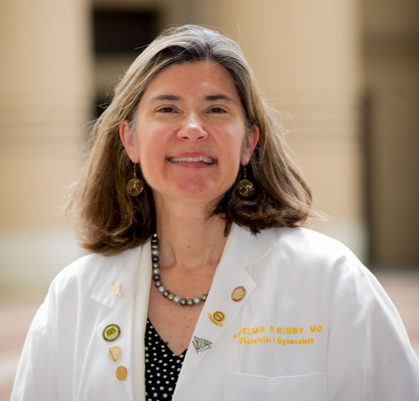
Richards and colleagues explored the effects of aspirin prophylaxis in women with chronic hypertension. They did not detect a lowered risk for preeclampsia but did note a significantly decreased risk for preterm birth in the aspirin group. This was a systematic review and meta-analysis of nine studies (including retrospective cohort and randomized controlled trials). The mixed quality of the source data did limit the meta-analysis. However, this finding suggests that further research is warranted, and we may have a new role for aspirin in helping to decrease preterm birth in women with chronic hypertension.
Cleary and colleagues investigated the use of 30 mg oral nifedipine ER given every 24 hours until delivery in patients with preE with SF. In this randomized, triple-blinded, placebo-controlled trial, 110 patients were randomly assigned to nifedipine treatment or placebo. The results suggest a role for this medication early in the treatment of preE with SF, as the treated patients were much less likely to require acute therapy for severe-range blood pressure. The researchers also noted a trend toward fewer cesarean deliveries (20.8% vs 34.7%) and lower neonatal intensive care unit admissions (29.1% vs 47.1%) in the nifedipine ER group. This favors the use of nifedipine ER in patients with preE with SF.
Stewart and colleagues examined the more common morbidities associated with MTOP after 20 weeks estimated gestational age using a 10-year retrospective cohort study involving 407 patients. They found that 99% of the women had a successful vaginal delivery; however, 25% had some morbidity. Additionally, 16% of the women needed manual removal of placental tissue, 11% had postpartum hemorrhage, and 1.3% experienced severe maternal morbidity (including amniotic fluid embolism), but no maternal deaths occurred. Increased surveillance for postpartum hemorrhage in this patient population should be considered.
Righini and colleagues provide reassurance regarding a commonly used test to rule out pulmonary embolism in pregnant women. They present ancillary data from a prospective management outcome study of 149 women who underwent CT pulmonary angiography testing in pregnancy. There have been concerns raised regarding potential harmful effects related to intravenous iodinated contrast agents on thyroid function. None of the infants born to these patients had evidence of neonatal hypothyroidism (assessed via thyroid-stimulating hormone measurements). This gives reassurance that the use of CT pulmonary angiography testing for pulmonary embolism in pregnancy is safe.
New consensus on managing nausea and vomiting in pregnancy
Although the nausea and vomiting associated with pregnancy are usually mild, they are more severe (hyperemesis gravidarum) in around one-third of women and require hospitalization in the first trimester for 0.3%-3.6% of these women in France. Given the diversity of practical care, a working group from the National College of French Gynecologists and Obstetricians (CNGOF) has established a consensus on the definition and management of these symptoms.
Definition and severity
Nausea and vomiting during pregnancy are defined as those emerging in the first trimester of pregnancy and for which there is no other etiology.
The severity of these symptoms should be assessed through weight loss from the beginning of the pregnancy, clinical signs of dehydration (thirst, skin turgor, hypotension, oliguria, etc.), and modified PUQE (Pregnancy-Unique Quantification of Emesis and Nausea) score. This is a three-question score rated from 0 to 15, available in the full text of the expert consensus.
Severe nausea and vomiting are not considered complicated when weight loss is < 5%, with no clinical signs of dehydration, and combined with a PUQE score of ≤ 6. In contrast, hyperemesis gravidarum is distinguished from nausea and vomiting during pregnancy by weight loss of ≥ 5 % or signs of dehydration or a PUQE score of ≥ 7.
Treating hyperemesis gravidarum
A laboratory workup should be ordered, along with an assay of blood potassium, blood sodium ions, and creatinine levels, as well as a complete dipstick urinalysis.
If symptoms persist or worsen despite well-managed treatment, an additional assessment is recommended, including an abdominal ultrasound and laboratory workup (white blood cell count, transaminases, lipase, CRP, TSH, T4).
Hospitalization is proposed when at least one of the following criteria is met: weight loss ≥ 10%, one or more clinical signs of dehydration, PUQE score of ≥ 13, hypokalemia < 3.0 mmol/L, hyponatremia < 120 mmol/L, elevated serum creatinine > 100 micromol/L, or resistance to treatment.
Which treatment?
Prenatal vitamins and iron supplementation should be stopped, as the latter seems to make symptoms worse. This step should be taken without stopping folic acid supplementation.
Women are free to adapt their diets and lifestyles according to their symptoms, since no such changes have been reported to improve symptoms.
If the PUQE score is < 6, even in the absence of proof of their benefit, ginger or B6 vitamin can be used. The same applies to acupressure, acupuncture, and electrical stimulation, which should only be considered in women without complications. Aromatherapy is not to be used, because of the potential risks associated with essential oils, and as no efficacy has been demonstrated.
It is proposed that drugs or combinations of drugs associated with the least severe and least frequent side effects should always be chosen in the absence of superiority of one class over another.
To prevent Gayet Wernicke encephalopathy, vitamin B1 must be administered systematically for hyperemesis gravidarum needing parenteral rehydration. Psychological support should be offered to all patients with hyperemesis gravidarum because of the negative impact of this pathology on mental well-being. Patients should be informed that there are patient associations involved in supporting these women and their families.
A version of this article first appeared on Medscape.com and was translated from Univadis France.
Menopause an independent risk factor for schizophrenia relapse
Investigators studied a cohort of close to 62,000 people with SSDs, stratifying individuals by sex and age, and found that starting between the ages of 45 and 50 years – when the menopausal transition is underway – women were more frequently hospitalized for psychosis, compared with men and women younger than 45 years.
In addition, the protective effect of antipsychotic medication was highest in women younger than 45 years and lowest in women aged 45 years or older, even at higher doses.

“Women with schizophrenia who are older than 45 are a vulnerable group for relapse, and higher doses of antipsychotics are not the answer,” lead author Iris Sommer, MD, PhD, professor, department of neuroscience, University Medical Center of Groningen, the Netherlands, told this news organization.
The study was published online in Schizophrenia Bulletin.
Vulnerable period
There is an association between estrogen levels and disease severity throughout the life stages of women with SSDs, with lower estrogen levels associated with psychosis, for example, during low estrogenic phases of the menstrual cycle, the investigators note.
“After menopause, estrogen levels remain low, which is associated with a deterioration in the clinical course; therefore, women with SSD have sex-specific psychiatric needs that differ according to their life stage,” they add.
“Estrogens inhibit an important liver enzyme (cytochrome P-450 [CYP1A2]), which leads to higher blood levels of several antipsychotics like olanzapine and clozapine,” said Dr. Sommer. In addition, estrogens make the stomach less acidic, “leading to easier resorption of medication.”
As a clinician, Dr. Sommer said that she has “often witnessed a worsening of symptoms [of psychosis] after menopause.” As a researcher, she “knew that estrogens can have ameliorating effects on brain health, especially in schizophrenia.”
She and her colleagues were motivated to research the issue because there is a “remarkable paucity” of quantitative data on a “vulnerable period that all women with schizophrenia will experience.”
Detailed, quantitative data
The researchers sought to provide “detailed, quantitative data on life-stage dependent clinical changes occurring in women with SSD, using an intra-individual design to prevent confounding.”
They drew on data from a nationwide, register-based cohort study of all hospitalized patients with SSD between 1972 and 2014 in Finland (n = 61,889), with follow-up from Jan. 1, 1996, to Dec. 31, 2017.
People were stratified according to age (younger than 45 years and 45 years or older), with the same person contributing person-time to both age groups. The cohort was also subdivided into 5-year age groups, starting at age 20 years and ending at age 69 years.
The primary outcome measure was relapse (that is, inpatient hospitalization because of psychosis).
The researchers focused specifically on monotherapies, excluding time periods when two or more antipsychotics were used concomitantly. They also looked at antipsychotic nonuse periods.
Antipsychotic monotherapies were categorized into defined daily doses per day (DDDs/d):
- less than 0.4
- 0.4 to 0.6
- 0.6 to 0.9
- 0.9 to less than 1.1
- 1.1 to less than 1.4
- 1.4 to less than 1.6
- 1.6 or more
The researchers restricted the main analyses to the four most frequently used oral antipsychotic monotherapies: clozapine, olanzapine, quetiapine, and risperidone.
The turning tide
The cohort consisted of more men than women (31,104 vs. 30,785, respectively), with a mean (standard deviation) age of 49.8 (16.6) years in women vs. 43.6 (14.8) in men.
Among both sexes, olanzapine was the most prescribed antipsychotic (roughly one-quarter of patients). In women, the next most common antipsychotic was risperidone, followed by quetiapine and clozapine, whereas in men, the second most common antipsychotic was clozapine, followed by risperidone and quetiapine.
When the researchers compared men and women younger than 45 years, there were “few consistent differences” in proportions hospitalized for psychosis.
Starting at age 45 years and continuing through the oldest age group (65-69 years), higher proportions of women were hospitalized for psychosis, compared with their male peers (all Ps < .00001).
Women 45 or older had significantly higher risk for relapse associated with standard dose use, compared with the other groups.
When the researchers compared men and women older and younger than 45 years, women younger than 45 years showed lower adjusted hazard ratios (aHRs) at doses between of 0.6-0.9 DDDs/d, whereas for doses over 1.1 DDDs/d, women aged 45 years or older showed “remarkably higher” aHRs, compared with women younger than 45 years and men aged 45 years or older, with a difference that increased with increasing dose.
In women, the efficacy of the antipsychotics was decreased at these DDDs/d.
“We ... showed that antipsychotic monotherapy is most effective in preventing relapse in women below 45, as compared to women above that age, and also as compared to men of all ages,” the authors summarize. But after age 45 years, “the tide seems to turn for women,” compared with younger women and with men of the same age group.
One of several study limitations was the use of age as an estimation of menopausal status, they note.
Don’t just raise the dose
Commenting on the research, Mary Seeman, MD, professor emerita, department of psychiatry, University of Toronto, noted the study corroborates her group’s findings regarding the effect of menopause on antipsychotic response.
“When the efficacy of previously effective antipsychotic doses wanes at menopause, raising the dose is not the treatment of choice because it increases the risk of weight gain, cardiovascular, and cerebrovascular events,” said Dr. Seeman, who was not involved with the current research.
“Changing to an antipsychotic that is less affected by estrogen loss may work better,” she continued, noting that amisulpride and aripiprazole “work well post menopause.”
Additional interventions may include changing to a depot or skin-patch antipsychotic that “obviates first-pass metabolism,” adding hormone replacement or a selective estrogen receptor modulator or including phytoestrogens (bioidenticals) in the diet.
The study yields research recommendations, including comparing the effectiveness of different antipsychotics in postmenopausal women with SSDs, recruiting pre- and postmenopausal women in trials of antipsychotic drugs, and stratifying by hormonal status when analyzing results of antipsychotic trials, Dr. Seeman said.
This work was supported by the Finnish Ministry of Social Affairs and Health through the developmental fund for Niuvanniemi Hospital and the Academy of Finland. The Dutch Medical Research Association supported Dr. Sommer. Dr. Sommer declares no relevant financial relationships. The other authors’ disclosures are listed on the original paper. Dr. Seeman declares no relevant financial relationships.
A version of this article first appeared on Medscape.com.
Investigators studied a cohort of close to 62,000 people with SSDs, stratifying individuals by sex and age, and found that starting between the ages of 45 and 50 years – when the menopausal transition is underway – women were more frequently hospitalized for psychosis, compared with men and women younger than 45 years.
In addition, the protective effect of antipsychotic medication was highest in women younger than 45 years and lowest in women aged 45 years or older, even at higher doses.
“Women with schizophrenia who are older than 45 are a vulnerable group for relapse, and higher doses of antipsychotics are not the answer,” lead author Iris Sommer, MD, PhD, professor, department of neuroscience, University Medical Center of Groningen, the Netherlands, told this news organization.
The study was published online in Schizophrenia Bulletin.
Vulnerable period
There is an association between estrogen levels and disease severity throughout the life stages of women with SSDs, with lower estrogen levels associated with psychosis, for example, during low estrogenic phases of the menstrual cycle, the investigators note.
“After menopause, estrogen levels remain low, which is associated with a deterioration in the clinical course; therefore, women with SSD have sex-specific psychiatric needs that differ according to their life stage,” they add.
“Estrogens inhibit an important liver enzyme (cytochrome P-450 [CYP1A2]), which leads to higher blood levels of several antipsychotics like olanzapine and clozapine,” said Dr. Sommer. In addition, estrogens make the stomach less acidic, “leading to easier resorption of medication.”
As a clinician, Dr. Sommer said that she has “often witnessed a worsening of symptoms [of psychosis] after menopause.” As a researcher, she “knew that estrogens can have ameliorating effects on brain health, especially in schizophrenia.”
She and her colleagues were motivated to research the issue because there is a “remarkable paucity” of quantitative data on a “vulnerable period that all women with schizophrenia will experience.”
Detailed, quantitative data
The researchers sought to provide “detailed, quantitative data on life-stage dependent clinical changes occurring in women with SSD, using an intra-individual design to prevent confounding.”
They drew on data from a nationwide, register-based cohort study of all hospitalized patients with SSD between 1972 and 2014 in Finland (n = 61,889), with follow-up from Jan. 1, 1996, to Dec. 31, 2017.
People were stratified according to age (younger than 45 years and 45 years or older), with the same person contributing person-time to both age groups. The cohort was also subdivided into 5-year age groups, starting at age 20 years and ending at age 69 years.
The primary outcome measure was relapse (that is, inpatient hospitalization because of psychosis).
The researchers focused specifically on monotherapies, excluding time periods when two or more antipsychotics were used concomitantly. They also looked at antipsychotic nonuse periods.
Antipsychotic monotherapies were categorized into defined daily doses per day (DDDs/d):
- less than 0.4
- 0.4 to 0.6
- 0.6 to 0.9
- 0.9 to less than 1.1
- 1.1 to less than 1.4
- 1.4 to less than 1.6
- 1.6 or more
The researchers restricted the main analyses to the four most frequently used oral antipsychotic monotherapies: clozapine, olanzapine, quetiapine, and risperidone.
The turning tide
The cohort consisted of more men than women (31,104 vs. 30,785, respectively), with a mean (standard deviation) age of 49.8 (16.6) years in women vs. 43.6 (14.8) in men.
Among both sexes, olanzapine was the most prescribed antipsychotic (roughly one-quarter of patients). In women, the next most common antipsychotic was risperidone, followed by quetiapine and clozapine, whereas in men, the second most common antipsychotic was clozapine, followed by risperidone and quetiapine.
When the researchers compared men and women younger than 45 years, there were “few consistent differences” in proportions hospitalized for psychosis.
Starting at age 45 years and continuing through the oldest age group (65-69 years), higher proportions of women were hospitalized for psychosis, compared with their male peers (all Ps < .00001).
Women 45 or older had significantly higher risk for relapse associated with standard dose use, compared with the other groups.
When the researchers compared men and women older and younger than 45 years, women younger than 45 years showed lower adjusted hazard ratios (aHRs) at doses between of 0.6-0.9 DDDs/d, whereas for doses over 1.1 DDDs/d, women aged 45 years or older showed “remarkably higher” aHRs, compared with women younger than 45 years and men aged 45 years or older, with a difference that increased with increasing dose.
In women, the efficacy of the antipsychotics was decreased at these DDDs/d.
“We ... showed that antipsychotic monotherapy is most effective in preventing relapse in women below 45, as compared to women above that age, and also as compared to men of all ages,” the authors summarize. But after age 45 years, “the tide seems to turn for women,” compared with younger women and with men of the same age group.
One of several study limitations was the use of age as an estimation of menopausal status, they note.
Don’t just raise the dose
Commenting on the research, Mary Seeman, MD, professor emerita, department of psychiatry, University of Toronto, noted the study corroborates her group’s findings regarding the effect of menopause on antipsychotic response.
“When the efficacy of previously effective antipsychotic doses wanes at menopause, raising the dose is not the treatment of choice because it increases the risk of weight gain, cardiovascular, and cerebrovascular events,” said Dr. Seeman, who was not involved with the current research.
“Changing to an antipsychotic that is less affected by estrogen loss may work better,” she continued, noting that amisulpride and aripiprazole “work well post menopause.”
Additional interventions may include changing to a depot or skin-patch antipsychotic that “obviates first-pass metabolism,” adding hormone replacement or a selective estrogen receptor modulator or including phytoestrogens (bioidenticals) in the diet.
The study yields research recommendations, including comparing the effectiveness of different antipsychotics in postmenopausal women with SSDs, recruiting pre- and postmenopausal women in trials of antipsychotic drugs, and stratifying by hormonal status when analyzing results of antipsychotic trials, Dr. Seeman said.
This work was supported by the Finnish Ministry of Social Affairs and Health through the developmental fund for Niuvanniemi Hospital and the Academy of Finland. The Dutch Medical Research Association supported Dr. Sommer. Dr. Sommer declares no relevant financial relationships. The other authors’ disclosures are listed on the original paper. Dr. Seeman declares no relevant financial relationships.
A version of this article first appeared on Medscape.com.
FROM SCHIZOPHRENIA BULLETIN
Study uncovers two molecular subgroups of cervical cancer
Scientists have discovered that cervical cancer can be divided into two distinct molecular subgroups – one far more aggressive than the other – offering hope of better understanding and treatment of the disease.
In the United Kingdom, there are over 3,000 new case of cervical cancer, with around 850 deaths each year. It is almost always caused by the human papillomavirus (HPV), and vaccination against this virus has successfully reduced the incidence of cervical cancer – in fact, the reduction has been by 87% among women in their 20s in England who were offered the vaccine when they were aged 12-13 years as part of the U.K. HPV vaccination program.
“Despite major steps forward in preventing cervical cancer, many women still die from the disease,” said Tim Fenton, MD, associate professor in cancer biology, School of Cancer Sciences Centre for Cancer Immunology, University of Southampton (England), and coauthor of the new study.
Two distinct subgroups
In the new study, published in Nature Communications, researchers described their breakthrough findings as a “major step forward” in understanding the disease, and said they provided a “tantalizing new clue” in determining the best treatments for individual patients.
For the observational study - part of the largest ‘omics’ study of its kind – researchers led by scientists at University College London and the University of Southampton began by applying a multiomics approach to identify combinations of molecular markers and characteristics associated with the biological processes involved in cervical cancer cells. The integrated multiomic analysis of 643 cervical squamous cell carcinomas (CSCC) – the most common histological variant of cervical cancer – represented patient populations from the United States, Europe, and sub-Saharan Africa.
To begin with they analysed and compared DNA, RNA, proteins, and metabolites in 236 CSCC cases in a publicly available U.S. database. They found that U.S. cancers fell into two distinct “omics” subgroups, which they named C1 and C2. After further investigation, the researchers identified that C1 tumors contained a much higher number of cytotoxic T cells. “The findings suggested that patients with C1 tumors would have a stronger immune response within the tumor micro-environment,” they said.
Weaker antitumor immune response
To determine if the two sub-types affect patients with cervical cancer in different ways, the team, which also included researchers from the University of Kent, the University of Cambridge, Oslo University Hospital, the University of Bergen (Norway), and the University of Innsbruck (Austria) derived molecular profiles and looked at clinical outcomes of a further 313 CSCC cases from Norway and Austria.
The researchers found that, just as in the US cohort, nearly a quarter of patients fell into the C2 subtype, and that again C1 tumors contained far more killer T cells than C2 tumors. “Importantly, the data also showed C2 was far more clinically aggressive with worse outcomes for patients,” the authors said.
Patients with C2 tumors were more than twice as likely (hazard ratio, 2.32) to die from their cervical cancer at any point during the follow-up period – up to 21 years – than those with C1 tumors. In terms of 5-year disease-specific survival, the rates were 79% survival for C1 and 66% survival for C2, the authors pointed out.
They highlighted that the difference in outcomes between patients with C1 and C2 tumors was very similar across the US and European cohorts.
Kerry Chester, PhD, professor of molecular medicine at UCL Cancer Institute, and coauthor, said: “Inclusion of patient cohorts in Norway and Austria, for which highly detailed clinical information was available to complement the molecular data, were key factors in the success of the study.”
Analyzing a further cohort of 94 Ugandan CSCC cases, the team found that C2 tumors were much more common than C1 tumors in patients who were also HIV-positive, “underlining the link to a weaker antitumor immune response” in this group.
Molecular subtyping offers better prognostic information
Cervical cancer can be caused by at least 12 different ‘high-risk’ HPV types, and there have been conflicting reports as to whether the HPV type present in a cervical cancer influences the prognosis for the patient. CSCCs can now also be categorized into two subtypes, C1 and C2, the authors explained, among which C1 tumors have a more favorable outcome.
“Although HPV16 is more likely to cause C1 tumors and HPV18 C2 tumors, HPV type is not an independent predictor of prognosis, suggesting it is the tumor type rather than the causative HPV type that is critical for the disease outcome,” they highlighted.
“Intriguingly, the C1/C2 grouping appeared to be more informative than the type of HPV present,” they added. “While certain HPV types were found more commonly in either C1 or C2 tumors, prognosis was linked to the group to which the tumor could be assigned, rather than the HPV type it contained.”
The reason that HPV16 and other alpha-9 HPV types have been associated with more favorable outcomes was possibly that these viruses are “more likely to cause C1-type tumors”, the authors suggested. Although larger numbers are needed for robust within-stage comparisons of C1 and C2 tumors, “we observe a clear trend in the survival rates between C1 and C2 by stage”, they said.
Taking molecular (C1/C2) subtyping into account may allow for more “accurate prognostication” than current staging and potentially different clinical management of patients with C1 versus C2 tumors, the authors said. This could include the identification of patients at risk of relapse who may require further adjuvant therapy after completion of up-front therapy.
New therapeutic targets
Dr. Fenton highlighted that the study findings suggested that determining whether a patient has a C1 or a C2 cervical cancer could help in planning their treatment, since it appeared to provide “additional prognostic information beyond that gained from clinical staging”. Given the differences in the antitumor immune response observed in C1 and C2 tumors, this classification might also be useful in predicting which patients are likely to benefit from emerging immunotherapy drugs, he said.
The study findings also found that CSCC can develop along “two trajectories” associated with differing clinical behavior that can be identified using defined gene expression or DNA methylation signatures, and this may guide “improved clinical management of cervical cancer patients”, they said.
“This collaborative multidisciplinary research is a major step forward in our understanding of cervical cancer,” said Dr. Chester. “Through careful molecular profiling and genetic analysis of cervical cancer tumors we have gained valuable new insight into the tumor microenvironment and factors potentially making the cancer less aggressive in some patients.”
The authors expressed hope that their study findings will stimulate functional studies of genes and their role in cervical cancer pathogenesis, potentially enabling identification of new therapeutic targets.
The study was funded by Debbie Fund (a UCL postgraduate research scholarship), Rosetrees Trust, Cancer Research UK, the Biotechnology and Biosciences Research Council, the Royal Society, and the Global Challenges Doctoral Centre at the University of Kent, MRC, PCUK, BBSRC, TUF, Orchid, and the UCLH BRC. The authors declared no competing interests.
A version of this article first appeared on Medscape UK.
FROM NATURE COMMUNICATIONS
Younger doctors call for more attention to patients with disabilities
As an undergraduate student at Northeastern University in Boston, Meghan Chin spent her summers working for a day program in Rhode Island. Her charges were adults with various forms of intellectual and developmental disabilities (IDD).
“I was very much a caretaker,” Ms. Chin, now 29, said. “It was everything from helping them get dressed in the morning to getting them to medical appointments.”
During one such visit Ms. Chin got a lesson about how health care looks from the viewpoint of someone with an IDD.
The patient was a woman in her 60s and she was having gastrointestinal issues; symptoms she could have articulated, if asked. “She was perfectly capable of telling a clinician where it hurt, how long she had experienced the problem, and what she had done or not done to alleviate it,” Ms. Chin said.
And of comprehending a response. But she was not given the opportunity.
“She would explain what was going on to the clinician,” Ms. Chin recalled. “And the clinician would turn to me and answer. It was this weird three-way conversation – as if she wasn’t even there in the room with us.”
Ms. Chin was incensed at the rude and disrespectful way the patient had been treated. But her charge didn’t seem upset or surprised. Just resigned. “Sadly, she had become used to this,” Ms. Chin said.
For the young aide, however, the experience was searing. “It didn’t seem right to me,” Ms. Chin said. “That’s why, when I went to medical school, I knew I wanted to do better for this population.”

Serendipity led her to Georgetown University, Washington, where she met Kim Bullock, MD, one of the country’s leading advocates for improved health care delivery to those with IDDs.
Dr. Bullock, an associate professor of family medicine, seeks to create better training and educational opportunities for medical students who will likely encounter patients with these disabilities in their practices.
When Dr. Bullock heard Ms. Chin’s story about the patient being ignored, she was not surprised.
“This is not an unusual or unique situation,” said Dr. Bullock, who is also director of Georgetown’s community health division and a faculty member of the university’s Center for Excellence for Developmental Disabilities. “In fact, it’s quite common and is part of what spurred my own interest in educating pre-med and medical students about effective communication techniques, particularly when addressing neurodiverse patients.”
More than 13% of Americans, or roughly 44 million people, have some form of disability, according to the National Institute on Disability at the University of New Hampshire, a figure that does not include those who are institutionalized. The Centers for Disease Control and Prevention estimates that 17% of children aged 3-17 years have a developmental disability.
Even so, many physicians feel ill-prepared to care for disabled patients. A survey of physicians, published in the journal Health Affairs, found that some lacked the resources and training to properly care for patients with disabilities, or that they struggled to coordinate care for such individuals. Some said they did not know which types of accessible equipment, like adjustable tables and chair scales, were needed or how to use them. And some said they actively try to avoid treating patients with disabilities.
Don’t assume
The first step at correcting the problem, Dr. Bullock said, is to not assume that all IDD patients are incapable of communicating. By talking not to the patient but to their caregiver or spouse or child, as the clinician did with Ms. Chin years ago, “we are taking away their agency, their autonomy to speak for and about themselves.”
Change involves altering physicians’ attitudes and assumptions toward this population, through education. But how?
“The medical school curriculum is tight as it is,” Dr. Bullock acknowledged. “There’s a lot of things students have to learn. People wonder: where we will add this?”
Her suggestion: Incorporate IDD all along the way, through programs or experiences that will enable medical students to see such patients “not as something separate, but as people that have special needs just as other populations have.”
Case in point: Operation House Call, a program in Massachusetts designed to support young health care professionals, by building “confidence, interest, and sensitivity” toward individuals with IDD.
Eight medical and allied health schools, including those at Harvard Medical School and Yale School of Nursing, participate in the program, the centerpiece of which is time spent by teams of medical students in the homes of families with neurodiverse members. “It’s transformational,” said Susan Feeney, DNP, NP-C, director of adult gerontology and family nurse practitioner programs at the graduate school of nursing at the University of Massachusetts, Worcester. “They spend a few hours at the homes of these families, have this interaction with them, and journal about their experiences.”
Dr. Feeney described as “transformational” the experience of the students after getting to know these families. “They all come back profoundly changed,” she told this news organization. “As a medical or health care professional, you meet people in an artificial environment of the clinic and hospital. Here, they become human, like you. It takes the stigma away.”
One area of medicine in which this is an exception is pediatrics, where interaction with children with IDD and their families is common – and close. “They’re going to be much more attuned to this,” Dr. Feeney said. “The problem is primary care or internal medicine. Once these children get into their mid and later 20s, and they need a practitioner to talk to about adult concerns.”
And with adulthood come other medical needs, as the physical demands of age fall no less heavily on individuals with IDDs than those without. For example: “Neurodiverse people get pregnant,” Dr. Bullock said. They also can get heart disease as they age; or require the care of a rheumatologist, a neurologist, an orthopedic surgeon, or any other medical specialty.
Generation gap
Fortunately, the next generation of physicians may be more open to this more inclusionary approach toward a widely misunderstood population.
Like Ms. Chin, Sarah Bdeir had experience with this population prior to beginning her training in medicine. She had volunteered at a school for people with IDD.
“It was one of the best experiences I’ve ever had,” Ms. Bdeir, now 23 and a first-year medical student at Wayne State University, Detroit, said. She found that the neurodiverse individuals she worked with had as many abilities as disabilities. “They are capable of learning, but they do it differently,” she said. “You have to adjust to the way they learn. And you have to step out of your own box.”
Ms. Bdeir also heard about Dr. Bullock’s work and is assisting her in a research project on how to better improve nutritional education for people with IDDs. And although she said it may take time for curriculum boards at medical schools to integrate this kind of training into their programs, she believes they will, in part because the rising cohort of medical students today have an eagerness to engage with and learn more about IDD patients.
As does Ms. Chin.
“When I talk to my peers about this, they’re very receptive,” Ms. Chin said. “They want to learn how to better support the IDD population. And they will learn. I believe in my generation of future doctors.”
A version of this article first appeared on Medscape.com.
As an undergraduate student at Northeastern University in Boston, Meghan Chin spent her summers working for a day program in Rhode Island. Her charges were adults with various forms of intellectual and developmental disabilities (IDD).
“I was very much a caretaker,” Ms. Chin, now 29, said. “It was everything from helping them get dressed in the morning to getting them to medical appointments.”
During one such visit Ms. Chin got a lesson about how health care looks from the viewpoint of someone with an IDD.
The patient was a woman in her 60s and she was having gastrointestinal issues; symptoms she could have articulated, if asked. “She was perfectly capable of telling a clinician where it hurt, how long she had experienced the problem, and what she had done or not done to alleviate it,” Ms. Chin said.
And of comprehending a response. But she was not given the opportunity.
“She would explain what was going on to the clinician,” Ms. Chin recalled. “And the clinician would turn to me and answer. It was this weird three-way conversation – as if she wasn’t even there in the room with us.”
Ms. Chin was incensed at the rude and disrespectful way the patient had been treated. But her charge didn’t seem upset or surprised. Just resigned. “Sadly, she had become used to this,” Ms. Chin said.
For the young aide, however, the experience was searing. “It didn’t seem right to me,” Ms. Chin said. “That’s why, when I went to medical school, I knew I wanted to do better for this population.”
Serendipity led her to Georgetown University, Washington, where she met Kim Bullock, MD, one of the country’s leading advocates for improved health care delivery to those with IDDs.
Dr. Bullock, an associate professor of family medicine, seeks to create better training and educational opportunities for medical students who will likely encounter patients with these disabilities in their practices.
When Dr. Bullock heard Ms. Chin’s story about the patient being ignored, she was not surprised.
“This is not an unusual or unique situation,” said Dr. Bullock, who is also director of Georgetown’s community health division and a faculty member of the university’s Center for Excellence for Developmental Disabilities. “In fact, it’s quite common and is part of what spurred my own interest in educating pre-med and medical students about effective communication techniques, particularly when addressing neurodiverse patients.”
More than 13% of Americans, or roughly 44 million people, have some form of disability, according to the National Institute on Disability at the University of New Hampshire, a figure that does not include those who are institutionalized. The Centers for Disease Control and Prevention estimates that 17% of children aged 3-17 years have a developmental disability.
Even so, many physicians feel ill-prepared to care for disabled patients. A survey of physicians, published in the journal Health Affairs, found that some lacked the resources and training to properly care for patients with disabilities, or that they struggled to coordinate care for such individuals. Some said they did not know which types of accessible equipment, like adjustable tables and chair scales, were needed or how to use them. And some said they actively try to avoid treating patients with disabilities.
Don’t assume
The first step at correcting the problem, Dr. Bullock said, is to not assume that all IDD patients are incapable of communicating. By talking not to the patient but to their caregiver or spouse or child, as the clinician did with Ms. Chin years ago, “we are taking away their agency, their autonomy to speak for and about themselves.”
Change involves altering physicians’ attitudes and assumptions toward this population, through education. But how?
“The medical school curriculum is tight as it is,” Dr. Bullock acknowledged. “There’s a lot of things students have to learn. People wonder: where we will add this?”
Her suggestion: Incorporate IDD all along the way, through programs or experiences that will enable medical students to see such patients “not as something separate, but as people that have special needs just as other populations have.”
Case in point: Operation House Call, a program in Massachusetts designed to support young health care professionals, by building “confidence, interest, and sensitivity” toward individuals with IDD.
Eight medical and allied health schools, including those at Harvard Medical School and Yale School of Nursing, participate in the program, the centerpiece of which is time spent by teams of medical students in the homes of families with neurodiverse members. “It’s transformational,” said Susan Feeney, DNP, NP-C, director of adult gerontology and family nurse practitioner programs at the graduate school of nursing at the University of Massachusetts, Worcester. “They spend a few hours at the homes of these families, have this interaction with them, and journal about their experiences.”
Dr. Feeney described as “transformational” the experience of the students after getting to know these families. “They all come back profoundly changed,” she told this news organization. “As a medical or health care professional, you meet people in an artificial environment of the clinic and hospital. Here, they become human, like you. It takes the stigma away.”
One area of medicine in which this is an exception is pediatrics, where interaction with children with IDD and their families is common – and close. “They’re going to be much more attuned to this,” Dr. Feeney said. “The problem is primary care or internal medicine. Once these children get into their mid and later 20s, and they need a practitioner to talk to about adult concerns.”
And with adulthood come other medical needs, as the physical demands of age fall no less heavily on individuals with IDDs than those without. For example: “Neurodiverse people get pregnant,” Dr. Bullock said. They also can get heart disease as they age; or require the care of a rheumatologist, a neurologist, an orthopedic surgeon, or any other medical specialty.
Generation gap
Fortunately, the next generation of physicians may be more open to this more inclusionary approach toward a widely misunderstood population.
Like Ms. Chin, Sarah Bdeir had experience with this population prior to beginning her training in medicine. She had volunteered at a school for people with IDD.
“It was one of the best experiences I’ve ever had,” Ms. Bdeir, now 23 and a first-year medical student at Wayne State University, Detroit, said. She found that the neurodiverse individuals she worked with had as many abilities as disabilities. “They are capable of learning, but they do it differently,” she said. “You have to adjust to the way they learn. And you have to step out of your own box.”
Ms. Bdeir also heard about Dr. Bullock’s work and is assisting her in a research project on how to better improve nutritional education for people with IDDs. And although she said it may take time for curriculum boards at medical schools to integrate this kind of training into their programs, she believes they will, in part because the rising cohort of medical students today have an eagerness to engage with and learn more about IDD patients.
As does Ms. Chin.
“When I talk to my peers about this, they’re very receptive,” Ms. Chin said. “They want to learn how to better support the IDD population. And they will learn. I believe in my generation of future doctors.”
A version of this article first appeared on Medscape.com.
Listen up: Birdsong may calm anxiety, paranoia
Investigators found that people who listened to recordings of birds singing experienced a significant reduction in anxiety and paranoia. In contrast, the researchers also found that recordings of traffic noises, including car engines, sirens, and construction, increased depressive states.
“The results suggest that it may be worthwhile to investigate the targeted use of natural sounds such as birdsong in a clinical setting – for example, in hospital waiting rooms or in psychiatric settings,” study investigator Emil Stobbe, MSc, a predoctoral fellow at the Max Planck Institute for Human Development, Berlin, said in an interview.
“If someone is seeking an easily accessible intervention to lower distress, listening to an audio clip of birds singing might be a great option,” he added.
The study was published online in Scientific Reports.
Nature’s calming effect
The aim of the research was “to investigate how the physical environment impact brain and mental health,” Mr. Stobbe said.
Mr. Stobbe said that there is significantly more research examining visual properties of the physical environment but that the auditory domain is not as well researched, although, he added, that the beneficial effects of interactions with nature are “well studied.”
He noted that anxiety and paranoia can be experienced by many individuals even though they may be unaware that they are experiencing these states.
“We wanted to investigate if the beneficial effects of nature can also exert their impact on these states. In theory, birds can be representational for natural and vital environment, which, in turn, transfer the positive effects of nature on birdsong listeners,” he said.
A previous study compared nature versus city soundscape conditions and showed that the nature soundscape improved participants’ cognitive performance but did not improve mood. The present study added diversity to the soundscapes and focused not only on cognition and general mood but also on state paranoia, “which can be measured in a change-sensitive manner” and “has been shown to increase in response to traffic noise.”
The researchers hypothesized that birdsong would have a greater beneficial effect on mood and paranoia and on cognitive performance compared with traffic noise. They also investigated whether greater versus lower diversity of bird species or noise sources within the soundscapes “would be a relevant factor modulating the effects.”
The researchers recruited participants (n = 295) from a crowdsourcing platform. Participants’ mean age was late 20s (standard deviations ranged from 6.30 to 7.72), with a greater proportion of male versus female participants.
To be included, participants were required to have no history of mental illness, hearing difficulties, substance/drug intake, or suicidal thoughts/tendencies.
The outcomes of interest (mood, paranoia, cognitive performance) were measured before and after soundscape exposure and each soundscape had a low- versus high-diversity version. This resulted in several analyses that compared two types of sounds (birdsongs vs. traffic noise) x two levels of diversity (low vs. high diversity) and two time points (pre- vs. post exposure).
The exposure to sounds lasted for 6 minutes, after which they were asked to report (on a 0-100 visual scale) how diverse/monotone, beautiful, and pleasant they perceived the soundscape to be.
Reduction in depressive symptoms
Participants were divided into four groups: low-diversity traffic noise soundscape (n = 83), high-diversity traffic noise soundscape (n = 60), low-diversity birdsong soundscape (n = 63), and high-diversity birdsong soundscape (n = 80)
In addition to listening to the sounds, participants completed questionnaires measuring mood (depression and anxiety) and paranoia as well as a test of digit span cognitive performance (both the forward and the backward versions).
The type, diversity, and type x diversity all revealed significant effect sizes (F[3, 276] = 78.6; P < .001; eta-squared = 0.461; F[3, 276] = 3.16; P = .025; eta-squared = 0.033; and F[3, 276] = 2.66; P = .028, respectively), “suggesting that all of these factors, as well as their interaction, had a significant impact on the perception of soundscapes (that is, ratings on monotony/diversity, beauty, and pleasantness).”
A post hoc examination showed that depressive symptoms significantly increased within the low- and high-diversity urban soundscapes but decreased significantly in the high-diversity birdsong soundscapes (T[1, 60] = –2.57; P = .012; d = –0.29).
For anxiety, the post hoc within-group analyses found no effects within low- and high-diversity traffic noise conditions (T[1, 82] = –1.37; P = .174; d = –0.15 and T[1, 68] = 0.49; P = .629; d = 0.06, respectively). By contrast, there were significant declines in both birdsong conditions (low diversity: T[1, 62] = –6.13; P < .001; d = –0.77; high diversity: T[1, 60] = –6.32; P < .001; d = –0.70).
Similarly, there were no changes in participants with paranoia when they listened to either low- or high-diversity traffic noises (T[1, 82] = –0.55; P = .583; d = –0.06 and T[1, 68] = 0.67; P = .507; d = 0.08, respectively). On the other hand, both birdsong conditions yielded reductions in paranoia (low diversity: T[1, 62] = –5.90; P < .001; d = –0.74; high diversity: T[1, 60] = –4.11; P < .001; d = –0.46).
None of the soundscapes had any effect on cognition.
“In theory, birds can be representational for natural and vital environments which, in turn, transfer the positive effects of nature on birdsong listeners,” said Mr. Stobbe.
“Taken together, the findings of the current study provide another facet of why interactions with nature can be beneficial for our mental health, and it is highly important to preserve nature,” he added.
Mr. Stobbe said that future research should focus on investigating mixed soundscapes including examining whether the presence of natural sounds in urban settings lower stressors such as traffic noise.
An understudied area
Commenting for this article, Ken Duckworth, MD, chief medical officer of the National Alliance on Mental Illness called the study “interesting but limited.”
Dr. Duckworth, who was not involved in the research said that the “benefits of nature are understudied” and agreed with the investigators that it is potentially important to study the use of birdsongs in psychiatric facilities. “Future studies could also correlate the role of birdsong with the mental health benefits/aspects of ‘being in nature,’ which has been found to have some effect.”
Open Access funding was enabled and organized by Projekt DEAL. The authors and Dr. Duckworth declared no competing interests.
A version of this article first appeared on Medscape.com.
FROM SCIENTIFIC REPORTS