User login
Dose-adjusted EPOCH and rituximab beneficial for selected older patients with high-risk DLBCL
Key clinical point: Sufficiently fit older patients with high-risk diffuse large B-cell lymphoma (DLBCL) achieve favorable outcomes with dose-adjusted etoposide, prednisone, vincristine, cyclophosphamide, doxorubicin, and rituximab (DA-EPOCH-R); patients with a poor performance status (PS) develop unacceptable toxicity and require less intensive therapy.
Major finding: At 3 years, the progression-free survival (PFS) and overall survival (OS) rates were 53% and 58%, respectively, and the treatment-related mortality (TRM) rate was 13%. The 3-year PFS (58% vs 32%; P < .001) and OS (64% vs 33%; P = .007) rates were significantly higher and TRM rates were significantly lower (6% vs 43%; P < .001) among patients with PS 0-2 vs 3-4.
Study details: This multicenter retrospective real-life study included 120 patients aged ≥60 years with newly diagnosed high-risk DLBCL treated with a median of 6 DA-EPOCH-R cycles per patient.
Disclosures: No information on the source of funding or conflicts of interest was provided.
Source: Mitrovic Z et al. Dose-adjusted EPOCH and rituximab (DA-EPOCH-R) in older patients with high-risk aggressive diffuse large B-cell lymphoma: A real-life multicenter study by the Croatian Cooperative Group for Hematologic diseases (KroHem). Eur J Haematol. 2023 (Mar 20). Doi: 10.1111/ejh.13957
Axi-cel a promising second-line treatment option for older patients with relapsed or refractory LBCL
Key clinical point: Axicabtagene ciloleucel (axi-cel) is an effective second-line curative-intent therapy with a manageable safety profile for patients aged ≥65 years with relapsed or refractory large B-cell lymphoma (LBCL).
Major finding: At a median follow-up of 24.3 months, the median event-free survival was significantly longer with axi-cel vs standard of care (SOC; 21.5 vs 2.5 months; hazard ratio 0.276; descriptive P < .0001). The grade ≥3 treatment-emergent adverse event rates were 94% and 82% with axi-cel and SOC, respectively.
Study details: Findings are from a preplanned analysis of 109 patients aged ≥65 years with relapsed or refractory LBCL from the ZUMA-7 trial who were randomly assigned to receive second-line axi-cel (n = 51) or SOC (n = 58; 2-3 cycles of chemoimmunotherapy followed by high-dose chemotherapy with autologous stem-cell transplantation).
Disclosures: This study was supported by Kite, a Gilead Company. Some authors reported ties with various organizations, including Kite and Gilead.
Source: Westin JR et al. Safety and efficacy of axicabtagene ciloleucel versus standard of care in patients 65 years of age or older with relapsed/refractory large B-cell lymphoma. Clin Cancer Res. 2023 (Mar 31). Doi: 10.1158/1078-0432.CCR-22-3136
Lenalidomide+rituximab+venetoclax a potential therapy option for untreated MCL
Key clinical point: The addition of venetoclax to lenalidomide plus rituximab therapy may provide an effective and safe combination for the treatment of unselected patients with untreated mantle cell lymphoma (MCL).
Major finding: All patients were escalated to the maximum tolerated dose of venetoclax (400 mg daily). The overall response and complete remission rates were 96% and 86%, respectively. After a median follow-up of 27.5 months, the median overall survival and progression-free survival were not reached. No dose-limiting toxicity event was observed.
Study details: This multicenter phase 1 study included 28 unselected adult patients with untreated MCL who received induction therapy with lenalidomide (daily on days 1-21 of each cycle), rituximab (weekly during cycle 1 until cycle 2 day 1), and venetoclax (escalated weekly up to 400 mg daily) for 6-12 cycles followed by maintenance therapy.
Disclosures: This study was funded by AbbVie and the University of Michigan Rogel Cancer Center. Some authors reported ties with various organizations, including AbbVie.
Source: Phillips TJ et al. Adding venetoclax to lenalidomide and rituximab is safe and effective in patients with untreated mantle cell lymphoma. Blood Adv. 2023 (Apr 4). Doi: 10.1182/bloodadvances.2023009992
Higher rates of hospitalization, blood disorders, and infections among patients with MCL
Key clinical point: Compared with matched control individuals, patients with mantle cell lymphoma (MCL) treated with or without high-dose chemotherapy with autologous stem cell transplantation (HD-ASCT) had higher hospitalization rates and relative risks for blood disorders and infections.
Major finding: Patients with MCL vs control individuals had a significantly increased incidence rate of outpatient (incidence rate ratio [IRR] 2.0; 95% CI 1.8-2.2) and inpatient (IRR 7.2; 95% CI 6.3-8.3) visits and relative risks for blood disorders (non-HD-ASCT: hazard ratio [HR] 9.84; 95% CI 6.91-14.00; HD-ASCT: HR 5.80; 95% CI 3.42-9.84) and infections (non-HD-ASCT: HR 4.66; 95% CI 3.62-5.99; HD-ASCT: HR 5.62; 95% CI 4.20-7.52).
Study details: Findings are from a population-based study including 620 adult patients with MCL who did (n = 247) or did not (n = 373) receive HD-ASCT and 6200 matched control individuals without MCL.
Disclosures: This study was supported by the Swedish Cancer Society. The authors reported ties with various organizations.
Source: Ekberg S et al. Late effects in patients with mantle cell lymphoma treated with or without autologous stem cell transplantation. Blood Adv. 2023;7(5):866-874 (Mar 14). Doi: 10.1182/bloodadvances.2022007241
Ibrutinib+BR a promising treatment option for newly diagnosed MCL ineligible for intensive therapy
Key clinical point: Compared with bortezomib, rituximab, cyclophosphamide, doxorubicin, and prednisone (VR-CAP) and rituximab, cyclophosphamide, doxorubicin, vincristine, and prednisone (R-CHOP), ibrutinib plus bendamustine and rituximab (Ibru+BR) prolongs progression-free survival (PFS) in patients with newly diagnosed mantle cell lymphoma (MCL) who are ineligible for intensive therapy.
Major finding: Ibru+BR significantly improved PFS compared with VR-CAP (hazard ratio [HR] 0.55; P = .03) and R-CHOP (HR 0.35; P < .001). Adverse event risks were not significantly different in the Ibru+BR, VR-CAP, R-CHOP, and BR treatment arms.
Study details: The data come from a network meta-analysis of 3 studies involving 1459 patients with newly diagnosed MCL who were ineligible for intensive therapy and received first-line Ibru+BR, VR-CAP, R-CHOP, or BR.
Disclosures: This study did not report the source of funding. The authors declared no conflicts of interest.
Source: Sheng Z and Wang L. Superiority of ibrutinib plus bendamustine and rituximab in newly diagnosed patients with mantle-cell lymphoma ineligible for intensive therapy: A network meta-analysis. Eur J Haematol. 2023 (Mar 14). Doi: 10.1111/ejh.13953
Real-world data support the continued use of second-line targeted therapies in CLL
Key clinical point: Compared with chemoimmunotherapy, second-line targeted therapies improved treatment-free survival (TFS) and tended to improve overall survival (OS) in patients with chronic lymphocytic leukemia (CLL), including those who were frail and had comorbidities.
Major finding: The 3-year TFS and estimated OS rates were higher in patients receiving targeted therapies (63%, 95% CI 50%-76%; and 79%, 95% CI 68%-91%, respectively) vs fludarabine, cyclophosphamide, and rituximab or bendamustine and rituximab (FCR/BR; 37%,; 95% CI 26%-48%; and 70%, 95% CI 60%-81%, respectively) or chlorambucil+/−CD20-antibody (CD20Clb/Clb; 22%, 95% CI 10%-33%; and 60%, 95% CI 47%-74%, respectively). The grade ≥3 adverse event rate was similar with targeted treatment and FCR/BR.
Study details: This retrospective population-based real-world study included 286 patients with CLL who relapsed or were refractory to first-line treatment and received second-line targeted treatment (ibrutinib+venetoclax, venetoclax, venetoclax+rituximab/obinutuzumab, idelalisib, or idelalisib+rituximab), FCR/BR, or CD20Clb/Clb.
Disclosures: This study was partly supported by grants from Rigshospitalets Foundation. Some authors reported ties with various sources.
Source: Vainer N et al. Real-world outcomes upon second-line treatment in patients with chronic lymphocytic leukaemia. Br J Haematol. 2023 (Mar 10). Doi: 10.1111/bjh.18715
Premenopausal women benefit from ovarian conservation with benign hysterectomies
Although bilateral salpingo-oophorectomy (BSO) with hysterectomy has been shown to reduce the risk for ovarian cancer in women at increased risk, current guidelines are touting ovarian conservation, especially in premenopausal women, wrote Mathilde Gottschau, MD, of the Danish Cancer Society Research Center, Copenhagen, and colleagues. However, post-hysterectomy outcomes in women with and without BSO have not been well examined.
In a study published in the Annals of Internal Medicine, the researchers reviewed data from a nationwide registry of women in Denmark aged 20 years and older who underwent benign hysterectomies with BSO (22,974 women) and without BSO (120,011 women) between 1977 and 2017. The women were divided into subgroups based on age; those younger than 45 years were defined as premenopausal, those aged 45-54 years were defined as perimenopausal, those aged 55-64 were defined as early postmenopausal, and those aged 65 and older were defined as late menopausal.
The primary outcomes were hospitalization for cardiovascular disease, cancer incidence, and all-cause mortality over a median follow-up period of 22 years.
For women younger than 45 years, the 10-year cumulative risk for all cancer was lower with BSO than without, but the risk of overall cardiovascular disease was higher with BSO, with higher levels of ischemic heart disease and stroke, compared with women without BSO. The 10-year cumulative mortality was higher with BSO than without (2.16% vs. 1.94%).
For women aged 45-54 years, the 10-year cumulative cancer risk was higher in those with BSO than those without BSO (risk difference, 0.73 percentage points) associated mainly with nonbreast cancer, and both 10-year and 20-year mortality were higher in those with BSO than those without.
For women aged 55-65 years, the 10-year cumulative cancer risk was higher in those with BSO than those without BSO. Cumulative overall mortality was higher at 10 years for those with BSO, but lower at 20 years.
For women aged 65 years and older, both 10-year and 20-year cumulative overall cancer risk was higher with BSO than without (RD, 2.54 and 4.57 percentage points, respectively). Cumulative mortality was higher in the BSO group at 10 years, but lower at 20 years.
The study findings were limited by several factors including the use of age to determine menopausal status and the lack of genetic predisposition data, and the focus only on a relatively homogeneous population that may not be generalizable to other populations, the researchers noted.
However, the results were strengthened by the use of a nationwide registry and the long-term follow-up period, they said. The current study indicates that the health risks outweigh the potential benefits of BSO with benign hysterectomy for premenopausal women and supports the current guidelines for ovarian conservation in these women with low lifetime ovarian cancer risk, they said. For postmenopausal women, the data support a cautious approach to BSO given the lack of a clear survival benefit and cancer excess, they concluded.
Delayed diagnosis of ovarian cancers favors BSO
“The question of removing ovaries at the time of benign hysterectomy to prevent ovarian cancer in low-risk women has been widely debated,” which has contributed to the variation in incidence rates of unilateral and bilateral oophorectomy over time, wrote Elizabeth Casiano Evans, MD, of the University of Texas, San Antonio, and Deslyn T.G. Hobson, MD, of Wayne State University, Detroit, in an accompanying editorial.
Ovarian cancer often goes undiagnosed until an advanced stage, and BSO can significantly reduce risk in women with BRCA1 and BRCA2 mutations, they noted.
For women without increased risk, those who are premenopausal may wish to preserve ovarian function, but women also may benefit from improvements in a range of menopause-related symptoms including vasomotor and urogenital symptoms, sexual dysfunction, and psychiatric and cognitive symptoms, they said.
“In addition, salpingectomy alone has a role in significantly reducing ovarian cancer incidence without compromising ovarian function because the fallopian tube has been found to be at the origin of many ovarian cancer cases,” they noted. In the current study, “the crude ovarian cancer risk was lower with BSO” across all age groups, the editorialists said.
The choice of whether to include BSO at the time of benign hysterectomy is complicated, with many factors to consider, the editorialists wrote, and the current study supports the need for informed, shared decision-making between clinicians and patients.
The study was supported by the Danish Cancer Society’s Scientific Committee and the Mermaid Project. The researchers had no financial conflicts to disclose. The editorial authors had no financial conflicts to disclose.
FROM THE ANNALS OF INTERNAL MEDICINE
Therapies for Advanced Parkinson's Disease
As Parkinson's disease progresses, dopamine and dopamine agonists that control tremor and stiffness can wear off too early or cause dyskinesia when they peak in the bloodstream.
In this ReCAP, Dr Michael Okun, professor and chair of neurology at the University of Florida, explains that adjustments to dose, timing, or delivery mode of these medications can smooth out some fluctuations. He reports that catechol-O-methyltransferase inhibitors, such as opicapone, tolcapone, and entacapone, can extend the effect of dopamine and the addition of amantadine or istradefylline can suppress dyskinesia.
He points out that for other patients, surgical options, such as deep brain stimulation or focused ultrasound, can alter how the brain controls disordered movement or implanted pumps can better regulate the delivery of medication either subcutaneously or directly into the gut.
--
Michael S. Okun, MD, Chair of Neurology, College of Medicine; Director Norman Fixel Institute, University of Florida, Gainesville, Florida
Michael S. Okun, MD, has disclosed no relevant financial relationships

Relapsed CLL: New approaches prolong survival
NEW YORK –
“In people who are genomically unstable, almost half will have their CLL progress on single agent BTK inhibitor therapy. These patients, I’d like to treat with combination of a Bruton’s tyrosine kinase inhibitor plus venetoclax,” said Richard R. Furman, MD, Morton Coleman MD Distinguished Professor of medicine at Weill Cornell Medicine/New York Presbyterian Hospital. He presented on treating relapsed CLL at the Great Debates and Updates Hematologic Malignancies Conference in New York, April 13-15.
The efficacy of venetoclax (VX) in treating high risk CLL patients was demonstrated in the 2019 CLL14 trial (NCT02242942) in which the main entry criteria were being treatment-naive and being considered “unfit” for treatment, meaning that a patient’s Cumulative Illness Rating Scale score was >6, or they presented with decreased kidney function. Study participants were treated with six cycles of either venetoclax-obinutuzumab (VO) or chlorambucil-obinutuzumab (CO); the 4-year PFS was 74% vs. 35.4% (P < .0001) respectively. After 12 cycles of treatment, the 74% of patients in the VO group achieved undetectable minimal residual disease rate (MRD) vs. 32% in the CO group.
Achievement of MRD did predict for outcome, but Dr. Furman concluded that while MRD is a powerful prognostic tool, it is not clinically useful given the inability of its results to guide therapy. He went on to emphasize that “of those patients who achieved MRD levels of 10-4 after 12 cycles of venetoclax, 50% had worsening and 50% had improvement of the residual disease, suggesting that we do not know the optimal duration of venetoclax treatment, as half of the patients still continue to derive benefit.”
Dr. Furman noted that ibrutinib (IB) and the second-generation drugs zanubrutinib (ZB) and acalabrutinib (AL) are all effective, but that the second-generation Bruton’s tyrosine kinase inhibitors enable patients to derive the benefit of remaining on treatment longer, because the rate of concerning cardiac complications is lower than with IB.
The ALPINE trial (NCT03734016) confirmed ZB’s significantly improved overall response rate at 78.3% vs. IB’s 62.5%, as well as a lower rate of atrial fibrillation (2.5%) vs. IB (10.1%).
Noninferiority of AL vs. IB in high-risk relapsed CLL patients was demonstrated in the ELEVATE-RR trial (NCT02477696) with both drugs having a median PFS of 38.4 months. With a median follow up of 41 months, AL demonstrated less atrial fibrillation/atrial flutter, compared with IB (9.4% vs. 16.0%) with no difference in grade > 3 infections. Furthermore, AL’s half-life being shorter than that of IB means that side effects are shorter lived.
“That’s the advantage of the second generation of Bruton’s tyrosine kinase inhibitors,” said Stefan Glück, MD, PhD, a hematologic oncologist and independent consultant who was formerly affiliated with the Sylvester Comprehensive Cancer Center and taught at the Miller School of Medicine at the University of Miami.
Dr. Glück added that “acalabrutinib and zanubrutinib have demonstrated strong efficacy and safety. When patients no longer respond to these drugs, the addition of VX is crucial. It has a completely different mechanism of action and can reverse resistance to Bruton’s tyrosine kinase inhibitors, allowing them to start working again.”
Dr. Furman also discussed proteolysis-targeting chimeras (PROTACs), one area that he expects to make a significant impact as a future therapy for CLL and many other malignancies. Despite the fact these agents are in phase I studies, he commented that “they appear, thus far, to have tremendous potential.”
Dr. Furman disclosed relationships with Abbvie, Acerta/AstraZeneca, Beigene, Jansen, TG Therapeutics, Genentech-Roche, Incyte Corporation, Loxo Oncology, MEI Pharma, Morphosys, Pharmacyclics, Sanofi/Genzyme, and X4 Pharmaceuticals. Dr. Glück reported no conflicts of interest.
AT 2023 GREAT DEBATES AND UPDATES HEMATOLOGIC MALIGNANCIES CONFERENCE
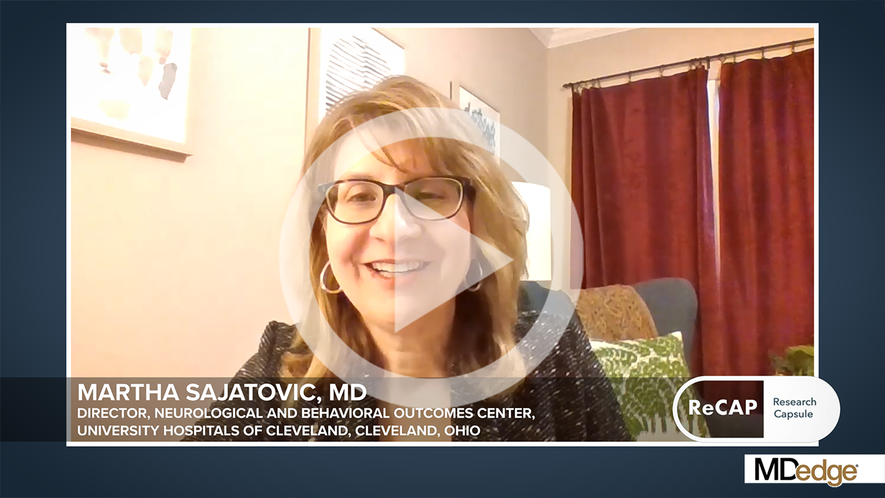
Long-Acting Injectables for the Treatment of Patients With Bipolar Disorder
Bipolar disorder is a multifaceted condition associated with an increased risk for hospitalization and suicide as well as high costs to society and family.
In this ReCAP, Dr Martha Sajatovic, of the University Hospitals of Cleveland, discusses evidence of the short- and long-term consequences of bipolar disorder, including progressive neurologic impact such as changes in brain structure.
She discusses two FDA-approved long-acting injectables for bipolar disorder and considerations for their use, including their potential for first-line maintenance treatment and benefits for medication adherence.
Finally, she considers challenges in the clinical use of the long-acting injectables, including insufficient caregiver involvement and lack of awareness of the drugs' availability.
--
Martha Sajatovic, MD, Director, Neurological and Behavioral Outcomes Center, University Hospitals of Cleveland, Cleveland, Ohio
Martha Sajatovic, MD, has disclosed the following relevant financial relationships:
Received research grant from: Otsuka; International Society for Bipolar Disorders; National Institutes of Health
Received income in an amount equal to or greater than $250 from: Otsuka; Janssen; Lundbeck; Teva; Neurelis
Received royalties from: Springer Press; Johns Hopkins University Press