User login
Set the Bar High
Newly elected board member Erin Stucky Fisher, MD, MHM, says that SHM should set its sights high when it comes to the quality of hospital care and the leadership role of hospitalists in improving the healthcare system.
A board-certified pediatrician and hospitalist at Rady Children's Hospital of San Diego, Dr. Fisher was elected via online voting in January to fill an at-large seat on SHM's 12-member board and starts her three-year term at SHM's annual meeting in Dallas in May.
"My vision of SHM is as the go-to medical society for clinical effectiveness in the hospital and for quality improvement and collaboration," Dr. Fisher says. "SHM represents systems, predominantly but not exclusively physician-based, and that's a huge strength for us. We have a different kind of opportunity because of what and who the society represents."
Key to SHM's leadership is promoting education, from workshops and conferences to supporting the Focused Practice in Hospital Medicine recertification process for hospitalists and, eventually, specific credentialing for pediatric hospitalists, she says.
"Working within this robust body, we need to leverage the great products and tools that are out there," she says. "How do we make sure we're offering more? How do we create that expectation from our members? We need to be hard on ourselves as leaders, and prove that we deserve to lead."
Dr. Fisher, a graduate of the University of California at San Francisco School of Medicine, completed her residency at UC San Diego and today serves there as professor of clinical pediatrics and director of a pediatric hospital medicine fellowship. She was awarded Masters in Hospital Medicine earlier this year and, in 2006, its recognition for outstanding service in hospital medicine.
Newly elected board member Erin Stucky Fisher, MD, MHM, says that SHM should set its sights high when it comes to the quality of hospital care and the leadership role of hospitalists in improving the healthcare system.
A board-certified pediatrician and hospitalist at Rady Children's Hospital of San Diego, Dr. Fisher was elected via online voting in January to fill an at-large seat on SHM's 12-member board and starts her three-year term at SHM's annual meeting in Dallas in May.
"My vision of SHM is as the go-to medical society for clinical effectiveness in the hospital and for quality improvement and collaboration," Dr. Fisher says. "SHM represents systems, predominantly but not exclusively physician-based, and that's a huge strength for us. We have a different kind of opportunity because of what and who the society represents."
Key to SHM's leadership is promoting education, from workshops and conferences to supporting the Focused Practice in Hospital Medicine recertification process for hospitalists and, eventually, specific credentialing for pediatric hospitalists, she says.
"Working within this robust body, we need to leverage the great products and tools that are out there," she says. "How do we make sure we're offering more? How do we create that expectation from our members? We need to be hard on ourselves as leaders, and prove that we deserve to lead."
Dr. Fisher, a graduate of the University of California at San Francisco School of Medicine, completed her residency at UC San Diego and today serves there as professor of clinical pediatrics and director of a pediatric hospital medicine fellowship. She was awarded Masters in Hospital Medicine earlier this year and, in 2006, its recognition for outstanding service in hospital medicine.
Newly elected board member Erin Stucky Fisher, MD, MHM, says that SHM should set its sights high when it comes to the quality of hospital care and the leadership role of hospitalists in improving the healthcare system.
A board-certified pediatrician and hospitalist at Rady Children's Hospital of San Diego, Dr. Fisher was elected via online voting in January to fill an at-large seat on SHM's 12-member board and starts her three-year term at SHM's annual meeting in Dallas in May.
"My vision of SHM is as the go-to medical society for clinical effectiveness in the hospital and for quality improvement and collaboration," Dr. Fisher says. "SHM represents systems, predominantly but not exclusively physician-based, and that's a huge strength for us. We have a different kind of opportunity because of what and who the society represents."
Key to SHM's leadership is promoting education, from workshops and conferences to supporting the Focused Practice in Hospital Medicine recertification process for hospitalists and, eventually, specific credentialing for pediatric hospitalists, she says.
"Working within this robust body, we need to leverage the great products and tools that are out there," she says. "How do we make sure we're offering more? How do we create that expectation from our members? We need to be hard on ourselves as leaders, and prove that we deserve to lead."
Dr. Fisher, a graduate of the University of California at San Francisco School of Medicine, completed her residency at UC San Diego and today serves there as professor of clinical pediatrics and director of a pediatric hospital medicine fellowship. She was awarded Masters in Hospital Medicine earlier this year and, in 2006, its recognition for outstanding service in hospital medicine.
AHA Targets Elevated Triglycerides; Sees Fructose Among Culprits
Triglyceride levels, which play a large role in both atherosclerotic risk and metabolic health, are highly responsive to decreases in dietary sugar intake and saturated and trans fat intake, along with increases in omega-3 acid intake and exercise, according to a scientific statement from the American Heart Association.
"What’s new is that we point out that triglycerides might be considered a marker for metabolic health," said Dr. Neil J. Stone of Northwestern University, Chicago, vice chair of the statement’s writing group, in an interview. "If you have a country where you’re seeing more obesity and more diabetes, it becomes important for people to start asking themselves ‘are there signs that I should be doing something different?’ and this is one," he said.

The scientific advisory, published online April 18 in the journal Circulation and citing some 528 sources, was not presented as a clinical guideline so much as a distillation of 30 years worth of evidence on the complex relationship among lifestyle factors, triglycerides, and cardiovascular and metabolic health (Circulation 2011 [doi:10.1161/ CIR.0b013e3182160726]).
However, the statement’s authors, led by Dr. Michael Miller, director of the Center for Preventive Cardiology at the University of Maryland, Baltimore, included a number of recommendations on diagnosing and treating hypertriglyceridemia, focusing on dietary and lifestyle changes.
The statement emphasizes the "increasingly crucial role" of triglycerides in the evaluation and management of cardiovascular disease, and the importance of diet – including consumption of sugars common in beverages – in contributing to unhealthy triglyceride levels.
Reductions of 50% or more are achievable without the use of medication – indeed medication is not a widely accepted strategy for reducing triglycerides except among people with extremely high values of greater than 500 mg/dL. "The subject of medication and triglycerides is still lacking crucial clinical trial evidence," Dr. Miller and colleagues wrote in their analysis, noting that certain medications, including hormonal treatments, can also contribute to elevated triglycerides.
About a third of American adults have elevated triglyceride levels, which are defined as fasting triglyceride of 150 mg/dL or higher. The authors recommended that optimal fasting triglyceride levels now be defined as 100 mg/dL – and that clinicians screen initially for nonfasting triglyceride, defining normal at below 200 mg/dL. People with higher nonfasting levels may then be further screened for fasting triglyceride.
The new dietary recommendations include restricting added dietary sugar to 5%-10% percent of calories consumed. In support of this, the authors cited a study of 6,113 U.S. adults showing that the lowest triglyceride levels were observed when added sugar represented less than 10% of total energy, and that higher triglyceride levels corresponded with added sugar accounting for a greater proportion of energy intake (JAMA 2010;303:1490-7).
The authors singled out fructose, a type of dietary sugar increasingly common in processed foods and soft drinks, as particularly problematic. Fructose in excess of 100 g/day, and possibly in excess of 50 g/day, has been associated with raised triglyceride levels. A typical can of cola or lemon-lime soda contains more than 20 grams of fructose, the authors noted.
Dr. Miller and his colleagues advocated weight loss of 5%-10% of body weight, which is associated with a 20% reduction in triglycerides, and regular aerobic exercise, to reduce triglyceride levels closer to optimal.
They also promoted increasing dietary fiber, keeping saturated fat below 7% of calories, eliminating trans fat from the diet, and increasing omega-3 polyunsaturated fatty acid consumption in the form of marine fish, though the authors said more research was needed to determine whether supplementing with fish-oil capsules provided equivalent benefits. Complete abstinence from alcohol was also recommended for people with very high triglycerides.
"Overall, optimization of nutrition-related practices can result in a marked triglyceride-lowering effect that ranges between 20% and 50%," they concluded.
Funding for the scientific advisory statement was provided by the American Heart Association. Dr. Miller declared no conflicts of interest affecting the drafting of the statement. However, Dr. Stone and the report’s third author, Dr. Christie Ballantyne of Baylor College of Medicine in Houston, disclosed support from pharmaceutical industry sources. Other coauthors and some reviewers disclosed additional support from pharmaceutical and agricultural firms.
Triglyceride levels, which play a large role in both atherosclerotic risk and metabolic health, are highly responsive to decreases in dietary sugar intake and saturated and trans fat intake, along with increases in omega-3 acid intake and exercise, according to a scientific statement from the American Heart Association.
"What’s new is that we point out that triglycerides might be considered a marker for metabolic health," said Dr. Neil J. Stone of Northwestern University, Chicago, vice chair of the statement’s writing group, in an interview. "If you have a country where you’re seeing more obesity and more diabetes, it becomes important for people to start asking themselves ‘are there signs that I should be doing something different?’ and this is one," he said.
The scientific advisory, published online April 18 in the journal Circulation and citing some 528 sources, was not presented as a clinical guideline so much as a distillation of 30 years worth of evidence on the complex relationship among lifestyle factors, triglycerides, and cardiovascular and metabolic health (Circulation 2011 [doi:10.1161/ CIR.0b013e3182160726]).
However, the statement’s authors, led by Dr. Michael Miller, director of the Center for Preventive Cardiology at the University of Maryland, Baltimore, included a number of recommendations on diagnosing and treating hypertriglyceridemia, focusing on dietary and lifestyle changes.
The statement emphasizes the "increasingly crucial role" of triglycerides in the evaluation and management of cardiovascular disease, and the importance of diet – including consumption of sugars common in beverages – in contributing to unhealthy triglyceride levels.
Reductions of 50% or more are achievable without the use of medication – indeed medication is not a widely accepted strategy for reducing triglycerides except among people with extremely high values of greater than 500 mg/dL. "The subject of medication and triglycerides is still lacking crucial clinical trial evidence," Dr. Miller and colleagues wrote in their analysis, noting that certain medications, including hormonal treatments, can also contribute to elevated triglycerides.
About a third of American adults have elevated triglyceride levels, which are defined as fasting triglyceride of 150 mg/dL or higher. The authors recommended that optimal fasting triglyceride levels now be defined as 100 mg/dL – and that clinicians screen initially for nonfasting triglyceride, defining normal at below 200 mg/dL. People with higher nonfasting levels may then be further screened for fasting triglyceride.
The new dietary recommendations include restricting added dietary sugar to 5%-10% percent of calories consumed. In support of this, the authors cited a study of 6,113 U.S. adults showing that the lowest triglyceride levels were observed when added sugar represented less than 10% of total energy, and that higher triglyceride levels corresponded with added sugar accounting for a greater proportion of energy intake (JAMA 2010;303:1490-7).
The authors singled out fructose, a type of dietary sugar increasingly common in processed foods and soft drinks, as particularly problematic. Fructose in excess of 100 g/day, and possibly in excess of 50 g/day, has been associated with raised triglyceride levels. A typical can of cola or lemon-lime soda contains more than 20 grams of fructose, the authors noted.
Dr. Miller and his colleagues advocated weight loss of 5%-10% of body weight, which is associated with a 20% reduction in triglycerides, and regular aerobic exercise, to reduce triglyceride levels closer to optimal.
They also promoted increasing dietary fiber, keeping saturated fat below 7% of calories, eliminating trans fat from the diet, and increasing omega-3 polyunsaturated fatty acid consumption in the form of marine fish, though the authors said more research was needed to determine whether supplementing with fish-oil capsules provided equivalent benefits. Complete abstinence from alcohol was also recommended for people with very high triglycerides.
"Overall, optimization of nutrition-related practices can result in a marked triglyceride-lowering effect that ranges between 20% and 50%," they concluded.
Funding for the scientific advisory statement was provided by the American Heart Association. Dr. Miller declared no conflicts of interest affecting the drafting of the statement. However, Dr. Stone and the report’s third author, Dr. Christie Ballantyne of Baylor College of Medicine in Houston, disclosed support from pharmaceutical industry sources. Other coauthors and some reviewers disclosed additional support from pharmaceutical and agricultural firms.
Triglyceride levels, which play a large role in both atherosclerotic risk and metabolic health, are highly responsive to decreases in dietary sugar intake and saturated and trans fat intake, along with increases in omega-3 acid intake and exercise, according to a scientific statement from the American Heart Association.
"What’s new is that we point out that triglycerides might be considered a marker for metabolic health," said Dr. Neil J. Stone of Northwestern University, Chicago, vice chair of the statement’s writing group, in an interview. "If you have a country where you’re seeing more obesity and more diabetes, it becomes important for people to start asking themselves ‘are there signs that I should be doing something different?’ and this is one," he said.
The scientific advisory, published online April 18 in the journal Circulation and citing some 528 sources, was not presented as a clinical guideline so much as a distillation of 30 years worth of evidence on the complex relationship among lifestyle factors, triglycerides, and cardiovascular and metabolic health (Circulation 2011 [doi:10.1161/ CIR.0b013e3182160726]).
However, the statement’s authors, led by Dr. Michael Miller, director of the Center for Preventive Cardiology at the University of Maryland, Baltimore, included a number of recommendations on diagnosing and treating hypertriglyceridemia, focusing on dietary and lifestyle changes.
The statement emphasizes the "increasingly crucial role" of triglycerides in the evaluation and management of cardiovascular disease, and the importance of diet – including consumption of sugars common in beverages – in contributing to unhealthy triglyceride levels.
Reductions of 50% or more are achievable without the use of medication – indeed medication is not a widely accepted strategy for reducing triglycerides except among people with extremely high values of greater than 500 mg/dL. "The subject of medication and triglycerides is still lacking crucial clinical trial evidence," Dr. Miller and colleagues wrote in their analysis, noting that certain medications, including hormonal treatments, can also contribute to elevated triglycerides.
About a third of American adults have elevated triglyceride levels, which are defined as fasting triglyceride of 150 mg/dL or higher. The authors recommended that optimal fasting triglyceride levels now be defined as 100 mg/dL – and that clinicians screen initially for nonfasting triglyceride, defining normal at below 200 mg/dL. People with higher nonfasting levels may then be further screened for fasting triglyceride.
The new dietary recommendations include restricting added dietary sugar to 5%-10% percent of calories consumed. In support of this, the authors cited a study of 6,113 U.S. adults showing that the lowest triglyceride levels were observed when added sugar represented less than 10% of total energy, and that higher triglyceride levels corresponded with added sugar accounting for a greater proportion of energy intake (JAMA 2010;303:1490-7).
The authors singled out fructose, a type of dietary sugar increasingly common in processed foods and soft drinks, as particularly problematic. Fructose in excess of 100 g/day, and possibly in excess of 50 g/day, has been associated with raised triglyceride levels. A typical can of cola or lemon-lime soda contains more than 20 grams of fructose, the authors noted.
Dr. Miller and his colleagues advocated weight loss of 5%-10% of body weight, which is associated with a 20% reduction in triglycerides, and regular aerobic exercise, to reduce triglyceride levels closer to optimal.
They also promoted increasing dietary fiber, keeping saturated fat below 7% of calories, eliminating trans fat from the diet, and increasing omega-3 polyunsaturated fatty acid consumption in the form of marine fish, though the authors said more research was needed to determine whether supplementing with fish-oil capsules provided equivalent benefits. Complete abstinence from alcohol was also recommended for people with very high triglycerides.
"Overall, optimization of nutrition-related practices can result in a marked triglyceride-lowering effect that ranges between 20% and 50%," they concluded.
Funding for the scientific advisory statement was provided by the American Heart Association. Dr. Miller declared no conflicts of interest affecting the drafting of the statement. However, Dr. Stone and the report’s third author, Dr. Christie Ballantyne of Baylor College of Medicine in Houston, disclosed support from pharmaceutical industry sources. Other coauthors and some reviewers disclosed additional support from pharmaceutical and agricultural firms.
FROM CIRCULATION
FDA explains dabigatran dose approval

Credit: Andre E.X. Brown
The medical community was surprised when the Food and Drug Administration (FDA) approved the higher dose of dabigatran and not the lower dose last October for patients with atrial fibrillation.
Now three reviewers from the FDA’s Center for Drug Evaluation and Research explain how the data gave them no other choice. Their account is published as a Perspective piece in the April 13 issue of The New England Journal of Medicine.
The FDA based its approval on the multicenter, active-control Randomized Evaluation of Long-term Anticoagulation Therapy (RE-LY) trial. It was designed to show the noninferiority of dabigatran, either 110 mg or 150 mg, compared to warfarin in reducing the risk of stroke and systemic embolism in patients with nonvalvular atrial fibrillation.
The study investigators found both dosages to be noninferior to warfarin. However, the 150-mg regimen was significantly superior to both warfarin (P=0.0001) and the 110-mg dose (P=0.004).
The FDA review team attempted to identify subsets within the RE-LY patient population that might benefit more from the lower dose. They focused on elderly patients, patients with renal function impairment, and those with previous bleeding episodes.
And in fact, the FDA reviewers said they were “unable to find any population for whom the availability of a lower dose would improve dabigatran’s benefit-risk profile.”
In the elderly, the rate of stroke or embolism was lower with the 150-mg dose, although the rate of major bleeding was higher. However, the reviewers assumed that “most people would agree...that the irreversible effects of strokes and emboli have greater clinical significance than nonfatal bleeding.”
In the subset of patients with moderate renal impairment, the rate of stroke or embolism with 150-mg was about half that of the lower-dose group and the rate of bleeding was similar. Patients with severe renal impairment were excluded from the trial.
And for those patients at higher risk for bleeding because of previous hemorrhage, they experienced similar rates of hemorrhage in both dabigatran groups and the warfarin group.
The review team commented, “[I]t appeared clear that most, if not all, patients should receive the higher dose.”
The review team pointed out that the noninferiority of the lower dose of dabigatran was less strong when it was compared with warfarin dosing that was well managed, which, they added, “is not always achieved.”
“Ultimately,” they said, the decision to approve only the higher dose “was based on our inability to identify any subgroup in which use of the lower dose would not represent a substantial disadvantage.”
The Perspective piece was written by B. Nhi Beasley, PharmD, Ellis F. Unger, MD, and Robert Temple, MD. 
Credit: Andre E.X. Brown
The medical community was surprised when the Food and Drug Administration (FDA) approved the higher dose of dabigatran and not the lower dose last October for patients with atrial fibrillation.
Now three reviewers from the FDA’s Center for Drug Evaluation and Research explain how the data gave them no other choice. Their account is published as a Perspective piece in the April 13 issue of The New England Journal of Medicine.
The FDA based its approval on the multicenter, active-control Randomized Evaluation of Long-term Anticoagulation Therapy (RE-LY) trial. It was designed to show the noninferiority of dabigatran, either 110 mg or 150 mg, compared to warfarin in reducing the risk of stroke and systemic embolism in patients with nonvalvular atrial fibrillation.
The study investigators found both dosages to be noninferior to warfarin. However, the 150-mg regimen was significantly superior to both warfarin (P=0.0001) and the 110-mg dose (P=0.004).
The FDA review team attempted to identify subsets within the RE-LY patient population that might benefit more from the lower dose. They focused on elderly patients, patients with renal function impairment, and those with previous bleeding episodes.
And in fact, the FDA reviewers said they were “unable to find any population for whom the availability of a lower dose would improve dabigatran’s benefit-risk profile.”
In the elderly, the rate of stroke or embolism was lower with the 150-mg dose, although the rate of major bleeding was higher. However, the reviewers assumed that “most people would agree...that the irreversible effects of strokes and emboli have greater clinical significance than nonfatal bleeding.”
In the subset of patients with moderate renal impairment, the rate of stroke or embolism with 150-mg was about half that of the lower-dose group and the rate of bleeding was similar. Patients with severe renal impairment were excluded from the trial.
And for those patients at higher risk for bleeding because of previous hemorrhage, they experienced similar rates of hemorrhage in both dabigatran groups and the warfarin group.
The review team commented, “[I]t appeared clear that most, if not all, patients should receive the higher dose.”
The review team pointed out that the noninferiority of the lower dose of dabigatran was less strong when it was compared with warfarin dosing that was well managed, which, they added, “is not always achieved.”
“Ultimately,” they said, the decision to approve only the higher dose “was based on our inability to identify any subgroup in which use of the lower dose would not represent a substantial disadvantage.”
The Perspective piece was written by B. Nhi Beasley, PharmD, Ellis F. Unger, MD, and Robert Temple, MD.
Credit: Andre E.X. Brown
The medical community was surprised when the Food and Drug Administration (FDA) approved the higher dose of dabigatran and not the lower dose last October for patients with atrial fibrillation.
Now three reviewers from the FDA’s Center for Drug Evaluation and Research explain how the data gave them no other choice. Their account is published as a Perspective piece in the April 13 issue of The New England Journal of Medicine.
The FDA based its approval on the multicenter, active-control Randomized Evaluation of Long-term Anticoagulation Therapy (RE-LY) trial. It was designed to show the noninferiority of dabigatran, either 110 mg or 150 mg, compared to warfarin in reducing the risk of stroke and systemic embolism in patients with nonvalvular atrial fibrillation.
The study investigators found both dosages to be noninferior to warfarin. However, the 150-mg regimen was significantly superior to both warfarin (P=0.0001) and the 110-mg dose (P=0.004).
The FDA review team attempted to identify subsets within the RE-LY patient population that might benefit more from the lower dose. They focused on elderly patients, patients with renal function impairment, and those with previous bleeding episodes.
And in fact, the FDA reviewers said they were “unable to find any population for whom the availability of a lower dose would improve dabigatran’s benefit-risk profile.”
In the elderly, the rate of stroke or embolism was lower with the 150-mg dose, although the rate of major bleeding was higher. However, the reviewers assumed that “most people would agree...that the irreversible effects of strokes and emboli have greater clinical significance than nonfatal bleeding.”
In the subset of patients with moderate renal impairment, the rate of stroke or embolism with 150-mg was about half that of the lower-dose group and the rate of bleeding was similar. Patients with severe renal impairment were excluded from the trial.
And for those patients at higher risk for bleeding because of previous hemorrhage, they experienced similar rates of hemorrhage in both dabigatran groups and the warfarin group.
The review team commented, “[I]t appeared clear that most, if not all, patients should receive the higher dose.”
The review team pointed out that the noninferiority of the lower dose of dabigatran was less strong when it was compared with warfarin dosing that was well managed, which, they added, “is not always achieved.”
“Ultimately,” they said, the decision to approve only the higher dose “was based on our inability to identify any subgroup in which use of the lower dose would not represent a substantial disadvantage.”
The Perspective piece was written by B. Nhi Beasley, PharmD, Ellis F. Unger, MD, and Robert Temple, MD.
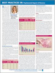
BEST PRACTICES IN: Psychosocial Impact of Rosacea
A supplement Family Practice News®. This supplement was sponsored by Galderma Laboratories, L.P.
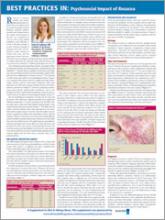
To view the supplement, click the image above.
Topics
- NRS Digital Perception Survey
- Presentation And Diagnosis
- Treatment Strategies
Faculty/Faculty Disclosure
Debra B. Luftman, MD
Coauthor of The Beauty Prescription:
The Complete Formula for Looking and Feeling Beautiful
Calabasas, California
Dr Luftman has received funding for clinical grants from and is a consultant for Galderma Laboratories, L.P.
Copyright © 2011 Elsevier Inc.
A supplement Family Practice News®. This supplement was sponsored by Galderma Laboratories, L.P.
To view the supplement, click the image above.
Topics
- NRS Digital Perception Survey
- Presentation And Diagnosis
- Treatment Strategies
Faculty/Faculty Disclosure
Debra B. Luftman, MD
Coauthor of The Beauty Prescription:
The Complete Formula for Looking and Feeling Beautiful
Calabasas, California
Dr Luftman has received funding for clinical grants from and is a consultant for Galderma Laboratories, L.P.
Copyright © 2011 Elsevier Inc.
A supplement Family Practice News®. This supplement was sponsored by Galderma Laboratories, L.P.
To view the supplement, click the image above.
Topics
- NRS Digital Perception Survey
- Presentation And Diagnosis
- Treatment Strategies
Faculty/Faculty Disclosure
Debra B. Luftman, MD
Coauthor of The Beauty Prescription:
The Complete Formula for Looking and Feeling Beautiful
Calabasas, California
Dr Luftman has received funding for clinical grants from and is a consultant for Galderma Laboratories, L.P.
Copyright © 2011 Elsevier Inc.
Rare Lymphoma Reports Continue in Young Patients on TNF Blockers
Cases of a rare, aggressive, and usually fatal lymphoma continue to be reported in people being treated with tumor necrosis factor blockers, azathioprine, and/or mercaptopurine, the Food and Drug Administration announced in an April 14 statement.
The reports of the lymphoma, hepatosplenic T-cell lymphoma (HSTCL), have primarily involved adolescents and young adults being treated with these agents for Crohn’s disease or ulcerative colitis. One patient, however, was being treated for psoriasis, and two others for rheumatoid arthritis.
Most patients were on a combination of treatments that are known to suppress the immune system, but there have been cases in patients taking azathioprine or mercaptopurine alone, the statement said.
"The risks and benefits of using TNF blockers, azathioprine, and/or mercaptopurine should be carefully weighed when prescribing these drugs to children and young adults, especially for the treatment of Crohn’s disease and ulcerative colitis," according to the FDA.
The statement recommends that health care professionals monitor patients on these treatments for malignancies and educate patients and their caregivers about the signs and symptoms of HSTCL, which can include splenomegaly, hepatomegaly, abdominal pain, persistent fever, night sweats, and weight loss.
The statement also notes that people with rheumatoid arthritis, Crohn’s, ankylosing spondylitis, psoriatic arthritis, and plaque psoriasis "may be more likely to develop lymphoma," compared with the general U.S. population, making it difficult to estimate the increased risk of malignancies associated with TNF blockers, azathioprine and/or mercaptopurine.
As of Dec. 31, 2010, the FDA’s Adverse Event Reporting System (AERS), the medical literature, and the Cancer Survivors Network had received the following unduplicated reports of HSTCL:
20 cases in patients taking infliximab (Remicade), including 18 patients also taking mercaptopurine or azathioprine.
• 1 case in a patient taking etanercept (Enbrel).
• 2 cases in patients taking adalimumab (Humira).
• 5 cases in patients taking a combination of infliximab and adalimumab (including 4 patients also taking mercaptopurine or azathioprine).
• 12 cases in patients taking azathioprine.
• 3 cases in patients taking mercaptopurine.
No cases have been reported in the TNF blockers certolizumab pegol (Cimzia) and golimumab (Simponi).
Reports of serious adverse events associated with these and other drugs should be reported online to the FDA’s MedWatch program or by phone to 800-332-1088.
Cases of a rare, aggressive, and usually fatal lymphoma continue to be reported in people being treated with tumor necrosis factor blockers, azathioprine, and/or mercaptopurine, the Food and Drug Administration announced in an April 14 statement.
The reports of the lymphoma, hepatosplenic T-cell lymphoma (HSTCL), have primarily involved adolescents and young adults being treated with these agents for Crohn’s disease or ulcerative colitis. One patient, however, was being treated for psoriasis, and two others for rheumatoid arthritis.
Most patients were on a combination of treatments that are known to suppress the immune system, but there have been cases in patients taking azathioprine or mercaptopurine alone, the statement said.
"The risks and benefits of using TNF blockers, azathioprine, and/or mercaptopurine should be carefully weighed when prescribing these drugs to children and young adults, especially for the treatment of Crohn’s disease and ulcerative colitis," according to the FDA.
The statement recommends that health care professionals monitor patients on these treatments for malignancies and educate patients and their caregivers about the signs and symptoms of HSTCL, which can include splenomegaly, hepatomegaly, abdominal pain, persistent fever, night sweats, and weight loss.
The statement also notes that people with rheumatoid arthritis, Crohn’s, ankylosing spondylitis, psoriatic arthritis, and plaque psoriasis "may be more likely to develop lymphoma," compared with the general U.S. population, making it difficult to estimate the increased risk of malignancies associated with TNF blockers, azathioprine and/or mercaptopurine.
As of Dec. 31, 2010, the FDA’s Adverse Event Reporting System (AERS), the medical literature, and the Cancer Survivors Network had received the following unduplicated reports of HSTCL:
20 cases in patients taking infliximab (Remicade), including 18 patients also taking mercaptopurine or azathioprine.
• 1 case in a patient taking etanercept (Enbrel).
• 2 cases in patients taking adalimumab (Humira).
• 5 cases in patients taking a combination of infliximab and adalimumab (including 4 patients also taking mercaptopurine or azathioprine).
• 12 cases in patients taking azathioprine.
• 3 cases in patients taking mercaptopurine.
No cases have been reported in the TNF blockers certolizumab pegol (Cimzia) and golimumab (Simponi).
Reports of serious adverse events associated with these and other drugs should be reported online to the FDA’s MedWatch program or by phone to 800-332-1088.
Cases of a rare, aggressive, and usually fatal lymphoma continue to be reported in people being treated with tumor necrosis factor blockers, azathioprine, and/or mercaptopurine, the Food and Drug Administration announced in an April 14 statement.
The reports of the lymphoma, hepatosplenic T-cell lymphoma (HSTCL), have primarily involved adolescents and young adults being treated with these agents for Crohn’s disease or ulcerative colitis. One patient, however, was being treated for psoriasis, and two others for rheumatoid arthritis.
Most patients were on a combination of treatments that are known to suppress the immune system, but there have been cases in patients taking azathioprine or mercaptopurine alone, the statement said.
"The risks and benefits of using TNF blockers, azathioprine, and/or mercaptopurine should be carefully weighed when prescribing these drugs to children and young adults, especially for the treatment of Crohn’s disease and ulcerative colitis," according to the FDA.
The statement recommends that health care professionals monitor patients on these treatments for malignancies and educate patients and their caregivers about the signs and symptoms of HSTCL, which can include splenomegaly, hepatomegaly, abdominal pain, persistent fever, night sweats, and weight loss.
The statement also notes that people with rheumatoid arthritis, Crohn’s, ankylosing spondylitis, psoriatic arthritis, and plaque psoriasis "may be more likely to develop lymphoma," compared with the general U.S. population, making it difficult to estimate the increased risk of malignancies associated with TNF blockers, azathioprine and/or mercaptopurine.
As of Dec. 31, 2010, the FDA’s Adverse Event Reporting System (AERS), the medical literature, and the Cancer Survivors Network had received the following unduplicated reports of HSTCL:
20 cases in patients taking infliximab (Remicade), including 18 patients also taking mercaptopurine or azathioprine.
• 1 case in a patient taking etanercept (Enbrel).
• 2 cases in patients taking adalimumab (Humira).
• 5 cases in patients taking a combination of infliximab and adalimumab (including 4 patients also taking mercaptopurine or azathioprine).
• 12 cases in patients taking azathioprine.
• 3 cases in patients taking mercaptopurine.
No cases have been reported in the TNF blockers certolizumab pegol (Cimzia) and golimumab (Simponi).
Reports of serious adverse events associated with these and other drugs should be reported online to the FDA’s MedWatch program or by phone to 800-332-1088.
ACOs Present HM Risk/Reward Opportunity
As the healthcare industry digests the Centers for Medicare & Medicaid Services’ (CMS) proposed regulations on accountable care organizations (ACOs), a leading hospitalist wants to ensure that physicians are duly compensated for risk in the process.
An ACO is a type of healthcare delivery model being piloted by CMS in which a group of providers band together to coordinate the care of beneficiaries. Reimbursement is shared by the group and is tied to the quality of care provided.
Under rules released March 31 and published in the Federal Register (PDF) last week, ACOs can enter a shared savings or a shared savings/losses model. According to Becker's Hospital Review, in the shared savings model, also called a "one-sided model," an ACO that creates at least 2% savings is then entitled to 50% of the revenue above that amount. The shared savings/losses construct, known as a "two-sided model," entitles an ACO to 60% of the threshold, but also penalizes them if the model increase costs, the review says.
"You can certainly start by taking a lower amount of risk, just upside risk," says Ron Greeno, MD, FCCP, SFHM, chief medical officer for Brentwood, Tenn.-based Cogent Healthcare and a senior member of SHM's Public Policy Committee. "But your plan should be not to stay there. Your plan should be to take more and more risk as soon as you can, as soon as you're capable."
By the third year of the program, all ACOs would become responsible for losses.
"I didn't see a lot with capitated risk," Dr. Greeno says. "That's where the opportunity is for providers. That's the opportunity to create the most savings in Medicare."
CMS will take comments on the proposed regulations until the first week of June. The program is set to go live Jan. 1, 2012.
As the healthcare industry digests the Centers for Medicare & Medicaid Services’ (CMS) proposed regulations on accountable care organizations (ACOs), a leading hospitalist wants to ensure that physicians are duly compensated for risk in the process.
An ACO is a type of healthcare delivery model being piloted by CMS in which a group of providers band together to coordinate the care of beneficiaries. Reimbursement is shared by the group and is tied to the quality of care provided.
Under rules released March 31 and published in the Federal Register (PDF) last week, ACOs can enter a shared savings or a shared savings/losses model. According to Becker's Hospital Review, in the shared savings model, also called a "one-sided model," an ACO that creates at least 2% savings is then entitled to 50% of the revenue above that amount. The shared savings/losses construct, known as a "two-sided model," entitles an ACO to 60% of the threshold, but also penalizes them if the model increase costs, the review says.
"You can certainly start by taking a lower amount of risk, just upside risk," says Ron Greeno, MD, FCCP, SFHM, chief medical officer for Brentwood, Tenn.-based Cogent Healthcare and a senior member of SHM's Public Policy Committee. "But your plan should be not to stay there. Your plan should be to take more and more risk as soon as you can, as soon as you're capable."
By the third year of the program, all ACOs would become responsible for losses.
"I didn't see a lot with capitated risk," Dr. Greeno says. "That's where the opportunity is for providers. That's the opportunity to create the most savings in Medicare."
CMS will take comments on the proposed regulations until the first week of June. The program is set to go live Jan. 1, 2012.
As the healthcare industry digests the Centers for Medicare & Medicaid Services’ (CMS) proposed regulations on accountable care organizations (ACOs), a leading hospitalist wants to ensure that physicians are duly compensated for risk in the process.
An ACO is a type of healthcare delivery model being piloted by CMS in which a group of providers band together to coordinate the care of beneficiaries. Reimbursement is shared by the group and is tied to the quality of care provided.
Under rules released March 31 and published in the Federal Register (PDF) last week, ACOs can enter a shared savings or a shared savings/losses model. According to Becker's Hospital Review, in the shared savings model, also called a "one-sided model," an ACO that creates at least 2% savings is then entitled to 50% of the revenue above that amount. The shared savings/losses construct, known as a "two-sided model," entitles an ACO to 60% of the threshold, but also penalizes them if the model increase costs, the review says.
"You can certainly start by taking a lower amount of risk, just upside risk," says Ron Greeno, MD, FCCP, SFHM, chief medical officer for Brentwood, Tenn.-based Cogent Healthcare and a senior member of SHM's Public Policy Committee. "But your plan should be not to stay there. Your plan should be to take more and more risk as soon as you can, as soon as you're capable."
By the third year of the program, all ACOs would become responsible for losses.
"I didn't see a lot with capitated risk," Dr. Greeno says. "That's where the opportunity is for providers. That's the opportunity to create the most savings in Medicare."
CMS will take comments on the proposed regulations until the first week of June. The program is set to go live Jan. 1, 2012.
In the Literature: Research You Need to Know
Clinical question: What is the in-hospital mortality risk associated with hospital-acquired Clostridium difficile infection after accounting for time to infection and baseline mortality risk at admission?
Background: Hospital-acquired C. diff infection (CDI) has been shown to be associated with a higher mortality rate and longer length of stay and cost. Previous studies have demonstrated an independent association of mortality with CDI, but have not incorporated time to infection and baseline mortality risk in the analyses.
Study design: Retrospective observational study.
Setting: Single-center, tertiary-care teaching hospital.
Synopsis: Patients who were hospitalized for more than three days were eligible. A baseline in-hospital mortality risk was estimated for each patient using an internally validated tool. A total of 136,877 admissions were identified. Mean baseline mortality risk was 1.8%. Overall rate of CDI was 1.02%.
Patients in the highest decile of baseline mortality risk had a higher rate of CDI than patients in the lowest decile (2.6% vs. 0.2%). Median time to diagnosis was 12 days. CDI was associated with an unadjusted fourfold higher risk of in-hospital death. When baseline mortality risk was included, the RR of death with CDI was 1.99 (95% CI 1.81-2.19).
Patients in the lowest decile of mortality risk had the highest risk of death (RR 45.70, 95% CI 11.35-183.98) compared with those in the highest decile (RR 1.29, 95% CI 1.11-1.50). Cox modeling estimated a threefold increase in death.
This study is limited by being single-site and the mortality risk model has not been validated externally. Results are also estimated from a small number of cases in the lower deciles.
Bottom line: CDI is associated with threefold higher in-hospital mortality. Patients with higher baseline mortality risk have a higher risk of CDI but have a lesser risk of dying compared with patients with lower baseline mortality risk. Hospitals should continue their efforts to reduce rates of CDI.
Citation: Oake N, Taljaard M, van Walraven C, Wilson K, Roth V, Forster AJ. The effect of hospital-acquired C. diff infection on in-hospital mortality. Arch Intern Med. 2010;170(20):1804-1810.
For more physician reviews of HM-related research, visit our website.
Clinical question: What is the in-hospital mortality risk associated with hospital-acquired Clostridium difficile infection after accounting for time to infection and baseline mortality risk at admission?
Background: Hospital-acquired C. diff infection (CDI) has been shown to be associated with a higher mortality rate and longer length of stay and cost. Previous studies have demonstrated an independent association of mortality with CDI, but have not incorporated time to infection and baseline mortality risk in the analyses.
Study design: Retrospective observational study.
Setting: Single-center, tertiary-care teaching hospital.
Synopsis: Patients who were hospitalized for more than three days were eligible. A baseline in-hospital mortality risk was estimated for each patient using an internally validated tool. A total of 136,877 admissions were identified. Mean baseline mortality risk was 1.8%. Overall rate of CDI was 1.02%.
Patients in the highest decile of baseline mortality risk had a higher rate of CDI than patients in the lowest decile (2.6% vs. 0.2%). Median time to diagnosis was 12 days. CDI was associated with an unadjusted fourfold higher risk of in-hospital death. When baseline mortality risk was included, the RR of death with CDI was 1.99 (95% CI 1.81-2.19).
Patients in the lowest decile of mortality risk had the highest risk of death (RR 45.70, 95% CI 11.35-183.98) compared with those in the highest decile (RR 1.29, 95% CI 1.11-1.50). Cox modeling estimated a threefold increase in death.
This study is limited by being single-site and the mortality risk model has not been validated externally. Results are also estimated from a small number of cases in the lower deciles.
Bottom line: CDI is associated with threefold higher in-hospital mortality. Patients with higher baseline mortality risk have a higher risk of CDI but have a lesser risk of dying compared with patients with lower baseline mortality risk. Hospitals should continue their efforts to reduce rates of CDI.
Citation: Oake N, Taljaard M, van Walraven C, Wilson K, Roth V, Forster AJ. The effect of hospital-acquired C. diff infection on in-hospital mortality. Arch Intern Med. 2010;170(20):1804-1810.
For more physician reviews of HM-related research, visit our website.
Clinical question: What is the in-hospital mortality risk associated with hospital-acquired Clostridium difficile infection after accounting for time to infection and baseline mortality risk at admission?
Background: Hospital-acquired C. diff infection (CDI) has been shown to be associated with a higher mortality rate and longer length of stay and cost. Previous studies have demonstrated an independent association of mortality with CDI, but have not incorporated time to infection and baseline mortality risk in the analyses.
Study design: Retrospective observational study.
Setting: Single-center, tertiary-care teaching hospital.
Synopsis: Patients who were hospitalized for more than three days were eligible. A baseline in-hospital mortality risk was estimated for each patient using an internally validated tool. A total of 136,877 admissions were identified. Mean baseline mortality risk was 1.8%. Overall rate of CDI was 1.02%.
Patients in the highest decile of baseline mortality risk had a higher rate of CDI than patients in the lowest decile (2.6% vs. 0.2%). Median time to diagnosis was 12 days. CDI was associated with an unadjusted fourfold higher risk of in-hospital death. When baseline mortality risk was included, the RR of death with CDI was 1.99 (95% CI 1.81-2.19).
Patients in the lowest decile of mortality risk had the highest risk of death (RR 45.70, 95% CI 11.35-183.98) compared with those in the highest decile (RR 1.29, 95% CI 1.11-1.50). Cox modeling estimated a threefold increase in death.
This study is limited by being single-site and the mortality risk model has not been validated externally. Results are also estimated from a small number of cases in the lower deciles.
Bottom line: CDI is associated with threefold higher in-hospital mortality. Patients with higher baseline mortality risk have a higher risk of CDI but have a lesser risk of dying compared with patients with lower baseline mortality risk. Hospitals should continue their efforts to reduce rates of CDI.
Citation: Oake N, Taljaard M, van Walraven C, Wilson K, Roth V, Forster AJ. The effect of hospital-acquired C. diff infection on in-hospital mortality. Arch Intern Med. 2010;170(20):1804-1810.
For more physician reviews of HM-related research, visit our website.
ASCO, NCCN Recommend EGFR Testing in Advanced Lung Cancer
Testing for epidermal growth factor receptor mutations is an important step in the evaluation process for systemic therapy in patients with metastatic or recurrent non–small cell lung cancer according to updated recommendations issued by the American Society of Clinical Oncology and the National Comprehensive Cancer Network.
ASCO issued a provisional clinical opinion (PCO) on April 7 that patients with advanced non–small cell lung cancer (NSCLC) who are being considered for treatment with one of the tyrosine kinase inhibitors (TKIs) that target the epidermal growth factor receptor (EGFR) should undergo EGFR-mutation testing.
Oncologists have learned that NSCLC is "really a collection of genetically distinct diseases," ASCO’s PCO panel cochair Dr. Vicki L. Keedy of Vanderbilt-Ingram Cancer Center in Nashville, Tenn., said in a press release. The goal is to "treat patients with drugs that target the molecular drivers of their specific tumors rather than using a one-size-fits-all approach."
The NCCN earlier updated its clinical management guidelines to include a category 1 recommendation that EGFR testing should be undertaken after histologic diagnosis of adenocarcinoma, large cell carcinoma, or undifferentiated carcinoma.
The NCCN recommendation does not extend to patients with squamous cell lung cancer, because the incidence of EGFR mutation in this patient subgroup is less than 3.6%, Dr. David S. Ettinger said in March at the organization’s annual conference.
Both groups based their endorsements on studies demonstrating that mutations in two regions of EGFR gene appear to predict tumor response to chemotherapy in general, and to TKIs specifically.
Among the research priorities that were identified by ASCO, Dr. Keedy noted the trials that are designed to discern whether first-line treatment with a TKI in EGFR mutation–negative patients delays chemotherapy or affects outcome; whether chemotherapy prior to TKI treatment in EGFR mutation–positive patients affects outcome; and whether there are clinically significant differences between erlotinib (Tarceva) and gefitinib (Iressa) among EGFR mutation–positive patients.
The last question is of particular interest, because gefitinib is not Food and Drug Administration approved outside a special program in the United States, whereas erlotinib is currently approved as second-line therapy, she said.
Dr. Ettinger, chair of the NCCN’s NSCLC guideline panel and professor of oncology at Johns Hopkins University in Baltimore, cited findings from the landmark IPASS (Iressa Pan-Asia Study) investigation that compared progression-free and overall survival in 1,217 East Asian patients with advanced NSCLC that was treated with the gefitinib or standard carboplatin and paclitaxel chemotherapy.
IPASS demonstrated that EGFR mutation strongly predicted a lower risk of progression on gefitinib vs. chemotherapy (hazard ratio, 0.48), whereas wild-type EGFR predicted a higher risk of progression on gefitinib relative to chemotherapy (HR, 2.85) (N. Engl. J. Med. 2009;361:947-57).
Similarly, in a pooled analysis of clinical outcomes of NSCLC patients who were treated with erlotinib, EGFR mutations were associated with a median progression-free survival of 13.2 vs. 5.9 months (J. Cell. Mol. Med. 2010;14:51-69). Neither study demonstrated a difference in overall survival among treated patients with and without EGFR mutations, Dr. Ettinger said.
The updated NCCN guidelines also state that the sequencing of KRAS (a G protein involved in the EGFR-related signal transmission) could be useful for the selection of patients as candidates for TKI therapy. The KRAS gene can harbor oncogenic mutations that may render a tumor resistant to EGFR-targeting agents, Dr. Ettinger explained, noting that studies have shown that a KRAS mutation in patients with NSCLC "confers a high level of resistance" to TKIs.
Although the data – which primarily come from retrospective reviews with small sample sizes – are insufficient to make a determination about an association between KRAS mutation status and survival, he said, they are sufficient to warrant a category 2A recommendation for sequencing, as well as a recommendation that patients with a known KRAS mutation should undergo first-line therapy with an agent other than a TKI.
Individuals who test negative for EGFR and KRAS should also be screened for a mutation of the anaplastic lymphoma kinase (ALK) fusion gene, Dr. Ettinger said. "Patients who screen positive may not benefit from EGFR TKIs, but they may be good candidates for an ALK-targeted therapy," he said, noting that the investigational ALK-targeting drug crizotinib, in particular, has demonstrated positive results in early studies of NSCLC patients with echinoderm microtubule-associated proteinlike 4 (EML4)-ALK translocations (N. Engl. J. Med. 2010;363:1693-703).
With respect to first-line systemic therapy, patients with adenocarcinoma, large cell carcinoma, or NSCLC "not otherwise specified" who have an Eastern Cooperative Oncology Group/World Health Organization performance status grade of 0-4 and who test positive for the EGFR mutation prior to first-line therapy should be treated with erlotinib, according to the NCCN guidelines. Alternatively, the guidelines state that gefitinib can be used in place of erlotinib "in areas of the world where it is available."
For patients in whom the EGFR mutation is discovered during chemotherapy, the guidelines recommend either adding erlotinib to the current chemotherapy protocol or switching to erlotinib as maintenance treatment."
For patients whose EGFR status is negative or unknown, even in the presence of clinical characteristics that might be suggestive of a mutation (for example, female, nonsmoker, Asian race), conventional chemotherapy is recommended, Dr. Ettinger said.
The updated NCCN guidelines for NSCLC are posted at www.nccn.org.
The guidelines take a conservative stance on the National Lung Screening Trial finding that screening with low-dose helical CT was associated with a 20% reduction in lung cancer deaths vs. screening with standard chest x-ray. Despite this positive finding, "the NCCN panel does not recommend the routine use of screening CT as a standard clinical practice," said Dr. Ettinger; more conclusive data from ongoing national trials are needed to define the associated risks and benefits. "High-risk patients should participate in a clinical trial evaluating CT screening or go to a center of excellence to discuss the potential risks and benefits of a screening CT," Dr. Ettinger said.
Other notable updates include the following:
• The addition of EBUS (endobronchial ultrasound) as a work-up recommendation.
• The recommendation that bevacizumab (Avastin) and chemotherapy or chemotherapy alone is indicated in performance status 0-1 patients with advanced or recurrent NSCLC, and that bevacizumab should be given until disease progression.
• The recommendation against systemic chemotherapy in performance status 3-4 NSCLC patients.
• The guidance that chemoradiation is better than chemotherapy alone in locally advanced NSCLC, and that concurrent chemoradiation is better than sequential chemoradiation.
• The addition of denosumab (Xgeva) as a treatment option for patients with bone metastases.
• The recommendation favoring cisplatin/pemetrexed (Alimta) vs. cisplatin/gemcitabine (Gemzar) in patients with nonsquamous histology.
• The recommendation against adding a third cytotoxic drug, with the exception of bevacizumab or cetuximab (Erbitux), in treatment-naive performance status 0-1 NSCLC patients.
• The guidance that cisplatin-based combinations are better than best supportive care in advanced, incurable disease, with improvement in median survival and 1-year survival rates.
The ASCO PCO is available online.
In an editorial that accompanied ASCO’s PCO announcement, Dr. Paul A. Bunn Jr. and Dr. Robert C. Doebele of the University of Colorado Cancer Center in Aurora wrote that the growing clinical importance of molecularly defined subgroups of adenocarcinoma signals a "new era of personalized medicine for patients with advanced lung cancer, in which it will be imperative to match the specific mutations of a patient’s tumor with a specific therapy."
The implementation of routine, simultaneous testing of multiple markers will likely be conducted on all patients prior to treatment initiation, regardless of clinical features, they stated, acknowledging certain procedural challenges, including obtaining adequate tumor material at the time of diagnostic biopsy and developing testing platforms "that simultaneously analyze for the presence of somatic mutations, gene fusions, or other genetic challenges."
Dr. Ettinger has consultancy agreements with the following companies: Biodesix, Boehringer Ingelheim, Daiichi Sankyo, Eli Lilly, Genentech, Merck, Novartis Pharmaceuticals, Poniard Pharmaceuticals, Prometheus Laboratories, Shin Nippon Biomedical Laboratories, and Telik. Dr. Keedy receives commercial research support from Ariad Pharmaceuticals, Ziopharm Oncology, and Amgen Oncology Therapeutics. Dr. Bunn has a consultant or advisory role with Amgen, AstraZeneca, Abraxis, Bayer, Boehringer Ingelheim, Bristol-Myers Squibb, Daiichi-Sankyo, Eli Lilly, GlaxoSmithKline, Syndax, Biodesix, Allos Therapeutics, Novartis, OSI/Genentech/Roche, Poniard, and Sanofi-Aventis. Dr. Doebele disclosed research funding from Lilly, ImClone Systems, and Pfizer.
Testing for epidermal growth factor receptor mutations is an important step in the evaluation process for systemic therapy in patients with metastatic or recurrent non–small cell lung cancer according to updated recommendations issued by the American Society of Clinical Oncology and the National Comprehensive Cancer Network.
ASCO issued a provisional clinical opinion (PCO) on April 7 that patients with advanced non–small cell lung cancer (NSCLC) who are being considered for treatment with one of the tyrosine kinase inhibitors (TKIs) that target the epidermal growth factor receptor (EGFR) should undergo EGFR-mutation testing.
Oncologists have learned that NSCLC is "really a collection of genetically distinct diseases," ASCO’s PCO panel cochair Dr. Vicki L. Keedy of Vanderbilt-Ingram Cancer Center in Nashville, Tenn., said in a press release. The goal is to "treat patients with drugs that target the molecular drivers of their specific tumors rather than using a one-size-fits-all approach."
The NCCN earlier updated its clinical management guidelines to include a category 1 recommendation that EGFR testing should be undertaken after histologic diagnosis of adenocarcinoma, large cell carcinoma, or undifferentiated carcinoma.
The NCCN recommendation does not extend to patients with squamous cell lung cancer, because the incidence of EGFR mutation in this patient subgroup is less than 3.6%, Dr. David S. Ettinger said in March at the organization’s annual conference.
Both groups based their endorsements on studies demonstrating that mutations in two regions of EGFR gene appear to predict tumor response to chemotherapy in general, and to TKIs specifically.
Among the research priorities that were identified by ASCO, Dr. Keedy noted the trials that are designed to discern whether first-line treatment with a TKI in EGFR mutation–negative patients delays chemotherapy or affects outcome; whether chemotherapy prior to TKI treatment in EGFR mutation–positive patients affects outcome; and whether there are clinically significant differences between erlotinib (Tarceva) and gefitinib (Iressa) among EGFR mutation–positive patients.
The last question is of particular interest, because gefitinib is not Food and Drug Administration approved outside a special program in the United States, whereas erlotinib is currently approved as second-line therapy, she said.
Dr. Ettinger, chair of the NCCN’s NSCLC guideline panel and professor of oncology at Johns Hopkins University in Baltimore, cited findings from the landmark IPASS (Iressa Pan-Asia Study) investigation that compared progression-free and overall survival in 1,217 East Asian patients with advanced NSCLC that was treated with the gefitinib or standard carboplatin and paclitaxel chemotherapy.
IPASS demonstrated that EGFR mutation strongly predicted a lower risk of progression on gefitinib vs. chemotherapy (hazard ratio, 0.48), whereas wild-type EGFR predicted a higher risk of progression on gefitinib relative to chemotherapy (HR, 2.85) (N. Engl. J. Med. 2009;361:947-57).
Similarly, in a pooled analysis of clinical outcomes of NSCLC patients who were treated with erlotinib, EGFR mutations were associated with a median progression-free survival of 13.2 vs. 5.9 months (J. Cell. Mol. Med. 2010;14:51-69). Neither study demonstrated a difference in overall survival among treated patients with and without EGFR mutations, Dr. Ettinger said.
The updated NCCN guidelines also state that the sequencing of KRAS (a G protein involved in the EGFR-related signal transmission) could be useful for the selection of patients as candidates for TKI therapy. The KRAS gene can harbor oncogenic mutations that may render a tumor resistant to EGFR-targeting agents, Dr. Ettinger explained, noting that studies have shown that a KRAS mutation in patients with NSCLC "confers a high level of resistance" to TKIs.
Although the data – which primarily come from retrospective reviews with small sample sizes – are insufficient to make a determination about an association between KRAS mutation status and survival, he said, they are sufficient to warrant a category 2A recommendation for sequencing, as well as a recommendation that patients with a known KRAS mutation should undergo first-line therapy with an agent other than a TKI.
Individuals who test negative for EGFR and KRAS should also be screened for a mutation of the anaplastic lymphoma kinase (ALK) fusion gene, Dr. Ettinger said. "Patients who screen positive may not benefit from EGFR TKIs, but they may be good candidates for an ALK-targeted therapy," he said, noting that the investigational ALK-targeting drug crizotinib, in particular, has demonstrated positive results in early studies of NSCLC patients with echinoderm microtubule-associated proteinlike 4 (EML4)-ALK translocations (N. Engl. J. Med. 2010;363:1693-703).
With respect to first-line systemic therapy, patients with adenocarcinoma, large cell carcinoma, or NSCLC "not otherwise specified" who have an Eastern Cooperative Oncology Group/World Health Organization performance status grade of 0-4 and who test positive for the EGFR mutation prior to first-line therapy should be treated with erlotinib, according to the NCCN guidelines. Alternatively, the guidelines state that gefitinib can be used in place of erlotinib "in areas of the world where it is available."
For patients in whom the EGFR mutation is discovered during chemotherapy, the guidelines recommend either adding erlotinib to the current chemotherapy protocol or switching to erlotinib as maintenance treatment."
For patients whose EGFR status is negative or unknown, even in the presence of clinical characteristics that might be suggestive of a mutation (for example, female, nonsmoker, Asian race), conventional chemotherapy is recommended, Dr. Ettinger said.
The updated NCCN guidelines for NSCLC are posted at www.nccn.org.
The guidelines take a conservative stance on the National Lung Screening Trial finding that screening with low-dose helical CT was associated with a 20% reduction in lung cancer deaths vs. screening with standard chest x-ray. Despite this positive finding, "the NCCN panel does not recommend the routine use of screening CT as a standard clinical practice," said Dr. Ettinger; more conclusive data from ongoing national trials are needed to define the associated risks and benefits. "High-risk patients should participate in a clinical trial evaluating CT screening or go to a center of excellence to discuss the potential risks and benefits of a screening CT," Dr. Ettinger said.
Other notable updates include the following:
• The addition of EBUS (endobronchial ultrasound) as a work-up recommendation.
• The recommendation that bevacizumab (Avastin) and chemotherapy or chemotherapy alone is indicated in performance status 0-1 patients with advanced or recurrent NSCLC, and that bevacizumab should be given until disease progression.
• The recommendation against systemic chemotherapy in performance status 3-4 NSCLC patients.
• The guidance that chemoradiation is better than chemotherapy alone in locally advanced NSCLC, and that concurrent chemoradiation is better than sequential chemoradiation.
• The addition of denosumab (Xgeva) as a treatment option for patients with bone metastases.
• The recommendation favoring cisplatin/pemetrexed (Alimta) vs. cisplatin/gemcitabine (Gemzar) in patients with nonsquamous histology.
• The recommendation against adding a third cytotoxic drug, with the exception of bevacizumab or cetuximab (Erbitux), in treatment-naive performance status 0-1 NSCLC patients.
• The guidance that cisplatin-based combinations are better than best supportive care in advanced, incurable disease, with improvement in median survival and 1-year survival rates.
The ASCO PCO is available online.
In an editorial that accompanied ASCO’s PCO announcement, Dr. Paul A. Bunn Jr. and Dr. Robert C. Doebele of the University of Colorado Cancer Center in Aurora wrote that the growing clinical importance of molecularly defined subgroups of adenocarcinoma signals a "new era of personalized medicine for patients with advanced lung cancer, in which it will be imperative to match the specific mutations of a patient’s tumor with a specific therapy."
The implementation of routine, simultaneous testing of multiple markers will likely be conducted on all patients prior to treatment initiation, regardless of clinical features, they stated, acknowledging certain procedural challenges, including obtaining adequate tumor material at the time of diagnostic biopsy and developing testing platforms "that simultaneously analyze for the presence of somatic mutations, gene fusions, or other genetic challenges."
Dr. Ettinger has consultancy agreements with the following companies: Biodesix, Boehringer Ingelheim, Daiichi Sankyo, Eli Lilly, Genentech, Merck, Novartis Pharmaceuticals, Poniard Pharmaceuticals, Prometheus Laboratories, Shin Nippon Biomedical Laboratories, and Telik. Dr. Keedy receives commercial research support from Ariad Pharmaceuticals, Ziopharm Oncology, and Amgen Oncology Therapeutics. Dr. Bunn has a consultant or advisory role with Amgen, AstraZeneca, Abraxis, Bayer, Boehringer Ingelheim, Bristol-Myers Squibb, Daiichi-Sankyo, Eli Lilly, GlaxoSmithKline, Syndax, Biodesix, Allos Therapeutics, Novartis, OSI/Genentech/Roche, Poniard, and Sanofi-Aventis. Dr. Doebele disclosed research funding from Lilly, ImClone Systems, and Pfizer.
Testing for epidermal growth factor receptor mutations is an important step in the evaluation process for systemic therapy in patients with metastatic or recurrent non–small cell lung cancer according to updated recommendations issued by the American Society of Clinical Oncology and the National Comprehensive Cancer Network.
ASCO issued a provisional clinical opinion (PCO) on April 7 that patients with advanced non–small cell lung cancer (NSCLC) who are being considered for treatment with one of the tyrosine kinase inhibitors (TKIs) that target the epidermal growth factor receptor (EGFR) should undergo EGFR-mutation testing.
Oncologists have learned that NSCLC is "really a collection of genetically distinct diseases," ASCO’s PCO panel cochair Dr. Vicki L. Keedy of Vanderbilt-Ingram Cancer Center in Nashville, Tenn., said in a press release. The goal is to "treat patients with drugs that target the molecular drivers of their specific tumors rather than using a one-size-fits-all approach."
The NCCN earlier updated its clinical management guidelines to include a category 1 recommendation that EGFR testing should be undertaken after histologic diagnosis of adenocarcinoma, large cell carcinoma, or undifferentiated carcinoma.
The NCCN recommendation does not extend to patients with squamous cell lung cancer, because the incidence of EGFR mutation in this patient subgroup is less than 3.6%, Dr. David S. Ettinger said in March at the organization’s annual conference.
Both groups based their endorsements on studies demonstrating that mutations in two regions of EGFR gene appear to predict tumor response to chemotherapy in general, and to TKIs specifically.
Among the research priorities that were identified by ASCO, Dr. Keedy noted the trials that are designed to discern whether first-line treatment with a TKI in EGFR mutation–negative patients delays chemotherapy or affects outcome; whether chemotherapy prior to TKI treatment in EGFR mutation–positive patients affects outcome; and whether there are clinically significant differences between erlotinib (Tarceva) and gefitinib (Iressa) among EGFR mutation–positive patients.
The last question is of particular interest, because gefitinib is not Food and Drug Administration approved outside a special program in the United States, whereas erlotinib is currently approved as second-line therapy, she said.
Dr. Ettinger, chair of the NCCN’s NSCLC guideline panel and professor of oncology at Johns Hopkins University in Baltimore, cited findings from the landmark IPASS (Iressa Pan-Asia Study) investigation that compared progression-free and overall survival in 1,217 East Asian patients with advanced NSCLC that was treated with the gefitinib or standard carboplatin and paclitaxel chemotherapy.
IPASS demonstrated that EGFR mutation strongly predicted a lower risk of progression on gefitinib vs. chemotherapy (hazard ratio, 0.48), whereas wild-type EGFR predicted a higher risk of progression on gefitinib relative to chemotherapy (HR, 2.85) (N. Engl. J. Med. 2009;361:947-57).
Similarly, in a pooled analysis of clinical outcomes of NSCLC patients who were treated with erlotinib, EGFR mutations were associated with a median progression-free survival of 13.2 vs. 5.9 months (J. Cell. Mol. Med. 2010;14:51-69). Neither study demonstrated a difference in overall survival among treated patients with and without EGFR mutations, Dr. Ettinger said.
The updated NCCN guidelines also state that the sequencing of KRAS (a G protein involved in the EGFR-related signal transmission) could be useful for the selection of patients as candidates for TKI therapy. The KRAS gene can harbor oncogenic mutations that may render a tumor resistant to EGFR-targeting agents, Dr. Ettinger explained, noting that studies have shown that a KRAS mutation in patients with NSCLC "confers a high level of resistance" to TKIs.
Although the data – which primarily come from retrospective reviews with small sample sizes – are insufficient to make a determination about an association between KRAS mutation status and survival, he said, they are sufficient to warrant a category 2A recommendation for sequencing, as well as a recommendation that patients with a known KRAS mutation should undergo first-line therapy with an agent other than a TKI.
Individuals who test negative for EGFR and KRAS should also be screened for a mutation of the anaplastic lymphoma kinase (ALK) fusion gene, Dr. Ettinger said. "Patients who screen positive may not benefit from EGFR TKIs, but they may be good candidates for an ALK-targeted therapy," he said, noting that the investigational ALK-targeting drug crizotinib, in particular, has demonstrated positive results in early studies of NSCLC patients with echinoderm microtubule-associated proteinlike 4 (EML4)-ALK translocations (N. Engl. J. Med. 2010;363:1693-703).
With respect to first-line systemic therapy, patients with adenocarcinoma, large cell carcinoma, or NSCLC "not otherwise specified" who have an Eastern Cooperative Oncology Group/World Health Organization performance status grade of 0-4 and who test positive for the EGFR mutation prior to first-line therapy should be treated with erlotinib, according to the NCCN guidelines. Alternatively, the guidelines state that gefitinib can be used in place of erlotinib "in areas of the world where it is available."
For patients in whom the EGFR mutation is discovered during chemotherapy, the guidelines recommend either adding erlotinib to the current chemotherapy protocol or switching to erlotinib as maintenance treatment."
For patients whose EGFR status is negative or unknown, even in the presence of clinical characteristics that might be suggestive of a mutation (for example, female, nonsmoker, Asian race), conventional chemotherapy is recommended, Dr. Ettinger said.
The updated NCCN guidelines for NSCLC are posted at www.nccn.org.
The guidelines take a conservative stance on the National Lung Screening Trial finding that screening with low-dose helical CT was associated with a 20% reduction in lung cancer deaths vs. screening with standard chest x-ray. Despite this positive finding, "the NCCN panel does not recommend the routine use of screening CT as a standard clinical practice," said Dr. Ettinger; more conclusive data from ongoing national trials are needed to define the associated risks and benefits. "High-risk patients should participate in a clinical trial evaluating CT screening or go to a center of excellence to discuss the potential risks and benefits of a screening CT," Dr. Ettinger said.
Other notable updates include the following:
• The addition of EBUS (endobronchial ultrasound) as a work-up recommendation.
• The recommendation that bevacizumab (Avastin) and chemotherapy or chemotherapy alone is indicated in performance status 0-1 patients with advanced or recurrent NSCLC, and that bevacizumab should be given until disease progression.
• The recommendation against systemic chemotherapy in performance status 3-4 NSCLC patients.
• The guidance that chemoradiation is better than chemotherapy alone in locally advanced NSCLC, and that concurrent chemoradiation is better than sequential chemoradiation.
• The addition of denosumab (Xgeva) as a treatment option for patients with bone metastases.
• The recommendation favoring cisplatin/pemetrexed (Alimta) vs. cisplatin/gemcitabine (Gemzar) in patients with nonsquamous histology.
• The recommendation against adding a third cytotoxic drug, with the exception of bevacizumab or cetuximab (Erbitux), in treatment-naive performance status 0-1 NSCLC patients.
• The guidance that cisplatin-based combinations are better than best supportive care in advanced, incurable disease, with improvement in median survival and 1-year survival rates.
The ASCO PCO is available online.
In an editorial that accompanied ASCO’s PCO announcement, Dr. Paul A. Bunn Jr. and Dr. Robert C. Doebele of the University of Colorado Cancer Center in Aurora wrote that the growing clinical importance of molecularly defined subgroups of adenocarcinoma signals a "new era of personalized medicine for patients with advanced lung cancer, in which it will be imperative to match the specific mutations of a patient’s tumor with a specific therapy."
The implementation of routine, simultaneous testing of multiple markers will likely be conducted on all patients prior to treatment initiation, regardless of clinical features, they stated, acknowledging certain procedural challenges, including obtaining adequate tumor material at the time of diagnostic biopsy and developing testing platforms "that simultaneously analyze for the presence of somatic mutations, gene fusions, or other genetic challenges."
Dr. Ettinger has consultancy agreements with the following companies: Biodesix, Boehringer Ingelheim, Daiichi Sankyo, Eli Lilly, Genentech, Merck, Novartis Pharmaceuticals, Poniard Pharmaceuticals, Prometheus Laboratories, Shin Nippon Biomedical Laboratories, and Telik. Dr. Keedy receives commercial research support from Ariad Pharmaceuticals, Ziopharm Oncology, and Amgen Oncology Therapeutics. Dr. Bunn has a consultant or advisory role with Amgen, AstraZeneca, Abraxis, Bayer, Boehringer Ingelheim, Bristol-Myers Squibb, Daiichi-Sankyo, Eli Lilly, GlaxoSmithKline, Syndax, Biodesix, Allos Therapeutics, Novartis, OSI/Genentech/Roche, Poniard, and Sanofi-Aventis. Dr. Doebele disclosed research funding from Lilly, ImClone Systems, and Pfizer.
FDA Keeping an Eye on New Malignancy Concerns With Lenalidomide
The Food and Drug Administration has alerted the public that the agency is currently reviewing all available information on the potential for increased risk of new malignancies associated with lenalidomide in patients treated for multiple myeloma or myelodysplatic syndromes.
The agency plans to communicate any new recommendations once it has completed its review of existing data, according to a safety announcement released on April 8, 2011. "At this time, [the] FDA recommends that patients continue their Revlimid [lenalidomide] treatment as prescribed by their health care provider," it said.
The concerns appear to be based in part on results from the phase III Cancer and Leukemia Group B (CALGB) 100104 trial of 460 patients with stage I-III multiple myeloma. In the trial, the estimated time to progression reached 42.3 months with lenalidomide maintenance following transplant vs. 21.8 months with placebo. (The results were reported at the 2010 annual meeting of the American Society of Hematology.)
As of late 2010, though, 25 patients had new malignancies: 15 patients in the lenalidomide group, 6 on placebo, and 4 who developed these before randomization. The second cancers included five cases of acute myeloid leukemia or myelodysplastic syndrome, three of which occurred in patients on lenalidomide maintenance.
Lenalidomide, a less-toxic thalidomide analogue, is one of the more important new therapies in multiple myeloma. In addition to the CALGB trial, results from the Intergroupe Francophone du Myélome (IFM) 2005-02 trial also support maintenance lenalidomide.
Lenalidomide is indicated for the treatment of multiple myeloma, in combination with dexamethasone, in patients who have received at least one prior therapy. It is also indicated for patients with transfusion-dependent anemia due to low- or intermediate-1-risk myelodysplastic syndromes associated with a deletion 5q abnormality with or without additional cytogenetic abnormalities.
"At this time, there is no recommendation to delay, modify, or restrict the use of Revlimid for patients being treated according to the FDA-approved indications," the agency noted. "[The] FDA believes the benefits of Revlimid continue to outweigh the potential risks."
The FDA is also currently reviewing all available information on this potential risk for thalidomide.
Physicians are encouraged to report adverse events involving lenalidomide to the FDA MedWatch program.
Cancer and Leukemia Group B, CALGB,
The Food and Drug Administration has alerted the public that the agency is currently reviewing all available information on the potential for increased risk of new malignancies associated with lenalidomide in patients treated for multiple myeloma or myelodysplatic syndromes.
The agency plans to communicate any new recommendations once it has completed its review of existing data, according to a safety announcement released on April 8, 2011. "At this time, [the] FDA recommends that patients continue their Revlimid [lenalidomide] treatment as prescribed by their health care provider," it said.
The concerns appear to be based in part on results from the phase III Cancer and Leukemia Group B (CALGB) 100104 trial of 460 patients with stage I-III multiple myeloma. In the trial, the estimated time to progression reached 42.3 months with lenalidomide maintenance following transplant vs. 21.8 months with placebo. (The results were reported at the 2010 annual meeting of the American Society of Hematology.)
As of late 2010, though, 25 patients had new malignancies: 15 patients in the lenalidomide group, 6 on placebo, and 4 who developed these before randomization. The second cancers included five cases of acute myeloid leukemia or myelodysplastic syndrome, three of which occurred in patients on lenalidomide maintenance.
Lenalidomide, a less-toxic thalidomide analogue, is one of the more important new therapies in multiple myeloma. In addition to the CALGB trial, results from the Intergroupe Francophone du Myélome (IFM) 2005-02 trial also support maintenance lenalidomide.
Lenalidomide is indicated for the treatment of multiple myeloma, in combination with dexamethasone, in patients who have received at least one prior therapy. It is also indicated for patients with transfusion-dependent anemia due to low- or intermediate-1-risk myelodysplastic syndromes associated with a deletion 5q abnormality with or without additional cytogenetic abnormalities.
"At this time, there is no recommendation to delay, modify, or restrict the use of Revlimid for patients being treated according to the FDA-approved indications," the agency noted. "[The] FDA believes the benefits of Revlimid continue to outweigh the potential risks."
The FDA is also currently reviewing all available information on this potential risk for thalidomide.
Physicians are encouraged to report adverse events involving lenalidomide to the FDA MedWatch program.
The Food and Drug Administration has alerted the public that the agency is currently reviewing all available information on the potential for increased risk of new malignancies associated with lenalidomide in patients treated for multiple myeloma or myelodysplatic syndromes.
The agency plans to communicate any new recommendations once it has completed its review of existing data, according to a safety announcement released on April 8, 2011. "At this time, [the] FDA recommends that patients continue their Revlimid [lenalidomide] treatment as prescribed by their health care provider," it said.
The concerns appear to be based in part on results from the phase III Cancer and Leukemia Group B (CALGB) 100104 trial of 460 patients with stage I-III multiple myeloma. In the trial, the estimated time to progression reached 42.3 months with lenalidomide maintenance following transplant vs. 21.8 months with placebo. (The results were reported at the 2010 annual meeting of the American Society of Hematology.)
As of late 2010, though, 25 patients had new malignancies: 15 patients in the lenalidomide group, 6 on placebo, and 4 who developed these before randomization. The second cancers included five cases of acute myeloid leukemia or myelodysplastic syndrome, three of which occurred in patients on lenalidomide maintenance.
Lenalidomide, a less-toxic thalidomide analogue, is one of the more important new therapies in multiple myeloma. In addition to the CALGB trial, results from the Intergroupe Francophone du Myélome (IFM) 2005-02 trial also support maintenance lenalidomide.
Lenalidomide is indicated for the treatment of multiple myeloma, in combination with dexamethasone, in patients who have received at least one prior therapy. It is also indicated for patients with transfusion-dependent anemia due to low- or intermediate-1-risk myelodysplastic syndromes associated with a deletion 5q abnormality with or without additional cytogenetic abnormalities.
"At this time, there is no recommendation to delay, modify, or restrict the use of Revlimid for patients being treated according to the FDA-approved indications," the agency noted. "[The] FDA believes the benefits of Revlimid continue to outweigh the potential risks."
The FDA is also currently reviewing all available information on this potential risk for thalidomide.
Physicians are encouraged to report adverse events involving lenalidomide to the FDA MedWatch program.
Cancer and Leukemia Group B, CALGB,
Cancer and Leukemia Group B, CALGB,
Serum Sickness with Clarithromycin
Serum sickness is an immunological condition characterized by fever, rash, arthralgia/arthritis, myalgia, edema, and localized lymphadenopathy. Historically, this syndrome was seen as an immunologic response to heterologous protein components administered for therapeutic purposes, such as in the treatment of diphtheria and scarlet fever. Following the decline in use of such heterologous proteins, this same condition is now seen with equine antitoxins, monoclonal antibodies, and some drugs.13 Specifically, the immunologic response to these drugs is referred to as serum sickness‐like reaction (SSLR). The classic serum sickness is described as a prototype Gell and Coombs type III or immune complex‐mediated hypersensitivity disease.4 When a foreign protein antitoxin is administered into human serum, immune system recognition and antibody production occurs. Antibodies become attached to antigens and, when there are sufficient antibody/antigen bonds, a lattice‐like aggregate called the immune complex forms. Normally these immune complexes are cleared from the blood by the reticulo‐endothelial system, but if the system is defective, or the complexes are in a sufficiently large quantity, then deposition into various tissues like the internal elastic lamina of arteries, perivascular regions, synovia, and glomeruli occurs. Following deposition, complement is activated, causing inflammation in these same tissues, resulting in fever, rash, arthralgia, and myalgia.5 A similar reaction has been seen with certain drug exposures as well. The mechanism for this reaction is less clear, but thought to be similar to haptens attaching to plasma proteins and inciting the immunological response.6
Case
A 57‐year‐old white female presented with rash and generalized body aches. She had no significant past medical history, except for sinusitis several years ago; she was prescribed clarithromycin but did not report any problem with this medication at that time. The patient was diagnosed with acute sinusitis 4 days before this presentation. She had visited a primary care physician for her sinusitis and had been prescribed clarithromycin 500 mg twice daily for 7 days. The patient did not use any prescribed or nonprescribed medications in the last 6 months, except the current use of clarithromycin. She used the medication for 3 days as directed, when she developed a generalized rash. The rash first developed on both arms and then migrated to involve the rest of the body within 1 day. The following day, she developed generalized weakness, muscle aches, and symmetric joint pain in the wrists, arms, fingers, and knees. She stopped taking the medication after her sixth dose because she thought her symptoms might be related to its use. Her rash began to fade away slightly. On the 4th day, her myalgias and arthralgias acutely worsened, limiting her normal activities. She developed shortness of breath, ultimately prompting her visit to the emergency department. On presentation, her temperature was 98F, pulse 76, blood pressure 115/73, and oxygen saturation 99% on room air. She was in no acute distress, had no signs of acute airway compromise, and was comfortable at rest. On examination, she had a pruritic morbilliform rash which was most prominent on her upper extremities. There was no muscular tenderness elicited on her body. The joint examination was entirely normal. Ear, nose, and throat examination was normal; there was no lip swelling, erythema, or swelling in the oral cavity or stridor. The chest was clear to auscultation, and the heart examination was normal. Pertinent labs (and normal ranges) included: C3, 83 mg/dL (79‐152 mg/dL); C4, 11 mg/dL (16‐38 mg/dL); total complement, 24 mg/dL (30‐75 mg/dL); erythrocyte sedimentation rate (ESR), 21 mm/hr (20 mm/hr); and C‐reactive protein (CRP), 0.8 mg/dL (normal, 0.8 mg/dL). Basic chemistries were unremarkable. Serum creatinine was 0.8 mg/dL, and blood urea nitrogen was 11 mg/dL. Creatine phosphokinase was 54 U/L. Liver function tests were normal. Complete blood count with differential showed: Hb, 12.5 g/dL; platelets, 228,000/mm3; polymorphonuclear cells, 76%; lymphocytes, 15%; and eosinophils, 5%. Given the history, the temporal association of symptoms with medication use, physical examination findings, low complement level, and elevated ESR, the diagnosis of serum sickness‐like reaction was made. The patient received intravenous dexamethasone 4 mg once and, following an observation period in the emergency department, was discharged on an oral prednisone taper, with diphenhydramine to use as needed. The patient responded well, and recovered uneventfully.
Discussion
Serum sickness‐like reaction has been described for many drugs, especially antibiotics.7 A clarithromycin‐associated reaction has not been reported previously. Diagnosis of SSLR in this case was suggested by several factors, including the temporal association between clarithromycin ingestion, as well as consistent physical examination and laboratory findings. The patient's past history of clarithromycin use caused the reaction to occur within 36 hours of drug ingestion. Important diagnoses that were considered included angioedema, systemic lupus erythematosus, StevensJohnson syndrome or other drug eruptions, viral exanthemata, reactive arthritis, and acute rheumatic fever. However, the typical morbilliform skin eruptions with mucosal sparing made both lupus and StevensJohnson syndrome unlikely. Without facial or lip edema, angioedema also seemed less probable. Typical features of viral exanthem were also not seen in this patient. The lack of a prior history of a similar reaction and prompt recovery with antiinflammatories also supported a diagnosis of SSLR. Clarithromycin is a very commonly prescribed antibiotic for the treatment of upper respiratory tract infections; this case emphasizes that clinicians should remain aware that its use may rarely be associated with SSLR.
- ,,,.Serum sickness‐like reactions in patients receiving intravenous infliximab.J Emerg Med.2006;30(1):41–44.
- ,,,.Severe serum sickness reaction to oral and intramuscular penicillin.Pharmacotherapy.2006;26(5):705–708.
- ,,,.Serum sickness‐like reactions to amoxicillin, cefaclor, cephalexin, and trimethoprim‐sulfamethoxazole.J Infect Dis.1988;158(2):474–477.
- ,,, et al.A prospective clinical and immunologic analysis of patients with serum sickness.N Engl J Med.1984;311(22):1407–1413.
- ,.Severe adverse cutaneous reactions to drugs.N Engl J Med.1994;331(19):1272–1285.
- ,,.Idiosyncratic drug reactions: the reactive metabolite syndromes.Lancet2000;356(9241):1587–1591.
- ,,,,.Cefaclor‐associated serum sickness‐like disease: eight cases and review of the literature.Ann Pharmacother.1992;26(7–8):910–914.
Serum sickness is an immunological condition characterized by fever, rash, arthralgia/arthritis, myalgia, edema, and localized lymphadenopathy. Historically, this syndrome was seen as an immunologic response to heterologous protein components administered for therapeutic purposes, such as in the treatment of diphtheria and scarlet fever. Following the decline in use of such heterologous proteins, this same condition is now seen with equine antitoxins, monoclonal antibodies, and some drugs.13 Specifically, the immunologic response to these drugs is referred to as serum sickness‐like reaction (SSLR). The classic serum sickness is described as a prototype Gell and Coombs type III or immune complex‐mediated hypersensitivity disease.4 When a foreign protein antitoxin is administered into human serum, immune system recognition and antibody production occurs. Antibodies become attached to antigens and, when there are sufficient antibody/antigen bonds, a lattice‐like aggregate called the immune complex forms. Normally these immune complexes are cleared from the blood by the reticulo‐endothelial system, but if the system is defective, or the complexes are in a sufficiently large quantity, then deposition into various tissues like the internal elastic lamina of arteries, perivascular regions, synovia, and glomeruli occurs. Following deposition, complement is activated, causing inflammation in these same tissues, resulting in fever, rash, arthralgia, and myalgia.5 A similar reaction has been seen with certain drug exposures as well. The mechanism for this reaction is less clear, but thought to be similar to haptens attaching to plasma proteins and inciting the immunological response.6
Case
A 57‐year‐old white female presented with rash and generalized body aches. She had no significant past medical history, except for sinusitis several years ago; she was prescribed clarithromycin but did not report any problem with this medication at that time. The patient was diagnosed with acute sinusitis 4 days before this presentation. She had visited a primary care physician for her sinusitis and had been prescribed clarithromycin 500 mg twice daily for 7 days. The patient did not use any prescribed or nonprescribed medications in the last 6 months, except the current use of clarithromycin. She used the medication for 3 days as directed, when she developed a generalized rash. The rash first developed on both arms and then migrated to involve the rest of the body within 1 day. The following day, she developed generalized weakness, muscle aches, and symmetric joint pain in the wrists, arms, fingers, and knees. She stopped taking the medication after her sixth dose because she thought her symptoms might be related to its use. Her rash began to fade away slightly. On the 4th day, her myalgias and arthralgias acutely worsened, limiting her normal activities. She developed shortness of breath, ultimately prompting her visit to the emergency department. On presentation, her temperature was 98F, pulse 76, blood pressure 115/73, and oxygen saturation 99% on room air. She was in no acute distress, had no signs of acute airway compromise, and was comfortable at rest. On examination, she had a pruritic morbilliform rash which was most prominent on her upper extremities. There was no muscular tenderness elicited on her body. The joint examination was entirely normal. Ear, nose, and throat examination was normal; there was no lip swelling, erythema, or swelling in the oral cavity or stridor. The chest was clear to auscultation, and the heart examination was normal. Pertinent labs (and normal ranges) included: C3, 83 mg/dL (79‐152 mg/dL); C4, 11 mg/dL (16‐38 mg/dL); total complement, 24 mg/dL (30‐75 mg/dL); erythrocyte sedimentation rate (ESR), 21 mm/hr (20 mm/hr); and C‐reactive protein (CRP), 0.8 mg/dL (normal, 0.8 mg/dL). Basic chemistries were unremarkable. Serum creatinine was 0.8 mg/dL, and blood urea nitrogen was 11 mg/dL. Creatine phosphokinase was 54 U/L. Liver function tests were normal. Complete blood count with differential showed: Hb, 12.5 g/dL; platelets, 228,000/mm3; polymorphonuclear cells, 76%; lymphocytes, 15%; and eosinophils, 5%. Given the history, the temporal association of symptoms with medication use, physical examination findings, low complement level, and elevated ESR, the diagnosis of serum sickness‐like reaction was made. The patient received intravenous dexamethasone 4 mg once and, following an observation period in the emergency department, was discharged on an oral prednisone taper, with diphenhydramine to use as needed. The patient responded well, and recovered uneventfully.
Discussion
Serum sickness‐like reaction has been described for many drugs, especially antibiotics.7 A clarithromycin‐associated reaction has not been reported previously. Diagnosis of SSLR in this case was suggested by several factors, including the temporal association between clarithromycin ingestion, as well as consistent physical examination and laboratory findings. The patient's past history of clarithromycin use caused the reaction to occur within 36 hours of drug ingestion. Important diagnoses that were considered included angioedema, systemic lupus erythematosus, StevensJohnson syndrome or other drug eruptions, viral exanthemata, reactive arthritis, and acute rheumatic fever. However, the typical morbilliform skin eruptions with mucosal sparing made both lupus and StevensJohnson syndrome unlikely. Without facial or lip edema, angioedema also seemed less probable. Typical features of viral exanthem were also not seen in this patient. The lack of a prior history of a similar reaction and prompt recovery with antiinflammatories also supported a diagnosis of SSLR. Clarithromycin is a very commonly prescribed antibiotic for the treatment of upper respiratory tract infections; this case emphasizes that clinicians should remain aware that its use may rarely be associated with SSLR.
Serum sickness is an immunological condition characterized by fever, rash, arthralgia/arthritis, myalgia, edema, and localized lymphadenopathy. Historically, this syndrome was seen as an immunologic response to heterologous protein components administered for therapeutic purposes, such as in the treatment of diphtheria and scarlet fever. Following the decline in use of such heterologous proteins, this same condition is now seen with equine antitoxins, monoclonal antibodies, and some drugs.13 Specifically, the immunologic response to these drugs is referred to as serum sickness‐like reaction (SSLR). The classic serum sickness is described as a prototype Gell and Coombs type III or immune complex‐mediated hypersensitivity disease.4 When a foreign protein antitoxin is administered into human serum, immune system recognition and antibody production occurs. Antibodies become attached to antigens and, when there are sufficient antibody/antigen bonds, a lattice‐like aggregate called the immune complex forms. Normally these immune complexes are cleared from the blood by the reticulo‐endothelial system, but if the system is defective, or the complexes are in a sufficiently large quantity, then deposition into various tissues like the internal elastic lamina of arteries, perivascular regions, synovia, and glomeruli occurs. Following deposition, complement is activated, causing inflammation in these same tissues, resulting in fever, rash, arthralgia, and myalgia.5 A similar reaction has been seen with certain drug exposures as well. The mechanism for this reaction is less clear, but thought to be similar to haptens attaching to plasma proteins and inciting the immunological response.6
Case
A 57‐year‐old white female presented with rash and generalized body aches. She had no significant past medical history, except for sinusitis several years ago; she was prescribed clarithromycin but did not report any problem with this medication at that time. The patient was diagnosed with acute sinusitis 4 days before this presentation. She had visited a primary care physician for her sinusitis and had been prescribed clarithromycin 500 mg twice daily for 7 days. The patient did not use any prescribed or nonprescribed medications in the last 6 months, except the current use of clarithromycin. She used the medication for 3 days as directed, when she developed a generalized rash. The rash first developed on both arms and then migrated to involve the rest of the body within 1 day. The following day, she developed generalized weakness, muscle aches, and symmetric joint pain in the wrists, arms, fingers, and knees. She stopped taking the medication after her sixth dose because she thought her symptoms might be related to its use. Her rash began to fade away slightly. On the 4th day, her myalgias and arthralgias acutely worsened, limiting her normal activities. She developed shortness of breath, ultimately prompting her visit to the emergency department. On presentation, her temperature was 98F, pulse 76, blood pressure 115/73, and oxygen saturation 99% on room air. She was in no acute distress, had no signs of acute airway compromise, and was comfortable at rest. On examination, she had a pruritic morbilliform rash which was most prominent on her upper extremities. There was no muscular tenderness elicited on her body. The joint examination was entirely normal. Ear, nose, and throat examination was normal; there was no lip swelling, erythema, or swelling in the oral cavity or stridor. The chest was clear to auscultation, and the heart examination was normal. Pertinent labs (and normal ranges) included: C3, 83 mg/dL (79‐152 mg/dL); C4, 11 mg/dL (16‐38 mg/dL); total complement, 24 mg/dL (30‐75 mg/dL); erythrocyte sedimentation rate (ESR), 21 mm/hr (20 mm/hr); and C‐reactive protein (CRP), 0.8 mg/dL (normal, 0.8 mg/dL). Basic chemistries were unremarkable. Serum creatinine was 0.8 mg/dL, and blood urea nitrogen was 11 mg/dL. Creatine phosphokinase was 54 U/L. Liver function tests were normal. Complete blood count with differential showed: Hb, 12.5 g/dL; platelets, 228,000/mm3; polymorphonuclear cells, 76%; lymphocytes, 15%; and eosinophils, 5%. Given the history, the temporal association of symptoms with medication use, physical examination findings, low complement level, and elevated ESR, the diagnosis of serum sickness‐like reaction was made. The patient received intravenous dexamethasone 4 mg once and, following an observation period in the emergency department, was discharged on an oral prednisone taper, with diphenhydramine to use as needed. The patient responded well, and recovered uneventfully.
Discussion
Serum sickness‐like reaction has been described for many drugs, especially antibiotics.7 A clarithromycin‐associated reaction has not been reported previously. Diagnosis of SSLR in this case was suggested by several factors, including the temporal association between clarithromycin ingestion, as well as consistent physical examination and laboratory findings. The patient's past history of clarithromycin use caused the reaction to occur within 36 hours of drug ingestion. Important diagnoses that were considered included angioedema, systemic lupus erythematosus, StevensJohnson syndrome or other drug eruptions, viral exanthemata, reactive arthritis, and acute rheumatic fever. However, the typical morbilliform skin eruptions with mucosal sparing made both lupus and StevensJohnson syndrome unlikely. Without facial or lip edema, angioedema also seemed less probable. Typical features of viral exanthem were also not seen in this patient. The lack of a prior history of a similar reaction and prompt recovery with antiinflammatories also supported a diagnosis of SSLR. Clarithromycin is a very commonly prescribed antibiotic for the treatment of upper respiratory tract infections; this case emphasizes that clinicians should remain aware that its use may rarely be associated with SSLR.
- ,,,.Serum sickness‐like reactions in patients receiving intravenous infliximab.J Emerg Med.2006;30(1):41–44.
- ,,,.Severe serum sickness reaction to oral and intramuscular penicillin.Pharmacotherapy.2006;26(5):705–708.
- ,,,.Serum sickness‐like reactions to amoxicillin, cefaclor, cephalexin, and trimethoprim‐sulfamethoxazole.J Infect Dis.1988;158(2):474–477.
- ,,, et al.A prospective clinical and immunologic analysis of patients with serum sickness.N Engl J Med.1984;311(22):1407–1413.
- ,.Severe adverse cutaneous reactions to drugs.N Engl J Med.1994;331(19):1272–1285.
- ,,.Idiosyncratic drug reactions: the reactive metabolite syndromes.Lancet2000;356(9241):1587–1591.
- ,,,,.Cefaclor‐associated serum sickness‐like disease: eight cases and review of the literature.Ann Pharmacother.1992;26(7–8):910–914.
- ,,,.Serum sickness‐like reactions in patients receiving intravenous infliximab.J Emerg Med.2006;30(1):41–44.
- ,,,.Severe serum sickness reaction to oral and intramuscular penicillin.Pharmacotherapy.2006;26(5):705–708.
- ,,,.Serum sickness‐like reactions to amoxicillin, cefaclor, cephalexin, and trimethoprim‐sulfamethoxazole.J Infect Dis.1988;158(2):474–477.
- ,,, et al.A prospective clinical and immunologic analysis of patients with serum sickness.N Engl J Med.1984;311(22):1407–1413.
- ,.Severe adverse cutaneous reactions to drugs.N Engl J Med.1994;331(19):1272–1285.
- ,,.Idiosyncratic drug reactions: the reactive metabolite syndromes.Lancet2000;356(9241):1587–1591.
- ,,,,.Cefaclor‐associated serum sickness‐like disease: eight cases and review of the literature.Ann Pharmacother.1992;26(7–8):910–914.