User login
2nd Annual Emerging Techniques in Orthopedics Meeting—Las Vegas
Article Type
Changed
Display Headline
2nd Annual Emerging Techniques in Orthopedics Meeting—Las Vegas
Article PDF
Issue
The American Journal of Orthopedics - 42(2)
Publications
Topics
Page Number
59-61
Legacy Keywords
ajo, the american journal of orthopedics, ETO, emerging techniques in orthopedics, muscle tendon, ligament, arthroscopy, kneeajo, the american journal of orthopedics, ETO, emerging techniques in orthopedics, muscle tendon, ligament, arthroscopy, knee
Sections
Article PDF
Article PDF
Issue
The American Journal of Orthopedics - 42(2)
Issue
The American Journal of Orthopedics - 42(2)
Page Number
59-61
Page Number
59-61
Publications
Publications
Topics
Article Type
Display Headline
2nd Annual Emerging Techniques in Orthopedics Meeting—Las Vegas
Display Headline
2nd Annual Emerging Techniques in Orthopedics Meeting—Las Vegas
Legacy Keywords
ajo, the american journal of orthopedics, ETO, emerging techniques in orthopedics, muscle tendon, ligament, arthroscopy, kneeajo, the american journal of orthopedics, ETO, emerging techniques in orthopedics, muscle tendon, ligament, arthroscopy, knee
Legacy Keywords
ajo, the american journal of orthopedics, ETO, emerging techniques in orthopedics, muscle tendon, ligament, arthroscopy, kneeajo, the american journal of orthopedics, ETO, emerging techniques in orthopedics, muscle tendon, ligament, arthroscopy, knee
Sections
Article Source
PURLs Copyright
Inside the Article
Article PDF Media
Document
Bicortical Fixation of Medial Malleolar Fractures
Article Type
Changed
Display Headline
Bicortical Fixation of Medial Malleolar Fractures
Article PDF
Issue
The American Journal of Orthopedics - 42(2)
Publications
Topics
Page Number
90-92
Legacy Keywords
ajo, the american journal of orthopedics, foot, ankle, fracture management, bone, Steinmann pinajo, the american journal of orthopedics, foot, ankle, fracture management, bone, Steinmann pin
Sections
Article PDF
Article PDF
Issue
The American Journal of Orthopedics - 42(2)
Issue
The American Journal of Orthopedics - 42(2)
Page Number
90-92
Page Number
90-92
Publications
Publications
Topics
Article Type
Display Headline
Bicortical Fixation of Medial Malleolar Fractures
Display Headline
Bicortical Fixation of Medial Malleolar Fractures
Legacy Keywords
ajo, the american journal of orthopedics, foot, ankle, fracture management, bone, Steinmann pinajo, the american journal of orthopedics, foot, ankle, fracture management, bone, Steinmann pin
Legacy Keywords
ajo, the american journal of orthopedics, foot, ankle, fracture management, bone, Steinmann pinajo, the american journal of orthopedics, foot, ankle, fracture management, bone, Steinmann pin
Sections
Article Source
PURLs Copyright
Inside the Article
Article PDF Media
Document
Industry Buzz
Article Type
Changed
Display Headline
Industry Buzz
Article PDF
Publications
Page Number
C3
Legacy Keywords
Effaclar MAT, Replenix MD Perfect 10 Peel, Xeomin
Article PDF
Article PDF
Page Number
C3
Page Number
C3
Publications
Publications
Article Type
Display Headline
Industry Buzz
Display Headline
Industry Buzz
Legacy Keywords
Effaclar MAT, Replenix MD Perfect 10 Peel, Xeomin
Legacy Keywords
Effaclar MAT, Replenix MD Perfect 10 Peel, Xeomin
Article Source
Citation Override
Originally published in Cosmetic Dermatology
PURLs Copyright
Inside the Article
Article PDF Media
Document
Retrospective Evaluation of the Long-term Antiaging Effects of BroadBand Light Therapy
Article Type
Changed
Display Headline
Retrospective Evaluation of the Long-term Antiaging Effects of BroadBand Light Therapy
Article PDF
Publications
Page Number
34-40
Legacy Keywords
light-based phototherapy, skin rejuvenation, treatment for skin aging, nonsurgical cosmetic procedures
Article PDF
Article PDF
Page Number
34-40
Page Number
34-40
Publications
Publications
Article Type
Display Headline
Retrospective Evaluation of the Long-term Antiaging Effects of BroadBand Light Therapy
Display Headline
Retrospective Evaluation of the Long-term Antiaging Effects of BroadBand Light Therapy
Legacy Keywords
light-based phototherapy, skin rejuvenation, treatment for skin aging, nonsurgical cosmetic procedures
Legacy Keywords
light-based phototherapy, skin rejuvenation, treatment for skin aging, nonsurgical cosmetic procedures
Article Source
Citation Override
Originally published in Cosmetic Dermatology
PURLs Copyright
Inside the Article
Article PDF Media
Document
Minimally Invasive Facial Rejuvenation: Maximizing Practice Revenue With Dermal Fillers
Article Type
Changed
Display Headline
Minimally Invasive Facial Rejuvenation: Maximizing Practice Revenue With Dermal Fillers
Article PDF
Publications
Topics
Page Number
29-32
Legacy Keywords
injectable fillers, dermal fillers, cosmetic rejuvenation techniques, PLLA versus CaHA, poly-L-lactic acid versus calcium hydroxylapatite
Article PDF
Article PDF
Page Number
29-32
Page Number
29-32
Publications
Publications
Topics
Article Type
Display Headline
Minimally Invasive Facial Rejuvenation: Maximizing Practice Revenue With Dermal Fillers
Display Headline
Minimally Invasive Facial Rejuvenation: Maximizing Practice Revenue With Dermal Fillers
Legacy Keywords
injectable fillers, dermal fillers, cosmetic rejuvenation techniques, PLLA versus CaHA, poly-L-lactic acid versus calcium hydroxylapatite
Legacy Keywords
injectable fillers, dermal fillers, cosmetic rejuvenation techniques, PLLA versus CaHA, poly-L-lactic acid versus calcium hydroxylapatite
Article Source
Citation Override
Originally published in Cosmetic Dermatology
PURLs Copyright
Inside the Article
Article PDF Media
Document
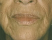
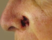
Maintaining Contour on Convex Surfaces: 2 Surgical Techniques for the Lateral Nasal Ala and Bald Scalp
Article Type
Changed
Display Headline
Maintaining Contour on Convex Surfaces: 2 Surgical Techniques for the Lateral Nasal Ala and Bald Scalp
Article PDF
Publications
Topics
Page Number
16-20
Legacy Keywords
facial reconstruction after Mohs surgery, skin tumor removal, cosmetic surgery techniques, cosmetic tissue repair, skin grafts
Article PDF
Article PDF
Page Number
16-20
Page Number
16-20
Publications
Publications
Topics
Article Type
Display Headline
Maintaining Contour on Convex Surfaces: 2 Surgical Techniques for the Lateral Nasal Ala and Bald Scalp
Display Headline
Maintaining Contour on Convex Surfaces: 2 Surgical Techniques for the Lateral Nasal Ala and Bald Scalp
Legacy Keywords
facial reconstruction after Mohs surgery, skin tumor removal, cosmetic surgery techniques, cosmetic tissue repair, skin grafts
Legacy Keywords
facial reconstruction after Mohs surgery, skin tumor removal, cosmetic surgery techniques, cosmetic tissue repair, skin grafts
Article Source
Citation Override
Originally published in Cosmetic Dermatology
PURLs Copyright
Inside the Article
Article PDF Media
Document
Defensive Management Strategies for Dermatology Billing and Coding
Article Type
Changed
Display Headline
Defensive Management Strategies for Dermatology Billing and Coding
Article PDF
Publications
Page Number
11-14
Legacy Keywords
dermatology billing, dermatology coding, practice management, health insurance companies
Article PDF
Article PDF
Page Number
11-14
Page Number
11-14
Publications
Publications
Article Type
Display Headline
Defensive Management Strategies for Dermatology Billing and Coding
Display Headline
Defensive Management Strategies for Dermatology Billing and Coding
Legacy Keywords
dermatology billing, dermatology coding, practice management, health insurance companies
Legacy Keywords
dermatology billing, dermatology coding, practice management, health insurance companies
Article Source
Citation Override
Originally published in Cosmetic Dermatology
PURLs Copyright
Inside the Article
Article PDF Media
Document
Comment on "Academic Physicians' Attitudes Toward Implementation of Multidisciplinary Cosmetic Centers and the Challenges of Subspecialties Working Together" (Cosmet Dermatol. 2012;25:327-332) [letter]
Article Type
Changed
Display Headline
Comment on "Academic Physicians' Attitudes Toward Implementation of Multidisciplinary Cosmetic Centers and the Challenges of Subspecialties Working Together" (Cosmet Dermatol. 2012;25:327-332) [letter]
Article PDF
Publications
Topics
Page Number
10
Legacy Keywords
MCC, cosmetic medicine, cosmetic departments, dermatology subspecialty, MCC, cosmetic medicine, cosmetic departments, dermatology subspecialty
Article PDF
Article PDF
Page Number
10
Page Number
10
Publications
Publications
Topics
Article Type
Display Headline
Comment on "Academic Physicians' Attitudes Toward Implementation of Multidisciplinary Cosmetic Centers and the Challenges of Subspecialties Working Together" (Cosmet Dermatol. 2012;25:327-332) [letter]
Display Headline
Comment on "Academic Physicians' Attitudes Toward Implementation of Multidisciplinary Cosmetic Centers and the Challenges of Subspecialties Working Together" (Cosmet Dermatol. 2012;25:327-332) [letter]
Legacy Keywords
MCC, cosmetic medicine, cosmetic departments, dermatology subspecialty, MCC, cosmetic medicine, cosmetic departments, dermatology subspecialty
Legacy Keywords
MCC, cosmetic medicine, cosmetic departments, dermatology subspecialty, MCC, cosmetic medicine, cosmetic departments, dermatology subspecialty
Article Source
Citation Override
Originally published in Cosmetic Dermatology
PURLs Copyright
Inside the Article
Article PDF Media
Document
Glycobiology and the Skin: A New Frontier
Article Type
Changed
Display Headline
Glycobiology and the Skin: A New Frontier
Article PDF
Publications
Topics
Page Number
8-9
Legacy Keywords
glycans, glycoconjugates, skin glycation, molecular biology and dermatology, cosmeceutical development
Article PDF
Article PDF
Page Number
8-9
Page Number
8-9
Publications
Publications
Topics
Article Type
Display Headline
Glycobiology and the Skin: A New Frontier
Display Headline
Glycobiology and the Skin: A New Frontier
Legacy Keywords
glycans, glycoconjugates, skin glycation, molecular biology and dermatology, cosmeceutical development
Legacy Keywords
glycans, glycoconjugates, skin glycation, molecular biology and dermatology, cosmeceutical development
Article Source
Citation Override
Originally published in Cosmetic Dermatology
PURLs Copyright
Inside the Article
Article PDF Media
Document
Managing Postinflammatory Hyperpigmentation
Article Type
Changed
Display Headline
Managing Postinflammatory Hyperpigmentation
Article PDF
Publications
Topics
Page Number
6-7, 14
Legacy Keywords
hyperpigmentation treatment, hyperpigmented skin, hydroquinone, melasma, skin of color, topical treatment for hyperpigmentation
Article PDF
Article PDF
Page Number
6-7, 14
Page Number
6-7, 14
Publications
Publications
Topics
Article Type
Display Headline
Managing Postinflammatory Hyperpigmentation
Display Headline
Managing Postinflammatory Hyperpigmentation
Legacy Keywords
hyperpigmentation treatment, hyperpigmented skin, hydroquinone, melasma, skin of color, topical treatment for hyperpigmentation
Legacy Keywords
hyperpigmentation treatment, hyperpigmented skin, hydroquinone, melasma, skin of color, topical treatment for hyperpigmentation
Article Source
Citation Override
Originally published in Cosmetic Dermatology
PURLs Copyright
Inside the Article
Article PDF Media
Document