User login
Anticoagulants sharply increase hematoma risk after thyroid, parathyroid surgery
SAN FRANCISCO – Patients undergoing thyroid or parathyroid surgery have a sharply higher risk of postoperative hematoma if they are on clopidogrel or anticoagulants – even if these agents are stopped in advance – researchers reported at the annual clinical congress of the American College of Surgeons.
“Patients with multiple factors considered high risk for postoperative hematoma formation after parathyroid or thyroid surgery should probably undergo a period of observation,” recommended lead investigator Dr. Sarah C. Oltmann, who at the time of the study was the director of endocrine surgery at Parkland Memorial Hospital, University of Texas Southwestern Medical Center, Dallas.
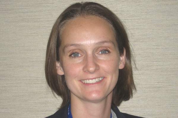
“The need specifically for anticoagulation in the perioperative period should really be carefully assessed, and decisions regarding their use in the perioperative period need to be made very cautiously,” she added. “This is particularly important when considering the need for an injectable bridge [anticoagulant], and discussions with the patient’s primary care provider or cardiologist should be prompted because obviously a hematoma risk of 11% [seen with injectable anticoagulants] is not insignificant.”
The researchers retrospectively studied 4,514 patients who underwent thyroid or parathyroid surgery at the center between 1994 and 2013. Most of the operations were performed by high-volume surgeons.
Overall, 25% of patients were using an antiplatelet agent and 3% were using an anticoagulant agent, defined in the study as current use or use up to 5-7 days before surgery. “We felt that there may be some alteration of hemostasis both at the time of surgery and in the days following surgery if they were resumed on their home meds,” explained Dr. Oltmann, who is now a clinical instructor of surgery at the University of Wisconsin–Madison.
Overall, 0.5% of patients developed a postoperative hematoma, with the majority of these events occurring in the first 24 hours. Three-fourths of the affected patients had to undergo repeat surgery.
In multivariate analyses, clopidogrel (Plavix) users had 5.6 times the odds of developing a hematoma. But neither lower-dose aspirin (less than 325 mg daily) alone nor higher-dose aspirin alone was associated with this complication.
Hematoma odds were elevated by an even greater extent, 7.5 and 29.5 times, for patients using oral and injectable anticoagulants, respectively. (Subcutaneous heparin was not included among injectable anticoagulants because surgeons at the center seldom use it in this setting, according to Dr. Oltmann.)
Patients also had increased odds of hematoma if they underwent thyroid surgery as compared with parathyroid surgery (odds ratio, 7.9), and had a bilateral procedure as compared with a unilateral one (OR, 4.9).
“Additional studies are needed to better clarify both the risk-benefit ratio of injectable anticoagulation in this patient population and potentially being able to better risk-stratify which patients would be better served with a period of overnight observation,” Dr. Oltmann concluded.
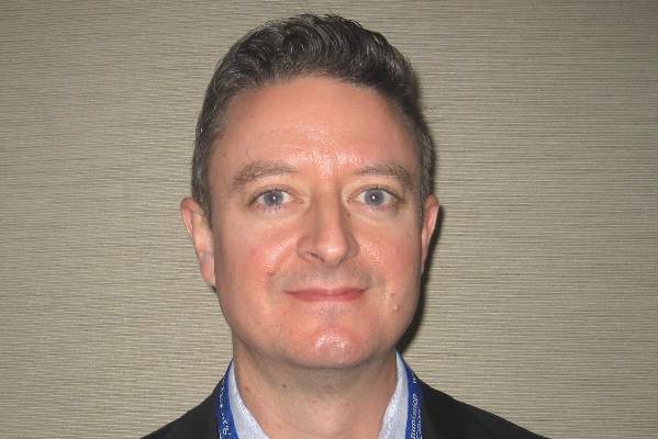
Invited discussant Dr. Raymon H. Grogan, director of the endocrine surgery research program at University of Chicago Medicine, commented, “I think this work represents a level of detail and granularity in regard to anticoagulants that we haven’t seen before in this literature, so it’s really important for us to see these data.
“We tend to get lulled into a false sense of security when we talk about complications related to thyroidectomy because they are so rare. But the truth of the matter is that this is a complication that causes deaths. A recent Nationwide Inpatient Sample study showed that about 1.3% of people who developed a hematoma will actually die in the United States, which is not an insignificant number of people who will die from a complication that’s directly caused by something we’ve done as surgeons,” he said.
Patients often have other risk factors for hematoma, Dr. Grogan noted. Therefore, he wondered, “Who can actually be sent home as a same-day patient after thyroidectomy? … When you combine your … people on these medications, along with all these other risk factors, as well as the risk of significant hypocalcemia postop, it starts to get to the point where, is it really safe to send anyone home the same day after thyroidectomy, given this overwhelming number of different factors that could cause problems?”
“That’s something we all struggle with to a certain degree, trying to be able to best determine who is safe to go home at night and who is not,” Dr. Oltmann replied, noting that risk in the study was greatest for the small proportion of patients on anticoagulants. “So I think a patient who is on some form of anticoagulant, I would definitely have significant reservations about sending home on the same day. They would be somebody I would at least want to keep overnight.”
“As far as the other variables – Graves disease, the size of the tumor, some people would also argue smoking and poorly controlled hypertension – it really becomes a conversation between the surgeon and the patient to know how reliable is the patient, how do you feel the operation went. … Hopefully, the next step is being able to find a way to weigh these different factors to be able to figure out, well, if my patient has A, B, and C, I must observe versus if they don’t, this might be somebody I can send home.”
Another attendee asked, “How do you [handle] aspirin use, given that it’s low risk as seen in your data set? How do you preop the patients, [do you] ask them to stop any low-risk agents, such as aspirin, or if they take the combination of aspirin and Plavix, which one do you hold and which do you continue in your practice?”
“After kind of combing through this data and becoming very familiar with it, I feel very comfortable with continuing aspirin use through the perioperative period,” Dr. Oltmann commented.
“For Plavix, obviously, you just have to juggle the risk-benefit ratio of why they are on that medication,” she said. “I think the most compelling situation is for our patients with atrial fibrillation, with the primary care provider wanting to … have them done on a Lovenox [enoxaparin] bridge, and now having some sort of objective data to get back with them and say, ‘Listen, they have an 11% risk of this really bad complication. Do you really think their risk of stroke trumps that?’ In most patients, that’s not the case, and I think [these data are] finally going to be able to give us some ammunition in that particular battle.”
Dr. Oltmann disclosed that she had no relevant conflicts of interest.
SAN FRANCISCO – Patients undergoing thyroid or parathyroid surgery have a sharply higher risk of postoperative hematoma if they are on clopidogrel or anticoagulants – even if these agents are stopped in advance – researchers reported at the annual clinical congress of the American College of Surgeons.
“Patients with multiple factors considered high risk for postoperative hematoma formation after parathyroid or thyroid surgery should probably undergo a period of observation,” recommended lead investigator Dr. Sarah C. Oltmann, who at the time of the study was the director of endocrine surgery at Parkland Memorial Hospital, University of Texas Southwestern Medical Center, Dallas.
“The need specifically for anticoagulation in the perioperative period should really be carefully assessed, and decisions regarding their use in the perioperative period need to be made very cautiously,” she added. “This is particularly important when considering the need for an injectable bridge [anticoagulant], and discussions with the patient’s primary care provider or cardiologist should be prompted because obviously a hematoma risk of 11% [seen with injectable anticoagulants] is not insignificant.”
The researchers retrospectively studied 4,514 patients who underwent thyroid or parathyroid surgery at the center between 1994 and 2013. Most of the operations were performed by high-volume surgeons.
Overall, 25% of patients were using an antiplatelet agent and 3% were using an anticoagulant agent, defined in the study as current use or use up to 5-7 days before surgery. “We felt that there may be some alteration of hemostasis both at the time of surgery and in the days following surgery if they were resumed on their home meds,” explained Dr. Oltmann, who is now a clinical instructor of surgery at the University of Wisconsin–Madison.
Overall, 0.5% of patients developed a postoperative hematoma, with the majority of these events occurring in the first 24 hours. Three-fourths of the affected patients had to undergo repeat surgery.
In multivariate analyses, clopidogrel (Plavix) users had 5.6 times the odds of developing a hematoma. But neither lower-dose aspirin (less than 325 mg daily) alone nor higher-dose aspirin alone was associated with this complication.
Hematoma odds were elevated by an even greater extent, 7.5 and 29.5 times, for patients using oral and injectable anticoagulants, respectively. (Subcutaneous heparin was not included among injectable anticoagulants because surgeons at the center seldom use it in this setting, according to Dr. Oltmann.)
Patients also had increased odds of hematoma if they underwent thyroid surgery as compared with parathyroid surgery (odds ratio, 7.9), and had a bilateral procedure as compared with a unilateral one (OR, 4.9).
“Additional studies are needed to better clarify both the risk-benefit ratio of injectable anticoagulation in this patient population and potentially being able to better risk-stratify which patients would be better served with a period of overnight observation,” Dr. Oltmann concluded.
Invited discussant Dr. Raymon H. Grogan, director of the endocrine surgery research program at University of Chicago Medicine, commented, “I think this work represents a level of detail and granularity in regard to anticoagulants that we haven’t seen before in this literature, so it’s really important for us to see these data.
“We tend to get lulled into a false sense of security when we talk about complications related to thyroidectomy because they are so rare. But the truth of the matter is that this is a complication that causes deaths. A recent Nationwide Inpatient Sample study showed that about 1.3% of people who developed a hematoma will actually die in the United States, which is not an insignificant number of people who will die from a complication that’s directly caused by something we’ve done as surgeons,” he said.
Patients often have other risk factors for hematoma, Dr. Grogan noted. Therefore, he wondered, “Who can actually be sent home as a same-day patient after thyroidectomy? … When you combine your … people on these medications, along with all these other risk factors, as well as the risk of significant hypocalcemia postop, it starts to get to the point where, is it really safe to send anyone home the same day after thyroidectomy, given this overwhelming number of different factors that could cause problems?”
“That’s something we all struggle with to a certain degree, trying to be able to best determine who is safe to go home at night and who is not,” Dr. Oltmann replied, noting that risk in the study was greatest for the small proportion of patients on anticoagulants. “So I think a patient who is on some form of anticoagulant, I would definitely have significant reservations about sending home on the same day. They would be somebody I would at least want to keep overnight.”
“As far as the other variables – Graves disease, the size of the tumor, some people would also argue smoking and poorly controlled hypertension – it really becomes a conversation between the surgeon and the patient to know how reliable is the patient, how do you feel the operation went. … Hopefully, the next step is being able to find a way to weigh these different factors to be able to figure out, well, if my patient has A, B, and C, I must observe versus if they don’t, this might be somebody I can send home.”
Another attendee asked, “How do you [handle] aspirin use, given that it’s low risk as seen in your data set? How do you preop the patients, [do you] ask them to stop any low-risk agents, such as aspirin, or if they take the combination of aspirin and Plavix, which one do you hold and which do you continue in your practice?”
“After kind of combing through this data and becoming very familiar with it, I feel very comfortable with continuing aspirin use through the perioperative period,” Dr. Oltmann commented.
“For Plavix, obviously, you just have to juggle the risk-benefit ratio of why they are on that medication,” she said. “I think the most compelling situation is for our patients with atrial fibrillation, with the primary care provider wanting to … have them done on a Lovenox [enoxaparin] bridge, and now having some sort of objective data to get back with them and say, ‘Listen, they have an 11% risk of this really bad complication. Do you really think their risk of stroke trumps that?’ In most patients, that’s not the case, and I think [these data are] finally going to be able to give us some ammunition in that particular battle.”
Dr. Oltmann disclosed that she had no relevant conflicts of interest.
AT THE ACS CLINICAL CONGRESS
Key clinical point: Clopidogrel and anticoagulants are risk factors for hematoma after thyroid or parathyroid surgery.
Major finding: The odds of hematoma were 5.6 times higher with clopidogrel use and 7.5 and 29.5 times higher with oral and injectable anticoagulant use, respectively.
Data source: A retrospective study of 4,514 patients undergoing thyroid or parathyroid surgery.
Disclosures: Dr. Oltmann disclosed that she had no relevant conflicts of interest.

Team discovers key aspects of HSC development

New research suggests proinflammatory signaling is crucial to the creation of hematopoietic stem cells (HSCs) during embryonic development, a finding that could help scientists reproduce HSCs for therapeutic use.
Researchers discovered that TNFR2, via TNFα, activates the Notch and NF-kB signaling pathways to establish HSC fate, which suggests inflammatory signaling is required for HSC generation.
The group also found that primitive neutrophils are the major source of TNFa. So it seems these cells are crucial to HSC development as well.
The researchers described these findings in Cell.
“The development of some mature cell lineages from iPSCs [induced pluripotent stem cells], such as cardiac and neural, has been reasonably straightforward, but not with HSCs,” said principal investigator David Traver, PhD, of the University of California, San Diego School of Medicine.
“This is likely due, at least in part, to not fully understanding all of the factors used by the embryo to generate HSCs. We believe the discovery that proinflammatory cues are important in vivo will help us recapitulate instruction of HSC fate in vitro from iPSCs.”
For this study, Dr Traver and his colleagues decided to examine the role of TNFα in HSC development, extending previous research by Victoriano Mulero, PhD, of the University of Murcia in Spain.
Dr Mulero reported that TNFα is important in the function of the embryonic vascular system. And, in animal models where TNF function was absent, blood defects resulted.
Raquel Espin-Palazon, PhD, a researcher in Dr Traver’s lab and a former colleague of Dr Mulero’s, determined that TNFα is required for the emergence of HSCs during embryogenesis in zebrafish.
Dr Traver said the finding was completely unexpected because HSCs emerge relatively early in embryonic formation, when the developing organism is considered to be largely sterile and devoid of infection.
“Thus, there was no expectation that proinflammatory signaling would be active at this time or in the blood-forming regions,” Dr Traver said. “Equally surprising, we found that a population of embryonic myeloid cells, which are transient cells produced before HSCs arise, are the producers of the TNFα needed to establish HSC fate.”
“So it turns out that a small subset of myeloid cells that persist for only a few days in development are necessary to help generate the lineal precursors of the entire adult blood-forming system.” ![]()
Agent reverses effects of edoxaban

Credit: Kevin MacKenzie
An investigational anticoagulant reversal agent known as PER977 can restore hemostasis following edoxaban administration, according to a phase 1/2 trial.
The study included 80 healthy subjects who received PER977 or placebo 3 hours after they received a dose of edoxaban.
PER977 decreased anticoagulation to within 10% of baseline levels within 10 to 30 minutes of administration, an effect that took 12 to 15 hours in placebo-treated subjects.
Jack Ansell, MD, of Hofstra North Shore–LIJ School of Medicine in Hempstead, New York, and his colleagues reported these results in a letter to NEJM. The research was funded by Perosphere, the company developing PER977.
PER977 is a small molecule that can bind to unfractionated heparin, low-molecular-weight heparin, edoxaban, rivaroxaban, apixaban, and dabigatran. In preclinical studies, PER977 reversed anticoagulation with each of the new oral anticoagulants.
Dr Ansell and his colleagues wanted to determine the safety and efficacy of PER977 when given to healthy subjects after edoxaban. So the team enrolled 80 subjects and gave them placebo or single, escalating doses of PER977 (5 mg to 300 mg) 3 hours after a 60 mg dose of edoxaban.
After edoxaban administration, the mean whole-blood clotting time (WBCT) increased 37% from baseline. In placebo-treated subjects, it took 12 to 15 hours for the mean WBCT to return to within 10% above the baseline level.
In subjects who received PER977 at 100 mg to 300 mg, it took 10 minutes or less to reach the same level. And the WBCT remained within 10% above or below the baseline value for 24 hours, with no rebound and no infusions needed.
The researchers also assessed clots while measuring WBCT to determine the mean fibrin-fiber diameter. They found that edoxaban significantly reduced the mean fibrin-fiber diameter relative to baseline, from about 250 nm to about 125 nm (P<0.001).
But the mean fibrin-fiber diameter was restored to normal 30 minutes after subjects received 100 mg to 300 mg of PER977.
In addition, the researchers saw no evidence of procoagulant activity after PER977 administration, as assessed by levels of D-dimer, prothrombin fragment 1.2, and tissue factor pathway inhibitor.
The team noted a few adverse events that may have been related to PER977, including transient, mild perioral and facial flushing, dysgeusia, and moderate headache.
One subject experienced a moderate muscle cramp and elevation in creatinine phosphokinase levels, but the researchers believe this was not related to PER977.
“[T]he fact that PER977 was shown to be safe, well tolerated, and, most importantly, effective in reversing edoxaban, one of the new factor Xa oral anticoagulants, is a major step forward in developing a readily available and simple-to-use reversal agent for the new oral anticoagulants,” Dr Ansell said. ![]()
Aspartame, sweetened drinks don’t increase risk of NHL

beverages at the supermarket
Consuming aspartame and drinking sweetened beverages do not increase a person’s risk of developing non-Hodgkin lymphoma (NHL), according to research published in the Journal of Nutrition.
Investigators analyzed information from more than 100,000 men and women in the US.
The results suggested that neither aspartame intake nor the consumption of sugar-sweetened or artificially sweetened beverages were associated with an increased risk of NHL.
Marjorie L. McCullough, SCD, RD, of the American Cancer Society in Atlanta, Georgia, and her colleagues conducted this research, analyzing data from the nutrition cohort of the Cancer Prevention Study II, an assessment of cancer incidence
and mortality in the US.
Study subjects first completed a questionnaire in 1992, noting information related to diet and other lifestyle factors. They completed follow-up questionnaires in 1999 and 2003, which included questions related to the consumption of sugar-sweetened and artificially sweetened beverages, as well as tabletop sweeteners containing aspartame.
Among the 100,442 adult men and women who provided information on diet and lifestyle factors in 1999, there were 1196 NHL cases verified during a 10-year follow-up period.
The investigators assessed the risk of NHL associated with sweetened beverage and aspartame consumption, adjusted for the subjects’ smoking status, body mass index, and history of diabetes.
The analysis revealed that, in women and men combined, there was no association between NHL risk and the consumption of 1 or more servings (355 mL) of artificially sweetened beverages. Compared to nondrinkers, subjects who drank artificially sweetened beverages had a risk ratio (RR) of 0.92 (P=0.14).
Similarly, there was no association between NHL risk and sugar-sweetened beverages. Compared to nondrinkers, the RR for sugar-sweetened beverage drinkers was 1.10 (P=0.62).
Furthermore, subjects’ overall aspartame intake, which was estimated from artificially sweetened carbonated beverage consumption and the use of aspartame packets, was not associated with NHL risk.
The RR was 1.02 (P=0.69) for the top quintile (which had a median aspartame intake of 145 mg per day) vs the bottom quintile (which had a median aspartame intake of 0 mg per day).
The investigators also found that associations between disease and sweetened beverage consumption or aspartame intake were generally null for specific NHL subtypes, including multiple myeloma, diffuse large B-cell lymphoma, chronic lymphocytic leukemia/small lymphocytic lymphoma, follicular lymphoma, and other B-cell lymphomas.
“The study supports the decades of research that have continued to find that aspartame is safe for use in foods and beverages,” said Haley Stevens, PhD, President of the Calorie Control Council. “It also supports the conclusions of the National Cancer Institute, who have determined that aspartame does not increase a person’s risk of developing cancer.” ![]()
Study reveals gap in patient blood management
PHILADELPHIA—Healthcare professionals may not be using blood management interventions in a majority of patients receiving red blood cell (RBC) transfusions, a large study suggests.
The research showed that 72 US hospitals have made strides in reducing the use of RBCs in patients undergoing orthopedic and cardiac surgery.
And smaller reductions have occurred in patients with gastrointestinal bleeding, obstetric patients, and those receiving bone marrow transplants or inpatient chemotherapy.
However, more than 60% of the transfusions studied were given to patients who did not belong to any of the aforementioned groups.
“So when we target our [patient blood management] interventions to these kind of surgical procedures, in fact, we’re looking at only about 40% of the red cell use, and 60% goes to a myriad of other patients,” said Robert L. Thurer, MD, of Haemonetics in Braintree, Massachusetts.
He added that the groups of specific patient populations within that 60% were so small—“2% of patients here and 3% of patients there”—that it was too difficult to examine them individually in this study.
Dr Thurer presented this research at the AABB Annual Meeting 2014 (abstract S65-030K).
He and his colleagues wanted to determine which specific groups of patients have been most affected by patient blood management, understand further opportunities for decreasing transfusion use, and project future blood needs.
So the researchers analyzed data from 3,946,428 inpatients at 72 US hospitals, comparing the use of RBC transfusions in 2009/2010 to use in 2013.
In 2009/2010, there were 1,378,581 patients admitted to the hospitals, the RBC transfusion rate was 11.5.%, and the utilization (total units/total patients) was 0.41. In 2013, there were 861,804 patients, the transfusion rate was 10%, and the utilization was 0.34.
So from 2009/2010 to 2013, there was a 13% reduction in transfusion rate and a 17% reduction in utilization.
The greatest decrease was in orthopedic surgery patients. In those undergoing hip and knee joint replacement surgery, there was a 45% reduction in transfusion rate and a 43% reduction in utilization. In patients with a hip fracture, there was a 20% decrease in transfusion rate and a 20% decrease in utilization.
There was a smaller, though still sizable, reduction in transfusion use among patients undergoing cardiac surgery—a 15% decrease in transfusion rate and an 18% decrease in utilization.
“My personal thoughts about this is that it represents, certainly, lower transfusion triggers, which are becoming more widely adopted, [and] the use of antifibrinolytic drugs, particularly in orthopedic surgeries,” Dr Thurer said.
“We like to think that comprehensive coagulation testing and better matching of coagulation abnormalities contributes to this. Perhaps correction of preoperative anemia [contributes] for elective patients. And, clearly, surgical techniques have evolved, and, as they do, blood loss goes down.”
Reductions in RBC use were also seen in patients with gastrointestinal bleeding, where there was a 3% decrease in transfusion rate and a 13% decrease in utilization.
“For gastrointestinal bleeding, I think the lower transfusion triggers [have made an impact], but there’s also . . . more interest in timely interventions to stop bleeding,” Dr Thurer said.
“So rather than the gastroenterologist saying, ‘Correct the hematocrit and the coagulation factors, and I’ll stop the bleeding in the morning,’ we’re seeing now much more interest in very prompt endoscopy to stop bleeding. And as you know, the way to stop giving transfusions is to stop the patient from bleeding.”
Obstetric patients saw a 5% reduction in transfusion rate and an 8% reduction in utilization. And patients undergoing bone marrow transplant or inpatient chemotherapy saw a 6% decrease in transfusion rate and an 8% decrease in utilization.
Other transplant patients actually saw an increase in RBC transfusions. In liver transplant recipients, there was a 2.2% increase in transfusion rate and a 6% increase in utilization. And in kidney transplant recipients, there was a 0.2% increase in transfusion rate and a 19% increase in utilization.
However, Dr Thurer noted that the majority of RBC transfusions are administered to patients outside of these groups. In 2013, 60.6% of transfusions went to patients who did not fit into any of the aforementioned categories.
“So clearly,” he concluded, “further studies are needed to determine whether these reductions that we’ve seen in some areas can be implemented in a wider variety of patients.” ![]()
Pyrethroid biomarker almost tripled odds of ADHD in boys
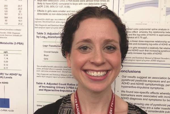
SAN DIEGO – Exposure to common household pesticides called pyrethroids almost tripled the odds of attention-deficit/hyperactivity disorder in boys, but not in girls, authors of a large cross-sectional study reported.
The results resemble findings from prior studies of mice, lead investigator Dr. Melissa L. Wagner-Schuman said in an interview. “Pyrethroids are the most commonly used pesticides for residential pest control and public health,” she and her associates said. “Given the growing use of pyrethroids and the perception that they are a safer insecticide alternative, our results may be of considerable public health import.”

Attention-deficit/hyperactivity disorder is more than twice as prevalent in boys as in girls, according to CDC data. The disorder is “highly heritable,” but environmental factors also play a role, the researchers said at the annual meeting of the American Academy of Child and Adolescent Psychiatry.
For the study, the investigators analyzed National Health and Nutrition Examination Survey data from 2001 to 2002 from 687 children aged 8-18 years. Children were categorized as having ADHD if they met DSM-IV criteria for the disorder on the Diagnostic Interview Schedule for Children caregiver module, had a diagnosis of ADHD reported by their caregivers, or both, the researchers said. Pyrethroid exposure was assessed by testing urine samples for a metabolite of several pyrethroids called 3-phenoxybenzoic acid (3-PBA), they noted.
Boys who had detectable levels of 3-PBA were 2.95 times more likely to have ADHD than were boys who lacked evidence of pyrethroid exposure (95% confidence interval, 1.07-8.08), said Dr. Wagner-Schuman, a pediatrics resident at Cincinnati Children’s Hospital Medical Center. “Effects in girls were smaller and nonsignificant,” she and her associates reported (adjusted odds ratio for girls with detectable biomarker levels, 1.54; 95% CI, 0.32-7.33). The analysis controlled for age; race or ethnicity; income; health insurance status; prenatal tobacco exposure; blood lead levels; urine organophosphate metabolite levels; and urine creatinine level.
Also in boys but not in girls, the odds of ADHD increased linearly with rising 3-PBA levels and did not plateau, the researchers reported.
In the mouse studies, pyrethroid exposure was found to trigger abnormalities in the dopamine system, which produced an “ADHD phenotype,” the investigators said. “Male animals appear to have a heightened vulnerability to exposure,” they added. Other studies have shown that prenatal pyrethroid exposure in humans can increase the risk of neurodevelopmental problems, the investigators noted.
The analysis was limited by its cross-sectional design, Dr. Wagner-Schuman said. Future studies should serially quantify pyrethroid exposure over time, she added.
The National Institute of Environmental Health Sciences funded the research. One coauthor reported having served as a consultant and expert witness for the California Attorney General’s Office, and having consulted for the California Department of Toxic Substances Control and the U.S. Environmental Protection Agency. The other investigators declared no conflicts of interest.
SAN DIEGO – Exposure to common household pesticides called pyrethroids almost tripled the odds of attention-deficit/hyperactivity disorder in boys, but not in girls, authors of a large cross-sectional study reported.
The results resemble findings from prior studies of mice, lead investigator Dr. Melissa L. Wagner-Schuman said in an interview. “Pyrethroids are the most commonly used pesticides for residential pest control and public health,” she and her associates said. “Given the growing use of pyrethroids and the perception that they are a safer insecticide alternative, our results may be of considerable public health import.”
Attention-deficit/hyperactivity disorder is more than twice as prevalent in boys as in girls, according to CDC data. The disorder is “highly heritable,” but environmental factors also play a role, the researchers said at the annual meeting of the American Academy of Child and Adolescent Psychiatry.
For the study, the investigators analyzed National Health and Nutrition Examination Survey data from 2001 to 2002 from 687 children aged 8-18 years. Children were categorized as having ADHD if they met DSM-IV criteria for the disorder on the Diagnostic Interview Schedule for Children caregiver module, had a diagnosis of ADHD reported by their caregivers, or both, the researchers said. Pyrethroid exposure was assessed by testing urine samples for a metabolite of several pyrethroids called 3-phenoxybenzoic acid (3-PBA), they noted.
Boys who had detectable levels of 3-PBA were 2.95 times more likely to have ADHD than were boys who lacked evidence of pyrethroid exposure (95% confidence interval, 1.07-8.08), said Dr. Wagner-Schuman, a pediatrics resident at Cincinnati Children’s Hospital Medical Center. “Effects in girls were smaller and nonsignificant,” she and her associates reported (adjusted odds ratio for girls with detectable biomarker levels, 1.54; 95% CI, 0.32-7.33). The analysis controlled for age; race or ethnicity; income; health insurance status; prenatal tobacco exposure; blood lead levels; urine organophosphate metabolite levels; and urine creatinine level.
Also in boys but not in girls, the odds of ADHD increased linearly with rising 3-PBA levels and did not plateau, the researchers reported.
In the mouse studies, pyrethroid exposure was found to trigger abnormalities in the dopamine system, which produced an “ADHD phenotype,” the investigators said. “Male animals appear to have a heightened vulnerability to exposure,” they added. Other studies have shown that prenatal pyrethroid exposure in humans can increase the risk of neurodevelopmental problems, the investigators noted.
The analysis was limited by its cross-sectional design, Dr. Wagner-Schuman said. Future studies should serially quantify pyrethroid exposure over time, she added.
The National Institute of Environmental Health Sciences funded the research. One coauthor reported having served as a consultant and expert witness for the California Attorney General’s Office, and having consulted for the California Department of Toxic Substances Control and the U.S. Environmental Protection Agency. The other investigators declared no conflicts of interest.
Key clinical point: Consider environmental factors such as exposure to pyrethroid pesticides when assessing boys for attention-deficit/hyperactivity disorder.
Major finding: Boys who had a biomarker for pyrethroids were 2.95 times more likely to have ADHD, compared with boys who lacked the metabolite.
Data source: Cross-sectional analysis of National Health and Nutrition Examination Survey data, and urine tests for 687 children and adolescents.
Disclosures: The National Institute of Environmental Health Sciences funded the research. One coauthor reported having served as a consultant and expert witness for the California Attorney General’s Office, and having consulted for the California Department of Toxic Substances Control and the U.S. Environmental Protection Agency. The other investigators declared no conflicts of interest.
Under My Skin: Neglect
Two disturbing patients came by last week.
The first was a frail old man. His daughter brought him. She said he’d been living in Florida and “shown up” on her doorstep.
As a dermatologist, I’m not often thrown by what I see, but this unfortunate man’s face was hard to look at, with a gaping hole where his left nasolabial fold should have been.
How long had the cancer been there to gouge that hole? How could he neglect it so long? What kind of relationship (or nonrelationship) with his child did it take for this to happen?
I didn’t pursue these questions. Instead, I referred him and his daughter to a skin oncology center where, I hoped, therapy could manage a situation whose severity could surely have been prevented.
Two days later, a Russian woman came in. Remarkably hale at the age of 95 years, she spoke no English. The man who accompanied her, a relative youngster in his mid-70’s, was not a relative, just a stranger who took pity on a fellow visitor to a Russian senior center. “She has two sons,” he explained, “but they live in Minnesota and Texas.”
Her problem was also a basal cell, but this one was on the back of her right ear, large but manageable. I arranged to remove the lesion and offered to speak with her sons. Neither ever called.
Disease is a physical problem in a social context. Patients often present with problems they ignored until other people insisted they take care of them. Parents bring their children. Women drag their husbands. Patients tolerate their itch until their coworkers get annoyed “at seeing me scratch like a monkey.” In situations like these – you can come up with many others – the problem is not just with the patients, but with the people in their vicinity. Sometimes there are people in patients’ lives who notice and care, who demand, “Have that looked at!” But what if nobody cares? Or what if there is no one around at all?
Factors like mental, family, and social dysfunction often underlie whether and to what extent the diseases we diagnose get treated. As practicing physicians, we have little control over such factors. We just try to manage what presents in our offices.
So we make assumptions– that patients can afford to see us, that they have the common sense to come, that they have family or friends who encourage them to come and make doing that possible.
In cases like the ones I’ve just described, these assumptions were wrong. The old man from Florida probably rarely left his apartment, and when he did people just looked away in disgust. He wasn’t their problem. In both cases family was nowhere to be found. How many such lonely and neglected people are there with no support systems, who don’t show up on our office doorstep until it is hard or impossible to take care of them?
I sometimes think back to a case that has haunted me since my early years, when I worked in several Boston-area health centers and sometimes made house calls in gritty neighborhoods. One day I was called to see a patient on the first floor of a rundown example of one of Boston’s wood-frame triple-deckers.
The front door was open. No one was around. I wandered past the parlor into a bedroom. There lay the patient: A woman in late middle age, lying on her back in a dirty nightgown, staring at the ceiling. That image has haunted me for 30 years.
I no longer remember what her skin problem was, just the pitiful sight of her and all the questions it raised: Where was everybody? Who looked after this woman? Who cooked for her, shopped for her? If I prescribed something, who would see that she got it and used it?
I didn’t know. Even if I did, there was nothing I could do about it. Doctors in practice can’t make families stay together, or weave a social safety net that neglected people don’t slip through.
When something lies beyond the scope of what you take to be your responsibility, it’s easier to look away. Now and then a neglected patient forces us to face our own limitations and pay attention to what we have been not looking at.
Dr. Rockoff practices dermatology in Brookline, Mass., and is a longtime contributor to Skin & Allergy News. He serves on the clinical faculty at Tufts University, Boston, and has taught senior medical students and other trainees for 30 years.
Shrink Rap News: California hospital system offers new resource for treating violence
For a clinician working in a forensic setting, the assessment and management of violence is a core clinical skill. Unfortunately, evidence-based practices are often lacking for those patients with serious, treatment-resistant psychoses who defy current guidelines, or for those with severe personality disorders.
This is why I was pleased to see a new resource put out by the California Department of State Hospitals entitled, appropriately enough, the California State Hospital Violence Assessment and Treatment (Cal-VAT) guidelines (CNS Spectrums 2014;19:449-65). The authors, led by Dr. Stephen M. Stahl, are respected experts in forensic psychiatry, psychopharmacology, and psychology. The document is a thorough and thoughtfully organized work that can be employed in both forensic and nonforensic settings.
Too often, the general public will view violence as the sine qua non of mental illness. Rather, violence is often attributable to an amalgam of risk factors, and even the best psychiatrist can be hard-pressed to tease out which aspect is the predominant driving force behind aggressive behavior. Dr. Stahl and his colleagues do an excellent job laying out the most common etiologies: psychosis and disorders associated with impulsivity, psychopathy, medical conditions, and adverse reactions to medication. They provide a structured and logical approach to pharmacologic management, complete with flowcharts and a discussion of general treatment principles relevant to each etiology. They suggest both primary or acute pharmacologic interventions as well as adjunctive therapies and long-term treatment options.
Compared with other papers I’ve read related to the treatment of violence, the Cal-VAT provides a more comprehensive approach by also considering environmental and staffing factors that contribute to violence, and by outlining nonpharmacologic strategies. I found these additional considerations to be just as important as the prescribing recommendations, so much so that I wanted to emphasize them in this column. Understaffing, high staff turnover, inexperience, and inadequate staff training will affect both morale and unit safety. The Cal-VAT guidelines recommend peer support for colleagues as well as postincident opportunities for staff to process violent incidents by patients. Training in de-escalation techniques and violence prevention, as well as good communication between team members, also can minimize the level of tension on a ward.
I was somewhat more skeptical regarding recommendations for the management of predatory aggression, but again, the Cal-VAT cautions that treatment gains in this domain might be modest or nonexistent. I strongly agree with the need for treatment in a highly structured environment for patients high in sociopathy, along with consistent strong boundaries and close external supervision. Good communication between security and clinical staff is also essential.
The guidelines were developed for hospitals, and in a hospital setting, security measures like management of contraband or adequate security staff are just as important as in the correctional setting. However, some of the recommendations in the Cal-VAT may not be practical for a correctional setting, where formularies may be limited and there may not be a means for providing PRN medication. For example, the use of clozapine in a correctional setting may not be practical when weekly blood draws can’t be guaranteed. Nevertheless, the guidelines provide a useful framework for risk assessment and violence risk management.
The Cal-VAT assumes that there are options for moving a patient to a higher level of care within a facility when the violence risk becomes too great. Given that many public hospitals now have a high proportion of forensic patients, a change in ward may not be sufficient to manage this risk. As a profession, we can move violence management forward by addressing this last remaining barrier – the resistance some systems put up against accepting the difficult-to-treat patient. Correctional facilities should be willing to provide the treatment resources needed for mentally ill prisoners high in sociopathy, and hospitals must be equally willing to accept severely ill offenders who cannot be medicated within a jail or prison. For a full spectrum of care, violence management requires a system-level commitment by both public mental health and corrections.
Dr. Hanson is a forensic psychiatrist and coauthor of “Shrink Rap: Three Psychiatrists Explain Their Work.” The opinions expressed are those of the author only, and do not represent those of any of Dr. Hanson’s employers or consultees, including the Maryland Department of Health and Mental Hygiene or the Maryland Division of Correction.

LISTEN NOW: Highlights of the November 2014 issue of The Hospitalist newsmagazine
The Right Choice? Paternalism, Autonomy, and the Incidental Finding
The case had been straightforward. My patient had primary hyperparathyroidism and her localization studies had shown a single parathyroid adenoma. In the operating room, with her under general anesthesia, I had found and removed the abnormal parathyroid gland. The intraoperative parathyroid hormone levels were being run outside the OR door. I was getting ready to close with my fellow when I happened to palpate the thyroid isthmus. There was a firm nodule right in the center of the isthmus. The thyroid looked fine, but the nodule was unmistakable.
This was a surprise. The patient had undergone an ultrasound in radiology the week before, and the study was notable for there being no thyroid nodules. We had performed our own ultrasound in the OR. We had confirmed the location of the parathyroid adenoma and saw no thyroid nodules. I was faced with the initial question of what to do with this incidental finding. Although I could not see the nodule, certainly by feel, it was suspicious, but it was also very small – several millimeters at most. One option was to simply ignore the finding – certainly a bad choice. I knew that the patient had come to the hospital with her sister and a close friend. They were both in the waiting room expecting my update as soon as we were finished. I could have discussed this unexpected finding with the sister and friend, but I felt certain that no one would object to me removing a small piece of thyroid when this added little or no risk. It seemed unnecessary to seek permission to do this small additional procedure.
We proceeded to resect the nodule within the thyroid gland, taking enough adjacent thyroid tissue that I never actually saw the nodule. Once it was removed, I faced another question: Should I send it for frozen section? This seemed to be an easy one to answer. If I was suspicious enough to remove it, I should also know what it is.
The frozen section report was called in a short time later. It was a 4-mm papillary thyroid cancer (PTC) that was within normal thyroid tissue. I had been expecting this possible result. Now I had more choices. I could talk with the family/friend in the waiting room and seek advice on what to do. Alternatively, I could simply say that the presence of PTC was enough of a reason to just take out the thyroid gland since I had not expected this finding and the negative preoperative ultrasound had certainly missed this small tumor (and there might even be others). Finally, I could simply close the patient based on the fact that a 4-mm PTC is of no real clinical significance. Certainly, if this small PTC had been removed with a thyroid lobe for other reasons, we would never go back to take out the rest of the thyroid gland.
As I considered these options, it seemed clear to me that if I went to talk with the family/friend with an unexpected diagnosis of cancer, it was very likely that the patient would wind up with a bigger operation than might be necessary. Ten or fifteen years ago, most surgeons would have removed the thyroid gland for almost any diagnosis of PTC so that patients could go on to receive radioactive iodine. However, today many patients with small incidental PTCs found on lobectomy are simply followed with surveillance ultrasounds because the risks of recurrence or spread are very low. It was clear that I had no basis to take out the whole thyroid gland for a small PTC that was already out. It also seemed unwise to ask what to do, when I felt certain that I knew what was best for the patient. Of course, the suggestion that “I knew what was best for the patient” is a very paternalistic thing to say. It suggests that the medical issues trump all others. It is also quite contrary to the movement of medical ethics in the last several decades that has emphasized shared decision making yet doing what is best for the patient is what surgical patients expect of their surgeons.
I decided to close the patient and then explain what I did and why I did it. She might have been angry with me that I had found a cancer and had not taken out her thyroid gland. However, I felt that the medical evidence supported a less-aggressive surgical approach. In addition, I could always take out her thyroid if she was too worried by the concept of surveillance but I could never put it back if I had removed it!
The patient was understandably surprised and concerned when talked to her. Her first response was one of concern about recurrence. She wanted to know how I knew that there was no more cancer in her thyroid gland. I explained that I actually could not know that, but based on the ultrasound, there was no evidence of any clinically significant thyroid cancer. Fortunately, she was ultimately relieved that the thyroid cancer had been found even though it raised concerns for the future that she had never considered previously.
Whenever surgeons operate on patients under general anesthesia, we are faced with the potential need to make decisions for our patients without the patient’s input. Sometimes it is appropriate to seek input from family when there are multiple good options. However, surgery requires surgeons to make many decisions on their patient’s behalf with no input from the patient – that is, surgeons are expected to act paternalistically in the OR. Rather than being detrimental to the ethical care of patients, such limited paternalism is sometimes the best that we can offer our patients and critical to our role as surgeon.
Dr. Angelos is an ACS Fellow; the Linda Kohler Anderson Professor of Surgery and Surgical Ethics; chief, endocrine surgery; and associate director of the MacLean Center for Clinical Medical Ethics at the University of Chicago.
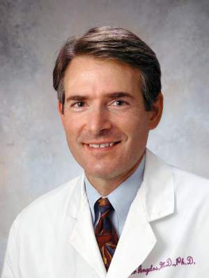