User login
Weekend Botox training: Shortcut to cash or risky business?
This transcript has been edited for clarity.
Dr. Patel: A friend recently joked with me and said, “I wish you were a dermatologist so you could hook me up with Botox and fillers.” Well, little does this friend know that I could be a certified cosmetic injector just after a weekend course. Botox parties, here I come?
I can’t blame any health care professional for having a side hustle. People are burned out, want to supplement their income, or scale back clinical hours. According to one Medscape survey, almost 40% of physicians do have some form of a side hustle, whether it is consulting, speaking engagements, being an expert witness, or moonlighting. I know plenty of doctors and nurses who have taken on Botox injecting as a way to make some extra cash.
Now, going back to me and smoothing out wrinkles. I’m a pediatric hospitalist. I’ve never injected an aesthetic product in anyone’s face. When it comes to sharp objects and faces, I’ve sewn lacerations and drained abscesses. In my world, when we talk about botulinum toxin, we’re usually talking about botulism or the therapeutic treatment of migraines and muscle spasms – pathology.
The National Laser Institute has a 2-day Botox and dermal filler training. “Our 2-day Botox and filler course will also teach you how to build a practice and capitalize on the enormous Botox and dermal filler market that exists in the United States.” That’s a lot to cover in 2 days. They even have lunch breaks.
Just from a quick search, I even found an online video course for $1,500. For an additional fee, you can have a live, hands-on component. There are so many trainings out there, including one that’s only 8 hours long, offered by Empire Medical. I also went and spoke with an employee at Empire Medical who told me that because I’m an MD, if I do the course, I can use my certificate and go directly to a manufacturer, buy Botox, and start injecting right away.
Now, is this training actually sufficient for me to go and get good results while minimizing adverse effects like brow ptosis, dry eyes, and asymmetry? I have no idea. According to a review from the Journal of Cosmetic Dermatology, it’s crucial to understand anatomic landmarks, muscle function, baseline asymmetry, potential migration of the toxin, and site-specific precautions.
Okay, that sounds really intimidating, but people still do it. I saw a Business Insider article about a hospitalist who took a 2-day Botox course and then, to her credit, she trained under supervision for an additional 6 months. She then started hosting Botox parties and each time was making $3,500 to upwards of $20,000.
Let’s do some quick mental math. If I were to go online and buy Botox for $3-$6 a unit and then charge patients $15 a unit, and then I consider that in areas like the forehead or in between the eyes – I read that could take 25-50 units – and I repeat this for multiple patients, I can make a few thousand dollars. Well, I may have to adjust my prices according to the market, obviously, because I did see some Groupons advertising $10 per unit.
Who can get in on some Botox cosmetic cash action? Well, physicians can right away. For other health care professionals, it depends on the state. For example, in California, dentists cannot get Botox solely for cosmetic purposes, whereas in Arizona, they can. Generally speaking, NPs and PAs require some type of physician oversight or supervision, but again, it depends on the state.
Oh, and fun fact: Connecticut outright banned Botox parties and said that Botox must be performed “in a medical spa or licensed health care facility and by a Connecticut-licensed health care provider within his or her scope of practice.”
It definitely worries me that someone could go online or go overseas, buy Botox, claim to be a health care professional, and literally commit fraud. I found stories out there such as a couple in San Jose who are giving out Botox from their home without a license. They got arrested. Also, a woman in Alabama who lied about being a licensed dermatologist and did the same, or another woman in Los Angeles who got arrested after selling counterfeit Botox to undercover law enforcement. Surely, there are plenty more cases out there like this.
I asked Dr. Jacqueline Watchmaker, a board-certified dermatologist at U.S. Dermatology Partners in Arizona who has an expertise in cosmetic procedures, what she thought about the booming med spa industry and what, if any, regulatory changes she wanted to see.
Jacqueline Watchmaker, MD: I do think the fact that people can just go to a 1- or 2-day injection course and inject filler and Botox is concerning. I think the lack of regulation surrounding this topic is also very concerning.
There’s so much that goes into being a skilled injector. It’s an intricate knowledge of facial anatomy, which takes weeks, if not months, to really master. There’s actually injection technique, which can be very complex depending on the part of the face that you’re injecting. Even more important, it’s how to prevent complications, but also how to deal with complications if they do occur. There’s no way that these weekend injection courses are able to cover those topics in a thorough and satisfactory manner.
I see complications from med spas all the time, and I think it’s people going to injectors who are not skilled. They don’t know their anatomy, they don’t know the appropriate filler to use, and then heaven forbid there is a complication, they don’t know how to manage the complication – and then those patients get sent to me.
I think patients sometimes forget that these cosmetic procedures are true medical procedures. You need sterile technique. Again, you need to know the anatomy. It can look easy on social media, but there’s a large amount of thought behind it. I think there needs to be more regulation around this topic.
Dr. Patel: In one study, out of 400 people who received a cosmetic procedure, 50 reported an adverse event, such as discoloration or burns, and these adverse events were more likely to occur if a nonphysician was doing the procedure. Granted, this was a small study. You can’t make a generalization out of it, but this does add to the argument that there needs to be more regulation and oversight.
Let’s be real. The cosmetic injection side hustle is alive and well, but I’m good. I’m not going there. Maybe there should be some more quality control. At Botox parties, do people even ask if their injectors are certified or where they bought their vials?
You might be thinking that this isn’t a big deal because it’s just Botox. Let me ask you all a question: If you or your family member were going to go get Botox or another cosmetic injection, would it still not be a big deal?
Dr. Patel is a pediatric hospitalist, television producer, media contributor, and digital health enthusiast. He splits his time between New York City and San Francisco, as he is on faculty at Columbia University/Morgan Stanley Children’s Hospital and UCSF Benioff Children’s Hospital. He reported conflicts of interest with Medumo.
A version of this article first appeared on Medscape.com.
Even one drink a day tied to increased BP in healthy adults
“A vexing question has been whether usual intake of small amounts of alcohol is associated with a higher level of BP. We identified a continuous, more or less linear association, with no evidence of a threshold for the association,” study coauthor Paul Whelton, MD, of Tulane University School of Public Health and Tropical Medicine, New Orleans, said in an interview.
For systolic BP (SBP), “the most important BP risk indicator for CVD [cardiovascular disease], the association was robust, being present in both men and women and in both North America as well as Asia,” Dr. Whelton noted.
Based on the results, “the lower the better, and no consumption even better, as we did not find any indication that human health may benefit from consumption of very small amounts of alcohol,” senior author Marco Vinceti, MD, PhD, of University of Modena and Reggio Emilia University in Italy, told this news organization.
“Clearly, alcohol is not the only or necessarily the main determinant of high blood pressure, and the effects of small intakes of alcohol emerging from our pooled analysis were certainly not biologically as relevant and meaningful as those induced by high intakes,” Dr. Vinceti added.
The study was published online in Hypertension.
The researchers analyzed data from seven large, observational studies conducted in the United States, Korea, and Japan involving 19,548 adults (65% men).
Participants ranged in age from 20 years to the early 70s at baseline and were followed for a median of 5.3 years (range, 4-12 years). None of the participants had previously been diagnosed with hypertension or other CVD, diabetes, liver disease, alcoholism, or binge drinking.
Compared with nondrinkers, SBP was 1.25 mm Hg higher in adults who consumed an average of 12 grams of alcohol per day, rising to 4.90 mm Hg in adults consuming an average of 48 grams of alcohol per day.
For reference, in the United States, 12 ounces of regular beer, 5 ounces of wine, or a 1.5-ounce shot of distilled spirits contains about 14 grams of alcohol.
Diastolic BP (DBP) was 1.14 mm Hg higher in adults who consumed an average of 12 grams of alcohol per day, rising to 3.10 mm Hg in those who consumed an average of 48 grams of alcohol per day.
Subgroup analyses by gender showed an almost linear association between baseline alcohol intake and SBP changes in men and women and for DBP in men, while in women, there was an inverted U-shaped association.
No safe level
“From a BP perspective, it’s best to avoid alcohol intake. This is what the WHO [World Health Organization] recommends,” Dr. Whelton said.
“If someone is already drinking alcohol and does not want to stop doing so, minimizing alcohol consumption is desirable; many guidelines recommend not starting to drink alcohol but in those already drinking alcohol, consumption of two or less standard drinks per day for men and one or less standard drinks of alcohol per day for women,” Dr. Whelton noted.
Commenting on the study for this article, Alberto Ascherio, MD, of Harvard T. H. Chan School of Public Health, Boston, said it’s been known for more than 30 years that alcohol intake is associated with increased systolic and diastolic BP. The added value of this new study is a “refinement of the estimate of the dose response.”
Dr. Ascherio noted that “moderate alcohol consumption is associated with a modest increase in risk of cancer, but, in spite of the adverse association with BP, with a potentially beneficial effect on cardiovascular disease.” However, “the causality of the latter association has been questioned, but there is no consensus on this.”
Also weighing in, Timothy Brennan, MD, MPH, chief of clinical services for the Addiction Institute of Mount Sinai Health System in New York City, said this new study represents “yet another piece of evidence suggesting that there simply is no ‘healthy’ amount of alcohol use in humans.
“Even small amounts of alcohol intake can have negative health effects, as demonstrated in this study,” Dr. Brennan said. “There is still a widely held belief among people that drinking in moderation is good for you. It is becoming more and more clear that this is simply not the case. As health authorities grapple with drinking ‘recommendations,’ additional datasets like these will be helpful.”
The study had no specific funding. Dr. Whelton, Dr. Vinceti, Dr. Ascherio, and Dr. Brennan have no relevant disclosures.
A version of this article first appeared on Medscape.com.
FROM HYPERTENSION
Erythematous Plaques on the Dorsal Aspect of the Hand
The Diagnosis: Majocchi Granuloma
Histopathology showed rare follicular-based organisms highlighted by periodic acid–Schiff staining. This finding along with her use of clobetasol ointment on the hands led to a diagnosis of Majocchi granuloma in our patient. Clobetasol and crisaborole ointments were discontinued, and she was started on oral terbinafine 250 mg daily for 4 weeks, which resulted in resolution of the rash.
Majocchi granuloma (also known as nodular granulomatous perifolliculitis) is a perifollicular granulomatous process caused by a dermatophyte infection of the hair follicles. Trichophyton rubrum is the most commonly implicated organism, followed by Trichophyton mentagrophytes and Epidermophyton floccosum, which also cause tinea corporis and tinea pedis.1 This condition most commonly occurs in women aged 20 to 35 years. Risk factors include trauma, occlusion of the hair follicles, immunosuppression, and use of potent topical corticosteroids in patients with tinea.2,3 Immunocompetent patients present with perifollicular papules or pustules with erythematous scaly plaques on the extremities, while immunocompromised patients may have subcutaneous nodules or abscesses on any hair-bearing parts of the body.3
Majocchi granuloma is considered a dermal fungal infection in which the disruption of hair follicles from occlusion or trauma allows fungal organisms and keratinaceous material substrates to be introduced into the dermis. The differential diagnosis is based on the types of presenting lesions. The papules of Majocchi granuloma can resemble folliculitis, acne, or insect bites, while nodules can resemble erythema nodosum or furunculosis.4 Plaques, such as those seen in our patient, can mimic cellulitis and allergic or irritant contact dermatitis.4 Additionally, the plaques may appear annular or figurate, which may resemble erythema gyratum repens or erythema annulare centrifugum.
The diagnosis of Majocchi granuloma often requires fungal culture and biopsy because a potassium hydroxide preparation is unable to distinguish between superficial and invasive dermatophytes.3 Histopathology will show perifollicular granulomatous inflammation. Fungal elements can be detected with periodic acid–Schiff or Grocott-Gomori methenamine-silver staining of the hairs and hair follicles as well as dermal infiltrates.4
Topical corticosteroids should be discontinued. Systemic antifungals are the treatment of choice for Majocchi granuloma, as topical antifungals are not effective against deep fungal infections. Although there are no standard guidelines on duration or dosage, recommended regimens in immunocompetent patients include terbinafine 250 mg/d for 4 weeks; itraconazole pulse therapy consisting of 200 mg twice daily for 1 week with 2 weeks off therapy, then repeat the cycle for a total of 2 to 3 pulses; and griseofulvin 500 mg twice daily for 8 to 12 weeks (Table).3 For immunocompromised patients, combination therapy with more than one antifungal may be necessary.

- James WD, Berger T, Elston DM. Diseases resulting from fungi and yeasts. In: James WD, Berger T, Elston D, eds. Andrews’ Diseases of the Skin: Clinical Dermatology. 12th ed. Saunders Elsevier; 2016:285-318.
- Li FQ, Lv S, Xia JX. Majocchi’s granuloma after topical corticosteroids therapy. Case Rep Dermatol Med. 2014;2014:507176.
- Boral H, Durdu M, Ilkit M. Majocchi’s granuloma: current perspectives. Infect Drug Resist. 2018;11:751-760.
- I˙lkit M, Durdu M, Karakas¸ M. Majocchi’s granuloma: a symptom complex caused by fungal pathogens. Med Mycol. 2012;50:449-457.
The Diagnosis: Majocchi Granuloma
Histopathology showed rare follicular-based organisms highlighted by periodic acid–Schiff staining. This finding along with her use of clobetasol ointment on the hands led to a diagnosis of Majocchi granuloma in our patient. Clobetasol and crisaborole ointments were discontinued, and she was started on oral terbinafine 250 mg daily for 4 weeks, which resulted in resolution of the rash.
Majocchi granuloma (also known as nodular granulomatous perifolliculitis) is a perifollicular granulomatous process caused by a dermatophyte infection of the hair follicles. Trichophyton rubrum is the most commonly implicated organism, followed by Trichophyton mentagrophytes and Epidermophyton floccosum, which also cause tinea corporis and tinea pedis.1 This condition most commonly occurs in women aged 20 to 35 years. Risk factors include trauma, occlusion of the hair follicles, immunosuppression, and use of potent topical corticosteroids in patients with tinea.2,3 Immunocompetent patients present with perifollicular papules or pustules with erythematous scaly plaques on the extremities, while immunocompromised patients may have subcutaneous nodules or abscesses on any hair-bearing parts of the body.3
Majocchi granuloma is considered a dermal fungal infection in which the disruption of hair follicles from occlusion or trauma allows fungal organisms and keratinaceous material substrates to be introduced into the dermis. The differential diagnosis is based on the types of presenting lesions. The papules of Majocchi granuloma can resemble folliculitis, acne, or insect bites, while nodules can resemble erythema nodosum or furunculosis.4 Plaques, such as those seen in our patient, can mimic cellulitis and allergic or irritant contact dermatitis.4 Additionally, the plaques may appear annular or figurate, which may resemble erythema gyratum repens or erythema annulare centrifugum.
The diagnosis of Majocchi granuloma often requires fungal culture and biopsy because a potassium hydroxide preparation is unable to distinguish between superficial and invasive dermatophytes.3 Histopathology will show perifollicular granulomatous inflammation. Fungal elements can be detected with periodic acid–Schiff or Grocott-Gomori methenamine-silver staining of the hairs and hair follicles as well as dermal infiltrates.4
Topical corticosteroids should be discontinued. Systemic antifungals are the treatment of choice for Majocchi granuloma, as topical antifungals are not effective against deep fungal infections. Although there are no standard guidelines on duration or dosage, recommended regimens in immunocompetent patients include terbinafine 250 mg/d for 4 weeks; itraconazole pulse therapy consisting of 200 mg twice daily for 1 week with 2 weeks off therapy, then repeat the cycle for a total of 2 to 3 pulses; and griseofulvin 500 mg twice daily for 8 to 12 weeks (Table).3 For immunocompromised patients, combination therapy with more than one antifungal may be necessary.
- James WD, Berger T, Elston DM. Diseases resulting from fungi and yeasts. In: James WD, Berger T, Elston D, eds. Andrews’ Diseases of the Skin: Clinical Dermatology. 12th ed. Saunders Elsevier; 2016:285-318.
- Li FQ, Lv S, Xia JX. Majocchi’s granuloma after topical corticosteroids therapy. Case Rep Dermatol Med. 2014;2014:507176.
- Boral H, Durdu M, Ilkit M. Majocchi’s granuloma: current perspectives. Infect Drug Resist. 2018;11:751-760.
- I˙lkit M, Durdu M, Karakas¸ M. Majocchi’s granuloma: a symptom complex caused by fungal pathogens. Med Mycol. 2012;50:449-457.
A 33-year-old woman presented with an asymptomatic rash on the left hand that was suspected by her primary care physician to be a flare of hand dermatitis. The patient had a history of irritant hand dermatitis diagnosed 2 years prior that was suspected to be secondary to frequent handwashing and was well controlled with clobetasol and crisaborole ointments for 1 year. Four months prior to the current presentation, she developed a flare that was refractory to these topical therapies; treatment with biweekly dupilumab 300 mg was initiated by dermatology, but the rash continued to evolve. A punch biopsy was performed to confirm the diagnosis.


Could your practice be more profitable if you outsource?
Outsourcing certain staff functions in a practice to outside contractors working in remote locations has become commonplace in many medical practices.
Health care outsourcing services, also known as virtual assistants (VAs), were already booming in 2017, when volume grew by 36%. Then, the COVID-19 pandemic in 2020 normalized off-site work, which was a boon to outsourcing providers.
The most popular services being outsourced today by medical practices include billing, scribes, telephone calls to patients, and processing prior authorizations.
“Outsourcing is not for everyone, but I’ve seen it work for many practices,” said Lara Hochman, MD, a practice management consultant in Austin, Tex. She said that practices have used outsourcing to solve problems like high staff turnover, tight budgets, and inefficient use of staff.
When in-house staffing is insufficient or not appropriately aligned with the task, outsourcing can produce big savings, said Teri Deabler, a practice management consultant with the Texas Medical Association.
For example, she said that a client was paying an in-house accountant $80,000 a year. When the accountant retired, she was replaced with a part-time bookkeeper earning $20,000 while her accounting work was outsourced at a cost of $20,000 a year. “The practice’s costs for this service were cut in half,” Ms. Deabler said.
What functions lend themselves to outsourcing?
Clinical services are rarely outsourced by individual practices – although hospitals now outsource numerous clinical services – but virtually any kind of administrative service can be contracted out. Outsourcing used to be limited mainly to billing and off-hours phone services, but today, more services are available, such as scribing, processing prior authorizations, accounting and bookkeeping, human resources (HR) and payroll, interactions with social media, recredentialing, medical transcription, and marketing.
Meanwhile, the original outsourced services have evolved. Billing and collections may now be handled by off-shore VAs, and phone services now deal with a wider variety of tasks, such as answering patients’ questions, scheduling appointments, and making referrals.
Ron Holder, chief operating officer of Medical Group Management Association in Englewood, Colo., said that some outsourcing services can also adjust the amount of work provided based on the customer’s needs. “For instance, an IT outsourcer may allow you to scale up IT support for a new big tech project, such as installing a new electronic health record,” he said.
The outsourced service provider, who might work in another state or another country, is connected to the practice by phone and electronically, and represents the practice when dealing with patients, insurers, or other vendors.
“No one, including patients and your physicians, should know that they are dealing with an outsourced company,” said Mr. Holder. “The work, look, and feel of the outsourced functions should be seamless. Employees at the outsourcer should always identify themselves as the practice, not the outsourcing service.”
Dr. Hochman said that many outsourcing companies dedicate a particular worker to a particular practice and train them to work there. One example of this approach is Provider’s Choice Scribe Services, based in San Antonio. On its website, the company notes that each scribe is paired with a doctor and learns his or her documentation preferences, EMR use, and charting requirements.
What medical practices benefit most from outsourcing?
All kinds and sizes of practices contract with outsourcing firms, but the arrangement is particularly useful for smaller practices, Mr. Holder said. “Larger practices have the economies of scale that allow services to be in-house,” he said, “but smaller practices don’t have that opportunity.”
Dr. Hochman added that outsourcing firms can be hired part-time when the practice doesn’t have enough work for a full-time position. Alternatively, a full-time outsourcing firm can perform two or more separate tasks, such as scribing while handling prior authorizations, she said.
Outsourcing is also useful for new practices, Ms. Deabler said. “A new practice is not earning much money, so it has to have a bare-bones staff,” she said. “Billing, for example, should be contracted out, but it won’t cost that much, because the outsourcer typically charges by volume, and the volume in a new practice is low.”
Meanwhile, Mr. Holder said that the outsourcing of prior authorization work can particularly benefit specialty practices because they typically have a lot of prior authorizations to deal with.
The pros and cons of outsourcing
Experts with experience in outsourcing agree there are both pluses and minuses. “Practices with outsourced workers have less overhead, don’t have to deal with staff turnover, and costs may be lower than for in-house staff,” Ms. Deabler said. “However, you have limited control over outsourced workers and the practice may seem more anonymous to patients, so you need to consider this option very carefully.”
“With outsourcing, you lose control,” said John Machata, MD, a recently retired solo family physician in Wickford, R.I. “You’re trusting someone else to do work that you could do anyway.”
When he briefly considered outsourcing the practice’s billing many years ago, he found that billing companies wouldn’t handle bills that took a lot of work, such as getting in touch with the insurance company and explaining the patient’s situation. “They would only handle the easy bills, which the practice could do anyway,” he said.
However, he does think that answering services may be useful to outsource. “Patients are more inclined to call an anonymous entity than the doctor,” he said. When he gave patients his cell phone number, he said that some patients held off from calling because they didn’t want to bother him.
“Outsourced staff should be less expensive than in-house staff,” said Daniel Shay, an attorney at Gosfield & Associates in Philadelphia. “On the other hand, you are liable for the outsourcer’s mistakes. If your outsourced billing company is upcoding claims, your practice would be on the hook for repayment and penalties.”
Mr. Holder said: “An outsourcer ought to be more efficient at its chosen task because that is what they know how to do. This is a plus at a small practice, where the practice manager may need to do the billing, HR, IT, marketing, some legal work, and accounting,” he said. “No one person can do all of those things well.”
He added, however, “If you choose outsourcing and then decide you don’t like it, it’s difficult to unwind the arrangement. Staff that have been dismissed can’t easily be hired back, so it shouldn’t be an easy decision to make.”
Also, sometimes the staff at offshore outsourcing firms may have accents that are harder for patients to understand, and the offshore staff may not readily understand a U.S. caller. However, Dr. Hochman said that practices often have a chance to interview and select specific persons on the offshore team who best fit their needs.
Offshore outsourcing
Outsourcing firms have been moving abroad, where costs are lower. Typical venues are India and the Philippines because there are larger percentages of people who speak English. Since 2020, demand at offshore medical billing companies has been growing faster than their domestic counterparts, according to a recent analysis.
The difference in price can be substantial. In 2020, the average salary for scribes in India was $500 a month, compared with $2,500 for scribes in the United States.
However, offshore outsourcing is starting to face limitations in some places because of privacy issues, according to David J. Zetter, a practice management consultant in Mechanicsburg, Pa. He pointed to a new Florida law that limits use of offshore vendors because they deal with confidential patient information. The law, which became effective July 1, requires that any protected health information must be maintained in the United States or Canada.
“This will make it very hard for many types of offshore vendors to operate in Florida,” he said. He noted that Florida is the only state with such a restriction, but similar proposals are under consideration in a few other states, such as Texas.
How to select the right company
Mr. Zetter said that the biggest mistake practices make when choosing a company is failing to take enough time to examine their choice. “Quite often, practices don’t validate that companies know what they are doing,” he said. “They get a recommendation and go with it.”
“Choose a company with experience in your specialty,” Mr. Zetter advised. “Speak with the company’s clients, not just the ones the company gives you to speak to. You should ask for the full list of clients and speak to all of them.”
Ms. Deabler said that it’s fairly easy to find respected outsourcing companies. “Colleagues can make recommendations, state and specialty societies can provide lists of preferred vendors, and you can visit vendors’ booths at medical conferences,” she said. She added that it’s also easy to find evaluations of each company. “You can Google the company and come up with all kinds of information about it,” she said.
Mr. Shay said that practices should make sure they understand the terms of the contract with a VA. “Depending on how the contract is worded, you may be stuck with the relationship for many years,” he said. “Before you sign an outsourcing contract, you need to make sure it has a reasonable termination provision.”
Because vetting companies properly can require extensive work, Ms. Deabler said, the work can be given to an experienced practice management consultant. “The consultant can start with a cost-benefit analysis that will show you whether outsourcing would be worthwhile,” she said.
Working with outsource service providers
Mr. Holder said that doctors should keep track of what the outsourcer is doing rather than simply let them do their work. “For example, doctors should understand the billing codes they use most often, such as the five levels of evaluation and management codes, and not just blindly rely on the billing company to code and bill their work correctly,” he noted.
Ms. Deabler said that companies provide monthly reports on their work. “Doctors should be reading these reports and contacting the company if expectations aren’t met,” she said.
Even in the reports, companies can hide problems from untrained eyes, Mr. Holder said. “For example, anyone can meet a metric like days in accounts receivable simply by writing off any charge that isn’t paid after 90 days.”
“You need to be engaged with the outsourcer,” he said. “It’s also a good idea to bring in a consultant to periodically check an outsourcer’s work.”
Will outsourcing expand in the future?
Mr. Holder said that the increasing use of value-based care may require practices to rely more on outsourcing in the future. “For instance, if a practice has a value-based contract that requires providing behavioral health services to patients, it might make sense to outsource that work rather than hire psychologists in-house,” he said.
Practices rarely outsource clinical services, but Mr. Holder said that this may happen in the future: “Now that Medicare is paying less for telehealth, practices have to find a way to provide it without using expensive examining room space,” he said. “Some practices may decide to outsource telehealth instead.”
Mr. Shay said that there are many reasons why outsourcing has a strong future. “It allows you to concentrate on your clinical care, and it is a solution to problems with turnover of in-house staff,” he said. “It can also be more efficient because the service is presumably an expert in areas like billing and collections, which means it may be able to ensure more efficient and faster reimbursements. And if the work is outsourced overseas, you can save money through lower worker salaries.”
A version of this article first appeared on Medscape.com.
ACP sticks with 50 as age to start CRC screening
Not so fast with earlier screening for colorectal cancer (CRC), at least according to one professional group.
The American College of Physicians published updated clinical guidance maintaining 50 as the age when clinicians should start screening for CRC in patients who are asymptomatic and at average risk.
The recommendation conflicts with guidelines from the American Cancer Society and the U.S. Preventive Services Task Force, which lowered the recommended age to start screening to age 45.
Although the rate of CRC has increased among adults aged 45-49, the incidence is 35.1 cases per 100,000 people, much lower than among persons aged 50-64 (71.9 per 100,000) and those aged 65-74 (128.9 per 100,000), the guidance notes.
“The net benefit of screening is much less favorable in average-risk adults between ages 45 and 49 years than in those aged 50-75 years,” the authors wrote. “Clinicians should discuss the uncertainty around benefits and harms of screening in this population.”
The ACP’s updated guidance is provocative and should be considered in the context of other groups’ recommendations, not as superseding them just because it is the most recently published document, according to Jeffrey A. Meyerhardt, MD, MPH, codirector of the Colon and Rectal Cancer Center at Dana-Farber Cancer Institute, Boston.
“As with a lot of the things we do in medicine, it is balancing risk and potential benefit,” Dr. Meyerhardt said in an interview. “If a patient is informed that at a younger age doing screening is very likely not to find anything and there are some risks to screening, that patient could then weigh the risks and benefit with their provider.”
Three screening approaches
The new guidance statement is based on a critical review of existing clinical guidelines, evidence reviews, and modeling studies. The guidance does not apply to patients who have long-standing inflammatory bowel disease and those with a family history of CRC.
The guideline also addresses when clinicians should stop screening – at age 75 – and what types of tests patients should choose from.
After discussing the benefits, harms, cost, availability, and patient preferences, clinicians and patients should select one of three screening approaches, according to the ACP: a fecal immunochemical or high-sensitivity guaiac fecal occult blood test every 2 years; colonoscopy every 10 years; or flexible sigmoidoscopy every 10 years plus a fecal immunochemical test every 2 years.
They should avoid CRC screening tests that use stool DNA, CT colonography, capsule endoscopy, urine, or serum, according to the guidance.
A balancing act
Some physicians view starting screenings at age 45 as a settled argument.
“The entire nation is now focused on increasing screening capacity and getting everyone screened,” said Richard C. Wender, MD, professor and chair of family medicine and community health at the University of Pennsylvania, Philadelphia, who was not involved in the new guidelines. “There is not a controversy about age to start, and I anticipate that this paper won’t create a new one.”
The epidemiology of CRC is changing rapidly, Dr. Wender said.
“While CRC mortality is going down in older age groups, mortality is now rising in younger people,” he said. “While cancer incidence is lower in the 45- to 49-years-old group, the precursors to cancer are present and can be found in a substantial percentage of patients – the same percentage as 50- to 55-year-olds.”
Dr. Meyerhardt said in an interview that the recommendation to start screening at age 45 was reasonable but that more people need to be screened to detect CRC than the older population.
“Ultimately, one’s going to have to consider the various recommendations from these different societies when having a patient in front of you as a primary care or other physician to discuss screening in someone who’s what we call average risk,” he said.
Younger patients who notice any possible symptoms of CRC such as blood in stool or changes in bowel habits should discuss them with a physician, he said.
The ACP also differs from other groups in not recommending stool DNA tests such as Cologuard (Exact Sciences). Dr. Wender said this test is the least cost effective based on comparing adherence for other options. “If Cologuard can lead to higher adherence and there are data suggesting it can, then relative cost-effectiveness looks better.”
Why 50
In weighing the risks and benefits of screening, the ACP noted that CRC screening can entail risk for serious bleeding and perforation in the case of colonoscopy.
Overdiagnosis and associated overtreatment, as well as costly follow-ups for findings that are clinically unimportant, are additional factors to consider with various cancer screening tests, said Amir Qaseem, MD, PhD, MHA, the ACP’s chief science officer and the corresponding author of the updated guidance.
Despite some differences between various groups’ recommendations, Dr. Qaseem saw important similarities.
“We need to get everyone between 50 and 75 screened,” Dr. Qaseem said. On that point, “there is no disagreement.”
One guideline author reported receiving salary from the ACP. Dr. Qaseem reported no conflicts of interest.
A version of this article first appeared on Medscape.com.
FROM ANNALS OF INTERNAL MEDICINE
FDA OKs dostarlimab plus chemo for endometrial cancer
The U.S. Food and Drug Administration has approved dostarlimab-gxly (Jemperli, GlaxoSmithKline) with carboplatin and paclitaxel, followed by single-agent dostarlimab, for primary advanced or recurrent endometrial cancer that is mismatch repair–deficient (dMMR), as determined by an FDA-approved test or microsatellite instability–high (MSI-H).
The approval was based on GSK’s RUBY trial. Across 122 patients with dMMR/MSI-H primary advanced or recurrent endometrial cancer, progression-free survival was 30.3 months in women randomly assigned to dostarlimab on a background of carboplatin and paclitaxel, followed by dostarlimab monotherapy, vs. 7.7 months among women randomly assigned to placebo (hazard ratio, 0.29; P < .0001), according to the FDA’s press release.
MMR/MSI tumor status was determined by local testing or by the Ventana MMR RxDx Panel when local testing was unavailable.
“Until now, chemotherapy alone has been the standard of care with many patients experiencing disease progression,” GSK executive Hesham Abdullah said in the company’s press release. The trial results “and today’s approval underscore our belief in the potential for Jemperli to transform cancer treatment as a backbone immuno-oncology therapy.”
that has progressed on or following a platinum-containing chemotherapy and is not a candidate for curative surgery or radiation. The latest approval means that the agent “is now indicated earlier in treatment in combination with chemotherapy,” GSK said.
Dostarlimab also carries an indication for dMMR recurrent or advanced solid tumors that have progressed on or following prior treatment when there are no satisfactory alternative treatment options.
Immune-mediated adverse reactions with dostarlimab include pneumonitis, colitis, hepatitis, endocrinopathies such as hypothyroidism, nephritis with renal dysfunction, and skin adverse reactions. The most common adverse reactions (≥ 20%) with carboplatin and paclitaxel in the Ruby trial were rash, diarrhea, hypothyroidism, and hypertension.
The recommended dostarlimab dose is 500 mg every 3 weeks for 6 doses with carboplatin and paclitaxel, followed by 1,000 mg monotherapy every 6 weeks until disease progression or unacceptable toxicity, or up to 3 years.
Drugs.com lists dostarlimab’s price at $11,712.66 for 500 mg/10 mL intravenous solution.
A version of this article first appeared on Medscape.com.
Cognitive problems transient after AFib ablation
Investigators randomly assigned 100 patients with symptomatic AFib who had failed at least one anti-arrhythmic drug (AAD) to ongoing therapy or to AFib catheter ablation. Patients were followed for 1 year, and changes in cognitive performance were assessed at baseline and at 3, 6, and 12 months.
Although patients in the ablation arm initially showed more cognitive dysfunction than those in the medical arm, at 6 months, the gap was smaller, and at 12 months, no patients in the ablation arm showed signs of cognitive dysfunction. In fact, more than 1 in 10 showed signs of cognitive improvement, compared with no patients in the medical arm.
The study was published online in the July issue of JACC: Clinical Electrophysiology.
Important pillar
Previous research has shown that AFib is associated with cognitive dysfunction independently of stroke, “suggesting that AFib is an additional risk factor for cognitive impairment,” the authors write.
Catheter ablation is an “important pillar” in the management of patients with AFib that is refractory to medical therapy, but postoperative cognitive dysfunction (POCD) may occur in the immediate aftermath of the procedure, they note. Little is known about whether these cognitive changes persist long term, and no randomized studies have investigated this issue.
The researchers randomly assigned 100 patients with symptomatic paroxysmal or persistent AFib who had failed greater than or equal to 1 AAD to receive either medical management or catheter ablation. The mean age of the patients was 59 plus or minus 12 years, 32% were women, and 46% had persistent AFib.
Medical management consisted of optimization of AADs to maintain sinus rhythm. For those who underwent ablation, AADs were discontinued five half-lives prior to the procedure (with the exception of amiodarone).
Participants were followed for 12 months after enrollment. Clinical reviews and cognitive testing were performed at 3, 6, and 12 months during that time.
AADs and oral anticoagulation were weaned and were discontinued 3 months after the procedure, depending on each patient’s individual risk profile.
Cognitive testing included the Consortium to Establish a Registry for Alzheimer’s Disease (CERAD) Auditory Verbal Learning Test and Semantic Fluency test; the Controlled Oral Word Association test; and the Trail Making Task (parts A and B).
Participants also completed the University of Toronto AFib Symptom Severity Scale at baseline and at all follow-up visits.
The primary endpoint was prevalence of new-onset cognitive dysfunction. Main secondary endpoints included improvement in cognitive function during follow-up; AFib recurrence and AFib function during follow-up; AAD use during follow-up; and changes to AFib symptom severity assessment scores during follow-up.
More research needed
Of the 100 participants, 96 completed the study protocol (52 in the ablation group and 48 in the medical management group). There were no significant differences between the groups regarding baseline demographics, clinical AFib risk factors, and echocardiographic parameters.
At 3 months, new-onset cognitive dysfunction was detected across a wide range of the neuropsychological tests in 14% of participants in the ablation arm, versus 2% of participants in the medical arm (P = .03)
But at 6 months, only 4% of patients in the ablation arm displayed cognitive dysfunction, compared again with 2% in the medical arm (P = .60). And by 12 months, there were no patients with detectable cognitive dysfunction in the ablation arm, compared with the same patient who showed cognitive impairment in the medical arm (P = .30).
Longer ablation time was an independent predictor of new-onset cognitive dysfunction (odds ratio, 1.30; 95% CI, 1.01-1.60; P = .003).
When patients with and those without new-onset cognitive dysfunction were compared, no differences were found in arrhythmia recurrence or AFib burden post ablation.
At 12 months, 14% of those in the ablation arm showed improvement in cognitive performance, compared with no participants in the medical arm (P = .007).
Compared with participants who had no change in cognitive performance, those who had a significant improvement had a trend toward lower AFib recurrence rates (29% vs. 48%; P = .30). However, both groups were found to have a low AFib burden over the 12 months. And the use of AADs at the 12-month mark was significantly lower among those with versus those without cognitive improvement (0% vs. 38%; P = .04).
As early as 3 months post procedure and then at 12 months, participants in the ablation group had significant improvement in AFib-related symptoms, compared with those in the medical arm (for both, P < .001).
“Among a contemporary cohort of symptomatic paroxysmal and persistent AFib patients, catheter ablation was associated with a transient decline in cognitive function in the short-term, followed by recovery at 12 months,” the authors conclude.
They note that further large studies “are required to determine with AFib ablation may prevent the longer-term neurocognitive decline and dementia development associated with AFib.”
‘Reassuring’ findings
In a comment, Andrea Natale, MD, executive medical director, Texas Cardiac Arrhythmia Institute at St. David’s Medical Center, Austin, said POCD is “very likely due to the vulnerable state of mind and the stress that the patients encounter while undergoing the cardiac procedure,” as well as postsurgical inflammation, which “can cause brief functional alterations in the brain, leading to temporary cognitive impairment.” Inadequate preprocedural anticoagulation may also play a role.
Dr. Natale, co-author of an accompanying editorial, said it’s “prudent” when evaluating cognitive function to use questionnaires that are “sensitive to mild cognitive impairment,” such as the Montreal Cognitive Assessment or the Mini-Mental State Examination.
Additionally, “post-ablation cognitive function should be assessed way after the blanking period to avoid any plausible impact of inflammation, medications, the feeling of being overwhelmed, and the stress of undergoing a cardiac procedure,” advised Dr. Natale, who was not involved with the study.
Also commenting, Matthew Hyman, MD, PhD, an electrophysiologist and assistant professor of medicine at the Hospital of the University of Pennsylvania, called it a “well-done and very reassuring study.”
Dr. Hyman, who was also not part of the research team, added that previous work has shown an association between AFib and dementia, “and it remains to be seen if patients with rhythm control over longer durations than the current studies have the best outcomes.”
The authors of the study are supported by the National Health and Medical Research Council research scholarship. One author of the study is supported by a practitioner fellowship from the National Health and Medical Research Council; has received research support from Biosense Webster, Boston Scientific, Abbott, and Medtronic; and has served on the advisory board of Boston Scientific and Biosense Webster. Dr. Natale is a consultant for Abbott, Baylis, Biosense Webster, Biotronik, Boston Scientific, and Medtronic. Dr. Hyman is a consultant/speaker for Abbott, Biosense Webster and Boston Scientific.
A version of this article first appeared on Medscape.com.
FROM JACC: CLINICAL ELECTROPHYSIOLOGY
Free teledermatology clinic helps underserved patients initiate AD care
A in other underserved areas in the United States.
Washington, D.C., has “staggering health disparities that are among the largest in the country,” and Ward 8 and surrounding areas in the southeastern part of the city are “dermatology deserts,” said Adam Friedman, MD, professor and chair of dermatology at George Washington University, Washington, who started the program in 2021 with a pilot project. Dr. Friedman spoke about the project, which has since been expanded to include alopecia areata, at the Revolutionizing Atopic Dermatitis conference in April and in an interview after the meeting.

Patients who attend the clinics – held at the Temple of Praise Church in a residential area of Ward 8, a predominantly Black community with a 30% poverty rate – are entered into the GW Medical Faculty Associates medical records system and educated on telemedicine best practices (such as not having light behind them during a session) and how to use telemedicine with their own device.
Those with AD who participate learn about the condition through an image-rich poster showing how it appears in various skin tones, handouts, National Eczema Association films, and discussion with medical students who staff the clinics under Dr. Friedman’s on-site supervision. Participants with alopecia areata similarly can view a poster and converse about the condition.
Patients then have a free 20-minute telehealth visit with a GWU dermatology resident in a private room, and a medical student volunteer nearby to assist with the technology if needed. They leave with a treatment plan, which often includes prescriptions, and a follow-up telemedicine appointment.
The program “is meant to be a stepping point for initiating care ... to set someone up for success for recurrent telehealth visits in the future” and for treatment before symptoms become too severe, Dr. Friedman said in an interview. “We want to demystify telemedicine and educate on the disease state and dispel myths ... so the patient understands why it’s happening” and how it can be treated.
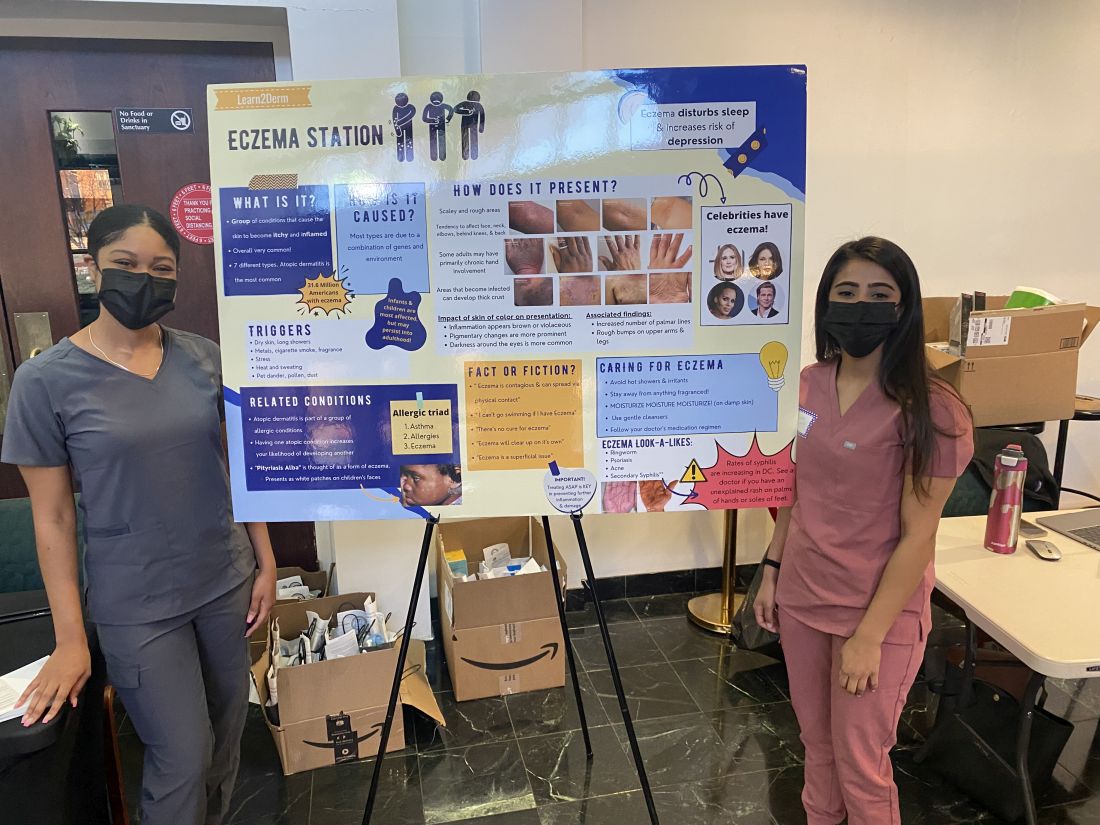
The pilot project, funded with a grant from Pfizer, involved five 2-hour clinics held on Mondays from 4 p.m. to 6 p.m., that together served almost 50 adult and pediatric patients. Grants from Pfizer and Eli Lilly enabled additional clinics in the spring of 2023 and into the summer. And in June, GWU and Pfizer announced a $1 million national grant program focused on broad implementation of what they’ve coined the “Teledermatology Help Desk Clinic” model.
Practices or organizations that secure grants will utilize GWU’s experience and meet with an advisory council of experts in dermatology telemedicine and community advocacy. Having a “long-term plan” and commitment to sustainability is an important element of the model, said Dr. Friedman, who is chairing the grant program.
Patients deem clinic ‘extremely’ helpful
As one of the most prevalent skin disorders – and one with a documented history of elevated risk for specific populations – AD was a good starting point for the teledermatology clinic program. Patients who identify as Black have a higher incidence and prevalence of AD than those who identify as White and Hispanic, and they tend to have more severe disease. Yet they account for fewer visits to dermatologists for AD.
One cross-sectional study of about 3,500 adults in the United States with AD documented that racial/ethnic and socioeconomic disparities reduce outpatient utilization of AD care and increase urgent care and hospital utilization. And in a longitudinal cohort study of children in the United States with AD, Black children with poorly controlled AD were significantly less likely than White children to see a dermatologist.
Like other programs, the GWU department of dermatology had pivoted to telehealth in 2020, and a published survey of patients who attended telehealth appointments during the early part of the pandemic showed that it was generally well liked – and not only for social distancing, but for time efficiency and because transportation was not needed. Only 10% of the 168 patients who completed the survey (out of 894 asked) reported they were unlikely to undertake another telehealth visit. For 10%, eczema was the reason for the visit.
However, only 1% of the survey respondents were from Ward 8, which “begged the question, did those who really need access know this was an option?” Dr. Friedman said at the RAD meeting. He wondered whether there was not only a dermatology desert in Ward 8, but a “technology desert” as well.
Findings from a patient satisfaction survey taken at the end of the pilot program are encouraging, Dr. Friedman said. While data on follow-up visits has not been collected yet, “what I do now have a sense of” is that “the entry point [afforded by the clinics] changed the course in terms of patients’ understanding of the disease and how they feel about its management.”
About 94% of survey respondents indicated the clinic was “extremely” helpful and the remainder said it was “very” helpful; 90% said telehealth significantly changed how they will manage their condition; and 97% said it is “extremely” important to continue the clinics. The majority of patients – 70% – indicated they did not have a dermatologist.
Education about AD at the clinics covers moisturizers/emollients, bathing habits, soaps and detergents, trigger avoidance, and the role of stress and environmental factors in disease exacerbation. Trade samples of moisturizers, mild cleansers, and other products have increasingly been available.
For prescriptions of topical steroids and other commonly prescribed medications, Dr. Friedman and associates combed GoodRx for coupons and surveyed local pharmacies for self-pay pricing to identify least expensive options. Patients with AD who were deemed likely candidates for more advanced therapies in the future were educated about these possibilities.
Alopecia areata
The addition of alopecia areata drew patients with other forms of hair loss as well, but “we weren’t going to turn anyone away who did not have that specific autoimmune form of hair loss,” Dr. Friedman said. Depending on the diagnosis, prescriptions were written for minoxidil and 5-alpha reductase inhibitors.
Important for follow-up is GWU’s acceptance of Medicaid and the availability of both a sliding scale for self-pay and services that assist patients in registering for Medicaid and, if eligible, other insurance plans.
Building partnerships, earning trust
Establishment of the teledermatology clinic program took legwork and relationship building. “You can’t just show up. That’s not enough,” said Dr. Friedman, who also directs the dermatology residency program at GWU. “You have to show through action and through investment of time and energy that you are legitimate, that you’re really there for the long haul.”
Dr. Friedman had assistance from the Rodham Institute, which was established at GWU (and until recently was housed there) and has a history of engagement with local stakeholders such as community centers, church leadership, politicians, and others in the Washington area. He was put in touch with Bishop Deborah Webb at the Temple of Praise Church, a community pillar in Ward 8, and from there “it was a courtship,” he said, with trust to be built and logistics to be worked out. (Budgets for the clinics, he noted, have included compensation to the church and gift cards for church volunteers who are present at the clinics.)
In the meantime, medical student volunteers from GWU, Howard University, and Georgetown University were trained in telemedicine and attended a “boot camp” on AD “so they’d be able to talk with anyone about it,” Dr. Friedman said.
Advertising “was a learning experience,” he said, and was ultimately multipronged, involving church service announcements, flyers, and, most importantly, Facebook and Instagram advertisements. (People were asked to call a dedicated phone line to schedule an appointment and were invited to register in the GW Medical Faculty Associates records system, though walk-ins to the clinics were still welcomed.)
In a comment, Misty Eleryan, MD, MS, a Mohs micrographic surgeon and dermatologist in Santa Monica, Calif., said dermatology deserts are often found in rural areas and/or areas “with a higher population of marginalized communities, such as Black, Brown, or poorer individuals” – communities that tend to rely on care from urgent care or ED physicians who are unaware of how skin conditions present on darker skin tones.
Programs that educate patients about various presentations of skin conditions are helpful not only for the patients themselves, but could also enable them to help friends, family members, and colleagues, said Dr. Eleryan, who did her residency training at GWU.
“Access,” she noted, is more than just physical access to a person, place, or thing. Referring to a “five A’s” framework described several decades ago, Dr. Eleryan said access to care is characterized by affordability, availability (extent to which the physician has the requisite resources, such as personnel and technology, to meet the patient’s needs), accessibility (geographic), accommodation (extent to which the physician can meet the patient’s constraints and preferences – such as hours of operation, how communications are handled, ability to receive care without prior appointments), and acceptability (extent to which the patient is comfortable with the “more immutable characteristics” of the physician and vice versa).
The GWU program, she said, “is a great start.”
Dr. Friedman said he’s fully invested. There has long been a perception, “rightfully so, that underserved communities are overlooked especially by large institutions. One attendee told me she never expected in her lifetime to see something like this clinic and someone who looked like me caring about her community. ... It certainly says a great deal about the work we need to put in to repair longstanding injury.”
Dr. Friedman disclosed that, in addition to being a recipient of grants from Pfizer and Lilly, he is a speaker for Lilly. Dr. Eleryan said she has no relevant disclosures.
A in other underserved areas in the United States.
Washington, D.C., has “staggering health disparities that are among the largest in the country,” and Ward 8 and surrounding areas in the southeastern part of the city are “dermatology deserts,” said Adam Friedman, MD, professor and chair of dermatology at George Washington University, Washington, who started the program in 2021 with a pilot project. Dr. Friedman spoke about the project, which has since been expanded to include alopecia areata, at the Revolutionizing Atopic Dermatitis conference in April and in an interview after the meeting.
Patients who attend the clinics – held at the Temple of Praise Church in a residential area of Ward 8, a predominantly Black community with a 30% poverty rate – are entered into the GW Medical Faculty Associates medical records system and educated on telemedicine best practices (such as not having light behind them during a session) and how to use telemedicine with their own device.
Those with AD who participate learn about the condition through an image-rich poster showing how it appears in various skin tones, handouts, National Eczema Association films, and discussion with medical students who staff the clinics under Dr. Friedman’s on-site supervision. Participants with alopecia areata similarly can view a poster and converse about the condition.
Patients then have a free 20-minute telehealth visit with a GWU dermatology resident in a private room, and a medical student volunteer nearby to assist with the technology if needed. They leave with a treatment plan, which often includes prescriptions, and a follow-up telemedicine appointment.
The program “is meant to be a stepping point for initiating care ... to set someone up for success for recurrent telehealth visits in the future” and for treatment before symptoms become too severe, Dr. Friedman said in an interview. “We want to demystify telemedicine and educate on the disease state and dispel myths ... so the patient understands why it’s happening” and how it can be treated.
The pilot project, funded with a grant from Pfizer, involved five 2-hour clinics held on Mondays from 4 p.m. to 6 p.m., that together served almost 50 adult and pediatric patients. Grants from Pfizer and Eli Lilly enabled additional clinics in the spring of 2023 and into the summer. And in June, GWU and Pfizer announced a $1 million national grant program focused on broad implementation of what they’ve coined the “Teledermatology Help Desk Clinic” model.
Practices or organizations that secure grants will utilize GWU’s experience and meet with an advisory council of experts in dermatology telemedicine and community advocacy. Having a “long-term plan” and commitment to sustainability is an important element of the model, said Dr. Friedman, who is chairing the grant program.
Patients deem clinic ‘extremely’ helpful
As one of the most prevalent skin disorders – and one with a documented history of elevated risk for specific populations – AD was a good starting point for the teledermatology clinic program. Patients who identify as Black have a higher incidence and prevalence of AD than those who identify as White and Hispanic, and they tend to have more severe disease. Yet they account for fewer visits to dermatologists for AD.
One cross-sectional study of about 3,500 adults in the United States with AD documented that racial/ethnic and socioeconomic disparities reduce outpatient utilization of AD care and increase urgent care and hospital utilization. And in a longitudinal cohort study of children in the United States with AD, Black children with poorly controlled AD were significantly less likely than White children to see a dermatologist.
Like other programs, the GWU department of dermatology had pivoted to telehealth in 2020, and a published survey of patients who attended telehealth appointments during the early part of the pandemic showed that it was generally well liked – and not only for social distancing, but for time efficiency and because transportation was not needed. Only 10% of the 168 patients who completed the survey (out of 894 asked) reported they were unlikely to undertake another telehealth visit. For 10%, eczema was the reason for the visit.
However, only 1% of the survey respondents were from Ward 8, which “begged the question, did those who really need access know this was an option?” Dr. Friedman said at the RAD meeting. He wondered whether there was not only a dermatology desert in Ward 8, but a “technology desert” as well.
Findings from a patient satisfaction survey taken at the end of the pilot program are encouraging, Dr. Friedman said. While data on follow-up visits has not been collected yet, “what I do now have a sense of” is that “the entry point [afforded by the clinics] changed the course in terms of patients’ understanding of the disease and how they feel about its management.”
About 94% of survey respondents indicated the clinic was “extremely” helpful and the remainder said it was “very” helpful; 90% said telehealth significantly changed how they will manage their condition; and 97% said it is “extremely” important to continue the clinics. The majority of patients – 70% – indicated they did not have a dermatologist.
Education about AD at the clinics covers moisturizers/emollients, bathing habits, soaps and detergents, trigger avoidance, and the role of stress and environmental factors in disease exacerbation. Trade samples of moisturizers, mild cleansers, and other products have increasingly been available.
For prescriptions of topical steroids and other commonly prescribed medications, Dr. Friedman and associates combed GoodRx for coupons and surveyed local pharmacies for self-pay pricing to identify least expensive options. Patients with AD who were deemed likely candidates for more advanced therapies in the future were educated about these possibilities.
Alopecia areata
The addition of alopecia areata drew patients with other forms of hair loss as well, but “we weren’t going to turn anyone away who did not have that specific autoimmune form of hair loss,” Dr. Friedman said. Depending on the diagnosis, prescriptions were written for minoxidil and 5-alpha reductase inhibitors.
Important for follow-up is GWU’s acceptance of Medicaid and the availability of both a sliding scale for self-pay and services that assist patients in registering for Medicaid and, if eligible, other insurance plans.
Building partnerships, earning trust
Establishment of the teledermatology clinic program took legwork and relationship building. “You can’t just show up. That’s not enough,” said Dr. Friedman, who also directs the dermatology residency program at GWU. “You have to show through action and through investment of time and energy that you are legitimate, that you’re really there for the long haul.”
Dr. Friedman had assistance from the Rodham Institute, which was established at GWU (and until recently was housed there) and has a history of engagement with local stakeholders such as community centers, church leadership, politicians, and others in the Washington area. He was put in touch with Bishop Deborah Webb at the Temple of Praise Church, a community pillar in Ward 8, and from there “it was a courtship,” he said, with trust to be built and logistics to be worked out. (Budgets for the clinics, he noted, have included compensation to the church and gift cards for church volunteers who are present at the clinics.)
In the meantime, medical student volunteers from GWU, Howard University, and Georgetown University were trained in telemedicine and attended a “boot camp” on AD “so they’d be able to talk with anyone about it,” Dr. Friedman said.
Advertising “was a learning experience,” he said, and was ultimately multipronged, involving church service announcements, flyers, and, most importantly, Facebook and Instagram advertisements. (People were asked to call a dedicated phone line to schedule an appointment and were invited to register in the GW Medical Faculty Associates records system, though walk-ins to the clinics were still welcomed.)
In a comment, Misty Eleryan, MD, MS, a Mohs micrographic surgeon and dermatologist in Santa Monica, Calif., said dermatology deserts are often found in rural areas and/or areas “with a higher population of marginalized communities, such as Black, Brown, or poorer individuals” – communities that tend to rely on care from urgent care or ED physicians who are unaware of how skin conditions present on darker skin tones.
Programs that educate patients about various presentations of skin conditions are helpful not only for the patients themselves, but could also enable them to help friends, family members, and colleagues, said Dr. Eleryan, who did her residency training at GWU.
“Access,” she noted, is more than just physical access to a person, place, or thing. Referring to a “five A’s” framework described several decades ago, Dr. Eleryan said access to care is characterized by affordability, availability (extent to which the physician has the requisite resources, such as personnel and technology, to meet the patient’s needs), accessibility (geographic), accommodation (extent to which the physician can meet the patient’s constraints and preferences – such as hours of operation, how communications are handled, ability to receive care without prior appointments), and acceptability (extent to which the patient is comfortable with the “more immutable characteristics” of the physician and vice versa).
The GWU program, she said, “is a great start.”
Dr. Friedman said he’s fully invested. There has long been a perception, “rightfully so, that underserved communities are overlooked especially by large institutions. One attendee told me she never expected in her lifetime to see something like this clinic and someone who looked like me caring about her community. ... It certainly says a great deal about the work we need to put in to repair longstanding injury.”
Dr. Friedman disclosed that, in addition to being a recipient of grants from Pfizer and Lilly, he is a speaker for Lilly. Dr. Eleryan said she has no relevant disclosures.
Heat waves plus air pollution tied to doubling of fatal MI
, a study from China suggests.

The researchers estimate that up to 3% of all deaths due to MI could be attributed to the combination of extreme temperatures and high levels of ambient fine particulate matter (PM2.5).
“Our findings provide evidence that reducing exposure to both extreme temperatures and fine particulate pollution may be useful to prevent premature deaths from heart attack,” senior author Yuewei Liu, MD, PhD, with Sun Yat-sen University in Guangzhou, China, said in a statement.
There is “long-standing evidence” of the harmful cardiovascular effects of air pollution, Jonathan Newman, MD, MPH, cardiologist at NYU Langone Heart in New York, who wasn’t involved in the study, said in an interview.
The added value of this study was finding an interaction between extreme hot temperatures and air pollution, “which is worrisome with global warming,” said Dr. Newman, assistant professor, department of medicine, the Leon H. Charney Division of Cardiology at NYU Langone Health.
The study was published online in Circulation.
Intensity and duration matter
The researchers analyzed data on 202,678 adults (mean age, 77.6 years; 52% male) who suffered fatal MI between 2015 and 2020 in Jiangsu province, a region with four distinct seasons and a wide range of temperatures and ambient PM2.5.
They evaluated the association of exposure to extreme temperature events, including both hot and cold spells, and PM2.5 with MI mortality, and their interactive effects.
Among the key findings:
- The risk of fatal MI was 18% higher during 2-day heat waves with heat indexes at or above the 90th percentile (ranging from 82.6° to 97.9° F) and 74% higher during 4-day heat waves with heat indexes at or above the 97.5th percentile (ranging from 94.8° to 109.4° F), compared with control days.
- The risk of fatal MI was 4% higher during 2-day cold snaps with temperatures at or below the 10th percentile (ranging from 33.3° to 40.5° F) and 12% higher during 3-day cold snaps with temperatures at or below the 2.5th percentile (ranging from 27.0° to 37.2° F).
- The risk of fatal MI was twice as high during 4-day heat waves that had PM2.5 above 37.5 mcg/m3. Days with high levels of PM2.5 during cold snaps did not have an equivalent increase in the risk of fatal MI.
- Up to 2.8% of MI deaths during the 5-year study period may be attributable to the combination of extreme temperature exposure and PM2.5 at levels exceeding World Health Organization air quality guidelines (37.5 mcg/m3).
- The risk of fatal MI was generally higher among women than men during heat waves and was higher among adults 80 years old and older than in younger adults during heat waves, cold snaps, or days with high levels of PM2.5.
The finding that adults over age 80 are particularly susceptible to the effects of heat and air pollution and the interaction of the two is “notable and particularly relevant given the aging of the population,” Dr. Newman told this news organization.
Mitigating both extreme temperature events and PM2.5 exposures “may bring health cobenefits in preventing premature deaths from MI,” the researchers write.
“To improve public health, it is important to take fine particulate pollution into consideration when providing extreme temperature warnings to the public,” Dr. Liu adds in the statement.
In an earlier study, Dr. Liu and colleagues showed that exposure to both large and small particulate matter, as well as nitrogen dioxide, was significantly associated with increased odds of death from MI.
This study was funded by China’s Ministry of Science and Technology. The authors and Dr. Newman have disclosed no relevant financial relationships.
A version of this article first appeared on Medscape.com.
, a study from China suggests.
The researchers estimate that up to 3% of all deaths due to MI could be attributed to the combination of extreme temperatures and high levels of ambient fine particulate matter (PM2.5).
“Our findings provide evidence that reducing exposure to both extreme temperatures and fine particulate pollution may be useful to prevent premature deaths from heart attack,” senior author Yuewei Liu, MD, PhD, with Sun Yat-sen University in Guangzhou, China, said in a statement.
There is “long-standing evidence” of the harmful cardiovascular effects of air pollution, Jonathan Newman, MD, MPH, cardiologist at NYU Langone Heart in New York, who wasn’t involved in the study, said in an interview.
The added value of this study was finding an interaction between extreme hot temperatures and air pollution, “which is worrisome with global warming,” said Dr. Newman, assistant professor, department of medicine, the Leon H. Charney Division of Cardiology at NYU Langone Health.
The study was published online in Circulation.
Intensity and duration matter
The researchers analyzed data on 202,678 adults (mean age, 77.6 years; 52% male) who suffered fatal MI between 2015 and 2020 in Jiangsu province, a region with four distinct seasons and a wide range of temperatures and ambient PM2.5.
They evaluated the association of exposure to extreme temperature events, including both hot and cold spells, and PM2.5 with MI mortality, and their interactive effects.
Among the key findings:
- The risk of fatal MI was 18% higher during 2-day heat waves with heat indexes at or above the 90th percentile (ranging from 82.6° to 97.9° F) and 74% higher during 4-day heat waves with heat indexes at or above the 97.5th percentile (ranging from 94.8° to 109.4° F), compared with control days.
- The risk of fatal MI was 4% higher during 2-day cold snaps with temperatures at or below the 10th percentile (ranging from 33.3° to 40.5° F) and 12% higher during 3-day cold snaps with temperatures at or below the 2.5th percentile (ranging from 27.0° to 37.2° F).
- The risk of fatal MI was twice as high during 4-day heat waves that had PM2.5 above 37.5 mcg/m3. Days with high levels of PM2.5 during cold snaps did not have an equivalent increase in the risk of fatal MI.
- Up to 2.8% of MI deaths during the 5-year study period may be attributable to the combination of extreme temperature exposure and PM2.5 at levels exceeding World Health Organization air quality guidelines (37.5 mcg/m3).
- The risk of fatal MI was generally higher among women than men during heat waves and was higher among adults 80 years old and older than in younger adults during heat waves, cold snaps, or days with high levels of PM2.5.
The finding that adults over age 80 are particularly susceptible to the effects of heat and air pollution and the interaction of the two is “notable and particularly relevant given the aging of the population,” Dr. Newman told this news organization.
Mitigating both extreme temperature events and PM2.5 exposures “may bring health cobenefits in preventing premature deaths from MI,” the researchers write.
“To improve public health, it is important to take fine particulate pollution into consideration when providing extreme temperature warnings to the public,” Dr. Liu adds in the statement.
In an earlier study, Dr. Liu and colleagues showed that exposure to both large and small particulate matter, as well as nitrogen dioxide, was significantly associated with increased odds of death from MI.
This study was funded by China’s Ministry of Science and Technology. The authors and Dr. Newman have disclosed no relevant financial relationships.
A version of this article first appeared on Medscape.com.
FROM CIRCULATION
Fatalities from breast cancer have ‘improved substantially’
Women diagnosed with early-stage breast cancer are more likely to become long-term survivors of the disease now than they were 20 years ago, a new study found.
Researchers at the University of Oxford (England) conducted an observational study that examined case fatality rates for women with breast cancer and found that the prognosis for women has “improved substantially” over the past few decades. For women diagnosed with early invasive breast cancer during the 1990s, the risk of death within 5 years of diagnosis was just over 14% on average. For women diagnosed during the 2010s, it was nearly 5% on average.
“The take-home message in our study is that it’s good news for women who are diagnosed with early breast cancer today because most of them can expect to become long-term cancer survivors, and so I think our results are reassuring,” said lead study author Carolyn Taylor, DPhil, a clinical oncologist from the Nuffield Department Of Population Health, University of Oxford.
The study was published online in the BMJ.
Although breast cancer survival has improved, recent estimates don’t incorporate detailed data on age, tumor size, tumor grade, and nodal and receptor status. In the current population-based study, researchers explored improvement in survival from early-stage breast cancer. They used nine patient and tumor characteristics as factors in their analysis.
The study is based on data from the National Cancer Registration for 512,447 women in England who were diagnosed with early-stage invasive breast cancer between 1993 and 2015. Women were broken into four groups: those diagnosed during 1993-1999, 2000-2004, 2005-2009, and 2010-2015.
The study focused on women who initially underwent either breast-conserving surgery or mastectomy as their first treatment. Data included age, tumor size, tumor grade, number of positive nodes, and estrogen receptor (ER) status. For women who were diagnosed from 2010 to 2015, HER2 status was included. Data regarding recurrence, receipt of neoadjuvant therapy, and patients who were diagnosed with more than one cancer were not included.
The major finding: Among women diagnosed with early-stage invasive breast cancer, the risk of dying decreased almost threefold between 1993 and 2015. The 5-year cumulative case fatality risk was 14.4% for women diagnosed in the 1990s (1993-1999) versus 4.9% for women diagnosed about 2 decades later (2010-2015).
Dr. Taylor and colleagues found that the case fatality rate was highest during the 5 years after diagnosis; within those years, the rates typically increased during the first 2 years, peaked during the third, and declined thereafter.
The 5-year risk of death, however, varied widely among women in the population. For most (62.8%) who were diagnosed between 2010 and 2015, the case fatality risk was 3% or less; however, for a small subset of women (4.6%), the risk reached 20% or higher.
Patients with ER-negative tumors tended to have worse prognoses in the first decade following their diagnosis. Overall, higher tumor size and grade, more positive nodes, and older age tended to be associated with worse prognoses.
Overall, the annual case fatality rates decreased over time in nearly every patient group.
While Dr. Taylor said these findings are encouraging, she added that the investigators did not analyze why survival rates have improved over 2 decades.
“We didn’t explain how much of the improvement was due to advances treatments, improved screening rates, etc,” Dr. Taylor said. Another limitation is that data on recurrence were not available.
Kathy Miller, MD, who specializes in breast cancer at the Melvin and Bren Simon Cancer Center at Indiana University, Indianapolis, said the 5-year mark for survival is great news for some patients with breast cancer but that the time frame doesn’t apply to all.
While the risk of case fatality from breast cancer may be higher during the first 5 years after diagnosis, Dr. Miller said that is not the case for women with ER-positive breast cancer. In the study, the researchers highlighted this trend for ER status: before the 10-year mark, survival rates for women with ER-positive disease were better, but after the 10-year mark, those with ER-negative tumors seemed to fare slightly better.
“Many patients have heard this very arbitrary 5-year mark, and for patients with ER-positive disease, that 5-year mark has no meaning, because their risk in any given year is very low and it stays at that very low consistent level for at least 15 years, probably longer,” Dr. Miller said in an interview. “I think a better way to think about this for ER-positive patients is that every day that goes by without a problem makes it a tiny bit less likely that you will ever have a problem.”
The authors took a similar view for the overall population, concluding that, “although deaths from breast cancer will continue to occur beyond this [5-year mark], the risk during each subsequent 5-year period is likely to be lower than during the first 5 years.”
The research was funded by Cancer Research UK, the National Institute for Health Research Oxford Biomedical Research Centre, the U.K. Medical Research Council, and the University of Oxford. Some study authors received support for several of these institutions, but they reported no financial relationships with organizations that might have had an interest in the submitted work during the previous 3 years.
A version of this article first appeared on Medscape.com.
FROM THE BMJ