User login
VIDEO: Get comfortable with screening for, treating CVD risk in RA
LAS VEGAS – When genetic risk and lifestyle risk factors collide with the baseline systemic inflammation of rheumatoid arthritis (RA), cardiovascular risk increases significantly. Helping patients to manage risk for cardiovascular disease (CVD) requires getting comfortable in making risk assessments and counseling patients about medication and lifestyle options, especially for patients who are not actively being managed by primary care physicians.
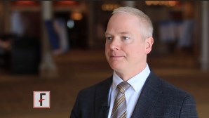
Jon Giles, MD, said that a large portion of the elevated risk for CVD in patients with RA is “driven by the fact that [RA] patients have more atherosclerosis.”
Dr. Giles, professor of medicine at Columbia University, New York, said that other CVD risk factors can boost the risk further. “If you have diabetes, smoking, high blood pressure, elevated lipids in your blood – if you have a combination of those plus inflammation, it makes that risk even higher,” Dr. Giles said at the annual Perspectives in Rheumatic Diseases held by Global Academy for Medical Education.
“There’s definitely a lot of data that suggests that, as rheumatologists, we’re not doing a very good job of screening and treating for cardiovascular disease and risk,” Dr. Giles said in an interview at the meeting. He suggests that his fellow rheumatologists become comfortable with screening and treatment guidelines for cardiovascular disease. For selected patients, coronary CT or carotid ultrasound may be valuable in guiding decision making, since very low LDL cholesterol may be correlated with an increased risk of CVD for some patients with RA.
Global Academy for Medical Education and this news organization are owned by the same parent company.
The video associated with this article is no longer available on this site. Please view all of our videos on the MDedge YouTube channel
On Twitter @karioakes
EXPERT ANALYSIS FROM THE ANNUAL PERSPECTIVES IN RHEUMATIC DISEASES
Observation Status Utilization by Hospitalist Groups Is Increasing
Editor's Note: Listen to Dr. Smith share more of his views on the State of Hospital Medicine report.
Hospitalist groups and their stakeholders must continually adapt to evolving reimbursement models and their attendant financial foci on quality. Even in the midst of care models that rely less heavily on volume of care as a marker for reimbursement, the use of criteria by insurers to separate hospital stays into inpatient or observation status remains widespread. Hospitalist groups vary in the reimbursement model environment they work in, and different reimbursement models can drive hospitalist group behavior in different ways.
SHM’s 2016 State of Hospital Medicine Report revisits the issue of observation status utilization raised in previous surveys.1 The 2012 survey’s methodology reports admissions classified as observation status based on CPT coding.2 The 2016 survey continues the 2014 survey methodology of using discharges classified as observation status based on CPT coding, along with same-day admission and discharge reported as a third hospitalization status category. In groups serving adults only, observation discharges accounted for 21.2% of all discharges, which represents an increase from 16.1% in the 2014 survey3 and a general return to the 2012-reported percentage of 20%. If same-day admissions and discharges, many of which are likely classified as observation status, are added, then observation status use in the 2016 survey may be as high as 24% of all admissions. This represents a considerable increase from the combined 19.6% rate in 2014.
Changes in non-academic status hospitalist groups largely account for this increase. Academic hospitalist groups reported an observation status utilization rate of 15.3% of admissions in 2012 and 19.4% in 2014, with a subsequent decrease to 17.5% reported in the 2016 survey. Inclusion of same-day admission and discharge with reported observation status use also reveals a decrease from 22.8% in 2014 to 20.8% in the new survey. In contrast, non-academic hospitalist groups now report a substantial change in observation status utilization, up to 21.4% in the 2016 survey from 15.6% in 2014 and similar to the 2012 level of 20.4%. When same-day admission and discharge codes are also included, the totals for non-academic hospitalist groups also evidence an increase, to 24.3% in the new survey from 19.2% in 2014.
I postulated in 2015 that the comparative increase in observation status utilization by academic groups as compared with non-academic groups in the 2014 survey may have been associated with greater proficiency in documentation and related billing inherent in a bedside clinical workforce entirely composed of physicians who have completed postgraduate training. Other phenomena may now potentially explain the increase in observation status use we see in the 2016 survey. These include adoption of the two-midnight rule by the Centers for Medicare & Medicaid Services, use of readmission rates in hospitalist group incentive structures, sharing of cost savings between hospitalist groups and healthcare organizations mutually engaged in third-party bundled payment arrangements, or risk-avoidant strategies executed by clinicians and institutional coders perhaps in excess of their institutions’ needs for risk avoidance. For many of these events, the 2016 State of Hospital Medicine Report provides further benchmark data, in a national and regional context, to inform understanding for hospitalist groups facing challenges associated with observation status utilization.
G. Randy Smith Jr., MD, MS, FRCP(Edin), SFHM, is an assistant professor in the Division of Hospital Medicine at Northwestern University Feinberg School of Medicine in Chicago.
References
- 2016 State of Hospital Medicine Report. Society of Hospital Medicine website. Accessed September 11, 2016.
- 2012 State of Hospital Medicine Report. Society of Hospital Medicine website. Accessed September 11, 2016.
- 2014 State of Hospital Medicine Report. Society of Hospital Medicine website.
Accessed September 11, 2016.
More U.S. Babies Born Addicted to Opiates
(Reuters Health) - The proportion of U.S. babies born suffering from withdrawal syndrome after exposure to heroin or prescription opiates in utero has more than doubled in less than a decade, a study suggests.
Nationally, the rate of neonatal abstinence syndrome involving mothers' use of opiates - which includes heroin as well as prescription narcotics like codeine and Vicodin - surged from 2.8 cases for every 1,000 births in 2009 to 7.3 cases for every 1,000 births in 2013, the study found.
At least some of this surge in the case count is due to drug policies designed to crack down on prescription drug abuse and combat the methamphetamine epidemic, said lead study author Dr. Joshua Brown, a pharmacy researcher at the University of Kentucky in Lexington.
"The drug policies of the early 2000s were effective in reducing supply - we have seen a decrease in methamphetamine abuse and there have been reductions in some aspects of prescription drug abuse," Brown said by email. "However, the indirect results, mainly the increase in heroin abuse, were likely not anticipated and we are just starting to see these."
The findings of the current study add to a growing body of evidence pointing to a surge in births of babies suffering from opiate withdrawal. One report last month from the U.S. Centers for Disease Control and Prevention found an even bigger spike over a longer period, from 1.5 cases for every 1,000 births in 1999 to 6 cases per 1,000 in 2013.
CDC researchers also found wide variation in neonatal abstinence syndrome by state, ranging in 2013 from 0.7 cases for every 1,000 births in Hawaii to 33.4 cases per 1,000 in West Virginia.
"We know that certain states are harder hit by the opioid/heroin abuse epidemic, with about 10 states contributing half of all neonatal abstinence syndrome cases," Brown said. "These states are often more rural and impoverished areas of the U.S. such as Mississippi, Alabama, and West Virginia."
Brown and colleagues looked at Kentucky in particular. Here, the rate of neonatal abstinence syndrome climbed from 5 cases for every 1,000 births in 2008 to 21.2 cases per 1,000 births in 2014, researchers report in JAMA Pediatrics, online September 26.
While the study didn't look at health outcomes for babies born suffering from drug withdrawal, these infants often require intensive medical care. (See Reuters' 2015 special report "Helpless and Hooked" here.)
These babies may have central nervous system issues like seizures and tremors, gastrointestinal problems and feeding difficulties, breathing challenges, as well as unstable body temperatures.
Typically, they remain in the hospital for several weeks after birth and receive low doses of methadone, Brown said.
Treatment can ease withdrawal symptoms in newborns, but can't necessarily address developmental problems these infants may have later on, said Dr. William Carey, a pediatrics researcher at pediatrics at Mayo Clinic Children's Center in Rochester, Minnesota.
"While abuse of prescription opiates has declined, the use of illicit opiates has increased such that there may be a zero-sum game at best," Carey, who wasn't involved in the study, said by email. "Since maternal use of either prescription opiates or illicit opiates is associated with withdrawal in newborns, it is reasonable to think that any increase in the overall use of opiates would be linked to an increase in the rate of neonatal abstinence syndrome."
Noninvasive Prenatal Testing: Practice Boosters
Diagnostic testing needed to confirm cfDNA results for aneuploidy and microdeletions, finds retrospective study
Researchers at the David Geffen School of Medicine at the University of California-Los Angeles, found a 73.5% positive predictive value (95% CI, 63%-82%) for cfDNA screening for autosomal aneuploidy, a lower-than-reported rate. The positive predictive value for microdeletion testing was found to be 0%.
This was a retrospective cohort study published in June in the American Journal of Obstetrics and Gynecology. It included 105 patients with abnormal or nonreportable cfDNA results for trisomies 21, 18, or 13, and 26 patients with positive or nonreportable microdeletions. Of the 105 patients with abnormal results for the trisomies, 92 results (87.6%) were positive for trisomy 21 (48, 52.2%), trisomy 18 (22, 23.9%), trisomy 13 (17, 18.5%), triploidy (2, 2.2%) and positive for >1 parameter (3, 3.3%). An additional 13 results (12.4%) were nonreportable.
Abnormal cfDNA results were associated with positive serum screening (by group: trisomy 21 [17/48]; trisomy 18 [7/22]; trisomy 13 [3/17]; nonreportable [2/13]; P = .004), and abnormal first-trimester ultrasound (trisomy 21 [25/45]; trisomy 18 [13/20]; trisomy 13 [6/14]; nonreportable [1/13]; P = .003). There was no association between false-positive rates and testing platform, but there was a difference between the 4 laboratories included in the study (P = .018).
In all, 26 patients had positive (n = 9) or nonreportable (n = 17) microdeletion results. Seven of 9 screens positive for microdeletions underwent confirmatory testing; all were false positives.
When is NIPT appropriate for use as a diagnostic tool? Large-scale systematic review evaluates the answer.
Many systematic reviews and meta-analyses evaluating the accuracy of cell-free fetal (cf) DNA−based testing have been published. This May 2016 study in the British Journal of Obstetrics and Gynecology was the most comprehensive of singleton pregnancies to date, say researchers, who aimed to reduce analysis bias by including only cohort studies (117 analyzed), undertaking bivariate meta-analysis when possible, and including all indications for antenatal use, which allowed for a uniform comparison of NIPT use in actual clinical practice.
The researchers concluded that cell-free fetal (cf) DNA−based NIPT is diagnostic for fetal sex and rhesus D (RHD) status. For trisomies 21, 18, and 13, however, they concluded that the lower sensitivities and specificities found and disease prevalence, combined with the biological influence of confined placental mosaicism, designates NIPT as a screening test (with invasive testing required to confirm a positive test result). They advised that these factors be considered not only when clinicians are counseling patients but also in the overall assessment of the cost of introducing NIPT into routine care.
2016 update on NIPT from the American College of Medical Genetics and Genomics
The American College of Medical Genetics and Genomics (ACMG) has published an updated position statement in Genetics in Medicine in July 2016 that replaces its 2013 statement. Among its recommendations, the ACMG advocates:
- providing up-to-date, balanced, and accurate information early in gestation to optimize patient decision making, independent of the screening approach used
- that laboratories work with public health officials, policymakers, and private payers to make NIPT, including the pretest and posttest education and counseling, accessible to all pregnant women
- informing all pregnant women that diagnostic testing (chorionic villus sampling or amniocentesis) is an option for the detection of chromosome abnormalities and clinically significant copy-number variants.
ACMG also recommends:
- informing all pregnant women that noninvasive prenatal screening (NIPS) is the most sensitive screening option for traditionally screened aneuploidies
- referring patients to a trained genetics professional when an increased risk of aneuploidy is reported after NIPS
- offering diagnostic testing when a positive screening test result is reported after NIPS
- laboratories provide readily visible and clearly stated detection rate (DR), clinical specificity (SPEC), and positive predictive values (PPV), and negative predictive values (NPVs) for conditions being screened
- laboratories not offer screening for Patau, Edwards, and Down syndromes if they cannot report DR, SPEC, and PPV values for these conditions.
ACMG does not recommend:
- NIPS to screen for autosomal aneuploidies other than those involving chromosomes 13, 18, and 21.
Public health perspective: Long-term study concludes implementing cfDNA in high-risk women could be cost-effective
Researchers in Italy retrospectively analyzed the performance of first-trimester screening (FTS) test in a general obstetrics population, including a cost-benefit analysis of a hypothetical model that implemented universal FTS testing using cfDNA. A 2-step strategy based on nuchal translucency, serum screening, and ultrasound assessment of the nasal bone was applied when analyzing the FTS results of 6,679 women. The researchers identified 3 groups: high-risk: >1:250; intermediate-risk: 1:251-1:999; and low-risk group: <1:1000. Women at intermediate-risk underwent NB assessment and recalculation of individual risk. All women at high-risk were offered fetal karyotyping.
The investigators concluded that the Italian public health system would not be able to sustain the cost of universal cfDNA testing in women undergoing FTS. However, a possible scenario based on use of cfDNA FTS in women at high-risk would result in a 6-fold reduction in the number of invasive procedures. It would also avoid 2 false-negative results (trisomy 21) diagnosed in women with intermediate-risk using the current screening strategy of combined invasive and noninvasive testing.
Early epilepsy increases risk of later comorbid ADHD in autism
VIENNA – Early-onset idiopathic epilepsy occurring before age 7 years nearly doubles the likelihood that a child with autism spectrum disorder will later develop comorbid attention-deficit/hyperactivity disorder, Johnny Downs, MD, reported at the annual congress of the European College of Neuropsychopharmacology.
Comorbid ADHD is common in the setting of autism spectrum disorder (ASD). In a search for risk factors for the comorbid condition, he and his coinvestigators reviewed the physical health records prior to age 7 years of 3,032 patients with ASD referred at ages 3-17 years to child and adolescent mental health services clinics serving South London.
“That’s information that often doesn’t make it into the clinical psychiatric record,” noted Dr. Downs, a child psychiatrist at King’s College London.
Half of the 3,032 subjects were diagnosed with ASD at age 6-12 years and another 39% at age 13-17. During 5 years of prospective follow-up after being diagnosed with ASD in this longitudinal observational study, 25.5% of patients were diagnosed with comorbid ADHD. Looking back through the early physical health records, 114 (3.76%) of study participants had experienced early-onset epilepsy before age 7 years.
This large sample size allowed for robust multivariate adjustment for potential confounders. In a multivariate analysis, ASD patients with a history of early-onset epilepsy were at a significant 1.75-fold increased risk for subsequent comorbid ADHD. The analysis was adjusted for family history of epilepsy, sociodemographic factors, intellectual disability, previous head injury, perinatal complications, central nervous system tumors, early meningitis, and other confounders.
“The take-home message would be if you’ve got social and communication difficulties in a young child appearing at the age of 5, 6, or 7 [years], and there’s a history of seizures, we are seeing from observational data that the child is at increased risk of ADHD over the age of 7,” Dr. Downs said in an interview.
Compared with white subjects with ASD, the risk of developing comorbid ADHD was reduced by 37% in black and by 52% in Asian patients with ASD.
He plans further studies aimed at determining whether conventional ADHD management strategies have the same risk/benefit ratios in children with ASD and comorbid ADHD as in those with ADHD alone.
Dr. Downs reported having no financial conflicts of interest regarding his study, which was conducted free of commercial support.
AT THE ECNP CONGRESS
Key clinical point: Youths with autism spectrum disorder and a history of early-onset epilepsy before age 7 years are at an increased risk of subsequent comorbid ADHD.
Major finding: Youths with autism spectrum disorder who have a history of early-onset epilepsy before age 7 years are at 1.75-fold increased likelihood of subsequently developing comorbid ADHD.
Data source: This longitudinal study included 3,032 children and adolescents with autism spectrum disorder, 26% of whom developed ADHD during 5 years of prospective follow-up.
Disclosures: The presenter reported having no financial conflicts of interest regarding this study, which was conducted free of commercial support.
Reimbursement hurdles hinder Entresto use in HFrEF
ORLANDO – Use of sacubitril/valsartan to treat patients with heart failure with reduced ejection fraction (HFrEF) became a class I recommendation in both the U.S. and European heart failure guidelines in May 2016, but virtually all U.S. health insurers continue to regard the potent and effective sacubitril/valsartan formulation as a second-line treatment that needs special preauthorization before patients receive reimbursement for the prescription.
“What is really morally sad is that U.S. payers are requiring physicians to fill out extensive, patient-by-patient paperwork” to allow patients with HFrEF to receive health insurance coverage for sacubitril/valsartan (Entresto), Milton Packer, MD, said at the annual scientific meeting of the Heart Failure Society of America.
“It is very difficult to understand why third-party payers would intentionally try to slow adoption of this life-saving drug simply because it is considered expensive. It is much cheaper than many drugs they cover for patients with cancer that don’t work half as well,” said Dr. Packer, a cardiologist and heart failure specialist at Baylor University Medical Center in Dallas.
The “excessive paperwork” for insurers when starting patients on sacubitril/valsartan is a “new and unique phenomenon among the cardiovascular drugs I prescribe,” agreed Nancy K. Sweitzer, MD, PhD, professor and chief of cardiology at the University of Arizona in Tuscon. “This approach by insurers seems based on cost; they do not want to pay” for sacubitril/valsartan, and to successfully arrange for coverage patients need to exactly match the enrollment criteria used in the PARADIGM-HF (Prospective Comparison of ARNI [Angiotensin Receptor – Neprilysin Inhibitor] with ACEI [Angiotensin-Converting Enzyme Inhibitor] to Determine Impact on Global Mortality and Morbidity in Heart Failure Trial ), the pivotal study that supplied the evidence base for making sacubitril/valsartan a class I agent for treating HFrEF.
“I don’t put some HFrEF patients on sacubitil/valsartan just because they don’t meet the trial’s entry criteria,” Dr. Sweitzer said in an interview. “Insurers seem to scrutinize every single parameter to make sure patients match the PARADIGM-HF patients. Coverage is denied if their BNP [brain natriuretic peptide] level is too low.” Dr. Sweitzer added that in one instance she had to submit a second preauthorization to simply uptitrate the dosage of sacubitril/valsartan she wanted a patient to receive.
“Based on the data it seems like you could easily identify HFrEF patients who are good candidates for sacubitril/valsartan, but your hands are tied by payers because you can’t prescribe it until you’ve first tried something else, and even then you still need to deal with a lot of paperwork,” agreed Robert O. Bonow, MD, professor of medicine at Northwestern University in Chicago. “The paperwork burden is really cumbersome for physicians with busy practices; it impedes taking care of patients,” Dr. Bonow said in an interview.
Sales figures for sacubitril/valsartan that the drug’s manufacturer, Novartis, has reported since the agent received U.S. marketing approval a little over a year ago reflect these challenges in prescribing the compound to patients. During the first quarter of 2016, Novartis reported $17 million in worldwide sales of the agent, followed by $32 million in worldwide sales during the second quarter of 2016, through June 30. With a total of $49 million in sacubitril/valsartan sales during the first 6 months of 2016, it seems like Novartis may be challenged to meet its stated target of $200 million in total sales of the compound during 2016. In April, one commentator called the $17 million sales figure for first quarter 2016 “an astonishingly small amount for a drug that was widely expected to be a blockbuster.”
Dr. Packer has in the past been a consultant to Novartis and was one of the lead investigators for the PARADIGM-HF trial. He said that currently he has no financial relationship with Novartis but he does serve as a consultant to several other drug companies. Dr. Sweitzer has received research support from Novartis and was an investigator for PARADIGM-HF. Dr. Bonow has been a consultant to Gilead.
On Twitter @mitchelzoler
EXPERT ANALYSIS FROM THE HFSA ANNUAL SCIENTIFIC MEETING
Database of research regulations gets update

for a clinical trial
Photo by Esther Dyson
The ClinRegs website has been updated and upgraded, according to the National Institute of Allergy and Infectious Diseases.
ClinRegs is an online database of country-specific information on clinical research regulations that was launched in 2014.
Now, the website houses regulatory information for 17 countries and has an interactive map on its homepage to provide a clearer picture of the countries included.
ClinRegs also has a hyperlinked table of contents on each country page that is intended to provide easier navigation.
Drop-down menus allow users to switch between country profiles and make comparisons between countries.
In addition, ClinRegs now provides a Quick Facts table with discrete pieces of information for each country.
And a feedback link has been added to the site to make it easy for users to submit comments or updates to regulations. ![]()
Combo disappoints in newly diagnosed MM
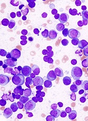
Top-line results from the phase 3 CLARION trial suggest that treatment with carfilzomib, melphalan, and prednisone (KMP) is not superior to treatment with bortezomib, melphalan, and prednisone (VMP).
The trial was designed to compare KMP and VMP in patients with newly diagnosed multiple myeloma (MM) who were ineligible for hematopoietic stem cell transplant.
The results showed that progression-free survival (PFS) rates were similar with the 2 regimens.
And although overall survival data are not yet mature, there seems to be a trend favoring the VMP regimen.
Amgen, the company developing carfilzomib, released these results yesterday.
“The CLARION results, generated in the context of a melphalan-containing regimen, are disappointing, especially given the robust data we’ve seen in the second-line setting,” said Sean E. Harper, MD, executive vice president of Research and Development at Amgen.
“However, the myeloma landscape has changed dramatically since the design of the CLARION study, with very few newly diagnosed patients treated with
melphalan-based regimens, particularly in the US. We remain committed to exploring Kyprolis in combination with other agents to advance the treatment of multiple myeloma.”
Dr Harper said he could not comment on whether the CLARION trial will continue, as Amgen hopes to present data from the trial at the 2016 ASH Annual Meeting.
The CLARION trial is a head-to-head, randomized study in transplant-ineligible patients with newly diagnosed MM. A total of 955 patients were randomized 1:1 to receive KMP or VMP for 54 weeks. The median patient age was 72.
The trial did not meet the primary endpoint of superiority in PFS. The median PFS was 22.3 months in the KMP arm and 22.1 months in the VMP arm. The hazard ratio was 0.91 (95% CI, 0.75-1.10), and the difference between the arms was not statistically significant.
The data for overall survival, a secondary endpoint, are not yet mature. But the observed hazard ratio was 1.21 (95% CI, 0.90-1.64), and there was no significant difference between the treatment arms.
The incidence of grade 3 or higher adverse events was 74.7% in the KMP arm and 76.2% in the VMP arm.
The incidence of grade 2 or higher peripheral neuropathy, a secondary endpoint, was 2.5% in the KMP arm and 35.1% in the VMP arm.
Fatal treatment-emergent adverse events occurred in 6.5% of patients in the KMP arm and 4.3% of those in the VMP arm. ![]()
KTE-C19 produces responses in aggressive NHL
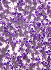
Interim results of a phase 1/2 trial suggest KTE-C19, a chimeric antigen receptor (CAR) T-cell therapy, can be effective against aggressive non-Hodgkin lymphoma (NHL).
KTE-C19, administered after conditioning chemotherapy, produced an overall response rate (ORR) of
79% and a complete response (CR) rate of 52%.
However, the therapy also caused severe adverse events (AEs), and there were 2 deaths resulting from KTE-C19-related AEs.
Kite Pharma, Inc., the company developing KTE-C19, released these results and said additional data from this trial, known as ZUMA-1, will be submitted for presentation at an upcoming scientific meeting.
ZUMA-1 has enrolled patients with chemo-refractory, aggressive NHL. The phase 1 portion of the trial included 7 patients with diffuse large B-cell lymphoma (DLBCL).
Thus far, the phase 2 portion includes 62 NHL patients—51 with DLBCL and 11 with transformed follicular lymphoma (TFL) or primary mediastinal B-cell lymphoma (PMBCL).
The patients received a conditioning chemotherapy regimen of fludarabine and cyclophosphamide, followed by a single infusion of KTE-C19 (at a target dose of 2 x 10⁶ CAR T cells/kg).
Responses
In the phase 1 portion of the trial (n=7), the initial ORR was 71%, and the CR rate was 57%. At 3 months of follow-up, the ORR and CR rate were both 43%. The response rates remained the same at 6 months and 9 months of follow-up.
In the phase 2 portion of the trial, for all 62 patients, the initial ORR was 79%, and the CR rate was 52%. At 3 months, the ORR was 44%, and the CR rate was 39%.
Among the 51 patients with DLBCL, the initial ORR was 76%, and the CR rate was 47%. At 3 months, the ORR was 39%, and the CR rate was 33%.
Among the 11 patients with TFL or PMBCL, the initial ORR was 91%, and the CR rate was 73%. At 3 months, the ORR and CR rates were 64%.
Longer follow-up data are not yet available for the phase 2 portion of the study.
Safety
For all 62 patients, the most common grade 3 or higher AEs were neutropenia (66%), anemia (40%), febrile neutropenia (29%), thrombocytopenia (29%), and encephalopathy (26%).
Grade 3 or higher cytokine release syndrome occurred in 18% of patients, and neurological toxicity occurred in 34%.
Two patients died from KTE-C19-related AEs—hemophagocytic lymphohistiocytosis and cardiac arrest in the setting of cytokine release syndrome.
Kite Pharma said the primary analysis from this study will include 101 patients with chemo-refractory NHL (DLBCL, TFL, and PMBCL), will have approximately 6 months of follow-up, and is expected in the first quarter of 2017.
ZUMA-1 is supported, in part, by funding from The Leukemia & Lymphoma Society Therapy Acceleration Program. ![]()
Visit the ACS Resource Center and Participate in the ACS Theatre Sessions
Make the most of your American College of Surgeons (ACS) Clinical Congress 2016 experience by visiting the ACS Resource Center to order the latest educational products; learn about the wide variety of programs, products, and services available to ACS members and meeting attendees; and meet ACS staff. You may update your ACS Member Profile and receive a flash drive with your own professional photo. The ACS Resource Center, located in the Exhibit Hall of the Walter E. Washington Convention Center, Washington, DC, will be open 9:00 am−4:30 pm, Monday through Wednesday. ACS volunteers are invited to visit the Volunteer Wall in the ACS Resource Center lounge where we will recognize and thank all of those members who contribute their time and expertise to the organization. Visit the ACS website at https://www.facs.org/clincon2016/about/resources/resource-center for more information about the ACS Resource Center.
Also located in the Exhibit Hall will be the ACS Theatre, which will be open to all attendees and will highlight select ACS programs during the lunch hour on Monday through Wednesday. Several short 30-minute programs will be featured each day, including the following:
Monday
Resources for International Members
Advanced Trauma Life Support (ATLS) 10th Edition: Demonstrating Innovation through Multimodal Education
ACS NSQIP: Tools for Improvement in Geriatric Surgery
Tuesday
The New AJCC [American Joint Committee on Cancer TNM Staging System [extent of the tumor (T), the extent of spread to the lymph nodes (N), and the presence of metastasis (M)]: Vision, What’s New, and Preparing for Implementation January 1, 2017
A Rising Tide Lifts All Boats: Developing Geriatric Surgical Standards [https://www.facs.org/quality-programs/geriatric-coalition] to Improve all Older Adult Care
Member Engagement through Operation Giving Back
Wednesday
The National Stop the Bleed [http://www.bleedingcontrol.org] Campaign: The Bleeding Control (B-Con) Course
Strong for Surgery
Look for the ACS Theatre schedule which will be posted in the ACS Resource Center and near the Theatre in the Exhibit Hall of the Walter E. Washington Convention Center. Find more information about the ACS Theatre Sessions on the ACS website at https://www.facs.org/clincon2016/about/resources/theatre.
