User login
Feds require more transparent reporting of clinical trial results
New federal regulations aim to strengthen the current requirements on reporting of clinical trial information.
The new rule offers a checklist to help determine which clinical trials are subject to regulations and who is responsible for information that appears on ClinicalTrials.gov. In addition, the rule does the following:
• Expands the scope of trials for which results must be submitted to include Food and Drug Administration–regulated products that have not yet been approved, licensed, or cleared.
• Requires additional information be submitted to ClinicalTrials.gov, including demographic information about trial participants.
• Expands adverse-event reporting.
• Adds potential penalties for noncompliance.
Current federal requirements for reporting of clinical trial information were originally enacted as part of the FDA Amendments Act of 2007. However, a 2015 analysis of trials listed on ClinicalTrials.gov from 2007 to 2012 found a woeful lack of compliance: Study results were posted for 13% of trials covered by reporting requirements (N Engl J Med. 2015;372:1031-9).
“Organizations will need to ensure that their systems, procedures, and organizational values all promote complete and timely clinical trial reporting,” wrote Deborah Zarin, MD, of the National Library of Medicine, Bethesda, Md., and her colleagues in a special report on the final rule (N Engl J Med. 2016 Sep 16. doi: 10.1056/NEJMsr1611785). “In the end, the parties responsible for clinical trials will be held accountable by the public.”
The final rule applies to most interventional studies of drugs, biologics, and devices regulated by FDA, but it will not apply to phase I trials of pharmaceuticals or small feasibility studies of devices. It specifies how and when information collected in a clinical trial should be collected and submitted to ClinicalTrials.gov but does not dictate how clinical trials should be designed or conducted.
“Expanding the registration information in ClinicalTrials.gov improves people’s ability to find clinical trials in which they may be able to participate and access investigational therapies,” NIH officials said in a statement. “More information about the scientific results of trials, whether positive or negative, may help inform healthcare providers and patients regarding medical decisions. Additional information will help researchers avoid unnecessary duplication of studies, focus on areas in need of study and improve study design, ultimately advancing the development of clinical interventions.”
“Many U.S. academic medical centers, including those that conduct the most clinical trials, will find that the majority of their clinical trials fall under the [FDA Amendments Act], the NIH policy, or both,” Dr. Zarin and her colleagues wrote. “We hope that sponsors and other relevant entities will go considerably above and beyond the minimum requirements and expectations, making an effort to honor the contributions of all study participants and ensure that others in the scientific community have access to complete and high-quality information about every clinical trial under their stewardship.”
The final rule was published in the Federal Register on Sept. 21 and becomes effective on Jan. 18, 2017.
AGA Resource
The AGA Center for Diagnostics and Therapeutics was created to provide objective, independent guidance to companies, regulators, investors and health-care professionals on the development of new therapies and diagnostics tests for digestive disorders. Learn more at http://www.gastro.org/about/initiatives/aga-center-for-diagnostics-and-therapeutics.
Type 2 diabetes in youth needs new treatment options
NEW ORLEANS – For adolescents and children with type 2 diabetes, there aren’t a lot of therapeutic options other than insulin and metformin. And the situation isn’t likely to change without extraordinary collaboration, Kristen J. Nadeau, MD, research director of the department of pediatric endocrinology at Children’s Hospital Colorado, Colorado Springs, said at the annual scientific sessions of the American Diabetes Association.
“Type 2 diabetes in youth appears to differ not only from pediatric type 1 diabetes, but also from adult type 2 diabetes, and current treatment options are limited,” Dr. Nadeau said. The estimated number of type 2 diabetes cases in the United States per year stands at 1,469,000 cases (12.3 per 100) in adults, compared with 5,100 (0.5 per 100) in youth. In adults there is a slight male predominance, whereas in kids girls are almost twice as likely as boys to be affected. Moreover, beta cell function declines faster in youth with type 2 diabetes.
The majority of insulins used by adults with type 2 diabetes are also approved for use in children and adolescents, but the only non-insulin medication approved for youth is metformin. According to Dr. Nadeau, 11 clinical safety and efficacy studies and 3 pharmacokinetic studies are ongoing for four DPP-4 inhibitors, two GLP-1 analogs, three SGLT2 inhibitors, colesevelam, bromocriptine, and insulins. A total of 5,000 youth are needed to complete current and planned trials, which “would require 100% participation from every child diagnosed in the next year, which is not feasible,” she said.
The required safety and efficacy studies are too difficult “because of the combination of unique challenges of the target population, study design concerns, and a lack of collaboration between agencies,” Dr. Nadeau said during a session that focused on the conclusions of the American Diabetes Association’s consensus conference on youth with type 2 diabetes, which took place on Oct. 20, 2015 in Alexandria, Va.
The consensus report was published online in Diabetes Care, and addresses the current status of type 2 diabetes in youth, the challenges of treatment, and priorities for research. Dr. Nadeau co-chaired the effort along with Dr. Philip Zeitler, section head of pediatric endocrinology at Children’s Hospital Colorado and medical director of the Children’s Hospital Colorado Clinical and Translational Research Center, Denver. Collaborators included the American Academy of Pediatrics, the International Society for Pediatric and Adolescent Diabetes, and the Pediatric Endocrine Society.
One example of the research challenges is evident in data from the Today trial, which found that only about 39% of kids with type 2 diabetes live with both parents (J Clin Endocrinol Metab. 2011 Jan; 96[1]:159-67). “Whenever you have only one parent in the home, there are difficulties with transportation by definition, so it’s a lot harder for these kids to participate in studies,” Dr. Nadeau said. “In addition, only 17% of their parents had a college or advanced education and 41% had a household income of less than $25,000 per year.”
The social environment is critical, she continued, because the lifestyle factors associated with type 2 diabetes often result in poor outcomes. “It’s very hard to make lifestyle changes if there is a socioeconomic challenge,” she said. “We can’t make change without understanding the community and culture that these youth live in. It’s also critical that we have participation of minorities and other research participants with diverse backgrounds in order for [clinical] trials to be effective for the population that this disease is affecting.”
Another issue keeping drug trials of youth with type 2 diabetes from being completed is the entry criteria. Some studies require youth to be drug naive and have a hemoglobin A1c greater than 7%. “This is difficult, because many youth that are referred to our diabetes center already come in on metformin, leaving only about 7% of subjects available for this criteria,” Dr. Nadeau explained. Another common study entry criterion is being on metformin and having a hemoglobin A1c of about 7%, “so basically being a metformin failure,” she said. “This is difficult to meet because metformin is relatively effective in the early stages of diabetes.”
“We need clear strategies for research, prevention, and treatment. Clarifying unique pathophysiology, complications, and psychosocial impact will enable industry, academia, funding agencies, advocacy groups, and regulators to collectively evaluate the best approaches to research, treatment, and prevention,” Dr. Nadeau said.
The consensus conference participants recommended the following objectives: clarify the biology of type 2 diabetes in youth, obtain new pediatric information on drugs, encourage the use of appropriate medications, and inform clinical decision-making. “We have a desperate need to understand the actions of drugs in type 2 diabetes youth,” Dr. Nadeau said. “Our current approach is not working. Potential solutions include considering efficacy outcomes besides A1c, potentially looking at improvement in insulin sensitivity, preservation of beta-cell function, trying to prevent the A1c increase instead of looking for an A1c reduction, and trying to extrapolate from effects in adults, if we can understand enough to do that.”
The conference participants also called for infrastructure changes, such as creating a resource for patients with type 2 diabetes in the model of the Type 1 Diabetes Exchange. “We need to have collaborations internationally,” she said. “We also need support for teams and clinical groups to work together to be able to accomplish these collaboratively.”
Dr. Nadeau reported having no financial disclosures.
Acoustic pharyngometry no additional benefit in OSA diagnosis
Assessment of upper airway cross-sectional area using acoustic pharyngometry is no better than the use of clinical variables to diagnose obstructive sleep apnea (OSA), according to a study in the Annals of the American Thoracic Society.
Tetyana Kendzerska, PhD, of the Institute for Clinical Evaluative Sciences, Toronto, and her colleagues found that the median upper airway cross-sectional area at functional residual capacity when sitting was significantly reduced in individuals with OSA, compared with those without the condition (3.3 cm2 vs. 3.7 cm2).
The researchers found that, at a cut-off value of 3.75 cm2, which struck the best balance of sensitivity and specificity, upper airway cross-sectional area had a sensitivity of 73% and specificity of 46%. Varying the apnea-hypopnea index to define OSA or varying the analysis of upper airway cross-sectional area did not improve its predictive or discriminative ability, nor was there any benefit to measuring upper airway cross-sectional area when an individual was supine, compared with sitting.
Dr. Kendzerska and her colleagues had hypothesized that acoustic pharyngometry could play a role in screening for OSA, based on previous studies suggesting significant differences in upper airway cross-sectional area measures in individuals with and without the condition. Their cross-sectional study included 576 subjects with suspected OSA who underwent acoustic pharyngometry within 35 days of standard diagnostic polysomnography (Ann Am Thorac Soc. 2016 Aug 16. doi: 10.1513/AnnalsATS.201601-056OC).
“Although the mean [upper airway cross-sectional area] at [functional residual capacity] when sitting was a significant predictor of OSA controlling for important confounders, it had only fair discriminant validity for identifying those with OSA in a clinic population and had no significantly greater discriminant value than the use of clinical variables,” the researchers reported. “Therefore, it is probably of no clinical utility in this setting.”
The investigators said that they had no conflicts of interest.
Key clinical point: .
Major finding: The addition of upper airway cross-sectional area to the clinical variables of age; sex; BMI; and heart, kidney, and lung disease only led to a very small and nonsignificant increase in predictive ability for obstructive sleep apnea.
Data source: Cross-sectional study in 576 subjects with suspected obstructive sleep apnea.
Disclosures: No conflicts of interest were declared.
Atopic dermatitis: Pivotal dupilumab results create sensation
VIENNA – The marquee event at this year’s annual congress of the European Academy of Dermatology and Venereology – the one everyone was eagerly awaiting – was the first presentation of two large, international, pivotal phase III randomized trials of dupilumab for treatment of inadequately controlled moderate to severe atopic dermatitis in adults.
Attendees at EADV 2016 understood that, if positive, these studies, known as SOLO 1 and SOLO 2, would be transformative. They would herald a new era of highly effective targeted biologic therapy for this common and often debilitating chronic relapsing skin disease, akin to what occurred in psoriasis therapy well over a decade ago.
The results did not disappoint.
“Dual targeting of interleukin-4 and -13 represents a therapeutic option for patients with moderate to severe atopic dermatitis,” added Dr. Simpson, professor of dermatology at Oregon Health and Science University, Portland.
These results have implications extending beyond atopic dermatitis. Asthma, chronic sinusitis with nasal polyposis, and eosinophilic esophagitis are other conditions where the type 2 inflammatory cytokines IL-4 and -13 are believed to be important drivers of disease activity. Clinical trials of dupilumab in those diseases are underway.
Dupilumab, a fully human monoclonal antibody that binds specifically to the shared alpha chain subunit of the IL-4 and -13 receptors, hit all of its primary and secondary outcome measures in SOLO 1 and SOLO 2. Moreover, some of these “secondary” endpoints are consistently reported in patient surveys to be among what they consider to be the most troublesome aspects of atopic dermatitis, including intense itching, disrupted sleep, clinically significant anxiety and/or depression, and generally diminished quality of life.
SOLO 1 and SOLO 2 were identically designed, independent, randomized, double-blind, placebo-controlled clinical trials of 16 weeks’ duration. Conducted in North America, Europe, and Asia, they included a total of 1,379 patients, split roughly 50/50 between those with moderate or severe atopic dermatitis. Their average disease duration was 26 years. Participants were randomized to subcutaneous injection of dupilumab at 300 mg once weekly or every 2 weeks or to matching placebo.
The primary endpoint was a score of clear or almost clear – 0 or 1 – on the Investigator’s Global Assessment (IGA) at week 16 accompanied by a reduction of at least 2 points from baseline. A key secondary endpoint was at least a 75% improvement in the Eczema Area and Severity Index (EASI-75), considered a coprimary endpoint by regulators in Japan and the European Union.
The use of topical agents for atopic dermatitis was not permitted except as rescue therapy for uncontrolled symptoms. An IGA of 0 or 1 with at least a 2-point drop from baseline was a high bar to reach, given that a median of 50% of participants’ body surface area was affected. But in SOLO 1, that target was achieved in 37.9% of subjects on dupilumab every other week, 37.2% with weekly therapy, and just 10.3% of placebo-treated controls. Similarly, in SOLO 2, the rates were 36.1%, 36.4%, and 8.5%, respectively.
Of note, there were essentially no differences in outcomes across the board with weekly versus biweekly dosing of dupilumab.
From a median baseline EASI score of 30, an EASI-75 was achieved at 16 weeks in 51.3% of patients on dupilumab every other week, 52.5% on weekly injections, and 14.7% of controls in SOLO 1. In SOLO 2, the corresponding EASI-75 rates were 44.2%, 48.1%, and 11.9%, respectively.
Itch is described by most patients with moderate to severe atopic dermatitis as their No. 1 issue. From a baseline median peak score of 7.7 on a 0-10 numerical rating scale for pruritus, week 16 scores dropped by a median of 51% in patients on dupilumab every 2 weeks, 48.9% with weekly therapy, and 26.1% with placebo in SOLO 1. Results in SOLO 2 mirrored those in SOLO 1.
Particularly noteworthy was the finding that a significant reduction in itch severity was documented by week 2 in both dupilumab treatment arms, Dr. Simpson observed.
Just under half of study participants had a baseline score of 8 or more on the Hospital Anxiety and Depression Scale Anxiety subscale or HADS Depression subscale, considered the cutoff for a clinically significant mood disorder. Among affected patients, a score of less than 8 was achieved at 16 weeks without the use of psychotropic medications in 12.4% of SOLO 1 participants on placebo, 41% on biweekly dupilumab, and 36.3% with weekly dupilumab. In SOLO 2, the rates were 6.1% with placebo, 39.5% with biweekly dupilumab, and 41.2% with once-weekly dupilumab.
The median baseline Dermatology Life Quality Index score was 15 across the two parallel trials. The collective proportion of patients who experienced at least a 4-point improvement, which is considered a clinically meaningful response, was 29.1% in controls, compared with 68.6% in patients dupilumab every other week and 60.2% with weekly dupilumab.
On the Patient-Oriented Eczema Measure, a composite yardstick that emphasizes sleep symptoms, the median baseline score was 22 out of a possible 28. An improvement of 4 points or more, defined as a minimal clinically important difference, was achieved in a collective 25.6% of controls, 69.6% of patients on biweekly dupilumab, and 63.6% on weekly dupilumab.
Regarding safety, no increase in infections was seen with dupilumab. In fact, only two adverse events were more frequent than with placebo. One was injection-site reactions, which were two- to threefold more common than in controls, and all of which were mild to moderate. The other safety issue was conjunctivitis, which occurred in three patients in the control arms of SOLO 1 and 2, compared with 36 in the dupilumab arms.
Asked about the mechanism of this conjunctivitis, Dr. Simpson said it remains unknown. There was no signal of an issue in the phase II studies.
“Ongoing studies are attempting to further characterize the affected patients. I would say the comforting thing is that most cases have been mild to moderate and have responded to topical steroids or topical cyclosporine. Only one patient had to discontinue dupilumab,” according to the dermatologist.
In any event, 16 weeks of treatment is not sufficient to determine the safety of long-term therapy. Long-term extension studies of SOLO 1 and 2 are well underway, as are earlier stage clinical trials in pediatric patients with moderate to severe atopic dermatitis.
In response to another audience question, Dr. Simpson said he and his coinvestigators plan to drill down into the data to see if patients with severe atopic dermatitis obtained significantly more benefits from weekly as compared with biweekly therapy, or if treatment every 2 weeks was as good as weekly therapy across the board. It’s an important question, but the study finished so recently that the investigators haven’t yet had time to conduct the analysis.
The pivotal phase III dupilumab findings met with an enthusiastic reception.
“Biologic therapy for atopic dermatitis is the light at the end of the tunnel,” declared session cochair Lajos Kemény, MD, professor and chairman of the department of dermatology and allergology at the University of Szeged, Hungary.
“Seminal work,” commented David M. Pariser, MD, professor of dermatology at Eastern Virginia Medical School in Norfolk.
Dr. Simpson’s presentation of the pivotal dupilumab studies was but one of the highlights of a horn-of-plenty late-breaking clinical trials session held on the final full day of EADV 2016. As attendees mingled in the hall afterward, a palpable sense of pride in their profession was evident. It was borne of the knowledge that their field not only includes basic and translational scientists capable of unraveling the inflammatory pathways involved in a challenging disease like atopic dermatitis, where there is a long-standing unmet need for new therapies, but also that their specialty includes experienced clinical trialists who can put those novel targeted therapies to the test.
There was also a sense of satisfaction that, although dermatology is a small specialty, these accomplishments are drawing favorable attention throughout the broader medical community. Pivotal trials of novel treatments for important dermatologic diseases are regularly getting published in prominent nondermatology journals. For instance, simultaneous with Dr. Simpson’s presentation in Vienna at EADV 2016, the SOLO 1 and 2 results were published online in the New England Journal of Medicine (doi. 10.1056/NEJMoa1610020).
“The online publication occurred a few minutes ago, at the start of my presentation. I didn’t say anything then because I didn’t want everybody looking at their cell phones,” he quipped.
The Food and Drug Administration has granted dupilumab a breakthrough therapy designation; a decision on the application for approval is expected by March 29, 2017.
The phase III dupilumab trials were funded by Sanofi and Regeneron Pharmaceuticals. Dr. Simpson reported having received research grants from and serving as a consultant to Regeneron and more than a dozen other pharmaceutical companies.
Key clinical point:
Major finding: After 16 weeks of weekly or biweekly subcutaneous injections of dupilumab, 36%-38% of patients with baseline moderate or severe atopic dermatitis were clear or almost clear, compared with 8%-10% of placebo-treated controls.
Data source: The SOLO 1 and SOLO 2 pivotal phase III randomized, double-blind, placebo-controlled clinical trials included a total of 1,379 adults with inadequately controlled moderate or severe atopic dermatitis on three continents.
Disclosures: The trials were funded by Sanofi and Regeneron Pharmaceuticals. The presenter reported having received research grants from and serving as a consultant to Regeneron and more than a dozen other pharmaceutical companies.
Ombitasvir, paritaprevir, ritonavir, and dasabuvir in CKD patients with HCV
Twelve weeks of ombitasvir, paritaprevir, ritonavir, and dasabuvir (Viekira Pak) achieved sustained viral response in 90% of patients with noncirrhotic chronic hepatitis C virus (HCV) genotype 1 infection and comorbid stage 4 or 5 chronic kidney disease, according to a small, single-arm, industry-sponsored trial reported in the November issue of Gastroenterology.
Adverse effects were usually mild or moderate, and serious adverse effects were considered unrelated to treatment, Paul Pockros, MD, at Scripps Clinic and Scripps Translational Science Institute in La Jolla, Calif., and his associates reported in Gastroenterology. No patients stopped direct-acting antivirals because of adverse effects, although nearly half had to interrupt or discontinue ribavirin because of worsening anemia. “The results of this study are important for hepatologists, gastroenterologists, and infectious disease specialists who are accustomed to treating HCV-infected patients with direct-acting antiviral therapy but who may not yet have seen sufficient data to initiate [it] in patients with end-stage renal disease,” the researchers said. “Nephrologists, who may not be accustomed to treating HCV, should also be aware that treatment options may now be available that can help prevent end-stage sequelae of HCV.”
All patients completed treatment, and 18 (90%) achieved sustained viral response (95% confidence interval, 70%-97%). The most common adverse events were anemia (45% of patients), fatigue (35%), diarrhea (25%), and nausea (25%). Among the two patients who did not achieve sustained viral response, one relapsed and the other died. The relapse occurred in a 49-year-old black man on hemodialysis who took about 91% of his medication doses, compared with about 97% for the rest of the cohort, the investigators said. This patient also had to interrupt ribavirin after his hemoglobin level dropped below 10 g/dL. The death occurred in a 60-year-old male hemodialysis patient who had hypertensive nephropathy and developed hypertensive urgency and cardiomyopathy soon after finishing treatment. His death, although considered unrelated to HCV treatment, underscores the need for close monitoring and collaboration between physicians treating HCV and nephrologists, the researchers said.
Most patients in this study were in stage 5 chronic kidney disease. However, the median baseline hemoglobin level was relatively high at 12 g/dL, implying that these patients would tolerate ribavirin better than would those with more pronounced anemia, the researchers noted. Nonetheless, 9 of 13 patients had to interrupt discontinue ribavirin because of worsening anemia. “Therefore, this study does not provide guidance for chronic kidney disease patients with much lower baseline hemoglobin levels, who might not tolerate even a small decrease,” the investigators cautioned.
AbbVie makes Viekira Pak and sponsored the study. Dr. Pockros disclosed ties to AbbVie, Bristol-Myers Squibb, Gilead, Janssen, and Merck.
FROM GASTROENTEROLOGY
Key clinical point: Twelve weeks of ombitasvir, paritaprevir, ritonavir, and dasabuvir with or without ribavirin was relatively well tolerated and cured most genotype 1 chronic hepatitis C virus infections in patients with severe or end-stage renal disease.
Major finding: All patients completed treatment and 18 (90%) achieved sustained viral response. The most common adverse effect was anemia (45% of patients).
Data source: A single-arm, multicenter study of 20 treatment-naive, noncirrhotic adults with HCV genotype 1 infection and stage 4 or 5 chronic kidney disease.
Disclosures: AbbVie makes Viekira Pak and sponsored the study. Dr. Pockros disclosed ties to AbbVie, Bristol-Myers Squibb, Gilead, Janssen, and Merck.
Clinicians call for expanded pulmonary palliative care
Patients with chronic obstructive pulmonary disease or interstitial lung disease have longer stays in the intensive care unit, yet are less likely than patients with metastatic cancer to receive comprehensive palliative care.
This finding, reported in Annals of the American Thoracic Society, underscores the need to expand palliative care programs, incorporate elements of palliative care into routine ICU practices, and identify the most effective components of palliative care, said several experts who were not involved in the study.
“Patients with metastatic cancer are more likely to discuss goals of therapy and code status with their inpatient physician and then receive referrals to palliative care,” said Dr. Michael J. Waxman, medical director of the intensive care unit at Research Medical Center in Kansas City. “I can share many anecdotes over the years where a patient is admitted to my ICU with metastatic cancer, or severe COPD [chronic obstructive pulmonary disease] or IPF [idiopathic pulmonary fibrosis],” he added. “The cognition of these patients in some cases may have been normal, but I learned during my review that they did not receive a good discussion of desires regarding resuscitation or intensity of care. It was regularly assumed that there would be no limits on intensity of care.”
Palliative care historically has focused on patients with cancer, even though mortality rates can be high in noncancer lung disease, Dr. Crystal Brown and her associates at the University of Washington in Seattle wrote in their article (Ann Am Thorac Soc. 2016;13:684-9.). Their secondary analysis of the randomized Integrating Palliative and Critical Care trial examined medical chart data for 592 patients with COPD, 158 patients with metastatic cancer, and 79 patients with interstitial lung disease (ILD) who died in the ICUs of 15 Seattle-area hospitals between 2003 and 2008. The investigators performed regression modeling to test associations between diagnosis and eight elements of palliative care – avoidance of cardiopulmonary resuscitation during the hour before death, pain assessment during the 24 hours before death, the presence of a do-not-resuscitate order at the time of death, discussion of prognosis within 72 hours of ICU admission, withdrawal of life support measures before death, involvement of a spiritual care provider, consultation with a palliative care specialist, and the presence of an advance directive. The statistical models controlled for many potential confounders, including age, sex, race and ethnicity, education level, hospital, and whether patients died before or after hospitals implemented a palliative care quality improvement intervention.
Even though median lengths of ICU stay were significantly longer for ILD patients (4.2 days) and COPD patients (2.9 days) than for metastatic cancer patients (2.3 days), patients with COPD were significantly less likely to avoid CPR in the hour before death (adjusted odds ratio, 0.43; 95% confidence interval, 0.20-0.90), while ILD patients were less likely to have a documented pain assessment in the 24 hours before death (OR, 0.43; 95% CI, 0.19-0.97), compared with metastatic cancer patients. Patients with ILD or COPD also were significantly less likely to have a do-not-resuscitate order in place or documentation of a discussion of their prognosis, Dr. Brown and her associates reported.
The findings raise several concerns. “Clearly, this points to both intensivists and palliative care consultants needing to do more to target patients with nonmalignant end-stage chronic lung diseases, such as some patients with COPD and ILD,” said Dr. Robert Hyzy, director of the critical care medicine unit at the University of Michigan Hospital, Ann Arbor. The difference in length of stay also suggests a need to recognize earlier when critically ill patients have not responded to an appropriate time period of treatment (sometimes called a “time-limited trial”), “which signals the transition from cure to comfort,” he added.
Vera De Palo, MD, MBA, FCCP, who is chief of medicine at Signature Healthcare Brockton (Mass.) Hospital, agreed. “While treatment plans for patients with end-stage ILD and COPD do at times include palliative care, the study points out what is often the experience for most patients,” she said. “Our oncology colleagues have better understood the time line of transition between curative care and palliative care than those of us who also manage noncancer chronic diseases. They are more likely to participate in the development of palliative care programs, ensuring that this avenue of care is also available to their patients.”
This is not the only study to reveal gaps in palliative care for advanced nonmalignant lung disease. In a recent analysis of the Nationwide Inpatient Sample, only 2.6% of COPD patients who were home on oxygen and then were hospitalized with an exacerbation received a palliative care referral (CHEST. 2016 Jul 4. doi:10.1016/j.chest.2016.06.023). Such findings belie the most recent palliative care guidelines from the American Thoracic Society for patients with respiratory diseases and critical illnesses, which not only emphasize most of the same palliative care elements as the study by Dr. Brown and her colleagues, but also recommend “early consultation” with palliative care experts to help manage difficult end-of-life discussions (Am J Respir Crit Care Med. 2008;177:912-27).
Oncology palliative care includes both primary and secondary (specialty-level) services, Dr. Arif Kamal of Duke Cancer Institute at Duke University Medical Center, Durham, N.C., and his associates wrote in a viewpoint published in JAMA. Primary services, such as assessing and managing symptoms, discussing priorities and what to expect, and ensuring continuity of care, are usually left to the oncology team. Secondary services are reserved for more complex or time-consuming cases and are provided by palliative care consultants. “This ‘manage first, refer second’ practice reflects the ethos of the oncology profession – the notion that ‘this is our job’ – while also reflecting a practical humility – ‘It’s hard to be everything to everyone all the time,’ ” Dr. Kamal and his associates wrote.
When it comes to palliative care for advanced nonmalignant lung disease, Dr. De Palo said, patients and families may not feel ready to discuss end-of-life issues, and providers may find it difficult to initiate these conversations. “From the moment of diagnosis, the focus of a patient’s care for providers is curative care.” Including a palliative focus can be difficult.
Nonmalignant pulmonary diseases often carry an “uncertain short-term prognosis,” the ATS guidelines stated, and experts echoed that point. “I believe our confidence in determination of prognosis is a key factor in hesitation or delay in engaging palliative care,” said David Bowton, MD, a professor specializing in critical care at Wake Forest School of Medicine, Winston-Salem, N.C. Oncology patients needing ICU care usually have “considerably higher” mortality than the rates of 20%-45% and 15%-30% that are cited for ILD and COPD patients, respectively, he said. Furthermore, there are seemingly accurate scoring systems for predicting short-term mortality in critically ill cancer patients, which is not the case for ILD or COPD, he added.
Such factors point to differences in disease trajectory. “In this study, it is likely that the patients with cancer diagnoses more often received the elements of palliative care in the ICU because it was clearly communicated to the intensive care providers that the opportunities for curative care were exhausted,” Dr. De Palo said. “With care for end-stage chronic respiratory diseases, ICU care can usually optimize breathing enough to get the patient off the vent and stabilized at their previous functional plateau or, more often, at a lower functional plateau, until the next shortness of breath episode.”
Given these challenges and uncertainties, how can clinicians improve palliative care for patients with advanced nonmalignant lung diseases? “Simple. Have a discussion with everyone about what their expectations are,” said Dr. Waxman. “Find out what is important to them and what their goals of therapy are. Help them understand the reality of what actually will be possible to accomplish in a hospitalization, a surgery, or a therapy.”
Dr. De Palo agreed. “For my patients with end-stage respiratory disease, we often discuss whether a sustaining therapy of mechanical ventilation would offer any benefit, and what role cardiopulmonary resuscitation should play in the context of their wishes for care as their disease progresses,” she said. “I believe that providers and health care organizations should offer patients the spectrum of curative and palliative care, and work together to develop a palliative care program where one does not exist,” she stressed. Access to “the full spectrum of care – from curative to palliative – will provide the compassion and quality of life at each stage of their chronic disease.”
Intensivists should also ensure that all ICU patients receive consultations with providers “who can look more at the big picture of their health care, not just at their admission diagnosis and the specific treatment they are receiving,” Dr. Waxman said. And Dr. Bowton offered a final caveat. “While it appears obvious that providing palliative care consultation or integrating elements of palliative care into our routine ICU care will improve the experience for our patients and their families, this has been difficult to demonstrate in well-designed studies,” he said. “Thus, rather than focusing solely on our apparent shortcomings in providing palliative care to our ICU patients with ILD and COPD, we should vigorously support efforts to ascertain what components of palliative care and what ‘dose’ are most effective in alleviating physical and emotional distress.”
The National Institute of Nursing Research funded the study by Dr. Brown and her associates, who reported no relevant financial conflicts of interest.
FROM ANNALS OF THE AMERICAN THORACIC SOCIETY
Study finds clues to fibrosis progression in chronic HCV infection
Fibrosis progression in hepatitis C virus (HCV)–infected individuals is not linear, is associated with alanine aminotransferase–related flares, and varies according to stage, with those who are least fibrotic tending to have the highest progression, according to a recent study. Learn more about what investigators discovered and how these results can help you identify patients who are most likely to progress to cirrhosis at Family Practice News: http://www.familypracticenews.com/specialty-focus/infectious-diseases/single-article-page/study-finds-clues-to-fibrosis-progression-in-chronic-hcv-infection/02ab40880ce75d60a0c7c645a5d32db6.html.
Fibrosis progression in hepatitis C virus (HCV)–infected individuals is not linear, is associated with alanine aminotransferase–related flares, and varies according to stage, with those who are least fibrotic tending to have the highest progression, according to a recent study. Learn more about what investigators discovered and how these results can help you identify patients who are most likely to progress to cirrhosis at Family Practice News: http://www.familypracticenews.com/specialty-focus/infectious-diseases/single-article-page/study-finds-clues-to-fibrosis-progression-in-chronic-hcv-infection/02ab40880ce75d60a0c7c645a5d32db6.html.
Fibrosis progression in hepatitis C virus (HCV)–infected individuals is not linear, is associated with alanine aminotransferase–related flares, and varies according to stage, with those who are least fibrotic tending to have the highest progression, according to a recent study. Learn more about what investigators discovered and how these results can help you identify patients who are most likely to progress to cirrhosis at Family Practice News: http://www.familypracticenews.com/specialty-focus/infectious-diseases/single-article-page/study-finds-clues-to-fibrosis-progression-in-chronic-hcv-infection/02ab40880ce75d60a0c7c645a5d32db6.html.
CABG reduces cardiovascular mortality in ischemic heart failure regardless of age
There should be no age cutoff in offering coronary artery bypass surgery (CABG) to older patients with ischemic heart failure, according to a secondary analysis from the landmark STICH trial. In fact, CABG provided an absolute 14.4% reduction in cardiovascular mortality, compared with medical management, in both the youngest and oldest quartiles of patients with heart failure due to ischemic cardiomyopathy. However, cardiovascular mortality was a secondary endpoint in STICH. Read about the primary endpoint, and the impact CABG has on it, by going to Cardiology News: http://www.ecardiologynews.com/specialty-focus/heart-failure/single-article-page/cabg-reduces-cardiovascular-mortality-in-ischemic-heart-failure-regardless-of-age/ff069be54ebbefc62c43bcc9afdbd907.html.
There should be no age cutoff in offering coronary artery bypass surgery (CABG) to older patients with ischemic heart failure, according to a secondary analysis from the landmark STICH trial. In fact, CABG provided an absolute 14.4% reduction in cardiovascular mortality, compared with medical management, in both the youngest and oldest quartiles of patients with heart failure due to ischemic cardiomyopathy. However, cardiovascular mortality was a secondary endpoint in STICH. Read about the primary endpoint, and the impact CABG has on it, by going to Cardiology News: http://www.ecardiologynews.com/specialty-focus/heart-failure/single-article-page/cabg-reduces-cardiovascular-mortality-in-ischemic-heart-failure-regardless-of-age/ff069be54ebbefc62c43bcc9afdbd907.html.
There should be no age cutoff in offering coronary artery bypass surgery (CABG) to older patients with ischemic heart failure, according to a secondary analysis from the landmark STICH trial. In fact, CABG provided an absolute 14.4% reduction in cardiovascular mortality, compared with medical management, in both the youngest and oldest quartiles of patients with heart failure due to ischemic cardiomyopathy. However, cardiovascular mortality was a secondary endpoint in STICH. Read about the primary endpoint, and the impact CABG has on it, by going to Cardiology News: http://www.ecardiologynews.com/specialty-focus/heart-failure/single-article-page/cabg-reduces-cardiovascular-mortality-in-ischemic-heart-failure-regardless-of-age/ff069be54ebbefc62c43bcc9afdbd907.html.
SUNRISE Program in India
Recently, CHEST completed the SUNRISE (Respiratory Initiative in Scientific Education) live learning program in India. More than 800 physicians attended and gained knowledge in asthma, COPD, ILD, and sleep over a 3-day period in three different cities – Bengaluru, Kolkata, and Delhi. According to the feedback report, more than half of the participants rated the program as highly above average, and approximately 70% will change something in their practice based on what they learned.
Suggestions for next year’s program, including content and speakers, are being considered.
Pulmonary Perspectives® The Sun Should Never Set on an “Un-ultrasound-ed” Pleural Effusion
The adage, “the sun should never set on an untapped pleural effusion,” was instilled in physicians for generations. However, anyone who practices medicine currently knows the sun often rises and sets several times before a pleural effusion is tapped. Why the change in mindset? Since the American Board of Internal Medicine removed the requirement for internal medicine residents to perform a minimum number of bedside procedures for certification, fewer graduating residents feel comfortable performing thoracentesis.
Additionally, the fear of litigation and institutional persecution from a postprocedure complication has caused many frontline clinicians to shy away from performing thoracentesis. Most important, we now appreciate that not all pleural effusions need to be tapped immediately, and the clinical decision making about the timing and technique to drain a pleural effusion is more complex than previously thought.
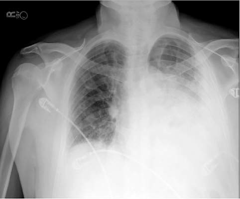
In recent years, the availability of portable ultrasound for bedside diagnostics and procedural guidance has revolutionized the practice of medicine, including the management of pleural effusions. When confronted with an obscured lower lobe on chest radiograph (Figure, left), clinicians were previously relegated to primitive bedside maneuvers, such as percussion or auscultation, to make critical decisions about the clinical management. Now, clinicians are able to look inside the body with point-of-care ultrasound and visually assess a pleural effusion before making any decisions. Point-of-care ultrasound has shifted the paradigm in the management of pleural effusions in several ways.
Ultrasound allows rapid detection and differentiation of pleural effusions from other pathologic findings.
Chest radiographs cannot accurately differentiate a pleural effusion from other common conditions, such as pneumonia, atelectasis, or an elevated hemidiaphragm. Ultrasound is the only bedside diagnostic modality that can rapidly differentiate these conditions within seconds and may reveal unsuspected findings, such as a mass or pericardial effusion.
For example, the pleural ultrasound exam of a patient in the confirmed the presence of a large, left-sided pleural effusion (Figure, right) but also revealed an unsuspected large pericardial effusion (asterisk) that was causing hemodynamic compromise. The management of this patient shifted focus from the pleural effusion to the pericardial effusion, and urgent pericardiocentesis was performed. The sensitivity of ultrasound to detect a pleural effusion is proportional to the volume of fluid, reaching 100% with as little as 100 mL of fluid (Kalokairinou-Motogna et al. Med Ultra. 2010;12[1]:12). The diagnostic accuracy of ultrasound for detection of pleural effusions is comparable to CT scans of the chest and superior to portable chest radiographs (Lichtenstein et al. Anesthesiology. 2004;100[1]:9).
Ultrasound characterizes pleural effusions to determine the most appropriate management strategy.
Any clinician with basic ultrasonography skills can learn to evaluate pleural effusions and categorize them as simple or complex based on the sonographic appearance. Visualization of fibrinous stranding or loculations increases the probability of pleural fluid being exudative and often drives the decision to drain the fluid. The density and distribution of loculations can guide decisions about the most appropriate type of drainage procedure – thoracentesis versus tube thoracostomy versus surgical intervention. Use of color flow Doppler ultrasound allows clinicians to assess whether or not pleural fluid is free flowing and amenable to drainage, potentially saving the patient from an unnecessary attempt at drainage.
Ultrasound affords frontline clinicians the ability to streamline consultation with the most appropriate specialist based on the type of drainage procedure indicated and potentially prevent duplicate procedures on the same patient from different specialists.
Ultrasound reduces the risk of postprocedure complications from thoracentesis.
The risk of postthoracentesis pneumothorax was reported to be as high as 20%-39% before the routine use of point-of-care ultrasound (Grogan et al. Arch Int Med. 1990;150:873). Ultrasound guidance has been shown to increase procedural success rates and decrease the risk of postprocedure pneumothorax (2.7%), cost of hospitalization, and length of stay (Mercaldi et al. Chest. 2015;143[2]:532).
Regardless of the chest radiograph or CT scan findings, if the ultrasound exam reveals a scant volume of pleural fluid, or densely loculated pleural fluid, clinicians can avoid unnecessary attempts at bedside drainage, which likely partly accounts for the reduction in postprocedure pneumothorax. Use of ultrasound for needle site selection may prevent up to 10% of potential accidental organ punctures and increases accurate site selection by 26%, compared with chest radiograph and physical examination findings combined (Diacon et al. Chest. 2003;123:436).
Ultrasound facilitates patient-centered care.
Point-of-care ultrasound is the only new technology that has taken clinicians back to the bedside to spend more time with patients. Clinicians can simultaneously perform an ultrasound exam and converse with patients to gather a medical history. The ultrasound image serves as a tool to help patients understand their condition and facilitates shared decision making with clinicians at the bedside.
As more specialties have gained expertise in thoracic ultrasonography, the use of ultrasound guidance for thoracentesis has evolved to become the standard of care in many hospitals in the United States. Besides pulmonary specialists, several acute care specialists, including hospitalists, intensivists, and emergency medicine physicians, are routinely using point-of-care ultrasound to guide diagnostic decision making and procedures. Over the past 10 years, nearly a dozen procedure services led by internal medicine-trained hospitalists have been created at academic institutions that are routinely performing ultrasound-guided thoracenteses with low complication rates (Franco-Sadud et al. SGIM Forum. 2016;39[5]:13). More important, ultrasound is being used on the front lines to expeditiously evaluate pleural effusions and perform a diagnostic thoracentesis or consult with the appropriate subspecialist. Even though demonstration of competency in bedside procedures is no longer required for board certification in internal medicine, many internal medicine residency programs have incorporated diagnostic and procedural point-of-care ultrasound training into their education curriculum (Schnobrich et al. JGME. 2013;5[3]:498). Further, approximately 62% of medical schools report integrating ultrasound education in their medical student curriculum, and in coming years, most medical students will likely graduate with a basic skill set in point-of-care ultrasonography (Bahner et al. Academic Med. 2014;89[12]:1681). As point-of-care ultrasound education becomes integrated in training of physicians and other health-care providers, use of ultrasound to guide management of pleural effusions could become universally practiced and accepted as the new standard of care. Thus, it is plausible that a day will come in the near future when the sun will not set on an “un-ultrasound-ed” pleural effusion.
Dr. Franco-Sadud is with the section of hospital medicine/division of general internal medicine, Medical College of Wisconsin, Milwaukee, Wisconsin; Dr. Soni is with the section of hospital medicine and the section of pulmonary and critical care medicine, South Texas Veterans Health Care System and University of Texas Health Science Center, San Antonio.
