User login
Jack Rozel II: Pittsburgh Shooting

Promising Results From Anti-HIV Combination Treatment
Reliable treatment with broadly neutralizing antibodies—bNAbs—could change the future for people living with HIV. But studies have found that infusions of a single bNAb did not suppress HIV because some patients developed resistance.
Rockefeller University researchers, however, theorized that combining multiple antibodies that target distinct regions of HIV would both suppress the virus and prevent resistance. So in an NIH-supported pilot study, researhcers recruited 15 volunteers whose HIV was suppressed with antiretroviral treatment (ART) and who were sensitive to 3BNC117 and 10-1074, both potent bNAbs.
Participants received infusions of both bNAbs, stopped taking ART 2 days later, and received additional infusions 3 and 6 weeks later.
Of the 11 people who completed the study, 9 maintained viral suppression without ART for an average of 15 weeks, until the amount of bNAbs in their bodies fell below protective levels. In 2 of the 9, virus was controlled through the end of the 30-week follow-up period. The remaining 2 participants were found to harbor HIV resistant to at least 1 bNAb and experienced viral rebound before 12 weeks after stopping ART.
The researchers are enrolling people with HIV in a larger study to determine an optimal regimen of bNAbs.
CARDIA: Smoke-free policies linked to lower blood pressure
Areas that have adopted smoke-free policies in their restaurants, bars, and workplaces have seen a corresponding drop in systolic blood pressure, according to data from the Coronary Artery Risk Development in Young Adults (CARDIA) study.

“Among a geographically diverse cohort of black and white nonsmoking adults followed for 15 years, we found that participants living in areas with smoke-free policies in restaurants, bars, and workplaces had lower systolic blood pressure at the end of follow-up, compared with participants living in areas without smoke-free policies,” wrote Stephanie L. Mayne, PhD, of the department of preventative medicine at Northwestern University, Chicago, and her coauthors in the Journal of the American Heart Association.
The study analyzed data from 2,606 CARDIA participants, all of whom enrolled in 1985-1986 and underwent follow-up exams after 2, 5, 7, 10, 15, 20, 25, and 30 years. Smoke-free policies were obtained from the American Non-Smokers’ Rights Foundation’s Local Ordinance Database and linked to participants based on their census tract and examination date. Systolic and diastolic blood pressure (SBP, DBP), along with physical activity and dietary quality, were measured at each examination.
By year 25, participants in areas with smoke-free restaurants had SBP values that were 1.14 mm Hg lower than participants who lived in areas with smoke-friendly restaurants (95% confidence interval, 2.15-0.12). Participants in areas with smoke-free bars returned similar results, with a SBP difference of 1.52 mm Hg (95% CI, 2.48-0.57). The data were less conclusive for DBP, though CARDIA indicated that SBP was more associated with cardiovascular disease risk than DBP and “even small reductions in SBP may result in meaningful reductions in CVD risk.”
The coauthors shared the study’s potential limitations, including an inability to control for antismoking campaigns and the possibility that participants did not report any infrequent smoking habits. However, they highlighted previous associations between smoke-free policies and reduced risk of hospitalization for CVD, noting the relation and suggesting “BP reduction as a potential mechanism through which smoke-free policies may reduce rates of CVD at the population level.”
This study was supported by the National Heart, Lung, and Blood Institute, in collaboration with the University of Alabama at Birmingham, Northwestern University, the University of Minnesota, Kaiser Foundation Research Institute, and Johns Hopkins University School of Medicine. It was partially supported by the Intramural Research Program of the National Institute on Aging. No conflicts of interest were reported.
SOURCE: Mayne SL et al. J Am Heart Assoc. 2018 Nov 21. doi: 10.1161/JAHA.118.009829.
Areas that have adopted smoke-free policies in their restaurants, bars, and workplaces have seen a corresponding drop in systolic blood pressure, according to data from the Coronary Artery Risk Development in Young Adults (CARDIA) study.
“Among a geographically diverse cohort of black and white nonsmoking adults followed for 15 years, we found that participants living in areas with smoke-free policies in restaurants, bars, and workplaces had lower systolic blood pressure at the end of follow-up, compared with participants living in areas without smoke-free policies,” wrote Stephanie L. Mayne, PhD, of the department of preventative medicine at Northwestern University, Chicago, and her coauthors in the Journal of the American Heart Association.
The study analyzed data from 2,606 CARDIA participants, all of whom enrolled in 1985-1986 and underwent follow-up exams after 2, 5, 7, 10, 15, 20, 25, and 30 years. Smoke-free policies were obtained from the American Non-Smokers’ Rights Foundation’s Local Ordinance Database and linked to participants based on their census tract and examination date. Systolic and diastolic blood pressure (SBP, DBP), along with physical activity and dietary quality, were measured at each examination.
By year 25, participants in areas with smoke-free restaurants had SBP values that were 1.14 mm Hg lower than participants who lived in areas with smoke-friendly restaurants (95% confidence interval, 2.15-0.12). Participants in areas with smoke-free bars returned similar results, with a SBP difference of 1.52 mm Hg (95% CI, 2.48-0.57). The data were less conclusive for DBP, though CARDIA indicated that SBP was more associated with cardiovascular disease risk than DBP and “even small reductions in SBP may result in meaningful reductions in CVD risk.”
The coauthors shared the study’s potential limitations, including an inability to control for antismoking campaigns and the possibility that participants did not report any infrequent smoking habits. However, they highlighted previous associations between smoke-free policies and reduced risk of hospitalization for CVD, noting the relation and suggesting “BP reduction as a potential mechanism through which smoke-free policies may reduce rates of CVD at the population level.”
This study was supported by the National Heart, Lung, and Blood Institute, in collaboration with the University of Alabama at Birmingham, Northwestern University, the University of Minnesota, Kaiser Foundation Research Institute, and Johns Hopkins University School of Medicine. It was partially supported by the Intramural Research Program of the National Institute on Aging. No conflicts of interest were reported.
SOURCE: Mayne SL et al. J Am Heart Assoc. 2018 Nov 21. doi: 10.1161/JAHA.118.009829.
FROM JOURNAL OF THE AMERICAN HEART ASSOCIATION
Key clinical point: As more restaurants, bars, and workplaces have introduced smoke-free policies, systolic blood pressure levels in those areas have fallen accordingly.
Major finding: At 25-year follow-up, participants in areas with smoke-free restaurants or bars had systolic blood pressure values that were 1.14 mm Hg and 1.52 mm Hg lower, respectively, than participants in areas without smoke-free options.
Study details: A longitudinal, multicenter cohort study of 2,606 nonsmoking adults who underwent follow-up exams after 2, 5, 7, 10, 15, 20, 25, and 30 years.
Disclosures: This study was supported by the National Heart, Lung, and Blood Institute, in collaboration with the University of Alabama at Birmingham, Northwestern University, the University of Minnesota, Kaiser Foundation Research Institute, and Johns Hopkins University School of Medicine. It was partially supported by the Intramural Research Program of the National Institute on Aging. No conflicts of interest were reported.
Source: Mayne SL et al. J Am Heart Assoc. 2018 Nov 21. doi: 10.1161/JAHA.118.009829.
Empagliflozin and left ventricular mass
Also today, symptomatic hyperuricemia may respond to urate-lowering therapy, the risk for cancer in non-alcoholic fatty liver disease is 91% higher than the general population, the link between antibiotic use and obesity is insignificant at age 10.
Amazon Alexa
Apple Podcasts
Google Podcasts
Spotify
Biosimilar deemed equivalent to rituximab in FL

Phase 3 results suggest the biosimilar product CT-P10 is equivalent to rituximab in patients with low-tumor-burden follicular lymphoma (FL).
Overall response rates were similar—both exceeding 80%—in patients who received CT-P10 and those who received rituximab.
In addition, adverse event (AE) profiles were comparable between the treatment arms.
Larry W. Kwak, MD, PhD, of City of Hope in Duarte, California, and his colleagues reported these results in The Lancet Haematology.
CT-P10 was approved by the European Commission in 2017 and was recommended for approval by the U.S. Food and Drug Administration’s Oncologic Drugs Advisory Committee last month.
The phase 3 trial of CT-P10 included 258 patients with stage II-IV low-tumor-burden FL. They were randomized to receive CT-P10 (n=130) or rituximab (n=128).
Patients received intravenous CT-P10 or rituximab weekly for 4 weeks as induction therapy. Patients experiencing disease control went on to a maintenance phase with their assigned treatment, given every 8 weeks for six cycles, followed by another year of maintenance therapy with CT-P10 for those still on study.
Efficacy
The overall response rate at 7 months was 83% in patients randomized to CT-P10 and 81% in those randomized to rituximab.
The complete response rates were 28% and 34%, respectively. The unconfirmed complete response rates were 5% and 2%, respectively. And the partial response rates were 51% and 46%, respectively.
The two treatments were deemed therapeutically equivalent, as the two-sided 90% confidence intervals for the difference in proportion of responders between CT-P10 and rituximab were within the prespecified equivalence margin of 17%.
Safety
Treatment-emergent AEs occurred in 71% of patients in the CT-P10 arm and 67% of those in the rituximab arm.
The most common treatment-emergent AEs (in the CT-P10 and rituximab arms, respectively) were:
- Infusion-related reactions (31% and 29%)
- Infections (27% and 21%)
- Worsening neutropenia (22% for both)
- Upper respiratory tract infection (12% and 11%)
- Worsening anemia (10% and 14%)
- Worsening thrombocytopenia (8% and 7%)
- Fatigue (7% and 9%)
- Diarrhea (5% for both)
- Nausea (5% for both)
- Urinary tract infection (4% and 5%)
- Headache (3% and 5%).
Serious AEs were reported in six patients in the CT-P10 arm and three patients in the rituximab arm.
Two serious AEs—myocardial infarction and constipation—in the CT-P10 arm were considered related to treatment. None of the serious AEs in the rituximab arm were considered treatment-related.
Two patients in the CT-P10 arm discontinued treatment due to AEs—one due to myocardial infarction and one due to dermatitis. There were no AE-related discontinuations in the rituximab arm.
There were two deaths in the CT-P10 arm as of the cutoff date (January 4, 2018). One was due to myocardial infarction, and one was due to respiratory failure. The myocardial infarction was considered possibly related to treatment.
This trial was sponsored by Celltrion, the company developing CT-P10. Three study authors are employees of the company.
Dr. Kwak and several other authors not employed by Celltrion reported disclosures related to the company. Authors also reported relationships with Novartis, Roche, AbbVie, Celgene, and Takeda, among other entities.
Mole near nose

The differential diagnosis for this lesion included a benign intradermal nevus and a basal cell carcinoma. So the FP recommended a shave biopsy to be sure that it was not cancer. (See the Watch & Learn video on “Shave biopsy.”)
After obtaining patient consent, he injected the 1% lidocaine with epinephrine and waited 5 minutes for the epinephrine to begin to work. He performed the shave with a Dermablade, and used a cotton-tipped applicator to apply aluminum chloride to the site. He used a twisting motion and pressure to achieve hemostasis. He dressed the lesion with petrolatum and some gauze.
Dermatopathology showed that this mole was a benign intradermal nevus. The FP reassured the patient and recommended that she be careful to avoid sun exposure.
Photos and text for Photo Rounds Friday courtesy of Richard P. Usatine, MD. This case was adapted from: Karnes J, Usatine R. Basal cell carcinoma. In: Usatine R, Smith M, Mayeaux EJ, et al. Color Atlas of Family Medicine. 2nd ed. New York, NY: McGraw-Hill; 2013:989-998.
To learn more about the Color Atlas of Family Medicine, see: www.amazon.com/Color-Family-Medicine-Richard-Usatine/dp/0071769641/.
You can now get the second edition of the Color Atlas of Family Medicine as an app by clicking on this link: usatinemedia.com.

Rivaroxaban for stroke prevention after embolic stroke
Clinical question: Does rivaroxaban prevent recurrent ischemic stroke in patients with embolic stroke from undetermined source?
Background: Embolic stroke of undetermined source (ESUS) represents approximately 20% of ischemic strokes. Rivaroxaban inhibits factor Xa and is shown to be effective for secondary stroke prevention in patients with nonvalvular atrial fibrillation. Strategies to prevent cryptogenic stroke caused by mechanisms other than cardioembolic sources are lacking.
Study design: International, double-blind, event-driven, randomized, phase III trial.

Setting: 459 hospitals in 31 countries, during 2014-2017.
Synopsis: 7,213 patients with ESUS were randomized to receive either 15 mg of rivaroxaban once daily or aspirin 100 mg once daily. The primary outcome was established as the first recurrent stroke of any type or systemic embolism. The primary safety endpoint was major bleeding.
Recurrent stroke was observed in 158 in the rivaroxaban group and 156 in the aspirin group, mostly ischemic. The trial was terminated early because of a higher incidence of intracranial hemorrhage and major bleeding among patients assigned to rivaroxaban at an annual rate of 1.8%, compared with 0.7% (hazard ratio, 2.72; 95% confidence interval, 1.68-4.39; P less than .001). Although the study utilized strict criteria to define ESUS, it is feasible that the lack of benefit may be due to heterogeneous arterial, cardiogenic, and paradoxical emboli with diverse composition and poor response to rivaroxaban.
Bottom line: Rivaroxaban does not confer protection from recurrent stroke in patients with embolic stroke of unknown origin and increases risk for intracranial hemorrhage and major bleeding.
Citation: Hart RG et al. Rivaroxaban for stroke prevention after embolic stroke of undetermined source. N Engl J Med. 2018;378(23):2191-201.
Dr. Vela-Duarte is assistant professor of neurology at the University of Colorado, Denver
Clinical question: Does rivaroxaban prevent recurrent ischemic stroke in patients with embolic stroke from undetermined source?
Background: Embolic stroke of undetermined source (ESUS) represents approximately 20% of ischemic strokes. Rivaroxaban inhibits factor Xa and is shown to be effective for secondary stroke prevention in patients with nonvalvular atrial fibrillation. Strategies to prevent cryptogenic stroke caused by mechanisms other than cardioembolic sources are lacking.
Study design: International, double-blind, event-driven, randomized, phase III trial.
Setting: 459 hospitals in 31 countries, during 2014-2017.
Synopsis: 7,213 patients with ESUS were randomized to receive either 15 mg of rivaroxaban once daily or aspirin 100 mg once daily. The primary outcome was established as the first recurrent stroke of any type or systemic embolism. The primary safety endpoint was major bleeding.
Recurrent stroke was observed in 158 in the rivaroxaban group and 156 in the aspirin group, mostly ischemic. The trial was terminated early because of a higher incidence of intracranial hemorrhage and major bleeding among patients assigned to rivaroxaban at an annual rate of 1.8%, compared with 0.7% (hazard ratio, 2.72; 95% confidence interval, 1.68-4.39; P less than .001). Although the study utilized strict criteria to define ESUS, it is feasible that the lack of benefit may be due to heterogeneous arterial, cardiogenic, and paradoxical emboli with diverse composition and poor response to rivaroxaban.
Bottom line: Rivaroxaban does not confer protection from recurrent stroke in patients with embolic stroke of unknown origin and increases risk for intracranial hemorrhage and major bleeding.
Citation: Hart RG et al. Rivaroxaban for stroke prevention after embolic stroke of undetermined source. N Engl J Med. 2018;378(23):2191-201.
Dr. Vela-Duarte is assistant professor of neurology at the University of Colorado, Denver
Tofacitinib and TNF inhibitors show similar VTE rates
CHICAGO – Rheumatoid arthritis patients treated with tofacitinib did not have a significantly increased incidence of hospitalization for venous thromboembolism, compared with patients treated with a tumor necrosis factor (TNF) inhibitor, in a study of more than 50,000 U.S. patients culled from a pair of health insurance databases.
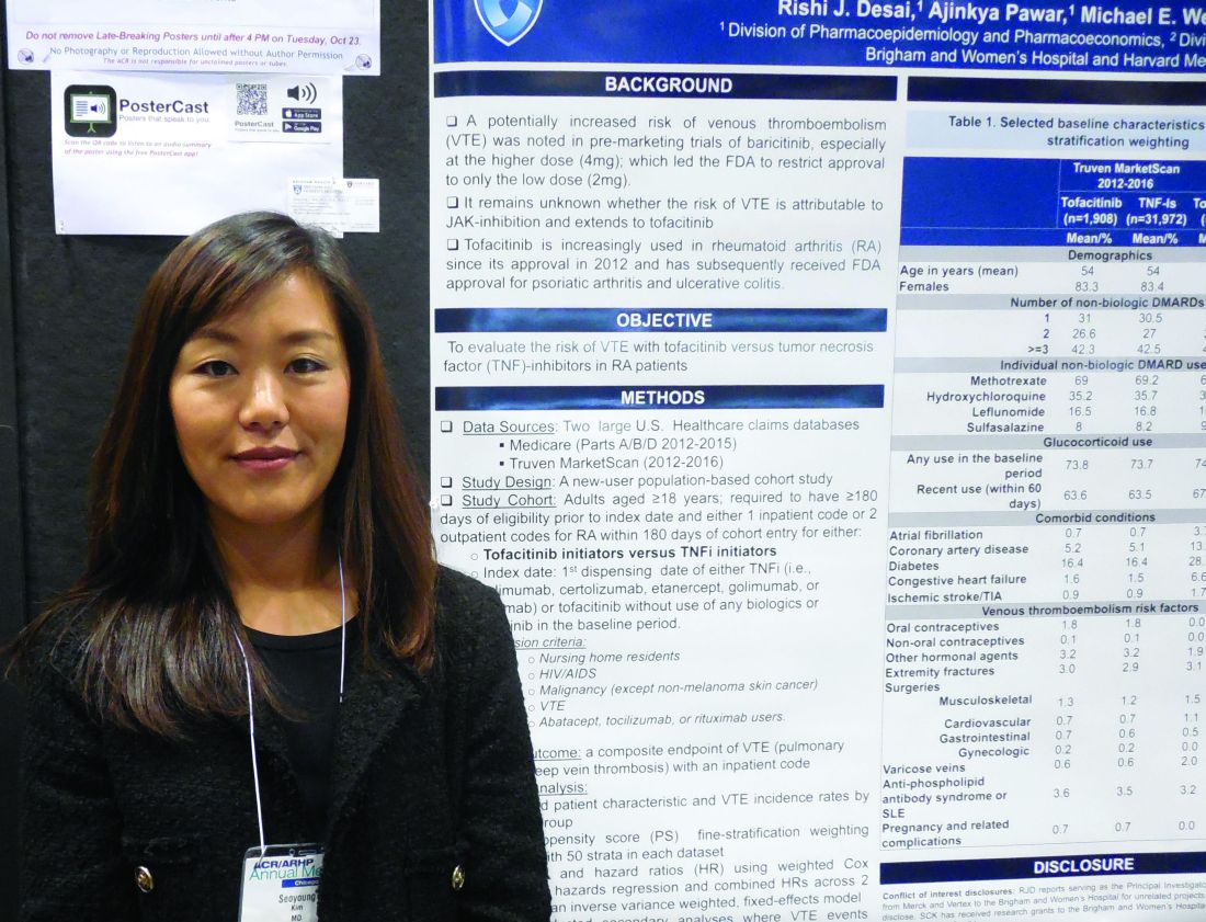
The Janus kinase inhibitor class of agents, including tofacitinib (Xeljanz), has acquired a reputation for causing an excess of venous thromboembolic events (VTE) (Drug Saf. 2018 Jul;41[7]:645-53). To assess this in a real-world setting Seoyoung C. Kim, MD, and her associates took data from Medicare patients during 2012-2015 and from Truven MarketScan for commercially insured patients during 2012-2016 and derived a database of 16,091 RA patients on newly begun treatment with a TNF inhibitor and 995 newly begun on tofacitinib in the Medicare data, and 32,164 RA patients newly started on a TNF inhibitor and 1,910 on tofacitinib in the Truven database. The analysis excluded patients with a history of VTE.
Using propensity score–adjusted matching of patients in the two treatment arms in both of these databases, and using a VTE event – either a pulmonary embolism or deep-vein thrombosis that resulted in hospitalization – as the primary endpoint, the results showed statistically nonsignificant excesses of VTE in the patients treated with tofacitinib, compared with a TNF inhibitor, Dr. Kim reported in a poster she presented at the annual meeting of the American College of Rheumatology.
In the adjusted comparison, the Medicare data showed a nonsignificant 12% higher VTE rate in the tofacitinib-treated patients, while the Truven data showed a nonsignificant 55% higher rate of VTE during tofacitinib treatment. When the data were pooled, the result was a 33% higher rate of VTE while on tofacitinib treatment, which was not statistically significant.
Dr. Kim cautioned that the low rate of VTE events, especially among the patients on tofacitinib, limited the precision of the results. The combined data included 2,905 patients on tofacitinib treatment who had 15 VTE events, a rate of 0.77 events/100 person-years of follow-up. This compared with a rate of 0.52/100 person-years among patients on a TNF inhibitor. Thus, in both treatment groups the absolute VTE rate was low.
The most reliable finding from the analysis is that it “rules out a large increase in the risk for VTE events with tofacitinib,” said Dr. Kim, a rheumatologist at Brigham and Women’s Hospital in Boston.
The researchers also ran an analysis that included not only VTE events that resulted in hospitalization but also VTE events managed on an outpatient basis. Dr. Kim did not report the specific numbers involved in this calculation, but she reported that, when her group included both types of VTE events, the patients treated with tofacitinib had a nonsignificant 12% lower rate of events, compared with patients treated with a TNF inhibitor.
Dr. Kim has received research support from Bristol-Myers Squibb, Pfizer, and Roche.
SOURCE: Desai RJ et al. Arthritis Rheumatol. 2018;70(suppl 10), Abstract L09.
CHICAGO – Rheumatoid arthritis patients treated with tofacitinib did not have a significantly increased incidence of hospitalization for venous thromboembolism, compared with patients treated with a tumor necrosis factor (TNF) inhibitor, in a study of more than 50,000 U.S. patients culled from a pair of health insurance databases.
The Janus kinase inhibitor class of agents, including tofacitinib (Xeljanz), has acquired a reputation for causing an excess of venous thromboembolic events (VTE) (Drug Saf. 2018 Jul;41[7]:645-53). To assess this in a real-world setting Seoyoung C. Kim, MD, and her associates took data from Medicare patients during 2012-2015 and from Truven MarketScan for commercially insured patients during 2012-2016 and derived a database of 16,091 RA patients on newly begun treatment with a TNF inhibitor and 995 newly begun on tofacitinib in the Medicare data, and 32,164 RA patients newly started on a TNF inhibitor and 1,910 on tofacitinib in the Truven database. The analysis excluded patients with a history of VTE.
Using propensity score–adjusted matching of patients in the two treatment arms in both of these databases, and using a VTE event – either a pulmonary embolism or deep-vein thrombosis that resulted in hospitalization – as the primary endpoint, the results showed statistically nonsignificant excesses of VTE in the patients treated with tofacitinib, compared with a TNF inhibitor, Dr. Kim reported in a poster she presented at the annual meeting of the American College of Rheumatology.
In the adjusted comparison, the Medicare data showed a nonsignificant 12% higher VTE rate in the tofacitinib-treated patients, while the Truven data showed a nonsignificant 55% higher rate of VTE during tofacitinib treatment. When the data were pooled, the result was a 33% higher rate of VTE while on tofacitinib treatment, which was not statistically significant.
Dr. Kim cautioned that the low rate of VTE events, especially among the patients on tofacitinib, limited the precision of the results. The combined data included 2,905 patients on tofacitinib treatment who had 15 VTE events, a rate of 0.77 events/100 person-years of follow-up. This compared with a rate of 0.52/100 person-years among patients on a TNF inhibitor. Thus, in both treatment groups the absolute VTE rate was low.
The most reliable finding from the analysis is that it “rules out a large increase in the risk for VTE events with tofacitinib,” said Dr. Kim, a rheumatologist at Brigham and Women’s Hospital in Boston.
The researchers also ran an analysis that included not only VTE events that resulted in hospitalization but also VTE events managed on an outpatient basis. Dr. Kim did not report the specific numbers involved in this calculation, but she reported that, when her group included both types of VTE events, the patients treated with tofacitinib had a nonsignificant 12% lower rate of events, compared with patients treated with a TNF inhibitor.
Dr. Kim has received research support from Bristol-Myers Squibb, Pfizer, and Roche.
SOURCE: Desai RJ et al. Arthritis Rheumatol. 2018;70(suppl 10), Abstract L09.
REPORTING FROM THE ACR ANNUAL MEETING
Key clinical point: Rheumatoid arthritis patients treated with tofacitinib showed no excess incidence of venous thromboembolism, compared with patients on a tumor necrosis factor inhibitor.
Major finding: Propensity score–adjusted rates of VTE were 33% higher with tofacitinib, compared with TNF inhibition, which was not a statistically significant difference.
Study details: Review of 51,160 rheumatoid arthritis patients from U.S. health insurance databases.
Disclosures: Dr. Kim has received research support from Bristol-Myers Squibb, Pfizer, and Roche.
Source: Desai RJ et al. Arthritis Rheumatol. 2018;70(suppl 10), Abstract L09.

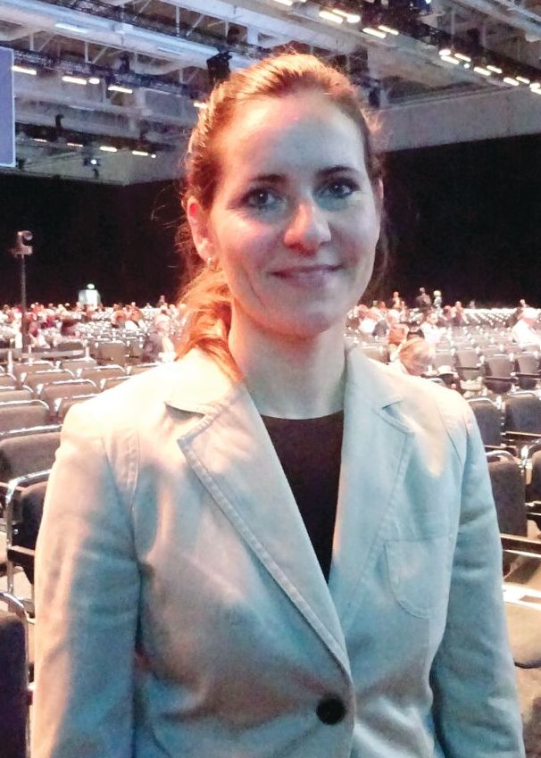
Nasal glucagon ‘viable alternative’ to intramuscular administration
BERLIN – A dry-powder nasal formulation of glucagon was as good as intramuscular delivery for the reversal of severe hypoglycemia in patients with type 1 diabetes mellitus (T1DM) in a randomized controlled trial.
A 100% treatment success rate (n = 66) was seen for both nasal glucagon and intramuscular glucagon, defined as an increase in plasma glucose to 70 mg/dL (3.9 mmol/L or greater) or more or an increase of 20 mg/L (1.1 mmol/L) or more from the glucose nadir within 30 minutes of administration.
Furthermore, slightly more than 97% of patients achieved treatment success within 15 minutes in both treatment groups, with mean times of 11.4 minutes for the nasal formulation and 9.8 minutes for intramuscular administration.
Similar glucose responses were observed within 40 minutes of glucagon administration, study investigator Leona Plum-Mörschel, MD, PhD, reported at the annual meeting of the European Association for the Study of Diabetes. Dr. Plum-Mörschel is the CEO of the clinical research organization Profil Mainz (Germany), which helped Eli Lilly conduct the trial.
“We are all aware and fully agree that severe hypoglycemia is a serious and potentially life-threatening complication of diabetes treatment with insulin and the sulfonylureas,” said Dr. Plum-Mörschel. At least one in three patients with diabetes reports one hypoglycemic episode per year, she added. While this is quite prevalent in patients with T1DM, it’s not uncommon for those with type 2 diabetes mellitus (T2DM) to experience hypoglycemic episodes.
“Outside of a hospital or emergency room setting, injectable glucagon is currently the only option to treat severe hypoglycemia,” Dr. Plum-Mörschel reminded delegates. “Thankfully, the available injectable glucagon emergency kits are highly effective for the treatment of hypoglycemic events,” she added.
However, for untrained caregivers they can be “really challenging to use,” according to Dr. Plum-Mörschel. This is because the available kits involve multiple steps, including reconstitution, before being ready to inject. A severe hypoglycemic event is stressful enough without them worrying about getting things right, she suggested.
This is where a nasal formulation would be advantageous, and it is something that’s been touted to be on the horizon for a few years. Studies have previously shown that nasal glucagon is comparable to intramuscular glucagon in both adult and pediatric populations. Turning the formulation used in those studies into a commercial product however, has meant more clinical testing before being licensed by the regulatory authorities.
The nasal formulation consists of a dry power containing 3 mg glucagon that is provided in a single-use, compact, and thus, portable, device. It’s been designed to be stored at room temperature and is ready to use immediately. Pressing the plunger on the device releases the fine powder that does not require inhalation to work, which means it can be given easily to an unconscious patient with severe hypoglycemia.
“The aim of the present study was to compare the efficacy and safety of commercially manufactured nasal glucagon with intramuscular glucagon for the treatment of insulin-induced hypoglycemia in adults with type 1 diabetes,” Dr. Plum-Mörschel said.
It was a randomized, single-dose, crossover study involving 70 adult patients with T1DM with hypoglycemia artificially induced by an intravenous insulin infusion during the two dosing visits. Five minutes after stopping insulin, nasal glucagon (3 mg) or intramuscular (1 mg) was given, with multiple plasma glucose measurements taken for up to 90 minutes.
The mean age of participants was 41.7 years old, 61% were male, the mean duration of diabetes was 19.8 years, with a baseline glycated hemoglobin (HbA1c) of 7.3%.
As for safety, Dr. Plum-Mörschel noted that nasal glucagon had a safety profile that was acceptable for emergency treatment. There was no difference between the nasal or intramuscular glucagon treatment groups in the percentages of patients experiencing treatment-emergent adverse events (at least 5% frequency): nausea was seen in 22% and 29%, vomiting in 10% and 12%, and headache in 12% and 7%, respectively.
Asking patients who had been treated with nasal glucagon about specific symptoms related to nasal administration showed around 63% had watery eyes, 49% nasal itching, 39% nasal congestion, 37% a runny nose, 24% sneezing, 20% itchy eyes, and 12.9% itchy throat. “All of these events were transient, generally mild or moderate in nature, and none were serious. Indeed, there were no deaths in the study and no other serious AEs [adverse events] occurred.”
Dr. Plum-Mörschel concluded: “These results support nasal glucagon as a viable alternative to intramuscular glucagon for the treatment of severe hypoglycemia.
“I personally would expect that, due to its simplicity of use, nasal glucagon will create a greater community who can render quick aid in a rescue situation.”
The study was funded by Eli Lilly. Dr. Plum-Mörschel is an employee of Profil Mainz and has received travel grants and speaker honoraria from Eli Lilly and Novo Nordisk.
SOURCE: Suico J et al. EASD 2018, Abstract 150.
REPORTING FROM EASD 2018
Key clinical point: Nasal glucagon was as good as intramuscular administration for reversing severe hypoglycemia in patients with T1DM.
Major finding: All (100%) of patients achieved treatment success; 97% or greater within 15 minutes in both treatment groups.
Study details: Randomized, single-dose, crossover study of nasal versus intramuscular glucagon in 70 adult patients with T1DM.
Disclosures: The study was funded by Eli Lilly. The presenting investigator Dr. Plum-Mörschel is an employee of Profil Mainz and has received travel grants and speaker honoraria from Eli Lilly and Novo Nordisk.
Source: Suico J et al. EASD 2018, Abstract 150.
Sponsored Video: TREMFYA® Patient Testimonial—Meet Patti
