User login
The Official Newspaper of the American Association for Thoracic Surgery
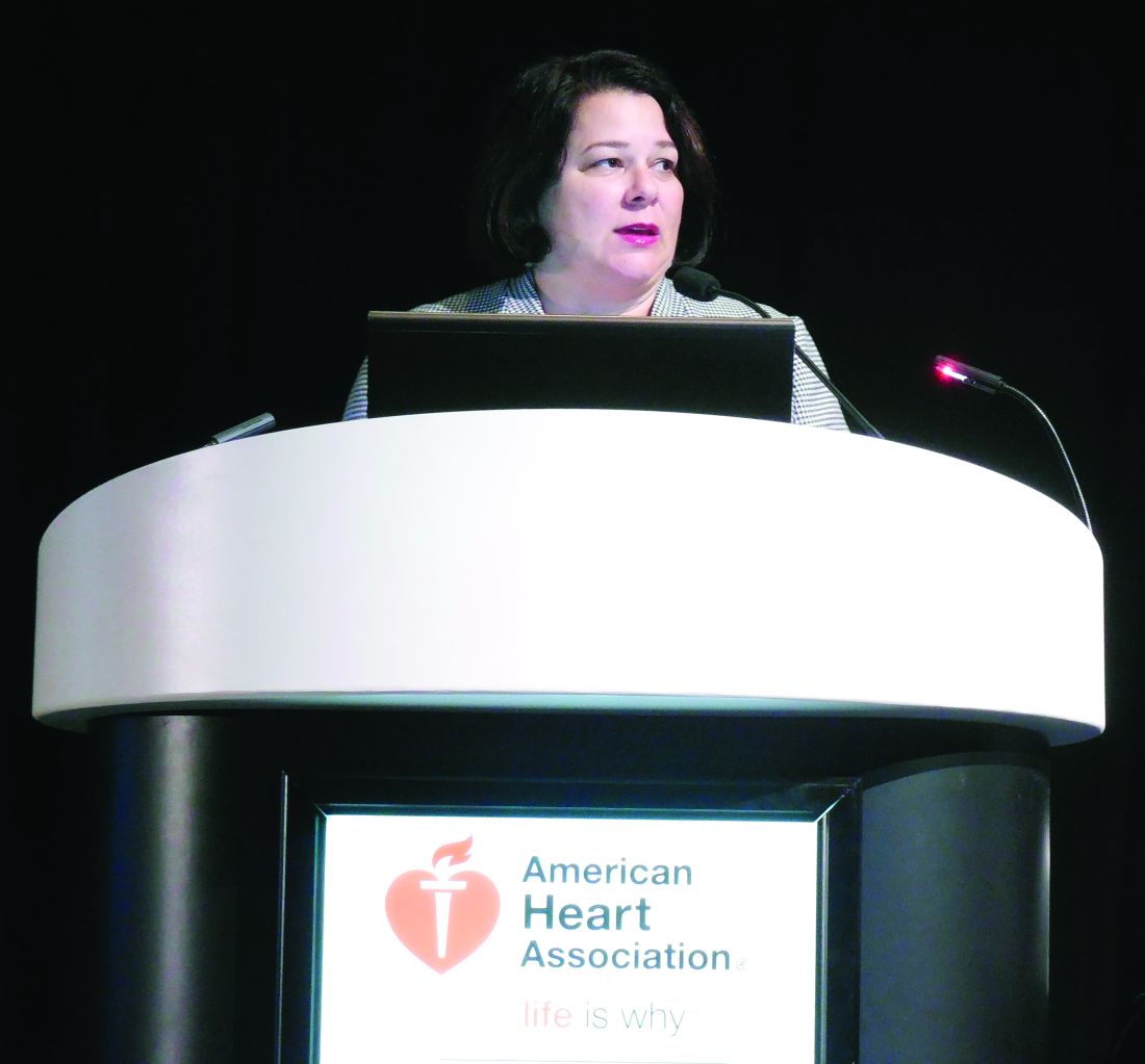
VIDEO: HeartMate 3 LVAD solves pump thrombosis
NEW ORLEANS – HeartMate 3, the latest left ventricular assist device in the HeartMate line, appears to have solved the problem of pump thrombosis, a complication that has dogged ventricular pumps since the issue leapt into medical awareness about 3 years ago (New Engl J Med. 2014 Jan 2;370:33-40).
During 6 months of follow-up, none of 152 heart failure patients assigned to receive a HeartMate 3 left ventricular assist device (LVAD) developed suspected or confirmed pump thrombosis, compared with 14 patients (10%) having pump thrombosis out of 138 recipients of the prior-generation HeartMate II LVAD who served as the control group for the study.

“Three years ago, when the issue of pump thrombosis was first revealed, there was a lot of consternation and some drop in LVAD use, especially as destination therapy. We think that seeing no pump thrombosis whatsoever will give people renewed confidence in this technology,” said Dr. Mehra, professor of medicine at Harvard Medical School and medical director of the Heart and Vascular Center of Brigham and Women’s Hospital, both in Boston.
Pump thrombosis has also been a problem for the patients who have received a competitor LVAD, the HeartWare HVAD device (Circulation. 2015 Nov 10;132[suppl 3]:A19675), approved for U.S. use as bridge to transplant. HeartMate II is approved for both bridge to transplant and for destination therapy.
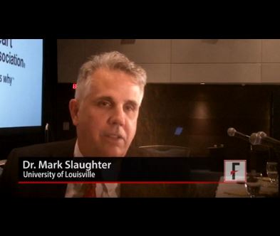
In addition to apparently eliminating pump thrombosis, HeartMate 3’s size and potential implantation approach should make its placement during routine use as quick and minimally invasive as the HeartWare device, features that should further help broader use of HeartMate 3, commented Mark Slaughter, MD, professor and chairman of cardiovascular and thoracic surgery at the University of Louisville (Ky.). But Dr. Slaughter and others were also quick to highlight the shortcomings that remain with both devices that will continue to hamper a broader role for LVAD treatment of patients with advanced heart failure.
“We thought that if there was less pump thrombosis we’d see less stroke, but that is not what the data suggest. It’s the big puzzle we need to figure out before we see widespread acceptance of this treatment,” Dr. Sweitzer said.
“This will not shift LVAD use substantially,” commented Christopher B. Granger, MD, a professor of medicine and a heart failure specialist at Duke University, Durham, N.C. “Reducing the need for reoperation is good for the field, and is an incremental advance, but it is not transformational,” he said in an interview.
The MOMENTUM 3 (Multicenter Study of MagLev Technology in Patients Undergoing Mechanical Circulatory Support Therapy with HeartMate 3) trial randomized 294 patients at 69 U.S. centers. The study’s primary endpoint of 6-month survival free from disabling stroke or reoperation to repair or replace the LVAD occurred in 86% of 152 patients who received a HeartMate 3 and 77% of 142 patients randomized to HeartMate II, a statistical difference that met the prespecified criteria for both noninferiority and superiority. Concurrently with Dr. Mehra’s report at the meeting, a journal article appeared online (New Engl J Med. 2016 Nov 16. doi: 10.1056/NEJMoa1610426). He stated that as far as he understood, St. Jude would submit the 6-month data he reported to the Food and Drug Administration in an application for marketing approval for HeartMate 3.
“I agree that there are still morbid evens [with HeartMate 3] that need to be surmounted, but this is a confidence-building step in the right direction,” Dr. Mehra said.
[email protected]
On Twitter @mitchelzoler
By eliminating all episodes of pump thrombosis during 6-month follow-up, the HeartMate 3 appeared to resolve one of the major issues that has stood in the way of patients and physicians feeling comfortable with left ventricular assist devices. The smaller size of the HeartMate 3 pump and its ability to be placed with minimally invasive and fairly rapid surgery is another big advance, putting this device on par with the rival pump, the HeartWare HVAD.
But the performance of the HeartMate 3 left ventricular assist device (LVAD) in MOMENTUM 3 also highlighted the shortcomings that still remain for these devices: the unchanged rates of stroke, gastrointestinal bleeds, and infections with HeartMate 3, compared with HeartMate II in this trial, and similar 6-month survival rates in the two arms of the study.
The HeartMate 3 can be implanted without sternotomy, using an 8 cm incision on the lateral chest wall, resulting in a shorter postoperative stay and fewer perisurgical adverse events. Despite the less invasive surgery and absence of pump thrombosis, some patients and physicians will remain hesitant to use an LVAD unless it is unavoidable because of concern about strokes. Until further design and procedural refinements change the rate of serious strokes and other adverse events, LVADs will not be fully competitive with heart transplantation.
The competition between HeartMate and the HeartWare devices will help drive this field forward, leading to further improvements in outcomes and expanded LVAD use.
Mark Slaughter, MD, is professor of surgery and chairman of cardiovascular and thoracic surgery at the University of Louisville (Ky.). He was an investigator in MOMENTUM 3, he has been a consultant to EvaHeart and Oregon Heart, and he has received research support from Carmat and HeartWare. He made these comments as designated discussant for the report and in a video interview.
The video associated with this article is no longer available on this site. Please view all of our videos on the MDedge YouTube channel
NEW ORLEANS – HeartMate 3, the latest left ventricular assist device in the HeartMate line, appears to have solved the problem of pump thrombosis, a complication that has dogged ventricular pumps since the issue leapt into medical awareness about 3 years ago (New Engl J Med. 2014 Jan 2;370:33-40).
During 6 months of follow-up, none of 152 heart failure patients assigned to receive a HeartMate 3 left ventricular assist device (LVAD) developed suspected or confirmed pump thrombosis, compared with 14 patients (10%) having pump thrombosis out of 138 recipients of the prior-generation HeartMate II LVAD who served as the control group for the study.
“Three years ago, when the issue of pump thrombosis was first revealed, there was a lot of consternation and some drop in LVAD use, especially as destination therapy. We think that seeing no pump thrombosis whatsoever will give people renewed confidence in this technology,” said Dr. Mehra, professor of medicine at Harvard Medical School and medical director of the Heart and Vascular Center of Brigham and Women’s Hospital, both in Boston.
Pump thrombosis has also been a problem for the patients who have received a competitor LVAD, the HeartWare HVAD device (Circulation. 2015 Nov 10;132[suppl 3]:A19675), approved for U.S. use as bridge to transplant. HeartMate II is approved for both bridge to transplant and for destination therapy.
In addition to apparently eliminating pump thrombosis, HeartMate 3’s size and potential implantation approach should make its placement during routine use as quick and minimally invasive as the HeartWare device, features that should further help broader use of HeartMate 3, commented Mark Slaughter, MD, professor and chairman of cardiovascular and thoracic surgery at the University of Louisville (Ky.). But Dr. Slaughter and others were also quick to highlight the shortcomings that remain with both devices that will continue to hamper a broader role for LVAD treatment of patients with advanced heart failure.
“We thought that if there was less pump thrombosis we’d see less stroke, but that is not what the data suggest. It’s the big puzzle we need to figure out before we see widespread acceptance of this treatment,” Dr. Sweitzer said.
“This will not shift LVAD use substantially,” commented Christopher B. Granger, MD, a professor of medicine and a heart failure specialist at Duke University, Durham, N.C. “Reducing the need for reoperation is good for the field, and is an incremental advance, but it is not transformational,” he said in an interview.
The MOMENTUM 3 (Multicenter Study of MagLev Technology in Patients Undergoing Mechanical Circulatory Support Therapy with HeartMate 3) trial randomized 294 patients at 69 U.S. centers. The study’s primary endpoint of 6-month survival free from disabling stroke or reoperation to repair or replace the LVAD occurred in 86% of 152 patients who received a HeartMate 3 and 77% of 142 patients randomized to HeartMate II, a statistical difference that met the prespecified criteria for both noninferiority and superiority. Concurrently with Dr. Mehra’s report at the meeting, a journal article appeared online (New Engl J Med. 2016 Nov 16. doi: 10.1056/NEJMoa1610426). He stated that as far as he understood, St. Jude would submit the 6-month data he reported to the Food and Drug Administration in an application for marketing approval for HeartMate 3.
“I agree that there are still morbid evens [with HeartMate 3] that need to be surmounted, but this is a confidence-building step in the right direction,” Dr. Mehra said.
[email protected]
On Twitter @mitchelzoler
AT THE AHA SCIENTIFIC SESSIONS
Key clinical point:
Major finding: During 6 months, suspected or confirmed pump thrombosis occurred in no HeartMate 3 patients and in 10% of HeartMate II recipients.
Data source: The MOMENTUM 3 trial, which randomized 294 patients at 69 U.S. centers.
Disclosures: MOMENTUM 3 was sponsored by St. Jude, the company developing the HeartMate 3 LVAD. Dr. Mehra has received travel reimbursements from St. Jude and has been a consultant to Medtronic, Stealth, and Teva. Dr. Sweitzer was an investigator in MOMENTUM 3 and has been a consultant to Acorda and Medtronic and received research support from Bayer, Corvia, and Novartis. Dr. Granger has been a consultant to Boehringer Ingelheim, and received research support from Medtronic and several other drug and device companies. Dr. Slaughter was an investigator in MOMENTUM 3, has been a consultant to EvaHeart and Oregon Heart, and has received research support from Carmat and HeartWare.
Hospital factors play key role in readmission risk after surgery
CORONADO, CALIF. – Variation in readmission risk across hospitals following certain surgical procedures is more attributable to hospital factors than to patient characteristics, results from a large analysis demonstrated.
Such is the impact of the care delivery macro environment (CDM), which Sarah A. Brownlee and coauthors defined as a series of complex interactions between patient characteristics and imposed hospital attributes than can impact patient outcomes postoperatively.

The purpose of the current study was to determine the relative contribution of various aspects of the CDM to 1-year readmission risk after surgery. Working with colleagues Anai Kothari, MD, and Paul Kuo MD, in the One:MAP Section of Clinical informatics and Analytics in the department of surgery at Loyola University Medical Center, Ms. Brownlee analyzed the Healthcare Cost and Utilization Project State Inpatient Databases from Florida, New York, and Washington between 2009 and 2013, which were linked to the American Hospital Association Annual Survey from that same time period.
The researchers used smoothed hazard estimates to determine all-cause readmission in the year after surgery, and multilevel survival models with shared frailty to determine the relative impact of hospital versus patient characteristics on the heterogeneity of readmission risk between hospitals. They limited the analysis to patients aged 18 years and older who underwent one the following procedures: abdominal aortic aneurysm repair, pancreatectomy, colectomy, coronary artery bypass graft, and total hip arthroplasty.
Ms. Brownlee reported results from 502,157 patients who underwent surgical procedures at 347 hospitals. The 1-year readmission rate was 23.5%, and ranged from 12% to 36% across procedures. After controlling for procedure, the researchers observed a 7.9% variation in readmission risk between hospitals. Staffing accounted for 9.8% of variance, followed by hospital structural characteristics such as teaching status and clinical programs (7.5%), patient ZIP code (3.8%), hospital perioperative resources such as inpatient rehab (2.9%), hospital volume (2.8%), and patient clinical characteristics (2.1%). The following hospital characteristics were significantly associated with a lower risk of 1-year readmission: high physician/bed ratio (hazard ratio 0.85; P = .00017); transplant status (HR 0.87; P = .022); high-income ZIP code (HR 0.89; P less than .001); high nurse bed/bed ratio (HR 0.90; P = .047), and cancer center designation (HR 0.93; P = .021).
“Compared to patient clinical characteristics, hospital factors such as staffing ratios, perioperative resources, and structural elements account for more variation in postoperative outcomes,” Ms. Brownlee concluded. “However, it’s important to note that in the present study, over 70% of variation in readmission rates is not explained by the covariates that we analyzed. It’s possible that there are other factors we need to consider. That’s where the direction of this research is going. Much of the variation in readmission risk across hospitals cannot be characterized with currently utilized administrative data.”
The National Institutes of Health provided funding for the study. Ms. Brownlee reported having no financial disclosures.
CORONADO, CALIF. – Variation in readmission risk across hospitals following certain surgical procedures is more attributable to hospital factors than to patient characteristics, results from a large analysis demonstrated.
Such is the impact of the care delivery macro environment (CDM), which Sarah A. Brownlee and coauthors defined as a series of complex interactions between patient characteristics and imposed hospital attributes than can impact patient outcomes postoperatively.
The purpose of the current study was to determine the relative contribution of various aspects of the CDM to 1-year readmission risk after surgery. Working with colleagues Anai Kothari, MD, and Paul Kuo MD, in the One:MAP Section of Clinical informatics and Analytics in the department of surgery at Loyola University Medical Center, Ms. Brownlee analyzed the Healthcare Cost and Utilization Project State Inpatient Databases from Florida, New York, and Washington between 2009 and 2013, which were linked to the American Hospital Association Annual Survey from that same time period.
The researchers used smoothed hazard estimates to determine all-cause readmission in the year after surgery, and multilevel survival models with shared frailty to determine the relative impact of hospital versus patient characteristics on the heterogeneity of readmission risk between hospitals. They limited the analysis to patients aged 18 years and older who underwent one the following procedures: abdominal aortic aneurysm repair, pancreatectomy, colectomy, coronary artery bypass graft, and total hip arthroplasty.
Ms. Brownlee reported results from 502,157 patients who underwent surgical procedures at 347 hospitals. The 1-year readmission rate was 23.5%, and ranged from 12% to 36% across procedures. After controlling for procedure, the researchers observed a 7.9% variation in readmission risk between hospitals. Staffing accounted for 9.8% of variance, followed by hospital structural characteristics such as teaching status and clinical programs (7.5%), patient ZIP code (3.8%), hospital perioperative resources such as inpatient rehab (2.9%), hospital volume (2.8%), and patient clinical characteristics (2.1%). The following hospital characteristics were significantly associated with a lower risk of 1-year readmission: high physician/bed ratio (hazard ratio 0.85; P = .00017); transplant status (HR 0.87; P = .022); high-income ZIP code (HR 0.89; P less than .001); high nurse bed/bed ratio (HR 0.90; P = .047), and cancer center designation (HR 0.93; P = .021).
“Compared to patient clinical characteristics, hospital factors such as staffing ratios, perioperative resources, and structural elements account for more variation in postoperative outcomes,” Ms. Brownlee concluded. “However, it’s important to note that in the present study, over 70% of variation in readmission rates is not explained by the covariates that we analyzed. It’s possible that there are other factors we need to consider. That’s where the direction of this research is going. Much of the variation in readmission risk across hospitals cannot be characterized with currently utilized administrative data.”
The National Institutes of Health provided funding for the study. Ms. Brownlee reported having no financial disclosures.
AT WSA 2016
Key clinical point:
Major finding: Staffing accounted for 9.8% of variance in readmission risk between hospitals, followed by hospital structural characteristics such as teaching status and clinical programs (7.5%).
Data source: Results from 502,157 patients who underwent surgical procedures at 347 hospitals in three states.
Disclosures: The National Institutes of Health provided funding for the study. Ms. Brownlee reported having no financial disclosures.
REBOA may be a safe alternative to RTACC in the acute care setting
WASHINGTON – Resuscitative endovascular balloon occlusion of the aorta (REBOA) could be an acceptable alternative to thoracotomy in traumatic arrest patients who are hemorrhaging below the diaphragm, according to the results of a small pilot study which were presented by William Teeter, MD, at the annual clinical congress of the American College of Surgeons.
Furthermore, virtual simulation training sufficiently prepares surgeons to safely use the REBOA technique in the acute care setting, a separate study found. Importantly, this training has the potential to allow REBOA to become a widespread tool for surgeons regardless of their endovascular surgical experience.
Dr. Teeter presented the preliminary results of a pilot study involving 19 patients who received RTACC between 2008 and 2013 and 17 patients who received REBOA between 2013 and 2015. All study participants were trauma patients who arrived at the R Adams Cowley Shock Trauma Center in arrest or arrested shortly after arrival.
Age, gender, Glasgow Coma Scale, and injury severity score were the same or similar between the two groups, Dr. Teeter reported. Mean systolic blood pressure at admission was 14 mmHg for the REBOA group and 28 mmHg for the RTACC group; however, the majority of patients (82% of REBOA patients and 73% of RTACC patients) arrived with a blood pressure of 0, reported Dr. Teeter.
Importantly, patients in the RTACC group who had penetrating chest injury were excluded for this analysis, Dr. Teeter noted, adding that there was a slightly higher incidence of blunt trauma within the REBOA group likely due to “a change in practice at the trauma center during this time.”
All resuscitations were captured with real-time videography. Continuous vitals were also collected and analyzed.
While more RTACC patients survived to the operating room (53% vs. 68%), among the REBOA group there were more patients who experienced return of spontaneous circulation (53% vs. 37%). However, neither of these results was statistically significant.
Following occlusion of the aorta, the blood pressure measures, taken from continuous vital signs and averaged over a 15-minute period, were 80 mmHg for the REBOA group and 46 mmHg for the RTACC group. Again, this result was statistically insignificant but trended toward favoring REBOA.
Overall, patient survival was dismal. Only one patient who received REBOA survived.
Following Dr. Teeter’s presentation, the study’s assigned discussant, Nicole A. Stassen, MD, of the University of Rochester Medical Center, N.Y., noted that while post-occlusion blood pressure was higher for the REBOA group it seemed not to matter as the majority of patients did not survive. Dr. Stassen also asked if these preliminary results were sufficient to inform or change clinical practice.
In response, Dr. Teeter explained that the pilot study was conducted at a time when the literature was unclear about how patients would respond to open versus endovascular occlusion, and this data helped guide further research and resuscitation efforts.
“At our center there has been a marked change in practice regarding which patients receive resuscitative thoracotomy and which get REBOA,” he added and concluded that “these and previous data suggest that the time performing thoracotomy for resuscitation purposes may be better spent performing CPR with REBOA.”
At the very least, this pilot study demonstrated that “REBOA may be an acceptable alternative to RTACC.” Further analysis of larger study populations will be published soon and will show that REBOA may be preferred over RTACC, according to Dr. Teeter.

A major hindrance to wider-spread REBOA use in the United States is the lack of endovascular training for surgeons during residency which has resulted in a limited number of surgeons who can perform the REBOA technique and a limited number of surgeons who can teach the procedure to others, said Dr. Hampton.
In lieu of experience, formalized 1- or 2-day endovascular simulation courses, such as BEST, were created to prepare surgeons to use techniques such as REBOA. Prior validation studies, including those conducted by researchers at the University of Maryland, demonstrated that surgeons who participated in these courses improved surgical technique and increased their surgical knowledge base, Dr. Hampton reported.
To further elucidate the benefits of these training courses on the successful use of REBOA in the acute care setting, Dr. Hampton and his associates selected nine acute care surgeons with varying endovascular surgical experience to complete the 1-day BEST course and then compared surgeons’ performances of the REBOA technique after successful course completion.
During the study, a total of 28 REBOA procedures were performed, 17 by the surgeons with no endovascular experience, and the remaining 11 by surgeons with endovascular surgical experience.
Overall, there was no difference in wire placements, sheath insertion, position or localization of balloons, or balloon inflation. In addition, there was no difference in mortality among patients, and there were no known REBOA complications during this study.
In conclusion, endovascular experience during residency is not a prerequisite for safe REBOA placement, Dr. Hampton commented.
Taken together, these two research studies are really helping to break ground on REBOA use in the acute care setting, commented an audience member.
The Department of Defense funded Dr. Teeter’s study. Dr. Teeter and Dr. Hampton both reported having no disclosures.
[email protected]
On Twitter @jessnicolecraig
WASHINGTON – Resuscitative endovascular balloon occlusion of the aorta (REBOA) could be an acceptable alternative to thoracotomy in traumatic arrest patients who are hemorrhaging below the diaphragm, according to the results of a small pilot study which were presented by William Teeter, MD, at the annual clinical congress of the American College of Surgeons.
Furthermore, virtual simulation training sufficiently prepares surgeons to safely use the REBOA technique in the acute care setting, a separate study found. Importantly, this training has the potential to allow REBOA to become a widespread tool for surgeons regardless of their endovascular surgical experience.
Dr. Teeter presented the preliminary results of a pilot study involving 19 patients who received RTACC between 2008 and 2013 and 17 patients who received REBOA between 2013 and 2015. All study participants were trauma patients who arrived at the R Adams Cowley Shock Trauma Center in arrest or arrested shortly after arrival.
Age, gender, Glasgow Coma Scale, and injury severity score were the same or similar between the two groups, Dr. Teeter reported. Mean systolic blood pressure at admission was 14 mmHg for the REBOA group and 28 mmHg for the RTACC group; however, the majority of patients (82% of REBOA patients and 73% of RTACC patients) arrived with a blood pressure of 0, reported Dr. Teeter.
Importantly, patients in the RTACC group who had penetrating chest injury were excluded for this analysis, Dr. Teeter noted, adding that there was a slightly higher incidence of blunt trauma within the REBOA group likely due to “a change in practice at the trauma center during this time.”
All resuscitations were captured with real-time videography. Continuous vitals were also collected and analyzed.
While more RTACC patients survived to the operating room (53% vs. 68%), among the REBOA group there were more patients who experienced return of spontaneous circulation (53% vs. 37%). However, neither of these results was statistically significant.
Following occlusion of the aorta, the blood pressure measures, taken from continuous vital signs and averaged over a 15-minute period, were 80 mmHg for the REBOA group and 46 mmHg for the RTACC group. Again, this result was statistically insignificant but trended toward favoring REBOA.
Overall, patient survival was dismal. Only one patient who received REBOA survived.
Following Dr. Teeter’s presentation, the study’s assigned discussant, Nicole A. Stassen, MD, of the University of Rochester Medical Center, N.Y., noted that while post-occlusion blood pressure was higher for the REBOA group it seemed not to matter as the majority of patients did not survive. Dr. Stassen also asked if these preliminary results were sufficient to inform or change clinical practice.
In response, Dr. Teeter explained that the pilot study was conducted at a time when the literature was unclear about how patients would respond to open versus endovascular occlusion, and this data helped guide further research and resuscitation efforts.
“At our center there has been a marked change in practice regarding which patients receive resuscitative thoracotomy and which get REBOA,” he added and concluded that “these and previous data suggest that the time performing thoracotomy for resuscitation purposes may be better spent performing CPR with REBOA.”
At the very least, this pilot study demonstrated that “REBOA may be an acceptable alternative to RTACC.” Further analysis of larger study populations will be published soon and will show that REBOA may be preferred over RTACC, according to Dr. Teeter.
A major hindrance to wider-spread REBOA use in the United States is the lack of endovascular training for surgeons during residency which has resulted in a limited number of surgeons who can perform the REBOA technique and a limited number of surgeons who can teach the procedure to others, said Dr. Hampton.
In lieu of experience, formalized 1- or 2-day endovascular simulation courses, such as BEST, were created to prepare surgeons to use techniques such as REBOA. Prior validation studies, including those conducted by researchers at the University of Maryland, demonstrated that surgeons who participated in these courses improved surgical technique and increased their surgical knowledge base, Dr. Hampton reported.
To further elucidate the benefits of these training courses on the successful use of REBOA in the acute care setting, Dr. Hampton and his associates selected nine acute care surgeons with varying endovascular surgical experience to complete the 1-day BEST course and then compared surgeons’ performances of the REBOA technique after successful course completion.
During the study, a total of 28 REBOA procedures were performed, 17 by the surgeons with no endovascular experience, and the remaining 11 by surgeons with endovascular surgical experience.
Overall, there was no difference in wire placements, sheath insertion, position or localization of balloons, or balloon inflation. In addition, there was no difference in mortality among patients, and there were no known REBOA complications during this study.
In conclusion, endovascular experience during residency is not a prerequisite for safe REBOA placement, Dr. Hampton commented.
Taken together, these two research studies are really helping to break ground on REBOA use in the acute care setting, commented an audience member.
The Department of Defense funded Dr. Teeter’s study. Dr. Teeter and Dr. Hampton both reported having no disclosures.
[email protected]
On Twitter @jessnicolecraig
AT THE ACS CLINICAL CONGRESS
Key clinical point:
Major finding: More RTACC patients survived to the operating room (53% vs. 68%), but more REBOA patients experienced return of spontaneous circulation (53% vs. 37%).
Data source: Pilot study involving 36 trauma patients who received either RTACC or REBOA.
Disclosures: The Department of Defense funded Dr. Teeter’s study. Dr. Teeter and Dr. Hampton both reported having no disclosures.
PPIs may boost ischemic stroke risk
NEW ORLEANS – The use of proton pump inhibitors (PPIs) was associated with significantly increased risk of having a first ischemic stroke in a large nationwide Danish cohort study, Thomas S. Sehested, MD, reported at the American Heart Association scientific sessions.
The relationship was dose dependent. At the lowest available dose of each of the four PPIs studied there was no significantly increased risk. At the intermediate doses of three of the four PPIs studied, the increased risk of ischemic stroke became statistically significant. And the highest dose of each drug was associated with the greatest ischemic stroke risk.
In Denmark, for instance, where most PPIs are prescription only and use is easily trackable, it’s estimated that, at any given time, 7% of the adult population is taking a PPI, often not as directed in the labeling.
The impetus for this study, Dr. Sehested explained, was the mounting evidence that PPIs may constitute an independent risk factor for acute MI and other cardiovascular events. For example, a recent meta-analysis of 17 randomized controlled trials totaling 7,540 participants published through mid-2015 concluded that the use of PPIs was associated with a 70% increase in cardiovascular risk (Neurogastroenterol Motil. 2016 Aug 30. doi: 10.1111/nmo.12926).
He reported on 245,676 Danes above age 30 who were free of prior MI or stroke when they underwent elective GI endoscopy during 1997-2012. After a 30-day postendoscopy grace period during which 1,476 patients had a first MI, stroke, or died of any cause, the final study population was 244,200, of whom 43.7% were PPI users during the grace period and beyond.
During a median 5.8 years of follow-up, 9,489 subjects (3.9%) had a first ischemic stroke. Because of the comprehensive nature of Denmark’s interlocking birth to death registries, there was virtually no loss to follow-up in this study.
The unadjusted incidence of ischemic stroke in PPI nonusers was 55.7 per 10,000 person-years, compared with 88.9 per 10,000 in PPI users.
The PPI users were slightly older than nonusers by roughly 3 years. They were also an absolute 5% more likely to be hypertensive and an absolute 1.7% more likely to be regular users of NSAIDs. All of these differences, while modest, were statistically significant because of the large patient numbers involved.
In a multivariate analysis adjusted for age, sex, calendar year, comorbid diabetes, hypertension, alcohol use disorder, heart failure, peptic ulcer, peripheral artery disease, kidney disease, aspirin, oral anticoagulants and other medications, and socioeconomic status, current users of PPIs were 19% more likely to have a first ischemic stroke than nonusers. That difference is statistically significant and clinically meaningful, Dr. Sehested said.
In contrast, when the same sort of nationwide analysis was repeated, comparing current users of histamine-2 receptor antagonists to nonusers of those drugs or PPIs, there was no difference in ischemic stroke risk between the two groups.
The message, according to Dr. Sehested, is that physicians should encourage more cautious use of PPIs. And especially in the United States, where most PPIs are available over the counter, it’s prudent during office visits to ask what nonprescription drugs a patient is taking.
Dr. Sehested presented his study findings in a session devoted to original research in cardiovascular epidemiology. Many top American epidemiologists were present in the audience, and several rose to congratulate him on his presentation of the latest elegant epidemiologic study to come out of Denmark, the only place in the world where this sort of nationwide comprehensive research is possible.
“Wow! I just love the work you do in Denmark. It’s really inspiring,” commented David Siscovick, MD, senior vice president for research at the New York Academy of Medicine and professor emeritus of medicine and epidemiology at the University of Washington in Seattle.
He had a question: “Did you deal with PPI starters and stoppers and compliance in any way?”
Dr. Sehested replied that he and his coinvestigators were able to see who was on a PPI at any given point in the study, and they accounted for that. One issue the researchers plan to examine but haven’t yet had a chance to, however, is the relationship between duration of PPI therapy and ischemic stroke risk. It’s likely that some patients had already been on a PPI for a lengthy time at elective endoscopy, which is when the study in its current form began.
“I think that would strengthen the study,” he said.
Comoderator Jorge Kizer, MD, of Albert Einstein College of Medicine in New York, commented, “Confounding by indication is clearly the elephant in the room. The guidelines actually recommend adding a PPI if a patient is on dual-antiplatelet therapy and has an NSAID added. Did you adjust for that? It would boost confidence that the results are actually due to the PPI.”
Dr. Sehested answered that the great majority of individuals with cardiovascular disease at baseline were excluded from the analysis.
“I don’t think we had that many on dual-antiplatelet therapy,” he added.
Preclinical studies suggest a possible mechanism by which PPIs may harm cardiovascular health. The drugs reduce nitric oxide synthase levels, with resultant endothelial dysfunction, he said.
Dr. Sehested is employed at the Danish Heart Foundation, which funded the study.
A growing number of retrospective studies have associated proton pump inhibitors with a host of serious adverse effects. These include chronic kidney disease, dementia, osteoporosis, cardiovascular events, pneumonia, enteric infections, and others. The authors of this large, retrospective Danish study have now added ischemic stroke to the list.
Nonetheless, this study should serve as wake-up call to closely examine the risks and benefits of ongoing PPI use for each individual patient. For example, guidelines clearly advocate the use of PPIs in patients at high risk for peptic ulcer disease (for example, use of aspirin and warfarin together), and these patients should continue PPIs unless more convincing evidence of serious side effects emerge. On the other hand, several studies have shown that many patients with uncomplicated gastroesophageal reflux disease symptoms can achieve symptom control with substitution of histamine2 blockers, p.r.n. dosing of PPIs, or without acid-reducing medications entirely. Still, many patients with confirmed pathologic acid reflux are likely to require ongoing PPIs. For patients who continue PPIs, they should use the lowest effective dose.
Surely, physicians will be discussing PPI adverse effects with increasing numbers of patients. Until higher-quality evidence in the form of a randomized controlled trial emerges, physicians should get used to explaining the principles of epidemiology.
Jacob Kurlander, MD, is a clinical lecturer in the division of gastroenterology, University of Michigan, Ann Arbor. He has received research funding from Ironwood Pharmaceuticals.
NEW ORLEANS – The use of proton pump inhibitors (PPIs) was associated with significantly increased risk of having a first ischemic stroke in a large nationwide Danish cohort study, Thomas S. Sehested, MD, reported at the American Heart Association scientific sessions.
The relationship was dose dependent. At the lowest available dose of each of the four PPIs studied there was no significantly increased risk. At the intermediate doses of three of the four PPIs studied, the increased risk of ischemic stroke became statistically significant. And the highest dose of each drug was associated with the greatest ischemic stroke risk.
In Denmark, for instance, where most PPIs are prescription only and use is easily trackable, it’s estimated that, at any given time, 7% of the adult population is taking a PPI, often not as directed in the labeling.
The impetus for this study, Dr. Sehested explained, was the mounting evidence that PPIs may constitute an independent risk factor for acute MI and other cardiovascular events. For example, a recent meta-analysis of 17 randomized controlled trials totaling 7,540 participants published through mid-2015 concluded that the use of PPIs was associated with a 70% increase in cardiovascular risk (Neurogastroenterol Motil. 2016 Aug 30. doi: 10.1111/nmo.12926).
He reported on 245,676 Danes above age 30 who were free of prior MI or stroke when they underwent elective GI endoscopy during 1997-2012. After a 30-day postendoscopy grace period during which 1,476 patients had a first MI, stroke, or died of any cause, the final study population was 244,200, of whom 43.7% were PPI users during the grace period and beyond.
During a median 5.8 years of follow-up, 9,489 subjects (3.9%) had a first ischemic stroke. Because of the comprehensive nature of Denmark’s interlocking birth to death registries, there was virtually no loss to follow-up in this study.
The unadjusted incidence of ischemic stroke in PPI nonusers was 55.7 per 10,000 person-years, compared with 88.9 per 10,000 in PPI users.
The PPI users were slightly older than nonusers by roughly 3 years. They were also an absolute 5% more likely to be hypertensive and an absolute 1.7% more likely to be regular users of NSAIDs. All of these differences, while modest, were statistically significant because of the large patient numbers involved.
In a multivariate analysis adjusted for age, sex, calendar year, comorbid diabetes, hypertension, alcohol use disorder, heart failure, peptic ulcer, peripheral artery disease, kidney disease, aspirin, oral anticoagulants and other medications, and socioeconomic status, current users of PPIs were 19% more likely to have a first ischemic stroke than nonusers. That difference is statistically significant and clinically meaningful, Dr. Sehested said.
In contrast, when the same sort of nationwide analysis was repeated, comparing current users of histamine-2 receptor antagonists to nonusers of those drugs or PPIs, there was no difference in ischemic stroke risk between the two groups.
The message, according to Dr. Sehested, is that physicians should encourage more cautious use of PPIs. And especially in the United States, where most PPIs are available over the counter, it’s prudent during office visits to ask what nonprescription drugs a patient is taking.
Dr. Sehested presented his study findings in a session devoted to original research in cardiovascular epidemiology. Many top American epidemiologists were present in the audience, and several rose to congratulate him on his presentation of the latest elegant epidemiologic study to come out of Denmark, the only place in the world where this sort of nationwide comprehensive research is possible.
“Wow! I just love the work you do in Denmark. It’s really inspiring,” commented David Siscovick, MD, senior vice president for research at the New York Academy of Medicine and professor emeritus of medicine and epidemiology at the University of Washington in Seattle.
He had a question: “Did you deal with PPI starters and stoppers and compliance in any way?”
Dr. Sehested replied that he and his coinvestigators were able to see who was on a PPI at any given point in the study, and they accounted for that. One issue the researchers plan to examine but haven’t yet had a chance to, however, is the relationship between duration of PPI therapy and ischemic stroke risk. It’s likely that some patients had already been on a PPI for a lengthy time at elective endoscopy, which is when the study in its current form began.
“I think that would strengthen the study,” he said.
Comoderator Jorge Kizer, MD, of Albert Einstein College of Medicine in New York, commented, “Confounding by indication is clearly the elephant in the room. The guidelines actually recommend adding a PPI if a patient is on dual-antiplatelet therapy and has an NSAID added. Did you adjust for that? It would boost confidence that the results are actually due to the PPI.”
Dr. Sehested answered that the great majority of individuals with cardiovascular disease at baseline were excluded from the analysis.
“I don’t think we had that many on dual-antiplatelet therapy,” he added.
Preclinical studies suggest a possible mechanism by which PPIs may harm cardiovascular health. The drugs reduce nitric oxide synthase levels, with resultant endothelial dysfunction, he said.
Dr. Sehested is employed at the Danish Heart Foundation, which funded the study.
AT THE AHA SCIENTIFIC SESSIONS
Key clinical point:
Major finding: Current use of a proton pump inhibitor was independently associated with a 19% increased risk of a first ischemic stroke, and the risk was greater at the top approved doses.
Data source: This retrospective nationwide Danish study involved 244,200 adults age 30 or older followed for a median of nearly 6 years following elective GI endoscopy.
Disclosures: The presenter is employed at the Danish Heart Foundation, which funded the study.
Allogeneic stem cells show promise for treating nonischemic dilated cardiomyopathy
NEW ORLEANS – Allogeneic stem cells appear to be a safe treatment option for nonischemic dilated cardiomyopathy and show somewhat greater efficacy than autologous stem cells, according to the results of the randomized POSEIDON-DCM trial.
“Nonischemic dilated cardiomyopathy is an incurable condition with significant genetic and immunologic underpinnings,” noted lead investigator Joshua M. Hare, MD, director of the Interdisciplinary Stem Cell Institute and professor of medicine at the University of Miami.

The phase I/II trial undertook a head-to-head comparison of allogeneic and autologous bone marrow–derived mesenchymal stem cells in 37 patients with nonischemic dilated cardiomyopathy.
Results presented at the American Heart Association scientific sessions and simultaneously published (J Am Coll Cardiol. 2016. doi: 10.1016/j.jacc.2016.11.009) showed that none of the patients in either group experienced a 30-day treatment-emergent serious adverse event, the trial’s primary endpoint.
The allogeneic group had a greater shift to a lesser inflammatory immune profile, and, at 12 months, a lower rate of major adverse cardiac events and more improvement in walk test distance. Additionally, half of patients in the allogeneic group no longer met the ejection fraction cutoff typically used to define dilated cardiomyopathy, compared with only about one-fifth of those in the autologous group.
“Immunomodulation may contribute to the efficacy of allogeneic human mesenchymal stem cells in nonischemic dilated cardiomyopathy, as we have shown suppression of immune activation to a greater degree with the allo versus auto cells,” Dr. Hare said.
“We argue that these data support the use of allogeneic mesenchymal stem cell therapy in future pivotal placebo-controlled clinical trials for this patient population, an important patient population with significant unmet need.”
Trial details
The patients enrolled in POSEIDON-DCM had left ventricular dysfunction due to nonischemic dilated cardiomyopathy and were randomized evenly to allogeneic or autologous stem cell therapy. Stem cells were delivered by transendocardial injection into 10 left ventricular sites using a catheter.
In the first 30 days after treatment, there were no treatment-emergent serious adverse events, defined as death, nonfatal myocardial infarction, stroke, hospitalization for worsening heart failure, cardiac perforation, pericardial tamponade, or sustained ventricular arrhythmias. “The 30-day safety and tolerability was excellent in both groups receiving either allogeneic or autologous therapy,” Dr. Hare said.
At 12 months, the allogeneic group had lower rates than the autologous group of major adverse cardiac events (20.3% vs. 57.1%, P = .0186) and all-cause rehospitalizations (28.2% vs. 70.0%, P = .0447).
In terms of efficacy, ejection fraction at 12 months had improved by a significant 8.0 Units in the allogeneic group and a nonsignificant 5.4 Units in the autologous group (P not significant for difference between groups). Roughly half of patients in the allogeneic group had achieved an ejection fraction of greater than 40%, compared with only two patients in the autologous group. “This is meaningful because the clinical definition of dilated cardiomyopathy typically uses an ejection fraction cutoff of 40%,” he noted.
The 6-minute walk test distance increased by a significant 37.0 m for the allogeneic group and by a nonsignificant 7.3 m for the autologous group (P = .0168 for difference between groups). Scores on the Minnesota Living With Heart Failure Questionnaire fell significantly in the former group and nonsignificantly in the latter group (P not significant for difference between groups).
Patients in the allogeneic group were more likely to have an improvement from baseline in New York Heart Association class (66.7% vs. 27.3%, P = .0527).
“An issue of concern in this field has been the formation of ectopic tissue with mesenchymal stem cells, so patients received whole-body CT scanning over 12 months,” Dr. Hare reported. “There was no ectopic tissue formation or tumor formation in any patient.”
In terms of biologic endpoints, two measures of endothelial function known to be suppressed in the setting of circulatory failure – endothelial progenitor cell colony-forming units and flow-mediated vasodilation – had increased significantly at 3 months in the allogeneic group only. Tumor necrosis factor–alpha levels fell by roughly 70% with allogeneic therapy versus 50% with autologous therapy (P = .05).
Both groups had a lessening of the immunosuppression that is common in heart failure, but benefit in several markers, such as the percentage of switched memory B cells, was greater with the allogeneic therapy. Additionally, there was a trend toward greater reduction of early T-cell activation in the allogeneic group.
“Of importance in the field of allogeneic cell therapy is [whether] the allogeneic cells mount a panel-reactive antigen [PRA],” commented Dr. Hare, who disclosed that he has an ownership interest in and is a consultant or advisory board member for Vestion.
Results showed that one patient in the allogeneic group developed a high-risk PRA, compared with none in the autologous group. Another four patients in the former group developed a moderate-risk PRA, compared with one in the latter group (P less than or equal to .05).
NEW ORLEANS – Allogeneic stem cells appear to be a safe treatment option for nonischemic dilated cardiomyopathy and show somewhat greater efficacy than autologous stem cells, according to the results of the randomized POSEIDON-DCM trial.
“Nonischemic dilated cardiomyopathy is an incurable condition with significant genetic and immunologic underpinnings,” noted lead investigator Joshua M. Hare, MD, director of the Interdisciplinary Stem Cell Institute and professor of medicine at the University of Miami.
The phase I/II trial undertook a head-to-head comparison of allogeneic and autologous bone marrow–derived mesenchymal stem cells in 37 patients with nonischemic dilated cardiomyopathy.
Results presented at the American Heart Association scientific sessions and simultaneously published (J Am Coll Cardiol. 2016. doi: 10.1016/j.jacc.2016.11.009) showed that none of the patients in either group experienced a 30-day treatment-emergent serious adverse event, the trial’s primary endpoint.
The allogeneic group had a greater shift to a lesser inflammatory immune profile, and, at 12 months, a lower rate of major adverse cardiac events and more improvement in walk test distance. Additionally, half of patients in the allogeneic group no longer met the ejection fraction cutoff typically used to define dilated cardiomyopathy, compared with only about one-fifth of those in the autologous group.
“Immunomodulation may contribute to the efficacy of allogeneic human mesenchymal stem cells in nonischemic dilated cardiomyopathy, as we have shown suppression of immune activation to a greater degree with the allo versus auto cells,” Dr. Hare said.
“We argue that these data support the use of allogeneic mesenchymal stem cell therapy in future pivotal placebo-controlled clinical trials for this patient population, an important patient population with significant unmet need.”
Trial details
The patients enrolled in POSEIDON-DCM had left ventricular dysfunction due to nonischemic dilated cardiomyopathy and were randomized evenly to allogeneic or autologous stem cell therapy. Stem cells were delivered by transendocardial injection into 10 left ventricular sites using a catheter.
In the first 30 days after treatment, there were no treatment-emergent serious adverse events, defined as death, nonfatal myocardial infarction, stroke, hospitalization for worsening heart failure, cardiac perforation, pericardial tamponade, or sustained ventricular arrhythmias. “The 30-day safety and tolerability was excellent in both groups receiving either allogeneic or autologous therapy,” Dr. Hare said.
At 12 months, the allogeneic group had lower rates than the autologous group of major adverse cardiac events (20.3% vs. 57.1%, P = .0186) and all-cause rehospitalizations (28.2% vs. 70.0%, P = .0447).
In terms of efficacy, ejection fraction at 12 months had improved by a significant 8.0 Units in the allogeneic group and a nonsignificant 5.4 Units in the autologous group (P not significant for difference between groups). Roughly half of patients in the allogeneic group had achieved an ejection fraction of greater than 40%, compared with only two patients in the autologous group. “This is meaningful because the clinical definition of dilated cardiomyopathy typically uses an ejection fraction cutoff of 40%,” he noted.
The 6-minute walk test distance increased by a significant 37.0 m for the allogeneic group and by a nonsignificant 7.3 m for the autologous group (P = .0168 for difference between groups). Scores on the Minnesota Living With Heart Failure Questionnaire fell significantly in the former group and nonsignificantly in the latter group (P not significant for difference between groups).
Patients in the allogeneic group were more likely to have an improvement from baseline in New York Heart Association class (66.7% vs. 27.3%, P = .0527).
“An issue of concern in this field has been the formation of ectopic tissue with mesenchymal stem cells, so patients received whole-body CT scanning over 12 months,” Dr. Hare reported. “There was no ectopic tissue formation or tumor formation in any patient.”
In terms of biologic endpoints, two measures of endothelial function known to be suppressed in the setting of circulatory failure – endothelial progenitor cell colony-forming units and flow-mediated vasodilation – had increased significantly at 3 months in the allogeneic group only. Tumor necrosis factor–alpha levels fell by roughly 70% with allogeneic therapy versus 50% with autologous therapy (P = .05).
Both groups had a lessening of the immunosuppression that is common in heart failure, but benefit in several markers, such as the percentage of switched memory B cells, was greater with the allogeneic therapy. Additionally, there was a trend toward greater reduction of early T-cell activation in the allogeneic group.
“Of importance in the field of allogeneic cell therapy is [whether] the allogeneic cells mount a panel-reactive antigen [PRA],” commented Dr. Hare, who disclosed that he has an ownership interest in and is a consultant or advisory board member for Vestion.
Results showed that one patient in the allogeneic group developed a high-risk PRA, compared with none in the autologous group. Another four patients in the former group developed a moderate-risk PRA, compared with one in the latter group (P less than or equal to .05).
AT THE AHA SCIENTIFIC SESSIONS
Key clinical point:
Major finding: At 12 months, the allogeneic group had lower rates than the autologous group of major adverse cardiac events (20.3% vs. 57.1%, P = .0186) and all-cause rehospitalizations (28.2% vs. 70.0%, P = .0447).
Data source: A randomized phase I/II trial among 37 patients with nonischemic dilated cardiomyopathy (POSEIDON-DCM trial).
Disclosures: Dr. Hare disclosed that he has an ownership interest in and is a consultant or advisory board member for Vestion.
TRUE-AHF: Urgent vasodilator therapy in acute HF provides no long-term benefit
NEW ORLEANS – An investigational synthetic natriuretic peptide given early during hospitalization for acute decompensated heart failure didn’t produce any of the hoped-for intermediate- or long-term clinical benefits in the phase III TRUE-AHF study, Milton Packer, MD, reported at the American Heart Association scientific sessions.
The failure of this investigational vasodilator, ularitide, to influence downstream cardiovascular mortality or early readmission for heart failure closes the door on the once-promising hypothesis that myocardial microinjury occurring during ADHF is due to ventricular distension, observed Dr. Packer, the Distinguished Scholar in Cardiovascular Science at Baylor University Medical Center, Dallas. “Ularitide did exactly what we expected it to do while we were giving it: we caused intravascular decompression, we reduced cardiac wall stress, but we did not affect cardiac microinjury, and we didn’t change long-term cardiovascular mortality or any of our secondary endpoints, including and in particular the 30-day risk of rehospitalization for heart failure,” he said.

During a median follow-up of 15 months there were 236 cardiovascular deaths in the ularitide group and 225 in the control group, a nonsignificant difference. Nor were there any differences between the two groups in secondary endpoints including length of stay in the ICU during the index hospitalization, rehospitalization for ADHF within 30 days of discharge, or the composite of all-cause mortality or cardiovascular hospitalization within 6 months, which occurred in 40.7% of the ularitide group and 37.2% of controls.
The explanation for this lack of long-term benefit lies in the finding that myocardial microinjury wasn’t prevented by the rapid reduction of cardiac distension produced by ularitide. This was evident in the therapy’s inability to dampen the rise in high-sensitivity cardiac troponin T which occurred in the initial 48 hours of the study.
“The trial demonstrated the effects and safety of ularitide. However, to gain long-term benefits on hospitalizations and death in patients following a hospital admission for heart failure, physicians must focus on the drugs that patients take as an outpatient rather than the drugs they receive as an inpatient,” Dr. Packer concluded.
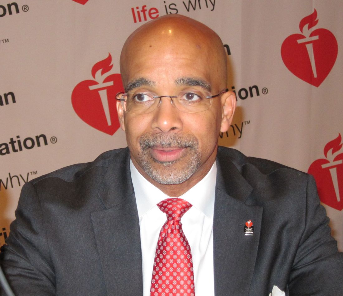
“Readmission rates remain stubbornly at 20% within 30 days and near 50% at 6 months. Acute decompensation is an inflection point in the natural history of heart failure with subsequent 1-year mortality rates consistently approximating 25%. Clearly there is something about the hospitalization that is a herald event which speaks to much worse outcomes, compared with chronic ambulatory heart failure,” said Dr. Yancy, professor of medicine and chief of cardiology at Northwestern University in Chicago.
He agreed with Dr. Packer that in light of the TRUE-AHF results, what Dr. Yancy termed “the early injury hypothesis” isn’t worth further pursuit.
Ularitide thus joins a long list of failed therapies for ADHF. Treatments that have convincingly been shown to have no significant impact on mortality and at best only modest impact on morbidity include continuous IV infusion of loop diuretics in the DOSE trial; the arginine vasopressin antagonists, which failed to impress in EVEREST, SECRET, and TACTICS; nesiritide (Natrecor) in the ASCEND-HF trial; and levosimendan (Simdax), which proved disappointing in the SURVIVE and REVIVE II studies, according to Dr. Yancy.
The jury is still out on serelaxin, he added. The drug showed a favorable signal in the RELAX-AHF trial. The results of RELAX II are awaited with interest.
“Today we still don’t have an effective single intervention for acute decompensated heart failure other than process-of-care improvement,” the cardiologist noted.
What holds promise for improved long-term outcomes in ADHF at this point? Dr. Yancy said sacubitril/valsartan (Entresto) is intriguing based upon the results of the PARADIGM-HF trial (N Engl J Med. 2014;371:993-1004). But the drug needs to be studied prospectively in patients in the throes of ADHF before it can appropriately be recommended for the purpose of changing the natural history of this disorder, Dr. Yancy stressed.
Devices such as the implantable pulmonary catheter are under study as a promising means of altering the natural history of ADHF by identifying actionable signals of impending decompensation weeks beforehand, he added.
The TRUE-AHF trial was sponsored by Cardiorentis. Dr. Packer reported serving as a consultant to that company and more than a dozen other pharmaceutical and medical device companies.
[email protected]
NEW ORLEANS – An investigational synthetic natriuretic peptide given early during hospitalization for acute decompensated heart failure didn’t produce any of the hoped-for intermediate- or long-term clinical benefits in the phase III TRUE-AHF study, Milton Packer, MD, reported at the American Heart Association scientific sessions.
The failure of this investigational vasodilator, ularitide, to influence downstream cardiovascular mortality or early readmission for heart failure closes the door on the once-promising hypothesis that myocardial microinjury occurring during ADHF is due to ventricular distension, observed Dr. Packer, the Distinguished Scholar in Cardiovascular Science at Baylor University Medical Center, Dallas. “Ularitide did exactly what we expected it to do while we were giving it: we caused intravascular decompression, we reduced cardiac wall stress, but we did not affect cardiac microinjury, and we didn’t change long-term cardiovascular mortality or any of our secondary endpoints, including and in particular the 30-day risk of rehospitalization for heart failure,” he said.
During a median follow-up of 15 months there were 236 cardiovascular deaths in the ularitide group and 225 in the control group, a nonsignificant difference. Nor were there any differences between the two groups in secondary endpoints including length of stay in the ICU during the index hospitalization, rehospitalization for ADHF within 30 days of discharge, or the composite of all-cause mortality or cardiovascular hospitalization within 6 months, which occurred in 40.7% of the ularitide group and 37.2% of controls.
The explanation for this lack of long-term benefit lies in the finding that myocardial microinjury wasn’t prevented by the rapid reduction of cardiac distension produced by ularitide. This was evident in the therapy’s inability to dampen the rise in high-sensitivity cardiac troponin T which occurred in the initial 48 hours of the study.
“The trial demonstrated the effects and safety of ularitide. However, to gain long-term benefits on hospitalizations and death in patients following a hospital admission for heart failure, physicians must focus on the drugs that patients take as an outpatient rather than the drugs they receive as an inpatient,” Dr. Packer concluded.
“Readmission rates remain stubbornly at 20% within 30 days and near 50% at 6 months. Acute decompensation is an inflection point in the natural history of heart failure with subsequent 1-year mortality rates consistently approximating 25%. Clearly there is something about the hospitalization that is a herald event which speaks to much worse outcomes, compared with chronic ambulatory heart failure,” said Dr. Yancy, professor of medicine and chief of cardiology at Northwestern University in Chicago.
He agreed with Dr. Packer that in light of the TRUE-AHF results, what Dr. Yancy termed “the early injury hypothesis” isn’t worth further pursuit.
Ularitide thus joins a long list of failed therapies for ADHF. Treatments that have convincingly been shown to have no significant impact on mortality and at best only modest impact on morbidity include continuous IV infusion of loop diuretics in the DOSE trial; the arginine vasopressin antagonists, which failed to impress in EVEREST, SECRET, and TACTICS; nesiritide (Natrecor) in the ASCEND-HF trial; and levosimendan (Simdax), which proved disappointing in the SURVIVE and REVIVE II studies, according to Dr. Yancy.
The jury is still out on serelaxin, he added. The drug showed a favorable signal in the RELAX-AHF trial. The results of RELAX II are awaited with interest.
“Today we still don’t have an effective single intervention for acute decompensated heart failure other than process-of-care improvement,” the cardiologist noted.
What holds promise for improved long-term outcomes in ADHF at this point? Dr. Yancy said sacubitril/valsartan (Entresto) is intriguing based upon the results of the PARADIGM-HF trial (N Engl J Med. 2014;371:993-1004). But the drug needs to be studied prospectively in patients in the throes of ADHF before it can appropriately be recommended for the purpose of changing the natural history of this disorder, Dr. Yancy stressed.
Devices such as the implantable pulmonary catheter are under study as a promising means of altering the natural history of ADHF by identifying actionable signals of impending decompensation weeks beforehand, he added.
The TRUE-AHF trial was sponsored by Cardiorentis. Dr. Packer reported serving as a consultant to that company and more than a dozen other pharmaceutical and medical device companies.
[email protected]
Key clinical point:
Major finding: Early administration of ularitide during hospitalization for acute decompensated heart failure failed to achieve any long-term clinical benefits.
Data source: The TRUE-AHF trial was a double-blind, placebo-controlled, randomized trial including 2,157 patients hospitalized for acute decompensated heart failure at 156 centers in 23 countries.
Disclosures: The study was sponsored by Cardiorentis. The presenter reported serving as a consultant to that company and more than a dozen other pharmaceutical and medical device companies.
Cardiac rehab also slashes stroke risk
ROME – Cardiac rehabilitation programs have a previously unappreciated benefit: participants enjoy a 60% reduction in the risk of stroke, Gijs van Halewijn, MD, reported at the annual congress of the European Society of Cardiology.
“I think cardiologists are really focused on cardiovascular deaths, especially from MI. But we’ve shown that cardiac rehabilitation also has an effect on cerebrovascular events,” said Dr. van Halewijn of Erasmus University in Rotterdam (the Netherlands).
He presented a meta-analysis of randomized controlled trials of cardiac rehab conducted during 2010-2015. The purpose was to assess the value provided by cardiac rehab in the contemporary era of acute coronary syndrome management featuring primary percutaneous coronary intervention, drug-eluting stents, and potent medications for secondary cardiovascular prevention. That hadn’t previously been looked at systematically.
“The standard meta-analyses cited in the field include randomized trials from as far back as just after World War II,” Dr. van Halewijn noted in an interview.
He employed the same search and analytic methods utilized by the Cochrane Collaboration in evaluating 18 randomized controlled trials of lifestyle- or exercise-based cardiac rehab, compared with usual care, in a total of 7,691 participants.
The results of the meta-analysis indicate cardiac rehab provides powerful secondary prevention benefits above and beyond those obtained through contemporary interventional procedures and preventive medications.
Cardiovascular mortality was reduced by 58% in cardiac rehab participants compared with usual care controls. The risk of acute MI was decreased by 30%. And in a new observation, cerebrovascular events were reduced by 60% in the four randomized trials in which that was an endpoint. All of these differences were highly statistically significant.
“Interestingly, the number needed to treat was 45 for MI, so if you have 45 patients included in your cardiac rehabilitation program, you can prevent one MI. And you can prevent one cerebrovascular event with 82 participants,” according to Dr. van Halewijn.
Cardiac rehab had no effect on all-cause mortality in the overall meta-analysis. However, in the trials involving comprehensive cardiac rehab programs targeting six or more of the components of secondary cardiovascular prevention described by the British Association for Cardiac Prevention and Rehabilitation (Heart. 2013 Aug;99[15]:1069-71), participation was associated with a 37% reduction in the risk of all-cause mortality, compared with usual care.
Those components are smoking, blood pressure, cholesterol, HbA1c, exercise training, counseling about the importance of exercise, stress management, and checking medications.
In addition, Dr. van Halewijn continued, cardiac rehab programs in which a physician or nurse made sure participants were on guideline-directed cardiovascular medications had a 65% reduction in all-cause mortality, compared with usual care.
“Cardiac rehabilitation’s opportunity is to evolve into comprehensive programs addressing all aspects of lifestyle, risk factor management, and prescription of medications to reduce death and nonfatal events,” the physician concluded.
This study was supported by Erasmus University Medical Center, Imperial College London, and the Dutch Heart Foundation. Dr. van Halewijn reported having no financial conflicts of interest.
[email protected]
AT THE ESC CONGRESS 2016
Key clinical point:
Major finding: The number of patients who need to participate in a cardiac rehabilitation program following an ACS in order to prevent one cerebrovascular event is 85.
Data source: This was a meta-analysis of 18 randomized trials carried out in 2010-2015 comparing contemporary cardiac rehabilitation to usual care in 7,691 patients.
Disclosures: This study was supported by Erasmus University (Rotterdam, the Netherlands) Medical Center, Imperial College London, and the Dutch Heart Foundation. The presenter reported having no financial conflicts of interest.
Tumor markers may predict anti–PD-L1 treatment response
NATIONAL HARBOR, MD. – Density in tumors of two key immune cells may be a biomarker for response to an inhibitor of programmed death–ligand 1 (PD-L1) among patients with non–small cell lung cancer (NSCLC).
Patients with NSCLC whose tumors bore high densities of both CD8-positive cytotoxic T lymphocytes and PD-L1 had significantly better outcomes when treated with the investigational PD-L1 inhibitor durvalumab than patients with high densities of either cell type alone, reported Sonja Althammer, PhD, a scientist at Definiens AG, maker of the anti–PD-L1 compound.

Dr. Althammer and her colleagues assessed the use of an automated image analysis and pattern recognition system to determine whether tumor-infiltrating CD8-positive cytotoxic T lymphocytes and PD-L1 densities could identify patients most likely to respond to the investigational PD-L1 inhibitor durvalumab.
“The hypothesis that we had in mind was that the interaction between both cell populations is important, so both cell populations should be present,” she said.
The investigators analyzed archived or fresh tumor biopsy samples from 163 patients with untreated or previously treated NSCLC (median 3 prior lines of therapy) who were enrolled in a nonrandomized phase I/II trial evaluating durvalumab in advanced NSCLC and other solid tumors.
They matched CD8 and PD-L1 immunohistochemistry-stained sections from tissues blocks. High PD-L1 expression was defined as a 25% or higher proportion of PD-L1–positive tumor cells with membrane staining at any intensity.
The images were then evaluated with an automated system, with results matched to clinical outcomes based on the densities of CD8-positive and PD-L1–positive cells, and PD-L1 alone from pre-treatment biopsy samples using a discovery set (84 samples) and validation set (79). The datasets were matched on baseline PD-L1 status, histology, Eastern Cooperative Oncology Group performance status, lines of therapy, and response.
In a training set looking at overall response rate (ORR), they found evidence to suggest that the combination of CD8 and PD-L1 positivity was associated with a higher ORR: 42%, compared with 31% for CD8-positive expression alone, 27% for PD-L1–positive alone, and 7% for CD8-positive PD-L1–negative (less than 25% expression) alone.
They then examined the predictive ability of the combined markers, and found that over approximately 28 months of follow-up, patients with CD8-positive and PD-L1–positive dense tumors had better overall survival (median overall survival, 24.3 months), compared with PD-L1–positive only patients (median overall survival, 17.1 months), CD8-positive only patients (median overall survival, 17.8 months), or the entire patient sample (median overall survival, 11.1 months).
Similarly, progression-free survival was also better among patients with high expression of both markers (respective median progression-free survival was 7.3, 3.6, 5.3, and 2.8 months).
Dr. Althammer acknowledged that the findings were preliminary and needed to be confirmed independently in larger studies.
The study was supported by MedImmune. Sonja Althammer is an employee of Definiens AG, which is a subsidiary of MedImmune/AstraZeneca.
NATIONAL HARBOR, MD. – Density in tumors of two key immune cells may be a biomarker for response to an inhibitor of programmed death–ligand 1 (PD-L1) among patients with non–small cell lung cancer (NSCLC).
Patients with NSCLC whose tumors bore high densities of both CD8-positive cytotoxic T lymphocytes and PD-L1 had significantly better outcomes when treated with the investigational PD-L1 inhibitor durvalumab than patients with high densities of either cell type alone, reported Sonja Althammer, PhD, a scientist at Definiens AG, maker of the anti–PD-L1 compound.
Dr. Althammer and her colleagues assessed the use of an automated image analysis and pattern recognition system to determine whether tumor-infiltrating CD8-positive cytotoxic T lymphocytes and PD-L1 densities could identify patients most likely to respond to the investigational PD-L1 inhibitor durvalumab.
“The hypothesis that we had in mind was that the interaction between both cell populations is important, so both cell populations should be present,” she said.
The investigators analyzed archived or fresh tumor biopsy samples from 163 patients with untreated or previously treated NSCLC (median 3 prior lines of therapy) who were enrolled in a nonrandomized phase I/II trial evaluating durvalumab in advanced NSCLC and other solid tumors.
They matched CD8 and PD-L1 immunohistochemistry-stained sections from tissues blocks. High PD-L1 expression was defined as a 25% or higher proportion of PD-L1–positive tumor cells with membrane staining at any intensity.
The images were then evaluated with an automated system, with results matched to clinical outcomes based on the densities of CD8-positive and PD-L1–positive cells, and PD-L1 alone from pre-treatment biopsy samples using a discovery set (84 samples) and validation set (79). The datasets were matched on baseline PD-L1 status, histology, Eastern Cooperative Oncology Group performance status, lines of therapy, and response.
In a training set looking at overall response rate (ORR), they found evidence to suggest that the combination of CD8 and PD-L1 positivity was associated with a higher ORR: 42%, compared with 31% for CD8-positive expression alone, 27% for PD-L1–positive alone, and 7% for CD8-positive PD-L1–negative (less than 25% expression) alone.
They then examined the predictive ability of the combined markers, and found that over approximately 28 months of follow-up, patients with CD8-positive and PD-L1–positive dense tumors had better overall survival (median overall survival, 24.3 months), compared with PD-L1–positive only patients (median overall survival, 17.1 months), CD8-positive only patients (median overall survival, 17.8 months), or the entire patient sample (median overall survival, 11.1 months).
Similarly, progression-free survival was also better among patients with high expression of both markers (respective median progression-free survival was 7.3, 3.6, 5.3, and 2.8 months).
Dr. Althammer acknowledged that the findings were preliminary and needed to be confirmed independently in larger studies.
The study was supported by MedImmune. Sonja Althammer is an employee of Definiens AG, which is a subsidiary of MedImmune/AstraZeneca.
AT SITC 2016
Key clinical point: Tumor expression of two cell types may predict responses to anti–PD-L1 immunotherapy.
Major finding: High tumor densities of CD8-postive cytotoxic T lymphocytes and the programmed death-ligand 1 were associated with improved overall response rates and survival in patients with advanced non–small cell lung cancer treated with a PD-L1 inhibitor.
Data source: Imaging study of samples from 163 patients enrolled in a nonrandomized phase I/II trial.
Disclosures: The study was supported by MedImmune. Sonja Althammer is an employee of Definiens AG, which is a subsidiary of MedImmune/AstraZeneca.
Risk stratification important for aortic valve disease in pregnancy
CHICAGO – Pregnancy-associated hemodynamic and physiologic changes can challenge hearts with diseased aortic valves. Understanding pregnancy’s toll on the body can help physicians take better care of pregnant women with aortic stenosis or aortic regurgitation, said Patrick T. O’Gara, MD. “This is a relatively infrequent condition, and it causes all of us a great deal of apprehension,” he said.
Dr. O’Gara, director of clinical cardiology at Brigham and Women’s Hospital, Boston, walked attendees through current recommendations for caring for pregnant women with aortic valve disease in a presentation at the annual Heart Valve Summit.
When talking to women with aortic valve disease, it’s important to let them know that a pregnancy during which they experience an adverse maternal cardiac event appears to increase their risk for later events as well, said Dr. O’Gara. One study of pregnancy in women with aortic valve disease, said Dr. O’Gara, found that the risk of later events for women who had had a cardiac event in pregnancy was 27% plus or minus 9 percentage points in the 5 years following pregnancy, while the risk was just 15% plus or minus 3 percentage points for women whose pregnancies were not complicated by cardiac adverse events (P = .02) (Heart. 2010 Oct;96(20):1656-61).
The World Health Organization classification of pregnancy risk, said Dr. O’Gara, provides some guidance. Patients who are WHO class III are considered to be high risk; pregnancy in these individuals calls for counseling and a multidisciplinary care team. Patients who fall into this class are those with mechanical heart valves, those with Marfan syndrome and aortic involvement with a valve diameter of 40-45 mm, and those with bicuspid aortic valve disease with a valve diameter of 45-50 mm.
WHO class IV patients, according to the guidelines, are at prohibitive risk. These patients should consider terminating a pregnancy if one occurs. Conditions that place women into WHO class IV include symptomatic severe aortic stenosis (AS), severe left ventricular dysfunction with an ejection fraction less than 40%, and severe coarctation of the aorta. Women with Marfan syndrome with aortic valve diameters greater than 45 mm and women with bicuspid aortic valves and a diameter over 50 mm also fall into this category of prohibitive risk.
If a physician is lucky enough to have a pre-conception relationship with a patient, then a host of considerations can come into play. In addition to cardiac risk stratification, obstetric and intrapartum risk to the mother as well as neonatal risk should be considered. Some factors to take into account are maternal life expectancy and potential long-term complications to the mother from pregnancy and childbirth.
Plasma volume increases rapidly through the first trimester, plateauing about halfway through pregnancy. With this normal dilutional anemia, “Most normal pregnancies are accompanied by a grade II systolic murmur, best heard at the upper left sternal border,” said Dr. O’Gara. Since stroke volume and heart rate both also increase through pregnancy, cardiac output goes up as well.
“Importantly, systemic vascular resistance is reduced throughout pregnancy,” said Dr. O’Gara. Consequently, “regurgitant heart valve problems are generally much better tolerated than stenotic lesions in pregnancy. That’s physiologic principle number one.”
In labor, abrupt hemodynamic changes include increases in cardiac output, heart rate, blood pressure, and venous return. “Labor and delivery are very stressful hemodynamic times for women,” said Dr. O’Gara.
In the immediate postpartum period, uterine contraction results in a return of a significant amount of blood to the maternal circulation. This effectively creates an autotransfusion, with resulting increased preload and cardiac output. The inferior vena cava is also freed from the pressure of a gravid uterus, increasing venous return further. Finally, there’s also a marked increase in systemic vascular resistance.
Aside from the risk of adverse cardiac events, other adverse outcomes for women can include an increased risk of premature rupture of membranes and postpartum hemorrhage. For the neonate, preterm birth and respiratory distress are more likely. Newborns are more likely to be small for gestational age, to suffer intraventricular hemorrhage, and to die. Depending on the parental genetic status, the infant may be at risk of congenital heart disease as well.
Neonatal risk may also be increased, said Dr. O’Gara. Some maternal conditions that can increase risk for the infant include a baseline New York Heart Association (NYHA) class of greater than II or having cyanosis; having a left heart obstruction; being a smoker; having a multiple gestation; having a mechanical heart valve; and taking an oral anticoagulant.
Ideally, a full discussion would include a genetic referral if indicated, as well as coordination with a gynecologist or primary care provider to provide contraception and to assist with planning for and optimizing outcomes of a pregnancy.
The American College of Cardiology and the American Heart Association have issued guidelines that include a class I recommendation regarding valve intervention before pregnancy for symptomatic patients with severe AS (those with aortic velocity of at least 4.0 m/sec, or mean pressure gradient greater than 41 mm Hg; stage D). For patients who meet the same hemodynamic criteria for severe AS but who are asymptomatic (stage C), the guidelines have a class IIa recommendation that pre-pregnancy valve intervention is “reasonable.” These recommendations have level C evidence supporting them.
However, another class IIa recommendation for patients with severe AS who are already pregnant advises valve intervention only in the case of hemodynamic deterioration, or the development of NYHA class III to IV heart failure symptoms. This is backed by grade B evidence, according to the associations.
For aortic regurgitation (AR), a level I recommendation advises valve repair or replacement before pregnancy if patients are symptomatic and have severe (stage D) regurgitation. Pregnant patients with severe regurgitation should only be considered for a valve operation if they have refractory NYHA class IV heart failure symptoms. These recommendations have level C evidence supporting them.
Since AR is better tolerated in pregnancy than AS, the nature of the valve disease is one of many factors to consider when deciding whether to perform intervention before the patient becomes pregnant. Plans for future pregnancy may also affect the choice of prosthesis, as anticoagulation management during pregnancy can be extremely tricky.
The recommendations overall make clear that, based on available evidence, “there should be a high threshold for intervention during pregnancy,” said Dr. O’Gara. And no one should go it alone when taking care of these complicated patients. “Care of pregnant women with heart disease requires coordination with a multidisciplinary team,” he said.
Dr. O’Gara reported no relevant disclosures.
[email protected]
On Twitter @karioakes
EXPERT ANALYSIS FROM THE HEART VALVE SUMMIT
ACGME seeks to return trainees’ maximum shifts to 24 hours
First-year residents may be permitted to work up to 24 consecutive hours – 8 hours longer than they can now – under a proposal from a task force of the Accreditation Council for Graduate Medical Education (ACGME).
The ACGME Duty Hour Task Force proposes to raise first-year trainees’ work hour limit from 16 hours, reverting to the 24-hour maximum that remained in effect until 2011 – and the existing limit in place for all other residents.
“There is better team continuity of care provided with less micromanagement of resident duty hours,” said Thomas J. Nasca, MD, ACGME chief executive and vice chairman of the task force.
The ACGME instituted the 16-hour cap for first-year residents in the wake of a December 2008 report released by the Institute of Medicine (IOM), “Resident Duty Hours: Enhancing Sleep, Supervision, and Safety.” The ACGME also prohibited 30-hour shifts, which some trainees had been clocking.
“Every 5 years, the ACGME reviews its program requirements,” Dr. Nasca said. “That’s a promise we made to the community and to the public” in July 2003. The most recent changes occurred in July 2011.
Since then, some faculty have contended that shorter work hour limits for the first-year residents are accelerating the frequency of patient transitions and disrupting continuity of care. Many educators also noted that residents would gain more knowledge by monitoring a hospitalized patient during the initial 24 hours.
The proposed changes are “encouraging and courageous,” according to R. James Valentine, MD, FACS, president of the Western Surgical Association.
“This is a real world scenario,” said Dr. Valentine, professor of vascular surgery at Vanderbilt University, Nashville, Tenn. “Medicine is such a complex system. It is not easily constrained by time limitations.”
Requiring a physician to transition a patient’s care at a specific time may help promote the trainee’s well-being; however, it also “robs the resident of the opportunity to see the disease progress and to see the response to the treatment that is being offered. The new rules help strike a balance between bedside education and rest,” he added.
The proposed revisions to training requirements also include phasing out the term “duty” hours in favor of “clinical experience and education,” highlighting that residents’ responsibility to patients takes precedence over any adherence to a schedule or clock. Working a certain number of hospital hours is only one aspect of delivering safe and quality care, the ACGME Duty Hour Task Force noted in its proposal.
The proposal does not change the following, which all are averaged over a period of 4 weeks: a maximum of 80 hours per week, 1 day free from clinical experience or education in 7, and in-house call no more often than every third night.
“It is important to note that the absence of a common 16-hour limit does not imply that programs may no longer configure their clinical schedules in 16-hour increments if that is the preferred option for a given setting or clinical context,” Dr. Nasca wrote in a Nov. 4 letter to the graduate medical education community.
In its December 2008 report, the IOM noted that revamping residents’ work hours alone offered no guarantee of patient safety. It called for increased supervision by seasoned physicians, restrictions on patient caseloads based on residents’ levels of experience and specialty, overlap in schedules around shift changes to decrease the possibility of error in transitioning patients from one provider to another, and broad-based research to examine the outcomes from these changes.
“Surgical residents, when they’re operating, come under very direct supervision by attending physicians,” said Jay Bhattacharya, MD, professor of medicine at Stanford (Calif.) University, who served on the 2008 IOM committee. “That supervision is generally pretty good. Even for a tired resident, the supervision makes it so that the fatigue the trainee faces doesn’t endanger the patient.”
Acknowledging the emerging evidence that physicians are at heightened risk for burnout and perhaps depression, the proposal underscores the need for residency programs and institutions to prioritize the well-being of residents as well as nurses and other hospital personnel.
“Burnout and depression impair a physician’s ability to provide excellent care. Self-care is an important aspect of professionalism, and a skill that must be learned and nurtured under the guidance and role modeling of faculty members,” Dr. Nasca wrote in his Nov. 4 letter.
If the proposal is approved by the ACGME board of directors in February, the changes will be rolled out in July.
The proposal is open to public comment through Dec. 19; comments will be reviewed and considered before a final set of proposed requirements are sent to the ACGME board for approval.
“We’re here to serve the public, and so the focus is on the comments that reflect the most recent and comprehensive evidence and educational outcomes rather than the individual opinions we receive,” Dr. Nasca said.