User login
AVAHO
div[contains(@class, 'header__large-screen')]
div[contains(@class, 'read-next-article')]
div[contains(@class, 'nav-primary')]
nav[contains(@class, 'nav-primary')]
section[contains(@class, 'footer-nav-section-wrapper')]
footer[@id='footer']
div[contains(@class, 'main-prefix')]
section[contains(@class, 'nav-hidden')]
div[contains(@class, 'ce-card-content')]
nav[contains(@class, 'nav-ce-stack')]
Genomic signature predicts safety of omitting RT in early breast cancer
A novel 16-gene panel based on the biology of locoregional recurrence in early invasive breast cancer can both identify patients with a low risk of recurrence if they were to skip postsurgery radiation therapy, and predict which patients would be unlikely to benefit from adjuvant radiation, investigators reported.
Among 354 patients with stage I or II invasive estrogen receptor–positive (ER+), HER2-negative breast cancers who did not receive adjuvant systemic therapy, the genomic signature, dubbed POLAR (Profile for the Omission of Local Adjuvant Radiation) was prognostic for locoregional recurrence in patients who did not undergo radiation therapy, reported Martin Sjöström, MD, PhD, of the University of California, San Francisco, and colleagues.
They reported their findings in a poster presented during the American Society of Clinical Oncology (ASCO) annual meeting (Abstract 512).
“This is to our knowledge the first radiation-omission signature that is both prognostic and predictive: prognostic for outcomes in the absence of radiation, and predictive of benefits,” coauthor Corey Speers MD, PhD, of the University of Michigan, Ann Arbor, said in an interview.
The investigators conducted a retrospective analysis of data on patients enrolled in the SweBCG 91 RT trial, in which 1,187 patients with T1-2N0M0 breast cancer underwent a standardized radical sector resection and were then randomly assigned to either postoperative radiotherapy or no further treatment.
As the investigators reported in a long-term follow-up study presented in 2010 at the San Antonio Breast Cancer Symposium, the addition of postoperative radiation did not significantly affect overall survival, but was associated with a significant improvement in recurrence-free survival.
In the study presented at ASCO 2021, Dr. Sjöström and colleagues in Sweden, the United States, and Canada sought to determine whether they could identify a genomic signature that would identify women at very low risk for recurrence who could safely be spared from radiotherapy.
They focused on those patients in the study with ER+, HER2-negative tumors who did not receive adjuvant systemic therapy. The patients were divided into a training cohort of 243, and a validation cohort of 354 patients.
The investigators performed transcriptome-wide profiling of tumors, and identified both biological gene sets and individuals genes associated with locoregional recurrence among patients in the training set who did not receive radiotherapy.
The final POLAR genomic signature, containing 16 genes, was locked prior to testing in the validation cohort.
In a multivariable Cox model adjusting for age, grade, tumor size and luminal A vs. luminal B subtype, the POLAR gene set was prognostic for locoregional recurrence, with a hazard ratio (HR) of 1.7 (P < .001).
The 10-year locoregional recurrence rate for patients in the POLAR low-risk category who had not received radiation was 7%, and there was no significant benefit for POLAR low-risk patients who did receive radiation (HR 1.1, P = ns).
In contrast, patients classified as POLAR high risk who received radiotherapy had significantly lower risk for locoregional recurrence than high-risk patients who did not receive radiotherapy (HR 0.43, P = .0053).
Dr. Speers said that the POLAR signature appears to be unique in its ability to discriminate radiation-omission risk.
“At least looking in this cohort in the Swedish trial, none of other previously derived signatures – Mammaprint, ProSigna, Oncotype – were prognostic or predictive of locoregional recurrence events with radiation,” he said.
The investigators are currently exploring the POLAR signature in other clinical trials in which patients were randomized to receive radiation or no radiation.
‘A true victory’
Invited discussant Benjamin D. Smith, MD, of the University of Texas MD Anderson Cancer Center in Houston, pointed out that in randomized clinical trials, approximately 60% of patients treated for early breast cancer did not experience recurrence in the absence of radiation, and that radiation prevented recurrence in about 30%, while about 10% experienced recurrence despite receiving radiation.
He said that the study by Dr. Sjöström and colleagues asks the question “can we use molecular factors to help identify patients who will not recur with lumpectomy alone without radiation therapy?”
The 60% of patients who will not experience recurrence in the absence of radiation can be categorized into two subpopulations: those with no residual malignant clonogens – the population included in this study by Dr. Sjöström and colleagues – and those with residual clonogens in the breast or elsewhere in the body that are sensitive to adjuvant endocrine therapy.
He said that the 7% 10-year risk of recurrence among patients in the POLAR low-risk group, who had neither radiation nor endocrine therapy, “is an exceptional outcome, which should be applauded, and I would point out that this risk of local recurrence of only 7% is at least in the same ballpark as the risk of recurrence that we accept every day when we treat early stage breast cancer patients with mastectomy alone, so this is a true victory.
“When we reflect on these provocative results from Dr. Sjöström and colleagues, it prompts in mind the question, could there be a group of patients with early breast cancer for whom a true ‘one-and-done’ strategy could be effective and safe?” Dr. Smith said.
Getting there will require a multidisciplinary, multimodality approach, involving imaging features of the primary tumor, clinical and pathologic features, and molecular information such as that provided by the POLAR genomic signature, he said.
The study was supported by PFS Genomics. Dr. Sjöström reported institutional funding from PFS Genomics. Dr. Speers disclosed stock and other ownership interests in the company, and he has applied for a patent on methods and genomic classifiers for prognosis of breast cancer and predicting benefit from adjuvant radiotherapy. Dr. Smith reported an equity interest in Oncora Medical, and an uncompensated relationship with the American Society for Radiation Oncology.
FROM ASCO 2021
Pregnancy effect on chemotherapy does not affect maternal breast cancer outcomes
Reassuring news for women who receive a diagnosis of breast cancer during pregnancy: Pregnancy-induced alterations in the pharmacokinetics of chemotherapy do not appear to compromise outcomes for the mother.
That’s according to investigators who reviewed registry data on 662 pregnant women and 2,081 nonpregnant women with a diagnosis of breast cancer. After a median follow-up of 66 months, there were no significant differences in either disease-free survival (DFS) or overall survival (OS), and women who received more than 60% of their chemotherapy doses during pregnancy had survival comparable to that of nonpregnant women, reported Frédéric Amant, MD, PhD, of University Hospitals Leuven (Belgium).
“These results support initiation of chemotherapy for breast cancer during pregnancy where indicated for oncological reasons,” they reported in a poster discussion session at the American Society of Clinical Oncology annual meeting. (Abstract 515).
Although in general a diagnosis of breast cancer during pregnancy does not appear to affect the mother’s prognosis when standard therapy is used, “caution is warranted as gestational changes in pharmacokinetics with respect to the distribution, metabolism, and excretion of drugs may lead to reduced chemotherapy concentration in pregnant patients,” the authors wrote.
To get a better picture of the prognosis for women diagnosed with breast cancer during pregnancy, the investigators created a cohort of patients from two multicenter registries: the International Network of Cancer, Infertility, and Pregnancy and the German Breast Group. Both registries collect data retrospectively and prospectively,
They used propensity scoring to smooth out differences in the baseline characteristics of pregnant women and nonpregnant controls.
The median age at diagnosis was 34 year for pregnant women, and 38 years for controls. Pregnant women were more likely than were controls to have stage II disease (60.1% vs. 56, 1%, P = .035), grade 3 tumors (74% vs. 62.2%, P < .001), hormone receptor–negative breast tumors (48.4% vs. 30%), and triple-negative breast cancer (38.9% vs. 26.9%, P < .001).
In Cox proportional hazard regression analysis controlling for age, stage, grade, hormone receptor status, HER2 status and histology, there were no significant differences between pregnant women and controls in either DFS (hazard ratio [HR] 1.02, P = .83) or OS (HR 1.08, P = .57).
As noted before, a subgroup analysis of 339 women who received more than 60% of their assigned chemotherapy doses during pregnancy also showed that survival was not significantly different from that of nonpregnant women (HR for DFS 0,71, P = .13; HR for OS 0.85, P = .39).
Termination does not benefit the mother
“Thanks to the important work of Dr. Amant in the INCIP [International Network on Cancer, Infertility, and Pregnancy] network and others around the world, we now have sufficient data to know that it’s safe to treat breast cancer during pregnancy, and that the prognosis of breast cancer during pregnancy is comparable to nonpregnant controls if we adjust for certain characteristics such as age and others,” said Fatima Cardoso, MD, of Champalimaud Clinical Center in Lisbon, Portugal, the invited discussant.
“With this and other studies, we can come to the conclusion that pregnancy-induced alterations in the chemotherapy concentration due to altered pharmacokinetics does not seem to affect maternal prognosis, and therefore we should initiate treatment of breast cancer during wherever it’s indicated for oncological reasons, knowing that you can only use chemotherapy during the second or third trimester,” she said.
Dr. Cardoso emphasized that breast cancer during pregnancy is a rare situation requiring that treatment be given in a specialized center by an experienced multidisciplinary team, and that interrupting the pregnancy does not improve the mother’s prognosis.
“We have to spread the word to all health professionals who come across these women to stop advising them to immediately terminate pregnancy. For the children, the most important take-home message is avoid prematurely delivery,” she said.
Treatment for women with a diagnosis of breast cancer during pregnancy should be similar to that for nonpregnant women, with the exception of endocrine therapy and anti-HER2 agents, which should be withheld until after delivery, she added.
The study was supported by the European Research Council, Research Foundation Flanders, and Kom op tegen kanker (Stand Up to Cancer). Dr. Amant disclosed a consulting or advisory role for AstraZeneca and Clovis Oncology. Dr. Cardoso disclosed consulting or advisory roles and travel support from multiple companies.
FROM ASCO 2021
Chemotherapy/local excision avoids proctectomy in rectal cancer
Chemotherapy and local excision led to organ preservation in over half of early-stage rectal cancer patients in a small study, but follow-up was only a median of 15.4 months.
Even so, “we believe that subsequent trials ... are warranted,” said lead investigator Hagen F. Kennecke, MD, medical director of GI oncology at Providence Cancer Institute, Portland, Ore., who presented the findings at the American Society of Clinical Oncology annual meeting.
“The results are quite promising,” said study discussant Karyn Stitzenberg, MD, a surgical oncologist at the University of North Carolina, Chapel Hill.
“The reported organ preservation rates of 57% to 79% compare favorably with the rates previously demonstrated in studies of neoadjuvant chemoradiation followed by local excision,” but longer-term follow up is needed “to know the true organ preservation rate,” she said.
Organ preservation – sparing the rectum during treatment – is a hot topic in rectal cancer. Total mesorectal excision (TME) is still the go-to option, but it’s fraught with bad GI, urinary, sexual, and other complications for patients. “Consequently, the concept of organ preservation ... is very appealing,” Dr. Stitzenberg explained.
The chemoradiation/local excision approach she referenced is gaining traction as an alternative, but the radiation component is itself associated with substantial short- and long-term problems, including sphincter dysfunction and wound healing complications.
The goal of Dr. Kennecke’s study, dubbed NEO [Neoadjuvant, Excision, Observation], was to see if the radiation could be left out altogether.
Recruited at eight centers in Canada and one in the United States, the 58 subjects had clinical stage T1-T3 A/B node-negative tumors with no pathologic high-risk features.
They received neoadjuvant FOLFOX (six cycles in 32 patients, 91% completion rate) or CAPOX (four cycles in 26 patients, 89% completion); 56 of the 58 subjects then went on to transanal endoscopic tumor excision; one of the other two patients wasn’t eligible because of tumor progression and the other one declined.
The 33 patients who were stage T0/T1N0 after treatment were spared organ removal and underwent observation every 3-6 months. TME was recommended for the 23 others who were stage 2 or higher or had nodal metastases following chemotherapy and excision.
The numbers translated to a per-protocol organ preservation rate of 57% over a median follow-up of 15.4 months; when the 13 patients who declined TME were added, the rate climbed to 79%.
Although “organ preservation in rectal cancer is becoming an increasingly promising and realistic option for a subset of patients,” Dr. Stitzenberg said, there are more reasons to be cautious beyond the short follow-up.
“The standard of care treatment for these patients would have been proctectomy ... Most would not have [had] systemic chemotherapy. As a result, the added morbidity of FOLFOX or CAPOX needs to be considered.” The study reported that there were no unexpected toxicities, but “what were the expected toxicities? How many patients experienced grade 3 to 5 complications?” she wondered.
Also, how realistic is it to expect patients to report for surveillance every few months outside of a trial? And how can they best be watched to make sure recurrence is caught “while salvage TME is still feasible? There are many longer-term follow-up questions that remain to be answered,” Dr. Stitzenberg said.
Even with short follow-up, there were two locoregional recurrences across the cohort (3.5%), both treated by TME to R0/1 resection. There were no distant relapses.
Subjects were a median of 67 years old, and over two-thirds were men. The majority had stage 2 disease at baseline. Tumors were well to moderately differentiated nonmucinous rectal adenocarcinomas with a median height of 6 cm.
The work was funded by the Canadian Cancer Trials Group. Dr. Kennecke disclosed relationships with Advanced Accelerator Applications, Ipsen, and Taiho Pharmaceutical. Dr. Stitzenberg had no relevant disclosures.
FROM ASCO 2021
KRAS inhibitor improved survival in phase 2 lung cancer trial
The first KRAS inhibitor approved for the treatment of lung cancer provided a clinically meaningful overall survival benefit in an updated analysis of a phase 2 study.
Treatment with sotorasib yielded a median overall survival (OS) of 12.5 months in patients with previously treated KRAS p.G12C-mutated non-small cell lung cancer (NSCLC), according to an analysis of the phase 2 CodeBreaK 100 trial data presented at the American Society of Clinical Oncology Annual Meeting.
Median progression-free survival (PFS) was 6.8 months in this update, which included a median follow-up of more than 15 months, according to investigator Ferdinandos Skoulidis, MD, PhD, assistant professor of thoracic/head and neck medical oncology at the University of Texas MD Anderson Cancer Center in Houston.
Efficacy responses
The confirmed objective response rate was 37.1%, including a 3.2% complete response rate and a median duration of response of 11.1 months, according to the report by Dr. Skoulidis.
In exploratory analyses, the benefit of sotorasib was consistent across patient subgroups, Dr. Skoulidis said in his presentation (Abstract 9003).
In particular, efficacy was observed in subgroups with co-occurring mutations in TP53, STK11, and KEAP1, which are molecular indicators of suboptimal outcomes on standard systemic treatments, according to Dr. Skoulidis.
This update on the registrational phase 2 CodeBreaK100 trial, published concurrently in the New England Journal of Medicine , came just one week after the U.S. Food and Drug Administration (FDA) granted accelerated approval to sotorasib.
Sotorasib was approved for the treatment of patients with previously treated KRAS G12C‑mutated locally advanced or metastatic NSCLC on the basis of previously reported results from CodeBreaK100.
This sotorasib indication represents a “historic milestone,” Dr. Skoulidis said in an interview.
No previously studied selective KRAS inhibitor has been approved despite scientific research efforts that stretch back nearly four decades, he explained.
“In a way, one can say that we have dealt KRAS-mutant lung cancer a knockdown blow, however, I should point out that the fight is not over,” he added.
“These clinical results will no doubt spearhead and galvanize further efforts to develop even more effective therapeutic combinations in the future, as well as identify and either forestall or overcome the eventual development of acquired resistance,” he said.
Only 1 out of 8 patients
The KRAS p.G12C mutation is present in about 13% of lung adenocarcinomas, or about one in every eight patients with nonsquamous NSCLC, Dr. Skoulidis said in the interview.
“We are estimating that this is in the region of 13,000 patients newly diagnosed every year in the U.S., and approximately 13,000 patients or so that are currently being treated in the second- or third-line setting,” he said.
The CodeBreaK100 trial included 126 patients with locally advanced or metastatic NSCLC and KRAS p.G12C mutation who had progressed on prior systemic therapies. About 43% had one prior line of treatment, while 35% had two lines, and 22% had three lines. A total of 81% had previously received both platinum-based chemotherapy and PD-1/PD-L1 axis inhibitors.
Most treatment-related adverse events in the study were grade 1-2 and generally manageable, according to Dr. Skoulidis. About 20% of patients experienced grade 3 treatment-related adverse events, which were mostly diarrhea or increases in aspartate aminotransferase and alanine aminotransferase levels. A grade 4 treatment-related adverse event, pneumonitis and dyspnea, was reported in one patient or approximately 1%.
Confirmatory trial
Although CodeBreak100 is not a randomized trial, the median OS of 12.5 months compares favorably to median OS times in the range of 7.9-10.3 months reported in randomized phase 3 clinical trials and subgroup analysis of randomized phase 3 trials of docetaxel for patients with KRAS-mutant lung adenocarcinoma, Dr. Skoulidis said in a question-and-answer session.
A confirmatory phase 3 CodeBreaK200 trial of sotorasib versus docetaxel in patients with previously treated KRAS p.G12C-mutated NSCLC is underway. That trial is evaluating PFS as a primary endpoint and OS as a secondary endpoint.
“If the same magnitude of benefit, 12.5 months median overall survival, is confirmed in the larger phase 3 clinical trial, as a clinician I would consider that beneficial for patients, compared to the standard of care,” Dr. Skoulidis said during the session.
Mature data
The updated analysis of the phase 2 CodeBreaK100 study is notable for its mature OS data, updated safety and the first molecular subgroup analyses, according to discussant Christine Marie Lovly, MD, PhD, of the division of hematology-oncology at Vanderbilt University Medical Center in Nashville.
“The objective response rate was 37.1%,” she added. “This is a little bit lower than we’re used to for targeted therapies, but remember, this is a different mutation and a very different class of drugs.”
The KRAS G12C inhibitors, several of which are under clinical development, are not tyrosine kinase inhibitors (TKIs), but rather allele-specific inhibitors that target mutant KRAS, trapping it in an inactive conformation, she explained.
Dr. Lovly referenced the exploratory analyses demonstrating efficacy in molecularly defined subgroups, calling it “interesting” that there was no difference in objective response rate between TP53 wild type and mutant tumors.
“We do have data that mutant TP53 seems to confer inferior outcomes for EGFR TKI-directed therapy in patients with EGFR-mutant lung cancer,” she said.
CodeBreaK100 was supported by Amgen, Inc. and partly by a National Institutes of Health Cancer Center Support Grant at Memorial Sloan Kettering Cancer Center.
Dr. Skoulidis reported honoraria from Bristol-Myers Squibb; research funding from AIMM Therapeutics and Amgen; and travel, accommodations, or expenses from Tango Therapeutics. Dr. Lovly reported disclosures related to Amgen, AstraZeneca, Genentech, Novartis, and Pfizer, among others.
FROM ASCO 2021
NSCLC: Immune-related AEs during checkpoint inhibitor therapy may predict outcomes
Experiencing an immune-related adverse event during checkpoint inhibitor treatment may predict outcomes in patients with non-small cell lung cancer, exploratory analyses of phase 3 trials suggest.
Immune-related adverse events (irAEs) were tied to longer overall survival (OS) in exploratory pooled analyses of three phase 3 clinical trials evaluating atezolizumab-based regimens, according to investigator Mark A. Socinski, MD, of AdventHealth Cancer Institute, Orlando, Fla.
Median OS approached 26 months for patients who received first-line atezolizumab and experienced an irAE, compared with just 13 months for those who did not experience an irAE, according to results reported at the American Society of Clinical Oncology Annual Meeting (Abstract 9002).
Atezolizumab-treated patients with grade 3 or greater irAEs had the shortest OS, shorter than those atezolizumab-treated patients who experienced grade 1-2 irAEs or no irAEs at all. That short OS may be due to treatment interruptions or discontinuations, said Dr. Socinski.
“Data from these analyses suggest an association between irAEs and efficacy in patients with [non-small cell cancer] NSCLC,” he stated in his presentation of the results.
A lot more to learn about irAEs
Similar linkages between irAEs and outcomes were observed in pooled analyses of patients enrolled in the control arms of the phase 3 trials, with a median OS of about 20 months for control patients experiencing an irAE, versus about 13 months for those who did not.
That linkage in the control arm prompted a question from an ASCO attendee about why an effect of irAEs, commonly associated with immune checkpoint inhibitor therapy, would be evident in analyses of patients who did not receive those agents.
In his response, Dr. Socinski characterized the finding as “a surprise” and said the finding may either reflect how adverse events are characterized or how chemotherapy impacts the immune system.
“I don’t know that our definition of irAEs is perfect,” he said, “and maybe we don’t understand what impact chemotherapy may have on the immune system, and may actually engender what historically we’ve always seen as an adverse event, but didn’t necessarily classify as an immune-related adverse event.”
More work is needed to better understand the connection between irAES and outcomes, and whether anything can be done as a result of that improved understanding, said discussant Mary Weber Redman, PhD.
“The question is, ‘what is actionable?’” added Dr. Redman, a biostatistician at the Fred Hutchinson Cancer Research Center, Seattle.
A firmer understanding of the relationship between irAEs and outcomes could change how clinicians monitor patients for irAEs, lead to better prediction of which patients may experience higher grade irAEs, and ultimately impact treatment selection potentially to avoid those higher grade events, Dr. Redman said in her remarks.
“Doing these types of analyses are quite important, because we have to look at the breadth of information that we have to be able to interpret that and think about what are future questions,” she said in the question-and-answer session accompanying Dr. Socinski’s presentation.
“I think the key is that we shouldn’t use these analyses to be definitive, but we should use them as to be hypothesis generating,” she added.
More evidence to link irAEs and outcomes
Immune-related AEs caused by off-target immune and inflammatory activity have been reported in up to 80% of patients receiving immune checkpoint inhibitors as monotherapy and up to 95% in combination regimens, Dr. Socinski said in his presentation.
“Increasing evidence suggests that the occurrence of immune-related adverse events with PD-L1 or PD-1 inhibitor therapy may be predictive of improved outcomes in cancers such as NSCLC, “ he added.
In their exploratory pooled analyses, Dr. Socinski and co-investigators looked at data from the phase 3 IMpower130 and IMpower132 trials, which evaluated first-line atezolizumab and chemotherapy for NSCLC, and the phase 3 IMpower150 trial, which evaluated atezolizumab plus chemotherapy with or without bevacizumab.
In all, they analyzed data for 1,557 atezolizumab-treated patients, and 900 patients who had been in the control arms of the studies.
Forty-eight percent of atezolizumab-treated patients experienced irAEs of any grade, while 11% experienced irAEs of grade 3-5, according to the presented data. In the control arm, 32% experienced irAEs of any grade and 5% experienced grade 3-5 irAEs.
The most common irAEs of any grade were rash, hepatitis, and hypothyroidism, occurring in 28%, 15%, and 12% of atezolizumab-treated patients, respectively.
Median OS in the atezolizumab arm was 25.7 months for patients with irAEs and 13.0 for patients with no irAEs, with a hazard ratio (HR) of 0.69 using a time-dependent Cox model.
Median OS in the control arm was 20.2 months for patients with irAEs and 12.8 months for patients with no irAEs, with an HR of 0.82.
The overall response rate (ORR) in the atezolizumab arm was 61.1% for patients with irAEs and 37.2% for those without irAEs; in the control arm, ORR was 42.2% for patients with irAEs and 34.0% for those with no irAEs.
Atezolizumab-treated patients who experienced grade 3-5 irAEs had the shortest OS, according to Dr. Socinski. The HRs for OS at 1, 3, 6, and 12 months in atezolizumab-treated patients with grade 3-5 irAEs (compared with those without irAEs) ranged from 1.25 to 0.87. By contrast, HRs at those time points for patients with grade 1-2 irAEs ranged from 0.78 to 0.72, Dr. Socinski said.
Dr. Socinski reported disclosures related to AstraZeneca/MedImmune, Bayer, Boehringer Ingelheim, Bristol-Myers Squibb, Celgene, Genentech, Guardant Health, Janssen, Lilly, Merck, Novartis, Roche/Genentech, and Spectrum Pharmaceuticals. Dr. Redman reported a consulting or advisory role with AstraZeneca.
REPORTING FROM ASCO 2021
Drug conjugate extends life in HER2+ end-stage metastatic colorectal cancer
, according to a phase 2 report.
Among the 53 patients with the highest expression in the study – defined as 3+ expression on immune histochemical staining or 2+ with positive in situ hybridization – median progression median progression-free survival (mPFS) was 6.9 months after failure of a median of four prior regimens.
With standard drugs, mPFS would be expected to be about 2 months or less, said investigator Kanwal Pratap Singh Raghav, MD, an associate professor of GI medical oncology at MD Anderson Cancer Center, Houston.
Many of the 86 study participants were enrolled at MD Anderson, and “they all derived some benefit from the conjugate. “It’s fairly well tolerated,” and “our experience has been pretty good; I think it’s actually a pretty good drug,” Dr. Raghav said shortly before presenting the findings at the American Society of Clinical Oncology Annual Meeting.
HER2 is over-expressed in about 5% of colorectal cancer patients. The conjugate is a kind of “smart bomb” for them that combines the anti-HER2 antibody trastuzumab (Herceptin) with a potent topoisomerase I inhibitor. The trastuzumab portion of the combination zeros in on cancer cells expressing HER2, delivering the cytotoxic agent directly to them.
“The amount of [cytotoxic] drug delivered by the antibody inside the cell is far in excess” to the standard approach of delivering chemotherapy agents individually, Dr. Raghav said.
“Single-agent treatments targeting HER2 only have modest activity. Seeing a response rate of [almost] 50% in colorectal cancer tumors that have high expression of HER2 is very exciting,” Muhammad Beg, MD, a GI oncologist and associate professor at UT Southwestern Medical Center, Dallas, said when asked for comment.
Trastuzumab deruxtecan already is approved for metastatic HER2-positive breast cancer after at least two anti-HER2-based regimens and locally advanced or metastatic HER2-positive gastric or gastroesophageal junction adenocarcinoma after a prior trastuzumab-based regime.
The phase 2 study, dubbed DESTINY-CRC01, divided patients by HER2 expression. In addition to the 53 “high-expressors,” there were 15 medium-expressors – defined as 2+ on immunohistochemical staining and no in situ hybridization – and 18 low-expressors with 1+ HER2 expression.
The patients had run out of other options, having experienced progression on 2 to 11 previous regimens. All participants had been on the topoisomerase I inhibitor irinotecan before, and almost a third of the high-expressors had been on anti-HER2 regimen.
They were treated with 6.4 mg/kg trastuzumab deruxtecan every 3 weeks for a median of 3 months. There was no control group.
The overall response rate was 45.3% among high-expressors. In addition to the mPFS of 6.9 months, median overall survival was 15.5 months. Among those on prior anti-HER2 therapy, the overall response rate was 43.8%.
Benefit was minimal in the lower-expression groups, with a mPFS of 2.1 months and overall survival of 7.3 months in medium-expressors and a mPFS of 1.4 months and overall survival of 7.7 months in low-expressors.
Sixty-five percent of patients (56) had treatment-emergent grade 3 or worse adverse events, most commonly hematologic and gastrointestinal; 13 subjects (15.1%) discontinued due to adverse events.
Eight patients (9.3%) developed interstitial lung disease, a particular concern with trastuzumab deruxtecan; it was fatal for three. “We need to study the lung toxicity. It will become a bigger factor as we think about using this drug for earlier lines of treatment,” Dr. Beg noted.
The median age in the study was 58.5 years, just over half the subjects were men, and more than 90% had left-sided colon or rectum cancer.
The next step in development is a randomized trial in unresectable/metastatic HER2-positive colorectal cancer dubbed DESTINY-CRC02, comparing the 6.4 mg dose with 5.4 mg. It’s already started recruiting.
The work was funded by trastuzumab deruxtecan maker Daiichi Sankyo. Dr. Raghav is an advisor and researcher for the company; Dr. Beg had no relationships with it.
FROM ASCO 2021
Noses can be electronic, and toilets can be smart
Cancer loses … by a nose
Since the human nose is unpredictable at best, we’ve learned to rely on animals for our detailed nozzle needs. But researchers have found the next best thing to man’s best friend to accurately identify cancers.
A team at the University of Pennsylvania has developed an electronic olfaction, or “e-nose,” that has a 95% accuracy rate in distinguishing benign and malignant pancreatic and ovarian cancer cells from a single blood sample. How?
The e-nose system is equipped with nanosensors that are able to detect the volatile organic compounds (VOCs) emitted by cells in a blood sample. Not only does this create an opportunity for an easier, noninvasive screening practice, but it’s fast. The e-nose can distinguish VOCs from healthy to cancerous blood cells in 20 minutes or less and is just as effective in picking up on early- and late-stage cancers.

The investigators hope that this innovative technology can pave the way for similar devices with other uses. Thanks to the e-nose, a handheld device is in development that may be able to sniff out the signature odor of people with COVID-19.
That’s one smart schnoz.
Do you think this is a (food) game?
Dieting and eating healthy is tough, even during the best of times, and it has not been the best of times. With all respect to Charles Dickens, it’s been the worst of times, full stop. Millions of people have spent the past year sitting around their homes doing nothing, and it’s only natural that many would let their discipline slide.
Naturally, the solution to unhealthy eating habits is to sit down and play with your phone. No, that’s not the joke, the Food Trainer app, available on all cellular devices near you, is designed to encourage healthy eating by turning it into a game of sorts. When users open the app, they’re presented with images of food, and they’re trained to tap on images of healthy food and pass on images of unhealthy ones. The process takes less than 5 minutes.
It sounds really simple, but in a study of more than 1,000 people, consumption of junk food fell by 1 point on an 8-point scale (ranging from four times per day to zero to one time per month), participants lost about half a kilogram (a little over one pound), and more healthy food was eaten. Those who used the app more regularly, along the lines of 10 times per month or more, saw greater benefits.

The authors did acknowledge that those who used the app more may have been more motivated to lose weight anyway, which perhaps limits the overall benefit, but reviews on Google Play were overall quite positive, and if there’s one great truth in this world, it’s that Internet reviewers are almost impossible to please. So perhaps this app is worth looking into if you’re like the LOTME staff and you’re up at the top end of that 8-point scale. What, pizza is delicious, who wouldn’t eat it four times a day? And you can also get it from your phone!
It’s time for a little mass kickin’
The universe, scientists tell us, is a big place. Really big. Chromosomes, scientists tell us, are small. Really small. But despite this very fundamental difference, the universe and chromosomes share a deep, dark secret: unexplained mass.
This being a medical publication, we’ll start with chromosomes. A group of researchers measured their mass with x-rays for the first time and found that “the 46 chromosomes in each of our cells weigh 242 picograms (trillionths of a gram). This is heavier than we would expect, and, if replicated, points to unexplained excess mass in chromosomes,” Ian K. Robinson, PhD, said in a written statement.
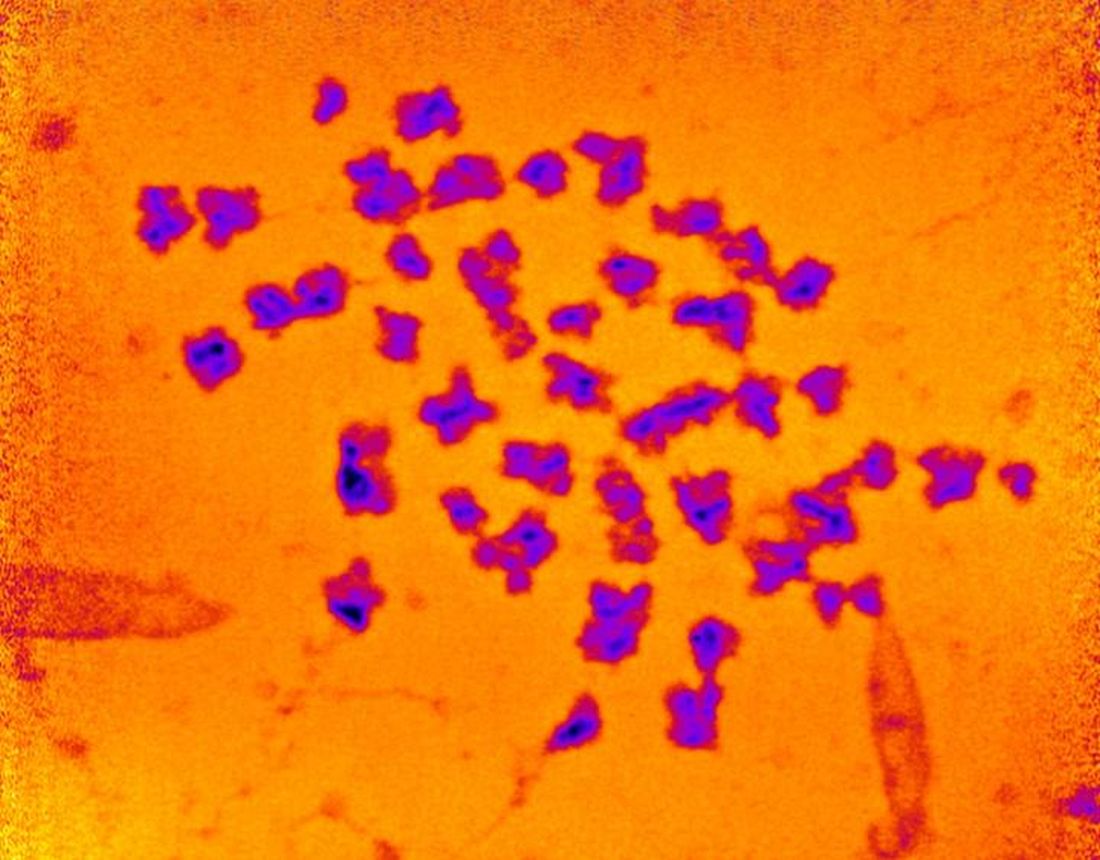
We’re not just talking about a bit of a beer belly here. “The chromosomes were about 20 times heavier than the DNA they contained,” according to the investigators.
Now to the universe. Here’s what CERN, the European Council for Nuclear Research, has to say about the mass of the universe: “Galaxies in our universe … are rotating with such speed that the gravity generated by their observable matter could not possibly hold them together. … which leads scientists to believe that something we cannot see is at work. They think something we have yet to detect directly is giving these galaxies extra mass.”
But wait, there’s more! “The matter we know and that makes up all stars and galaxies only accounts for 5% of the content of the universe!”
So chromosomes are about 20 times heavier than the DNA they contain, and the universe is about 20 times heavier than the matter that can be seen. Interesting.
We are, of course, happy to share this news with our readers, but there is one catch: Don’t tell Neil deGrasse Tyson. He’ll want to reclassify our genetic solar system into 45 chromosomes and one dwarf chromosome.
A photo finish for the Smart Toilet
We know that poop can tell us a lot about our health, but new research by scientists at Duke University is really on a roll. Their Smart Toilet has been created to help people keep an eye on their bowel health. The device takes pictures of poop after it is flushed and can tell whether the consistency is loose, bloody, or normal.
The Smart Toilet can really help people with issues such as irritable bowel syndrome and inflammatory bowel disease by helping them, and their doctors, keep tabs on their poop. “Typically, gastroenterologists have to rely on patient self-reported information about their stool to help determine the cause of their gastrointestinal health issues, which can be very unreliable,” study lead author Deborah Fisher said.

Not many people look too closely at their poop before it’s flushed, so the fecal photos can make a big difference. The Smart Toilet is installed into the pipes of a toilet and does its thing when the toilet is flushed, so there doesn’t seem to be much work on the patient’s end. Other than the, um, you know, usual work from the patient’s end.
Cancer loses … by a nose
Since the human nose is unpredictable at best, we’ve learned to rely on animals for our detailed nozzle needs. But researchers have found the next best thing to man’s best friend to accurately identify cancers.
A team at the University of Pennsylvania has developed an electronic olfaction, or “e-nose,” that has a 95% accuracy rate in distinguishing benign and malignant pancreatic and ovarian cancer cells from a single blood sample. How?
The e-nose system is equipped with nanosensors that are able to detect the volatile organic compounds (VOCs) emitted by cells in a blood sample. Not only does this create an opportunity for an easier, noninvasive screening practice, but it’s fast. The e-nose can distinguish VOCs from healthy to cancerous blood cells in 20 minutes or less and is just as effective in picking up on early- and late-stage cancers.
The investigators hope that this innovative technology can pave the way for similar devices with other uses. Thanks to the e-nose, a handheld device is in development that may be able to sniff out the signature odor of people with COVID-19.
That’s one smart schnoz.
Do you think this is a (food) game?
Dieting and eating healthy is tough, even during the best of times, and it has not been the best of times. With all respect to Charles Dickens, it’s been the worst of times, full stop. Millions of people have spent the past year sitting around their homes doing nothing, and it’s only natural that many would let their discipline slide.
Naturally, the solution to unhealthy eating habits is to sit down and play with your phone. No, that’s not the joke, the Food Trainer app, available on all cellular devices near you, is designed to encourage healthy eating by turning it into a game of sorts. When users open the app, they’re presented with images of food, and they’re trained to tap on images of healthy food and pass on images of unhealthy ones. The process takes less than 5 minutes.
It sounds really simple, but in a study of more than 1,000 people, consumption of junk food fell by 1 point on an 8-point scale (ranging from four times per day to zero to one time per month), participants lost about half a kilogram (a little over one pound), and more healthy food was eaten. Those who used the app more regularly, along the lines of 10 times per month or more, saw greater benefits.
The authors did acknowledge that those who used the app more may have been more motivated to lose weight anyway, which perhaps limits the overall benefit, but reviews on Google Play were overall quite positive, and if there’s one great truth in this world, it’s that Internet reviewers are almost impossible to please. So perhaps this app is worth looking into if you’re like the LOTME staff and you’re up at the top end of that 8-point scale. What, pizza is delicious, who wouldn’t eat it four times a day? And you can also get it from your phone!
It’s time for a little mass kickin’
The universe, scientists tell us, is a big place. Really big. Chromosomes, scientists tell us, are small. Really small. But despite this very fundamental difference, the universe and chromosomes share a deep, dark secret: unexplained mass.
This being a medical publication, we’ll start with chromosomes. A group of researchers measured their mass with x-rays for the first time and found that “the 46 chromosomes in each of our cells weigh 242 picograms (trillionths of a gram). This is heavier than we would expect, and, if replicated, points to unexplained excess mass in chromosomes,” Ian K. Robinson, PhD, said in a written statement.
We’re not just talking about a bit of a beer belly here. “The chromosomes were about 20 times heavier than the DNA they contained,” according to the investigators.
Now to the universe. Here’s what CERN, the European Council for Nuclear Research, has to say about the mass of the universe: “Galaxies in our universe … are rotating with such speed that the gravity generated by their observable matter could not possibly hold them together. … which leads scientists to believe that something we cannot see is at work. They think something we have yet to detect directly is giving these galaxies extra mass.”
But wait, there’s more! “The matter we know and that makes up all stars and galaxies only accounts for 5% of the content of the universe!”
So chromosomes are about 20 times heavier than the DNA they contain, and the universe is about 20 times heavier than the matter that can be seen. Interesting.
We are, of course, happy to share this news with our readers, but there is one catch: Don’t tell Neil deGrasse Tyson. He’ll want to reclassify our genetic solar system into 45 chromosomes and one dwarf chromosome.
A photo finish for the Smart Toilet
We know that poop can tell us a lot about our health, but new research by scientists at Duke University is really on a roll. Their Smart Toilet has been created to help people keep an eye on their bowel health. The device takes pictures of poop after it is flushed and can tell whether the consistency is loose, bloody, or normal.
The Smart Toilet can really help people with issues such as irritable bowel syndrome and inflammatory bowel disease by helping them, and their doctors, keep tabs on their poop. “Typically, gastroenterologists have to rely on patient self-reported information about their stool to help determine the cause of their gastrointestinal health issues, which can be very unreliable,” study lead author Deborah Fisher said.
Not many people look too closely at their poop before it’s flushed, so the fecal photos can make a big difference. The Smart Toilet is installed into the pipes of a toilet and does its thing when the toilet is flushed, so there doesn’t seem to be much work on the patient’s end. Other than the, um, you know, usual work from the patient’s end.
Elevated factor VIII troughs can lead to a higher proportion of zero bleeds in hemophilia
Rurioctocog alfa pegol prophylaxis was linked to fewer bleeding episodes in people with hemophilia A when it targeted higher levels of factor VIII (FVIII) troughs, according to a report published in Blood (2021;137[13]:1818-27).
Earlier studies demonstrated that the treatment effectively prevented bleeds with an acceptable safety profile in people with hemophilia A. The current prospective, randomized, open label PROPEL trial compared safety and efficacy of two target FVIII troughs in this population. Targeting 1%-3% and 8%-12% FVIII troughs was efficacious, with fewer bleeds in the latter arm and acceptable safety across both, according to Robert Klamroth, MD, of Vivantes Klinikum Friedrichshain, Berlin, and colleagues.
The PROPEL trial (NCT02585960) population comprised 155 patients with hepatitis A, aged 12-65 years, with severe disease and an annualized bleeding rate of at least 2 during the 12 months before enrollment in the study. All had previous FVIII treatment. Patients were randomized to 12 months’ pharmacokinetic rurioctocog alfa pegol prophylaxis targeting FVIII troughs of 1%-3% (reference arm) or 8%-12%.
The primary endpoint was absence of bleeds during the second 6-month period. A total of 95 patients completed the protocol.
Promising results
In the 1%-3% and 8%-12% arms, the proportions of patients who completed the protocol and had no bleeds were 40% and 67% respectively (P = .015). Serious adverse events occurred in 7 of 115 (6%) patients, including one treatment-related event in the 8%-12% arm. There were no deaths, serious thrombotic events, or adverse event-related discontinuations.
“Targeting 8% to 12% FVIII troughs resulted in a higher proportion of [patients] with no bleeds than prophylaxis that targeted 1% to 3% FVIII troughs. These results support the hypothesis that an elevated FVIII trough can benefit [patients]without changing the safety profile,” the researchers reported. Personalized treatment in this patient population should be considered, they added.
Problems remain
In an invited commentary, Christine L. Kempton, MD, of Emory University, Atlanta, pointed out that the study did not answer the question of what trough level is best, and that the target trough level may be up to a patient’s individual clinician to decide. “Many participants (42%) treated with the target trough level of 1% to 3% had no bleeding events during the study period, but some (38%) continued to have bleeding events despite higher target trough levels,” Dr. Kempton wrote. She added that, beyond this concern, the presence of subclinical bleeding is difficult to study and quantify, but its presence is supported in the literature by magnetic resonance imaging that demonstrated joint damage despite a lack of clinically evident bleeding.
“Thus, targeting zero clinical bleeding events does not mean that all joint disease, dysfunction, and pain will be eliminated. This reality underscores the need for better, not just more convenient, therapies,” she concluded.
The authors reported numerous relationships with a variety of pharmaceutical companies including grants, honoraria, and participation in speakers bureaus. Dr. Kempton reported honoraria from Takeda, Spark, Octapharma, and Pfizer, and research grants from Novo Nordisk.
FROM BLOOD
Freefall: How Skydiving Is Like Cancer Surgery
Cancer surgery is filled with moments of fear, even for doctors. Our cameras follow as an oncologist confronts this anxiety in the second episode of our video series The Oncologists.
Medscape Oncology © 2021 WebMD, LLC
Any views expressed above are the author's own and do not necessarily reflect the views of WebMD or Medscape.
Cite this: A Pivotal Moment in Cancer Surgery, Captured on Film - Medscape - Feb 18, 2021.
First drug for lung cancer with KRAS mutation gains FDA approval
The first drug to target KRAS mutations in non–small cell lung cancer (NSCLC) has been approved by the Food and Drug Administration.
KRAS mutations are the most common mutations to occur in NSCLC tumors, accounting for about 25% of them, but for a long time they appeared to be resistant to drug therapy.
The new drug, sotorasib (Lumakras), specifically targets the KRAS G12C mutation, which accounts for about 13% of NSCLC mutations.
It is considered to be something of a breakthrough in cancer research. When clinical data on the new drug (from 126 patients) were presented last year at the World Conference on Lung Cancer, lung cancer experts greeted the results enthusiastically, as reported by Medscape Medical News at the time.
“This is a historic milestone in lung cancer therapy. After four decades of scientific efforts in targeting KRAS, sotorasib has potential to be the first targeted treatment option for this patient population with a high unmet need,” Bob T. Li, MD, PhD, of Memorial Sloan Kettering Cancer Center in New York, said at the time.
Now, in a press release from the manufacturer, Amgen, he said: “Sotorasib represents a major advancement in oncology and changes the treatment paradigm for patients with KRAS G12C-mutated non–small cell lung cancer.
“Patients with non–small cell lung cancer who have progressed beyond first-line treatment face a poor prognosis and have limited treatment options available to them. Sotorasib delivers a new option for these patients, and it is the first KRAS-targeted therapy to be approved after nearly four decades of research,” he added.
Details of clinical data
This is an accelerated approval based on response rate data.
The FDA notes that the clinical data come from a study of 124 patients with locally advanced or metastatic KRAS G12C-mutated NSCLC with disease progression after receiving an immune checkpoint inhibitor and/or platinum-based chemotherapy.
The major outcome measured was overall response rate (ORR), which was 36%. Of the patients who responded, 58% had a duration of response of 6 months or longer.
Sotorasib was approved at a dose of 960 mg, and this dose was based on available clinical data as well as pharmacokinetic and pharmacodynamic modeling, the FDA noted. As part of the evaluation for this accelerated approval, the agency is requiring a postmarketing trial to investigate whether a lower dose will have a similar clinical effect.
The most common side effects include diarrhea, musculoskeletal pain, nausea, fatigue, liver damage, and cough. Sotorasib should not be used if patients develop symptoms of interstitial lung disease, and should be permanently discontinued if interstitial lung disease is confirmed.
Patients on sotorasib should have liver function tests prior to starting and while taking the drug; if liver damage develops, the drug should be stopped or the dose reduced. Patients should avoid taking acid-reducing agents, drugs that induce or are substrates for certain enzymes in the liver, and drugs that are substrates of P-glycoprotein (P-gp).
Companion diagnostic tests also approved
Along with the new drug, the FDA approved two companion diagnostic tests – the QIAGEN therascreen KRAS RGQ PCR kit (approval granted to QIAGEN GmbH) for analyzing tumor tissue and the Guardant360 CDx (approval granted to Guardant Health) for analyzing plasma specimens to determine if the KRAS G12C mutation is present. The agency notes that if the plasma test comes back negative, the patient’s tumor tissue should be tested.
A version of this article first appeared on Medscape.com.