User login
Bringing you the latest news, research and reviews, exclusive interviews, podcasts, quizzes, and more.
div[contains(@class, 'header__large-screen')]
div[contains(@class, 'read-next-article')]
div[contains(@class, 'nav-primary')]
nav[contains(@class, 'nav-primary')]
section[contains(@class, 'footer-nav-section-wrapper')]
footer[@id='footer']
div[contains(@class, 'main-prefix')]
section[contains(@class, 'nav-hidden')]
div[contains(@class, 'ce-card-content')]
nav[contains(@class, 'nav-ce-stack')]
Genomic analysis reveals possible role of AMH in PCOS infertility
A genomic study has revealed new insights into the function of anti-Müllerian hormone (AMH) in the context of reproductive biology and fertility.
Insights into the physiological, and potentially therapeutic, function were identified based on data from single-cell RNA sequencing, derived from transcriptomic analysis and immunolabeling of antral follicles.
“The specific contribution of elevated AMH to the molecular pathology of polycystic ovary syndrome (PCOS) and its defining clinical features is unclear, as no study, to date, has examined the effect of chronically elevated AMH in an experimentally controlled in vivo model,” study author Limor Man, MD, of Weill Cornell Medicine, New York, and colleagues wrote. The group’s findings were published in Science Advances.
The researchers used ovarian cortical xenografts with cotransplantation of engineered endothelial cells to examine the effect of chronic paracrine AMH stimulus on human folliculogenesis.
They cotransplanted human ovarian cortex with control or AMH-expressing endothelial cells in immunocompromised mice and recovered antral follicles for purification and subsequent analysis. Overall, 38 antral follicles were observed (19 control and 19 AMH) at long-term intervals, defined as intervals greater than 10 weeks.
The researchers found that long-term xenografts showed an accelerated growth rate in the setting of chronically elevated AMH and exhibited a molecular signature indicative of more advanced stages of follicle maturation, including that of luteinization.
In mice, exogenous AMH follicles showed a decreased ratio of primordial to growing follicles and antral follicles of increased diameter.
In addition, transcriptomic and immunolabeling analyses revealed that chronic high AMH had a marked influence on the growth and transcriptomic signature of antral-stage follicles, with a universal increase in factors related to the synthesis and/or metabolism of cholesterol and sex steroid hormones, as well as early expression of factors often seen at later stages of folliculogenesis.
“These data decouple elevated AMH from the metabolic and hyperandrogenic conditions that define PCOS and suggest that chronically elevated AMH induces a molecular cascade that contributes, at least in part, to the anovulatory phenotype in these patients,” the researchers wrote.
Furthermore, they found evidence to suggest that chronic high AMH can induce expression of the luteinizing hormone receptor at earlier stages of folliculogenesis, thereby worsening the disruptive effect of elevated luteinizing hormone from the pituitary.
“[These] findings underscore the broad influence of AMH on transcriptional activity and maturation state of follicles and support an independent role for dysregulation of AMH signaling in driving anovulation in women with PCOS,” they wrote.
While these findings are intriguing, the researchers cautioned against drawing conclusions from the study since elevated AMH is almost always seen in combination with one or more symptomatic hallmarks in PCOS.
“Despite [some] limitations, [our] analysis provides a deep and high-resolution examination of AMH action on human folliculogenesis and suggests a prominent effect on antral follicle maturation,” they explained.
Expert perspective
“From age 25, AMH levels begin their decline until reaching undetectable levels at menopause,” Mark P. Trolice, MD, director of the IVF Center in Winter Park, Fla., and professor of obstetrics and gynecology at the University of Central Florida, Orlando, said in an interview. “Women with PCOS experience a chronic and frustrating pathophysiologic problem whose origins and mechanism have evaded researchers for decades.

“As AMH elevations in utero may contribute to fetal susceptibility to PCOS, this study provides another potential link by suggesting that chronically elevated AMH induces anovulation,” he added. “We await, with great anticipation, future clinical studies to potentially further illustrate the apparent and intriguing role of AMH in the development of PCOS.”
This study was supported by the Queenie Victorina Neri Scholarship and a Research Grant from the American Society for Reproductive Medicine. One author reported financial relationships with Oviva Therapeutics; no other conflicts of interest were reported.
A genomic study has revealed new insights into the function of anti-Müllerian hormone (AMH) in the context of reproductive biology and fertility.
Insights into the physiological, and potentially therapeutic, function were identified based on data from single-cell RNA sequencing, derived from transcriptomic analysis and immunolabeling of antral follicles.
“The specific contribution of elevated AMH to the molecular pathology of polycystic ovary syndrome (PCOS) and its defining clinical features is unclear, as no study, to date, has examined the effect of chronically elevated AMH in an experimentally controlled in vivo model,” study author Limor Man, MD, of Weill Cornell Medicine, New York, and colleagues wrote. The group’s findings were published in Science Advances.
The researchers used ovarian cortical xenografts with cotransplantation of engineered endothelial cells to examine the effect of chronic paracrine AMH stimulus on human folliculogenesis.
They cotransplanted human ovarian cortex with control or AMH-expressing endothelial cells in immunocompromised mice and recovered antral follicles for purification and subsequent analysis. Overall, 38 antral follicles were observed (19 control and 19 AMH) at long-term intervals, defined as intervals greater than 10 weeks.
The researchers found that long-term xenografts showed an accelerated growth rate in the setting of chronically elevated AMH and exhibited a molecular signature indicative of more advanced stages of follicle maturation, including that of luteinization.
In mice, exogenous AMH follicles showed a decreased ratio of primordial to growing follicles and antral follicles of increased diameter.
In addition, transcriptomic and immunolabeling analyses revealed that chronic high AMH had a marked influence on the growth and transcriptomic signature of antral-stage follicles, with a universal increase in factors related to the synthesis and/or metabolism of cholesterol and sex steroid hormones, as well as early expression of factors often seen at later stages of folliculogenesis.
“These data decouple elevated AMH from the metabolic and hyperandrogenic conditions that define PCOS and suggest that chronically elevated AMH induces a molecular cascade that contributes, at least in part, to the anovulatory phenotype in these patients,” the researchers wrote.
Furthermore, they found evidence to suggest that chronic high AMH can induce expression of the luteinizing hormone receptor at earlier stages of folliculogenesis, thereby worsening the disruptive effect of elevated luteinizing hormone from the pituitary.
“[These] findings underscore the broad influence of AMH on transcriptional activity and maturation state of follicles and support an independent role for dysregulation of AMH signaling in driving anovulation in women with PCOS,” they wrote.
While these findings are intriguing, the researchers cautioned against drawing conclusions from the study since elevated AMH is almost always seen in combination with one or more symptomatic hallmarks in PCOS.
“Despite [some] limitations, [our] analysis provides a deep and high-resolution examination of AMH action on human folliculogenesis and suggests a prominent effect on antral follicle maturation,” they explained.
Expert perspective
“From age 25, AMH levels begin their decline until reaching undetectable levels at menopause,” Mark P. Trolice, MD, director of the IVF Center in Winter Park, Fla., and professor of obstetrics and gynecology at the University of Central Florida, Orlando, said in an interview. “Women with PCOS experience a chronic and frustrating pathophysiologic problem whose origins and mechanism have evaded researchers for decades.
“As AMH elevations in utero may contribute to fetal susceptibility to PCOS, this study provides another potential link by suggesting that chronically elevated AMH induces anovulation,” he added. “We await, with great anticipation, future clinical studies to potentially further illustrate the apparent and intriguing role of AMH in the development of PCOS.”
This study was supported by the Queenie Victorina Neri Scholarship and a Research Grant from the American Society for Reproductive Medicine. One author reported financial relationships with Oviva Therapeutics; no other conflicts of interest were reported.
FROM SCIENCE ADVANCES

Selling your practice
My previous column on practice valuation prompted a number of questions on the mechanics of selling a private practice. As usual, I cannot hope to cover this complex topic comprehensively in only 750 words, but here are the basics.
A generation ago, the sale of a medical practice was much like the sale of any other business: A retiring physician would sell his or her practice to a young doctor and the practice would continue on as before. Occasionally, that still happens, but changes in the business of medicine – most significantly the growth of managed care – have had a big impact on the way medical practices are bought and sold.

For one thing, there are far fewer solo practitioners these days, and polls indicate that most young physicians intend to continue that trend. .
For another, because the rules governing such sales have become so numbingly complex, the services of expert (and expensive) third parties are essential.
While these issues may complicate matters, there is still a market for the sale of medical practices. However, you must do everything possible to ensure you identify the best possible buyer and structure the best deal.
The first hurdle is the accurate valuation of your practice, which was covered in some detail in my last column. Briefly, for the protection of both parties, it is important that the appraisal be done by an experienced and neutral financial consultant, that all techniques used in the valuation be divulged and explained, and that documentation be supplied to support the conclusions reached.
Keep in mind that the valuation will not necessarily equal the purchase price; other factors may need to be considered before a final price can be agreed upon. Keep in mind, too, that there may be legal constraints on the purchase price. For example, if the buyer is a nonprofit corporation such as a hospital or HMO, by law it cannot pay in excess of fair market value for the practice – which may rule out any valuation of “good will.” In some states, the purchase of private practices by hospitals is prohibited altogether – so you might need to consider a long-term lease rather than a sale.
Once a value has been agreed upon, you must consider how the transaction will be structured. The most popular structures include purchase of assets, purchase of corporate stock, and merger.
Many buyers prefer to purchase assets, because it allows them to pick and choose only those items that have value to them. This can leave you with a bunch of “odd lot” assets to dispose of. But depending on the circumstances, an asset sale may still be to your advantage.
Sellers typically prefer to sell stock, because it allows them to sell their entire practice, which is often worth more than the sum of its parts, and often provides tax advantages.
The third option, merger, continues to grow in popularity and is a column subject in itself, and I will address it separately next month.
Tax issues must always be considered. Most private practices are corporations, and the sale of corporate stock will result in a long-term capital gain that will be taxed – currently at 15%-20%. As the saying goes, it’s not what you earn, it’s what you keep. So it may benefit you to accept a slightly lower price if the sale can be structured to provide significantly lower tax treatment. However, any gain that does not qualify as a long-term capital gain will be taxed as regular income – currently in the 32%-37% percent range – plus a Social Security tax of about 15%.
Payment in installments is a popular way to defer taxes, since they are incurred on each installment as it is paid; but such payments may be mistaken by the IRS for payments for referrals, which is illegal. And there is always the problem of making certain all payments are eventually made.
You may wish to continue working at the practice as an employee for an agreed-upon period of time, and this is often to the buyer’s advantage as well. Transitioning to new ownership in stages often maximizes the value of the business by improving patient retention, and allows patients to become accustomed to the transition. However, care must be taken, with the aid of good legal advice, to structure such an arrangement in a way that minimizes concerns of fraud and abuse.
Dr. Eastern practices dermatology and dermatologic surgery in Belleville, N.J. He is the author of numerous articles and textbook chapters, and is a longtime monthly columnist for Dermatology News. Write to him at [email protected].
My previous column on practice valuation prompted a number of questions on the mechanics of selling a private practice. As usual, I cannot hope to cover this complex topic comprehensively in only 750 words, but here are the basics.
A generation ago, the sale of a medical practice was much like the sale of any other business: A retiring physician would sell his or her practice to a young doctor and the practice would continue on as before. Occasionally, that still happens, but changes in the business of medicine – most significantly the growth of managed care – have had a big impact on the way medical practices are bought and sold.
For one thing, there are far fewer solo practitioners these days, and polls indicate that most young physicians intend to continue that trend. .
For another, because the rules governing such sales have become so numbingly complex, the services of expert (and expensive) third parties are essential.
While these issues may complicate matters, there is still a market for the sale of medical practices. However, you must do everything possible to ensure you identify the best possible buyer and structure the best deal.
The first hurdle is the accurate valuation of your practice, which was covered in some detail in my last column. Briefly, for the protection of both parties, it is important that the appraisal be done by an experienced and neutral financial consultant, that all techniques used in the valuation be divulged and explained, and that documentation be supplied to support the conclusions reached.
Keep in mind that the valuation will not necessarily equal the purchase price; other factors may need to be considered before a final price can be agreed upon. Keep in mind, too, that there may be legal constraints on the purchase price. For example, if the buyer is a nonprofit corporation such as a hospital or HMO, by law it cannot pay in excess of fair market value for the practice – which may rule out any valuation of “good will.” In some states, the purchase of private practices by hospitals is prohibited altogether – so you might need to consider a long-term lease rather than a sale.
Once a value has been agreed upon, you must consider how the transaction will be structured. The most popular structures include purchase of assets, purchase of corporate stock, and merger.
Many buyers prefer to purchase assets, because it allows them to pick and choose only those items that have value to them. This can leave you with a bunch of “odd lot” assets to dispose of. But depending on the circumstances, an asset sale may still be to your advantage.
Sellers typically prefer to sell stock, because it allows them to sell their entire practice, which is often worth more than the sum of its parts, and often provides tax advantages.
The third option, merger, continues to grow in popularity and is a column subject in itself, and I will address it separately next month.
Tax issues must always be considered. Most private practices are corporations, and the sale of corporate stock will result in a long-term capital gain that will be taxed – currently at 15%-20%. As the saying goes, it’s not what you earn, it’s what you keep. So it may benefit you to accept a slightly lower price if the sale can be structured to provide significantly lower tax treatment. However, any gain that does not qualify as a long-term capital gain will be taxed as regular income – currently in the 32%-37% percent range – plus a Social Security tax of about 15%.
Payment in installments is a popular way to defer taxes, since they are incurred on each installment as it is paid; but such payments may be mistaken by the IRS for payments for referrals, which is illegal. And there is always the problem of making certain all payments are eventually made.
You may wish to continue working at the practice as an employee for an agreed-upon period of time, and this is often to the buyer’s advantage as well. Transitioning to new ownership in stages often maximizes the value of the business by improving patient retention, and allows patients to become accustomed to the transition. However, care must be taken, with the aid of good legal advice, to structure such an arrangement in a way that minimizes concerns of fraud and abuse.
Dr. Eastern practices dermatology and dermatologic surgery in Belleville, N.J. He is the author of numerous articles and textbook chapters, and is a longtime monthly columnist for Dermatology News. Write to him at [email protected].

Inside insulin (Part 2): Approaching a cure for type 1 diabetes?
Editor’s note: This is the second in a two-part series commemorating the 100-year anniversary of the first use of insulin in humans. Part 1 of this series examined the rivalry behind the discovery and use of insulin.
One hundred years ago, teenager Leonard Thompson was the first patient with type 1 diabetes to be successfully treated with insulin, granting him a reprieve from what was a certain death sentence at the time.
Since then, research has gathered pace. In the century since insulin’s discovery and first use, recombinant DNA technology has allowed for the engineering of the insulin molecule, providing numerous short- and long-acting analog versions. At the same time, technological leaps in automated insulin delivery and monitoring of blood glucose ensure more time with glucose in range and fewer life-threatening complications for those with type 1 diabetes fortunate enough to have access to the technology.
In spite of these advancements, there is still scope for further evolution of disease management, with the holy grail being the transplant of stem cell–derived islet cells capable of making insulin, ideally encased in some kind of protective device so that immunosuppression is not required.
Indeed, it is not unreasonable to “hope that type 1 diabetes will be a curable disease in the next 100 years,” said Elizabeth Stephens, MD, an endocrinologist who has type 1 diabetes and practices in Portland, Ore.
Type 1 diabetes: The past 100 years
The epidemiology of type 1 diabetes has shifted considerably since 1922. A century ago, given that average life expectancy in the United States was around 54 years, it was pretty much the only type of diabetes that doctors encountered. “There was some type 2 diabetes about in heavier people, but the focus was on type 1 diabetes,” noted Dr. Stephens.
Originally called juvenile diabetes because it was thought to only occur in children, “now 50% of people are diagnosed with type 1 diabetes ... over [the age of] 20,” explained Dr. Stephens.
In the United States, around 1.4 million adults 20 years and older, and 187,000 children younger than 20, have the disease, according to data from the National Diabetes Statistics Report 2020 by the Centers for Disease Control and Prevention. This total represents an increase of nearly 30% from 2017.
Over the years, theories as to the cause, or trigger, for type 1 diabetes “have included cow’s milk and [viral] infections,” said Dr. Stephens. “Most likely, there’s a genetic predisposition and some type of exposure, which creates the perfect storm to trigger disease.”
There are hints that COVID-19 might be precipitating type 1 diabetes in some people. Recently, the CDC found SARS-CoV-2 infection was associated with an increased risk for diabetes (all types) among youth, but not other acute respiratory infections. And two further studies from different parts of the world have recently identified an increase in the incidence of type 1 diabetes in children since the COVID-19 pandemic began, but the reasons remain unclear.
The global CoviDiab registry has also been established to collect data on patients with COVID-19–related diabetes.
The million-dollar question: Is COVID-19 itself is propagating type 1 diabetes or unmasking a predisposition to the disease sooner? The latter might be associated with a lower type 1 diabetes rate in the future, said Partha Kar, MBBS, OBE, national specialty advisor, diabetes, for National Health Service England.
“Right now, we don’t know the answer. Whichever way you look at it, it is likely there will be a rise in cases, and in countries where insulin is not freely available, healthcare systems need to have supply ready because insulin is lifesaving in type 1 diabetes,” Dr. Kar emphasized.
CGMs and automated insulin delivery: A ‘godsend’
A huge change has also been seen, most notably in the past 15 to 20 years, in the technological advancements that can help those with type 1 diabetes live an easier life.
Continuous glucose monitors (CGMs) and automated ways of delivering insulin, such as smart pens and insulin pumps, have made the daily life of a person with type 1 diabetes in the Western world considerably more comfortable.
CGMs provide a constant stream of data to an app, often wirelessly in sync with the insulin pump. However, on a global level, they are only available to a lucky few.
In England, pending National Institute for Health and Care Excellence) approval, any CGM should be available to all eligible patients with type 1 diabetes within the NHS from April 2022, Dr. Kar pointed out. In the United States, CGMs are often unaffordable and access is mostly dependent on a person’s health insurance.
Kersten Hall, PhD, a scientist and U.K.-based medical historian who recently wrote a book, “Insulin, the Crooked Timber” (Oxford, England: Oxford University Press, 2022) uncovering the lesser-known story behind the discovery of insulin, was diagnosed with adult-onset type 1 diabetes at the age of 41. Dr. Hall had always found the finger-prick blood glucose test to be a chore but now has a CGM.
“It’s a total game changer for me: a godsend. I can’t sing its praises enough,” he said. “All it involves is the swipe of the phone and this provides a reading which tells me if my glucose is too low, so I eat something, or too high, so I might [go for] a run.”
Brewing insulin at scale
As described by Dr. Hall in his book, the journey from treating Mr. Thompson in 1922 to treating the masses began when biochemist James Collip, MD, PhD, discovered a means of purifying the animal pancreas extracts used to treat the teenager.
But production at scale presented a further challenge. This was overcome in 1924 when Eli Lilly drew on a technique used in the beer brewing process – where pH guides bitterness – to purify and manufacture large amounts of insulin.
By 1936, a range of slower-acting cattle and pig-derived insulins, the first produced by Novo Nordisk Pharmaceuticals, were developed.
However, it took 8,000 lb (approximately 3,600 kg) of pancreas glands from 23,500 animals to make 1 lb (0.5 kg) of insulin, so a more efficient process was badly needed.
Dr. Hall, who is a molecular biologist as well as an author, explains that the use of recombinant DNA technology to produce human insulin, as done by Genentech in the late 70s, was a key development in the story of modern insulin products. Genentech then provided synthetic human insulin for Eli Lilly to conduct clinical trials.
Human insulin most closely resembles porcine insulin structure and function, differing by only one amino acid, while human insulin differs from bovine insulin by three amino acid residues. This synthetic human insulin eliminated the allergies that the animal-derived products sometimes caused.
In the early 1980s, Eli Lilly produced Humulin, the first biosynthetic (made in Escherichia coli, hence the term, “bio”) human insulin.
This technology eventually “allowed for the alteration of specific amino acids in the sequence of the insulin protein to make insulin analogs [synthetic versions grown in E. coli and genetically altered for various properties] that act faster, or more slowly, than normal human insulin. By using the slow- and fast-acting insulins in combination, a patient can control their blood sugar levels with a much greater degree of finesse and precision,” Dr. Hall explained.
Today, a whole range of insulins are available, including ultra–rapid-acting, short-acting, intermediate-acting, long-acting, ultra–long-acting, and even inhaled insulin, although the latter is expensive, has been associated with side effects, and is less commonly used, according to Dr. Stephens.
Oral insulin formulations are even in the early stages of development, with candidate drugs by Generex and from the Oralis project.
“With insulin therapy, we try to reproduce the normal physiology of the healthy body and pancreas,” Dr. Stephens explained.
Insulin analogs are only made by three companies (Eli Lilly, Novo Nordisk, and Sanofi), and they are generally much more expensive than nonanalog human insulin. In the United Kingdom through the NHS, they cost twice as much.
In the United States today, one of the biggest barriers to proper care of type 1 diabetes is the cost of insulin, which can limit access. With the market controlled by these three large companies, the average cost of a unit of insulin in the United States, according to RAND research, was $98.17 in January 2021, compared with $7.52 in the United Kingdom and $12.00 in Canada.
Several U.S. states have enacted legislation capping insulin copayments to at, or under, $100 a month. But the federal Build Back Better Framework Act – which would cap copayments for insulin at $35 – currently hangs in the balance.
Alongside these moves, in 2020 the Food and Drug Administration approved the first interchangeable biosimilar insulin for type 1 diabetes (and insulin-dependent type 2 diabetes) in children and adults, called Semglee (Mylan Pharmaceuticals).
Biosimilars (essentially generic versions of branded insulins) are expected to be less expensive than branded analogs, but the indications so far are that they will only be around 20% cheaper.
“I totally fail to understand how the richest country in the world still has a debate about price caps, and we are looking at biosimilar markets to change the debate. This makes no sense to me at all,” stressed Dr. Kar. “For lifesaving drugs, they should be funded by the state.”
Insulin also remains unaffordable for many in numerous low- and middle-income countries, where most patients pay out-of-pocket for medicines. Globally, there are estimated to be around 30 million people who need insulin but cannot afford it.
How near to a cure in the coming decades?
Looking ahead to the coming years, if not the next 100, Dr. Stephens highlighted two important aspects of care.
First, the use of a CGM device in combination with an insulin pump (also known as a closed-loop system or artificial pancreas), where the CGM effectively tells the insulin pump how much insulin to automatically dispense, should revolutionize care.
A number of such closed-loop systems have recently been approved in both the United States, including systems from Medtronic and Omnipod, and Europe.
“I wear one of these and it’s been a life changer for me, but it doesn’t suit everyone because the technology can be cumbersome, but with time, hopefully things will become smaller and more accurate in insulin delivery,” Dr. Stephens added.
The second advance of interest is the development and transplantation of cells that produce insulin.
Dr. Stephens explained that someone living with type 1 diabetes has a lot to think about, not least, doing the math related to insulin requirement. “If we just had cells from a pancreas that could be transplanted and would do that for us, then it would be a total game changer.”
To date, Vertex Pharmaceuticals has successfully treated one patient – who had lived with type 1 diabetes for about 40 years and had recurrent episodes of severe hypoglycemia – with an infusion of stem cell–derived differentiated islet cells into his liver. The procedure resulted in near reversal of type 1 diabetes, with his insulin dose reduced from 34 to 3 units, and his hemoglobin A1c falling from 8.6% to 7.2%.
And although the patient, Brian Shelton, still needs to take immunosuppressive agents to prevent rejection of the stem cell–derived islets, “it’s a whole new life,” he recently told the New York Times.
Another company called ViaCyte is also working on a similar approach.
Whether this is a cure for type 1 diabetes is still debatable, said Anne Peters, MD, of the University of Southern California, Los Angeles. “Is it true? In a word, no. But we are part of the way there, which is much closer than we were 6 months ago.”
There are also ongoing clinical trials of therapeutic interventions to prevent or delay the trajectory from presymptomatic to clinical type 1 diabetes. The most advanced is the anti-CD3 monoclonal antibody teplizumab (Tzield, Provention Bio), which was rejected by the FDA in July 2021, but has since been refiled. The company expects to hear from the agency by the end of March 2022 as to whether the resubmission has been accepted.
Diabetes specialist nurses/educators keep it human
Dr. Hall said he concurs with the late eminent U.K. diabetes specialist Robert Tattersall’s observation on what he considers one of the most important advances in the management and treatment of type 1 diabetes: the human touch.
Referring to Dr. Tattersall’s book, “Diabetes: A Biography,” Dr. Hall quoted: “If asked what innovation had made the most difference to their lives in the 1980s, patients with type 1 diabetes in England would unhesitatingly have chosen not human insulin, but the spread of diabetes specialist nurses ... these people (mainly women) did more in the last two decades of the 20th century to improve the standard of diabetes care than any other innovation or drug.”
In the United States, diabetes specialist nurses were called diabetes educators until recently, when the name changed to certified diabetes care and education specialist.
“Above all, they have humanized the service and given the patient a say in the otherwise unequal relationship with all-powerful doctors,” concluded Dr. Hall, again quoting Dr. Tattersall.
A version of this article first appeared on Medscape.com.
Death of pig heart transplant patient is more a beginning than an end
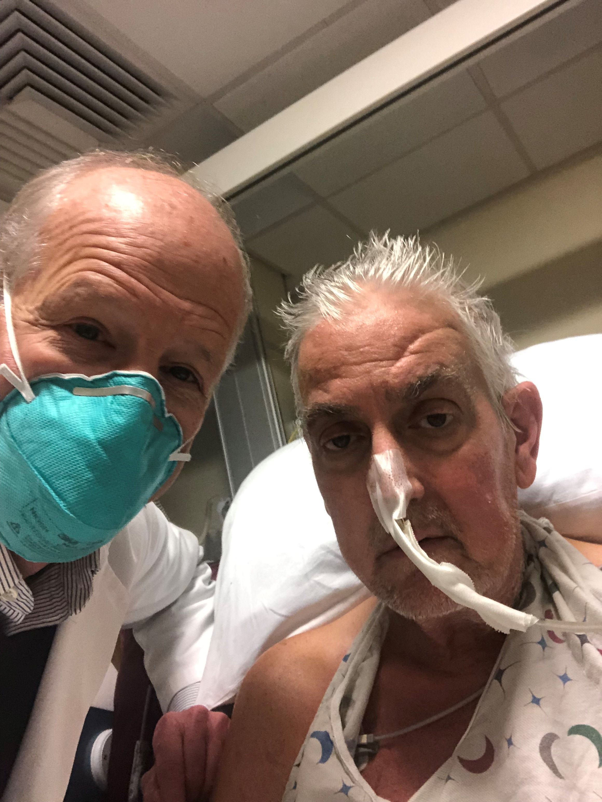
The genetically altered pig’s heart “worked like a rock star, beautifully functioning,” the surgeon who performed the pioneering Jan. 7 xenotransplant procedure said in a press statement on the death of the patient, David Bennett Sr.
“He wasn’t able to overcome what turned out to be devastating – the debilitation from his previous period of heart failure, which was extreme,” said Bartley P. Griffith, MD, clinical director of the cardiac xenotransplantation program at the University of Maryland, Baltimore.
Representatives of the institution aren’t offering many details on the cause of Mr. Bennett’s death on March 8, 60 days after his operation, but said they will elaborate when their findings are formally published. But their comments seem to downplay the unique nature of the implanted heart itself as a culprit and instead implicate the patient’s diminished overall clinical condition and what grew into an ongoing battle with infections.
The 57-year-old Bennett, bedridden with end-stage heart failure, judged a poor candidate for a ventricular assist device, and on extracorporeal membrane oxygenation (ECMO), reportedly was offered the extraordinary surgery after being turned down for a conventional transplant at several major centers.
“Until day 45 or 50, he was doing very well,” Muhammad M. Mohiuddin, MD, the xenotransplantation program’s scientific director, observed in the statement. But infections soon took advantage of his hobbled immune system.
Given his “preexisting condition and how frail his body was,” Dr. Mohiuddin said, “we were having difficulty maintaining a balance between his immunosuppression and controlling his infection.” Mr. Bennett went into multiple organ failure and “I think that resulted in his passing away.”
Beyond wildest dreams
The surgeons confidently framed Mr. Bennett’s experience as a milestone for heart xenotransplantation. “The demonstration that it was possible, beyond the wildest dreams of most people in the field, even, at this point – that we were able to take a genetically engineered organ and watch it function flawlessly for 9 weeks – is pretty positive in terms of the potential of this therapy,” Dr. Griffith said.
But enough questions linger that others were more circumspect, even as they praised the accomplishment. “There’s no question that this is a historic event,” Mandeep R. Mehra, MD, of Harvard Medical School, and director of the Center for Advanced Heart Disease at Brigham and Women’s Hospital, both in Boston, said in an interview.
Still, “I don’t think we should just conclude that it was the patient’s frailty or death from infection,” Dr. Mehra said. With so few details available, “I would be very careful in prematurely concluding that the problem did not reside with the heart but with the patient. We cannot be sure.”
For example, he noted, “6 to 8 weeks is right around the time when some cardiac complications, like accelerated forms of vasculopathy, could become evident.” Immune-mediated cardiac allograft vasculopathy is a common cause of heart transplant failure.
Or, “it could as easily have been the fact that immunosuppression was modified at 6 to 7 weeks in response to potential infection, which could have led to a cardiac compromise,” Dr. Mehra said. “We just don’t know.”
“It’s really important that this be reported in a scientifically accurate way, because we will all learn from this,” Lori J. West, MD, DPhil, said in an interview.
Little seems to be known for sure about the actual cause of death, “but the fact there was not hyperacute rejection is itself a big step forward. And we know, at least from the limited information we have, that it did not occur,” observed Dr. West, who directs the Alberta Transplant Institute, Edmonton, and the Canadian Donation and Transplantation Research Program. She is a professor of pediatrics with adjunct positions in the departments of surgery and microbiology/immunology.
Dr. West also sees Mr. Bennett’s struggle with infections and adjustments to his unique immunosuppressive regimen, at least as characterized by his care team, as in line with the experience of many heart transplant recipients facing the same threat.
“We already walk this tightrope with every transplant patient,” she said. Typically, they’re put on a somewhat standardized immunosuppressant regimen, “and then we modify it a bit, either increasing or decreasing it, depending on the posttransplant course.” The regimen can become especially intense in response to new signs of rejection, “and you know that that’s going to have an impact on susceptibility to all kinds of infections.”
Full circle
The porcine heart was protected along two fronts against assault from Mr. Bennett’s immune system and other inhospitable aspects of his physiology, either of which could also have been obstacles to success: Genetic modification (Revivicor) of the pig that provided the heart, and a singularly aggressive antirejection drug regimen for the patient.
The knockout of three genes targeting specific porcine cell-surface carbohydrates that provoke a strong human antibody response reportedly averted a hyperacute rejection response that would have caused the graft to fail almost immediately.
Other genetic manipulations, some using CRISPR technology, silenced genes encoded for porcine endogenous retroviruses. Others were aimed at controlling myocardial growth and stemming graft microangiopathy.
Mr. Bennett himself was treated with powerful immunosuppressants, including an investigational anti-CD40 monoclonal antibody (KPL-404, Kiniksa Pharmaceuticals) that, according to UMSOM, inhibits a well-recognized pathway critical to B-cell proliferation, T-cell activation, and antibody production.
“I suspect the patient may not have had rejection, but unfortunately, that intense immunosuppression really set him up – even if he had been half that age – for a very difficult time,” David A. Baran, MD, a cardiologist from Sentara Advanced Heart Failure Center, Norfolk, Va., who studies transplant immunology, said in an interview.
“This is in some ways like the original heart transplant in 1967, when the ability to do the surgery evolved before understanding of the immunosuppression needed. Four or 5 years later, heart transplantation almost died out, before the development of better immunosuppressants like cyclosporine and later tacrolimus,” Dr. Baran said.
“The current age, when we use less immunosuppression than ever, is based on 30 years of progressive success,” he noted. This landmark xenotransplantation “basically turns back the clock to a time when the intensity of immunosuppression by definition had to be extremely high, because we really didn’t know what to expect.”
Emerging role of xeno-organs
Xenotransplantation has been touted as potential strategy for expanding the pool of organs available for transplantation. Mr. Bennett’s “breakthrough surgery” takes the world “one step closer to solving the organ shortage crisis,” his surgeon, Dr. Griffith, announced soon after the procedure. “There are simply not enough donor human hearts available to meet the long list of potential recipients.”
But it’s not the only proposed approach. Measures could be taken, for example, to make more efficient use of the human organs that become available, partly by opening the field to additional less-than-ideal hearts and loosening regulatory mandates for projected graft survival.
“Every year, more than two-thirds of donor organs in the United States are discarded. So it’s not actually that we don’t have enough organs, it’s that we don’t have enough organs that people are willing to take,” Dr. Baran said. Still, it’s important to pursue all promising avenues, and “the genetic manipulation pathway is remarkable.”
But “honestly, organs such as kidneys probably make the most sense” for early study of xenotransplantation from pigs, he said. “The waiting list for kidneys is also very long, but if the kidney graft were to fail, the patient wouldn’t die. It would allow us to work out the immunosuppression without putting patients’ lives at risk.”
Often overlooked in assessments of organ demand, Dr. West said, is that “a lot of patients who could benefit from a transplant will never even be listed for a transplant.” It’s not clear why; perhaps they have multiple comorbidities, live too far from a transplant center, “or they’re too big or too small. Even if there were unlimited organs, you could never meet the needs of people who could benefit from transplantation.”
So even if more available donor organs were used, she said, there would still be a gap that xenotransplantation could help fill. “I’m very much in favor of research that allows us to continue to try to find a pathway to xenotransplantation. I think it’s critically important.”
Unquestionably, “we now need to have a dialogue to entertain how a technology like this, using modern medicine with gene editing, is really going to be utilized,” Dr. Mehra said. The Bennett case “does open up the field, but it also raises caution.” There should be broad participation to move the field forward, “coordinated through either societies or nationally allocated advisory committees that oversee the movement of this technology, to the next step.”
Ideally, that next step “would be to do a safety clinical trial in the right patient,” he said. “And the right patient, by definition, would be one who does not have a life-prolonging option, either mechanical circulatory support or allograft transplantation. That would be the goal.”
Dr. Mehra has reported receiving payments to his institution from Abbott for consulting; consulting fees from Janssen, Mesoblast, Broadview Ventures, Natera, Paragonix, Moderna, and the Baim Institute for Clinical Research; and serving on a scientific advisory board NuPulseCV, Leviticus, and FineHeart. Dr. Baran disclosed consulting for Getinge and LivaNova; speaking for Pfizer; and serving on trial steering committees for CareDx and Procyrion, all unrelated to xenotransplantation. Dr. West has declared no relevant conflicts.
A version of this article first appeared on Medscape.com.
CPAP has only small effect on metabolic syndrome
Continuous positive airway pressure (CPAP) may be only modestly effective for ameliorating metabolic syndrome in patients with moderate to severe obstructive sleep apnea (OSA).
That conclusion comes from investigators in a randomized controlled, trial, who found that, among 100 patients with OSA and a recent diagnosis of metabolic syndrome (MS), 18% of those assigned to use CPAP at night had a reversal of MS at 6 months of follow-up, compared with 4% of controls who were assigned to use nasal strips at night (P = .04).
The majority of patients assigned to CPAP still retained their MS diagnoses at 6 months, and CPAP did not significantly reduce individual components of the syndrome. Use of CPAP was, however, associated with small reductions in visceral fat and improvement in endothelial function, reported Sara Q.C. Giampa, PhD, from the University of São Paulo, and colleagues.
“Despite a significant rate of MS reversibility after CPAP therapy, most of the patients maintained the MS diagnosis. The modest effects of CPAP on MS reversibility underscore the need for combined therapy with CPAP, aiming to maximize metabolic syndrome recovery in parallel with improvements in OSA severity and related symptoms,” according to their study, reported in the journal CHEST®.
Asked whether he still recommends CPAP to patients with OSA and the metabolic syndrome, given the findings, corresponding author Luciano F. Drager, MD, PhD, replied “yes, definitely.”
“Despite the modest rate in reversing metabolic syndrome after CPAP, the rate was 5-fold higher than non-effective treatment (18% vs. 4%),” he said in an interview.
Dr. Drager noted that studies of other single interventions such as physical exercise to reverse MS in patients with OSA also had modest results.
A researcher who studies the relationship between sleep, circadian rhythms, and metabolism commented that, although the patients in the CPAP group were compliant with the assigned equipment and had both reductions in apneic events and improvement in oxygen saturation, the effect of CPAP on the metabolic syndrome was rather small.
“The CPAP was doing what we thought it was supposed to do, but it didn’t have the magnitude of effect on the metabolic syndrome as I expected or I think as the authors expected,” said Deanna Arble, PhD, assistant professor of biological science at Marquette University, Milwaukee.
She noted that the study also failed to detect a significant improvement in the blood pressure component of metabolic syndrome.
“In my experience and my review of the literature, blood pressure tends to be the one that’s improved most dramatically with CPAP,” she said.
Dr. Arble was not involved in the study.
Study details
In the trial, titled TREATOSA-MS, the investigators enrolled 100 patients with a recent diagnosis of metabolic syndrome and moderate to severe OSA, defined as 15 or more apnea-hypopnea index events per hour. The patients were stratified by body mass index and then randomized to undergo therapeutic CPAP or to use nasal strips for 6 months.
At baseline and at the end of each intervention investigators measured anthropometric variables, blood pressure, glucose, and lipid profiles. They also leptin and adiponectin, body composition, food intake, physical activity, subcutaneous and abdominal fat (visceral and hepatic), and endothelial function to control for potential confounders.
As noted previously, they found that after 6 months “most patients with OSA randomized to CPAP retained the MS diagnosis, but the rate of MS reversibility was higher than observed in the placebo group.” The difference in metabolic syndrome reversal, 18% with CPAP versus 4% with nasal strips, translated into a hazard ratio favoring CPAP of 5.27 (P = .04).
Also as noted, in analyses adjusted for baseline values, CPAP did not significantly improve either weight, liver fat, lip profiles, or the adiposity biomarkers leptin and adiponectin, but did have “very modest” influence on reducing visceral fat and improving endothelial function.
Rigorous study
Dr. Arble said that most studies of the association between OSA and metabolic syndrome have focused on only one or two of the parameters that were included in the TREATOSA-MS study, giving the findings additional weight.
“This could potentially be a very good, carefully controlled first insight into how obstructive sleep apnea is related to the metabolic syndrome,” she said.
The study was funded by grants Fundação de Amparo Q22 à Pesquisa do Estado de São Paulo and Coordenação de Aperfeiçoamento de Pessoal de Nível Superior. The authors and Dr. Arble reported having no conflicts of interest to disclose.
FROM CHEST
Lights on during sleep can play havoc with metabolism
“The most important finding” is that, compared with one night in a dim light environment, “one night of exposure to a moderate level of room light while sleeping with eyes closed increased heart rate and sympathetic [nervous system] activity during the entire sleep period,” said senior author Phyllis C. Zee, MD, PhD.
And on the morning following the moderate room light condition, a higher amount of insulin secretion was required to normalize glucose levels following ingestion of a bolus of glucose in an oral glucose tolerance test, consistent with higher insulin resistance, Dr. Zee, director of the center for circadian and sleep medicine at Northwestern University, Chicago, told this news organization in an email.
The study by Ivy C. Mason, PhD, also of Northwestern University, and colleagues was published March 14 in the Proceedings of the National Academy of Sciences.
Melatonin levels were similar under the two light conditions, Dr. Zee added, which “suggests that the effect of light during sleep on these cardiometabolic measures were more likely due to activation of the sympathetic [nervous] system and less likely due to changes in sleep or suppression of melatonin by light.”
“Attention to avoiding exposure to light at night during sleep may be beneficial for cardiometabolic health,” the researchers conclude.
That means “turn lights off before sleeping,” Dr. Zee elaborated. If a light is needed for safety reasons, keep it as dim as possible, she advises, and avoid exposure to blue or green light, but instead try red-amber colors.
How light during sleep may affect insulin, melatonin, heart rate
Several studies have investigated the effect of light on sleep and metabolic outcomes, the researchers explain.
In one study, light in the bedroom was associated with obesity in women, and in another study, it was associated with risk of type 2 diabetes in an elderly population.
Research has suggested that nighttime light exposure may alter glucose metabolism by increasing insulin resistance; lowering melatonin levels, which alters insulin secretion; and having an arousing effect on the sympathetic autonomic nervous system (increasing the stress hormone cortisol or heart rate, and decreasing heart rate variability).
However, the effect of a single night of moderate room light exposure across the entire nighttime sleep period has not been fully investigated.
The researchers enrolled and randomized 20 healthy young adults who were 18-40 years old and regularly went to sleep between 9 p.m. and 1 a.m. and slept 6.5-8.5 hours, to sleep 2 nights in the sleep laboratory under two conditions.
Ten participants (eight women, two men) slept in a dim light condition on night 1 and in a moderate light condition on night 2. The other 10 participants (six women, four men) slept 2 nights in the dim light condition.
The moderate light condition consisted of four 60-watt incandescent overhead ceiling light bulbs (a total of 100 lux), which “is bright enough to see, but not to read comfortably,” Dr. Zee explained. “It’s like hallway light in an apartment. But the people were sleeping, so about 90% of the light would be blocked by the eyelids.”
The dim light condition was less than 3 lux, which is dimmer than a night light.
When participants were awake, the room lighting was 240 lux.
Participants in each group were a mean age of 27 years and had a mean body mass index of 23 and 24 kg/m2.
The week before the study, participants went to bed at 11 p.m. and slept for 7 hours (based on actigraphy measures). During the laboratory stay, the participants were allowed to sleep 8 hours, during which polysomnography was performed.
They received standard meals at 2.5, 5, and 11 hours after waking and had 30 minutes to eat them. Snacking and caffeine were not permitted.
Participants were instructed to remain seated or standing in their room, but not exercise, when they were not sleeping. Blood samples to determine melatonin levels were collected hourly during wake and sleep via an intravenous line.
Participants slept for a similar time, around 7 hours, in both conditions.
Although melatonin levels were similar in both conditions, this was a relatively small sample, the researchers caution.
In the room light condition, participants spent proportionately more time in stage N2 sleep and less in slow-wave and rapid eye movement sleep. There was no increase in sleep fragmentation or arousals.
The research was partly supported by the Center for Circadian and Sleep Medicine at Northwestern University, the National Center for Advancing Translational Sciences, the National Institutes of Health, and the American Heart Association. The researchers have reported no relevant financial relationships.
A version of this article first appeared on Medscape.com.
FROM PROCEEDINGS OF THE NATIONAL ACADEMY OF SCIENCES
Guidance seeks to improve statin treatment adherence
International experts have created recommendations on ways to improve adherence to statin therapy by offering doctors guidance on how to distinguish between true side effects of statins and those arising due to patients’ expectations of side effects.
A position paper from the International Lipid Expert Panel (ILEP), a group of over 70 experts worldwide, provides a step-by-step approach to diagnosing and managing symptoms, such as muscle aches, and encourages patients to continue the statin therapy they have been prescribed.
The authors described in their paper, published in the Journal of Cachexia, Sarcopenia, and Muscle, how statins are among the most commonly prescribed drugs globally, with “strong and unambiguous evidence” that statin treatment makes a significant difference in preventing cardiovascular disease and dying from it.
They said how, although a recent meta-analysis showed the prevalence of statin intolerance is less than 10%, “as many as 1 in 2 patients stop taking statins, reduce the dose, or take them irregularly because they believe they are responsible for side effects.”
In addition to misattribution of aches and pains, a substantial proportion of statin-associated muscle symptoms (SAMS) result from the action of taking medicines and the expectation that medicines cause side effects. A systematic review of trials estimated that between 38% and 78% of SAMS-related statin intolerance could be attributed to expectation alone.
Nocebo/drucebo effect
President of the ILEP, Professor Maciej Banach, of the Medical University of Lodz and the University of Zielona Góra, both in Poland, who originated these recommendations, said: “There is an enormous worldwide problem with diagnosing statin intolerance correctly. In addition, we know that most diagnosed statin side effects should not, in fact, be attributed to statin therapy.”
He highlighted how as much as 70% of statin side effect symptoms may be due to a psychological phenomenon called the “nocebo” or “drucebo” effect.
“The ‘nocebo/drucebo’ effect is when patients’ expectations that they will experience side effects from the statins result in them actually experiencing these symptoms,” Professor Banach explained. Knowledge gained from the internet, leaflets, friends and family, and other sources, for example, about the most common side effects – muscle pain and liver complaints – can “result in them discontinuing their therapy and, therefore, increasing their risk of heart problems, stroke, and death,” he cautioned.
First author of the paper, Dr. Peter Penson, a reader in Cardiovascular Pharmacology at Liverpool John Moores University, England, said “the benefits of statins are not seen immediately by patients, whilst the associated adverse effects are more tangible, and so many patients stop taking statins, thereby putting themselves at risk of serious illness or death.”
A practical evidence-based guide
The authors expressed hope that their recommendations would help doctors improve patient-centered care for those patients at risk of cardiovascular disease and help these patients understand the reason for their treatment, the benefits, including that statins may prolong their lives, and the potential harms, thus enabling the patient to “make a fully informed decision about commencing and continuing therapy.”
The recommendations include:
- That health care professionals should consider the nocebo/drucebo effect when they first prescribe statins and provide information to patients about the rationale and benefits of the therapy
- The Personalized Lipid Intervention Plan (PLIP) should be used to help this process. It estimates the patient’s 10-year risk of cardiovascular disease with and without statin therapy, as well as providing clear information on adverse side effects, including that muscle symptoms are common but rarely caused by statins
- How to effectively diagnose statin intolerance and exclude nocebo/drucebo effect
- Routine follow-up to check the safety and efficacy of the therapy is recommended, and strategies for managing patients with complete statin intolerance are provided, within the recommendations. Also offered is advice about improving adherence to statin therapy and suggestions for the identification and management of the “relatively small number of patients who have true statin intolerance.”
Dr. Penson emphasized how this was the first paper to deal explicitly with the nocebo/drucebo effect and offers “practical and evidence-based suggestions” to help support individuals who are at risk of cardiovascular disease but who experience adverse effects attributable to their medicines. He added how the PLIP summarizes important lifestyle advice to help patients reduce their risk of heart attacks and strokes and also discusses the evidence for non-statin drugs that can be used to lower cholesterol.
Dr. Penson pointed out how “the vast majority of patients can take statins safely and that the benefits greatly outweigh the potential risk of side effects” and, therefore, an individual’s risk of heart problems, stroke, and death, can be reduced.
A version of this article first appeared on Medscape.com.
FROM THE JOURNAL OF CACHEXIA, SARCOPENIA, AND MUSCLE
Big missed opportunities for BP control in premenopausal women
A new report shows considerable gaps in the awareness, treatment, and control of hypertension in premenopausal women in the United States, with a key driver being regular access to health care.
In a nationally representative sample of women ages 35-54 with no prior cardiovascular disease, the prevalence of hypertension increased 8% from an estimated 15.2 million women between 2011 and 2014 to 16.4 million women between 2015 and 2018.
What’s more, the percentage of women with controlled hypertension dropped over the two time periods from 55% to 50%, which is well below the government’s Million Hearts target of 70%.
Missed opportunities for hypertension control in these premenopausal women were a lack of awareness of their hypertension in 23%, ineffective treatment in 34%, and a lack of health care access in 43%; increasing to 51% in non-Hispanic Black patients and 56% in Hispanic patients.
Notably, lack of health care access affected an estimated 3.1 million women (45%) in 2011-2014 and 3.5 million women (43%) in 2015-2018.
Equally stubborn over the two time periods was the lack of effective treatment, affecting 2.1 million (31%) versus 2.8 million (34%) women, and lack of awareness, affecting 1.6 million (24%) versus 1.9 million (23%) women.
“There’s been no improvement over the past decade, and there is evidence of race/ethnic disparities,” study author Susan Hennessy, PhD, said at the recent Epidemiology, Prevention/Lifestyle & Cardiometabolic Health (EPI|Lifestyle) 2022 conference sponsored by the American Heart Association.
The prevalence of uncontrolled hypertension among non-Hispanic Whites was less than that of the U.S. population, at 44%, and most of the missed opportunities were due to uncontrolled blood pressure (BP), noted Dr. Hennessy, a researcher with the University of California, San Francisco School of Medicine.
However, the uncontrolled prevalence was 54% in non-Hispanic Black women and 66% in Hispanic women. “In both of these subgroups, over half of the missed opportunities occur because these women have no regular access to health care,” she said.
In women who identified as “other,” which includes non-Hispanic Asian and mixed-race populations, the uncontrolled prevalence reached 70%, and the biggest missed opportunity was in those who were untreated.
Raising awareness, empowering women, and delivery of guideline-concordant care will help premenopausal women gain control of their blood pressure, Dr. Hennessy said. “But underpinning all of this is ensuring equitable health care access, because if we fail to get women into the system, then we have no opportunity to help them lower their blood pressure.”
She reminded the audience that cardiovascular disease (CVD) is the number one killer of women in the United States and that CVD risk, mediated through hypertension, increases after menopause. Thus, managing hypertension prior to this life event is an important element of primary prevention of CVD and should be a priority.
Session moderator Sadiya S. Khan, MD, Northwestern University Feinberg School of Medicine, Chicago, told this news organization that the findings should raise “alarm and concern that hypertension is not just a disease of the old but very prevalent in younger women, particularly around the time of pregnancy. And this is a clear driver of maternal morbidity and mortality as well.”
“This idea that patients should ‘Know Your Numbers’ is really important, and we talk a lot about that for hypertension, but if you don’t have a doctor, if you don’t have someone to go to, it’s very hard to know or understand what your numbers mean,” she said. “I think that’s really the main message.”
Speaking to this news organization, Dr. Hennessy said there’s no simple solution to the problem, given that some women are not even in the system, whereas others are not being treated effectively, but that increasing opportunities to screen BP would be a start. That could be through community programs, similar to the Barbershop Hypertension trial, or by making BP devices available for home monitoring.
“Again, this is about empowering ourselves to take some level of control, but, as a system, we have to be able to make it equitable for everyone and make sure they have the right equipment, the right cuff size,” she said. “The disparities arise because of the social determinants of health, so if these women are struggling to put food on the table, they aren’t going to be able to afford a blood pressure cuff.”
During a discussion of the findings, audience members noted that the National Health and Nutrition Examination Survey (NHANES) data used for the analysis were somewhat dated. Dr. Hennessy also pointed out that NHANES blood pressure is measured up to three times during a single visit, which differs from clinical practice, and that responses were based on self-report and thus subject to recall bias.
The sample included 3,343 women aged 35-54 years with no prior cardiovascular disease, representing an estimated 31.6 million American women. Hypertension was defined by a systolic BP of at least 140 mm Hg or a diastolic BP of at least 90 mm Hg or current BP medication use.
The authors and Dr. Khan report no relevant financial relationships.
A version of this article first appeared on Medscape.com.
Cardiologist pleads guilty to abusive sexual contact
John Giacomini, MD, has pleaded guilty to one count of abusive sexual contact of a female physician he was supervising, the Department of Justice (DOJ) has announced.
Dr. Giacomini, 73, of Atherton, California, had practiced medicine and cardiology for more than 30 years and served as chief of the cardiology section at the VA Hospital in Palo Alto from 1985 to 2018.
According to the statement from DOJ, starting in the fall of 2017, Dr. Giacomini repeatedly subjected a subordinate doctor to unwanted and unwelcome sexual contact, which included hugging, kissing, and intimate touching while on VA premises.
The victim explicitly told Dr. Giacomini she was not interested in a romantic or sexual relationship with him and forcibly resisted his repeated attempts to kiss her, the statement notes.
The abuse continued, culminating in December 2017 with the incident of abusive sexual contact, the DOJ says.
Afterward, the victim resigned from her position at the VA, citing Dr. Giacomini’s behavior as her principal reason for leaving.
“As a federal employee for well over 30 years, [Dr.] Giacomini was trained throughout his career on the prevention of workplace sexual assault and sexual harassment,” the DOJ says.
“As a supervisor and manager, [Dr.] Giacomini had an obligation to the VA and to his subordinates to prevent workplace sexual harassment and disclose any harassing behavior of which he became aware. He failed to do this,” the DOJ says.
A federal grand jury indicted Dr. Giacomini in March 2020, charging him with one count of abusive sexual contact. Dr. Giacomini has now pleaded guilty to the charge, a felony.
Sentencing is scheduled for July 12. Dr. Giacomini faces a maximum sentence of 2 years in prison, a fine of $250,000, restitution, and supervised release.
A version of this article first appeared on Medscape.com.
Resistance exercise may be best workout for a good night’s sleep
CHICAGO – A randomized trial suggests resistance exercise promotes better sleep than other workouts among inactive adults, particularly those who are poor sleepers.
“We thought resistance exercise would be somewhere in the same neighborhood as aerobic exercise or that maybe combined exercise would be a little bit better but, no, it was consistently resistance exercise, on its own, that seemed to show the most benefits across the board,” Angelique Brellenthin, PhD, told this news organization.

The results were presented at the recent Epidemiology, Prevention/Lifestyle & Cardiometabolic Health meeting sponsored by the American Heart Association.
Even before the pandemic and bedtime “doom scrolling” took hold, research showed that a third of Americans regularly get less than 7 hours of sleep. The AHA recommends aerobic exercise to improve sleep and promote cardiovascular health, yet little is known on how it compares with other types of exercise in the general population, she said.
Dr. Brellenthin and coinvestigator Duck-chul Lee, PhD, both of Iowa State University in Ames, recruited 406 inactive adults, aged 35-70 years, who had obesity or overweight (mean body mass index, 31.2 kg/m2) and had elevated or stage 1 hypertension and randomly assigned them to no exercise or 60 minutes of supervised aerobic, resistance, or combination exercise three times per week for 12 months.
The aerobic exercise group could choose among treadmills, upright or recumbent bikes, and ellipticals, and the participants had their heart rate monitored to ensure they were continuously getting moderate- to vigorous-intensity exercise.
The resistance exercise group performed three sets of 8-16 repetitions at 50%-80% of their one-rep maximum on 12 resistance machines: a leg press, chest press, lat pulldown, leg curl, leg extension, biceps curl, triceps pushdown, shoulder press, abdominal crunch, lower back extension, torso rotation, and hip abduction.
The combination group did 30 minutes of aerobic exercise at moderate to vigorous intensity, and then two sets of 8-16 repetitions of resistance exercise on 9 machines instead of 12.
Exercise adherence over the year was 84%, 77%, and 85%, respectively.
Participants also completed the Pittsburgh Sleep Quality Index (PSQI) at baseline and 12 months. Among the 386 participants (53% women) with evaluable data, 35% had poor-quality sleep, as indicated by a global PSQI score of more than 5, and 42% regularly slept less than 7 hours per night.
In adjusted analyses, sleep duration at 12 months, on average, increased by 13 minutes in the resistance-exercise group (P = .009), decreased by 0.6 minute in the aerobic-exercise group, and increased by 2 minutes in the combined-exercise group and by 4 minutes in the control group.
Among participants who got less than 7 hours of sleep at baseline, however, sleep duration increased by 40 minutes (P < .0001), compared with increases of 23 minutes in the aerobic group, 17 minutes in the combined group, and 15 minutes in the control group.
Overall sleep efficiency, or the ratio of total sleep time to time in bed, improved in the resistance (P = .0005) and combined (P = .03) exercise groups, but not in the aerobic or control groups.
Sleep latency, or the time needed to fall asleep, decreased by 3 minutes in the resistance-exercise group, with no notable changes in the other groups.
Sleep quality and the number of sleep disturbances improved in all groups, including the control group. This could be due to simply being part of a health intervention, which included a month of lifestyle education classes, Dr. Brellenthin suggested.
It’s unclear why the aerobic-exercise group didn’t show greater gains, given the wealth of research showing it improves sleep, she said, but it had fewer poor sleepers at baseline than the resistance group (33% vs. 42%). “So it may be that people who were already getting good sleep didn’t have much room to improve.”
Among the poor-quality sleepers at baseline, resistance exercise significantly improved sleep quality (-2.4 vs. -1.0 points; P = .009) and duration (+36 vs. +3 minutes; P = .02), compared with the control group. It also improved sleep efficiency by 9.0%, compared with 0.9% in the control group (P = .002) and 8.0% for the combined-exercise group (P = .01).
“For a lot of people who know their sleep could be a bit better, this could be a place to start without resorting to medications, if they wanted to focus on a lifestyle intervention,” Dr. Brellenthin said.
It’s not fully understood how resistance exercise improves sleep, but it might contribute to better overall mental health and it might enhance the synthesis and release of certain hormones, such as testosterone and human growth hormone, which are associated with better sleep, Dr. Brellenthin said. Another hypothesis is that it causes direct microscopic damage to muscle tissue, forcing that tissue to adapt and grow over time. “So potentially that microscopic damage could provide that extra signal boost to the brain to replenish and repair, and get this person sleep.”
The study was limited by the use of self-reported sleep outcomes and a lack of detailed information on sleep medications, although 81% of participants reported taking no such medications.
The research was supported by a National Institutes of Health/National Heart, Lung, and Blood Institute grant to Dr. Lee. Dr. Brellenthin reports no relevant financial relationships.
A version of this article first appeared on Medscape.com.
CHICAGO – A randomized trial suggests resistance exercise promotes better sleep than other workouts among inactive adults, particularly those who are poor sleepers.
“We thought resistance exercise would be somewhere in the same neighborhood as aerobic exercise or that maybe combined exercise would be a little bit better but, no, it was consistently resistance exercise, on its own, that seemed to show the most benefits across the board,” Angelique Brellenthin, PhD, told this news organization.
The results were presented at the recent Epidemiology, Prevention/Lifestyle & Cardiometabolic Health meeting sponsored by the American Heart Association.
Even before the pandemic and bedtime “doom scrolling” took hold, research showed that a third of Americans regularly get less than 7 hours of sleep. The AHA recommends aerobic exercise to improve sleep and promote cardiovascular health, yet little is known on how it compares with other types of exercise in the general population, she said.
Dr. Brellenthin and coinvestigator Duck-chul Lee, PhD, both of Iowa State University in Ames, recruited 406 inactive adults, aged 35-70 years, who had obesity or overweight (mean body mass index, 31.2 kg/m2) and had elevated or stage 1 hypertension and randomly assigned them to no exercise or 60 minutes of supervised aerobic, resistance, or combination exercise three times per week for 12 months.
The aerobic exercise group could choose among treadmills, upright or recumbent bikes, and ellipticals, and the participants had their heart rate monitored to ensure they were continuously getting moderate- to vigorous-intensity exercise.
The resistance exercise group performed three sets of 8-16 repetitions at 50%-80% of their one-rep maximum on 12 resistance machines: a leg press, chest press, lat pulldown, leg curl, leg extension, biceps curl, triceps pushdown, shoulder press, abdominal crunch, lower back extension, torso rotation, and hip abduction.
The combination group did 30 minutes of aerobic exercise at moderate to vigorous intensity, and then two sets of 8-16 repetitions of resistance exercise on 9 machines instead of 12.
Exercise adherence over the year was 84%, 77%, and 85%, respectively.
Participants also completed the Pittsburgh Sleep Quality Index (PSQI) at baseline and 12 months. Among the 386 participants (53% women) with evaluable data, 35% had poor-quality sleep, as indicated by a global PSQI score of more than 5, and 42% regularly slept less than 7 hours per night.
In adjusted analyses, sleep duration at 12 months, on average, increased by 13 minutes in the resistance-exercise group (P = .009), decreased by 0.6 minute in the aerobic-exercise group, and increased by 2 minutes in the combined-exercise group and by 4 minutes in the control group.
Among participants who got less than 7 hours of sleep at baseline, however, sleep duration increased by 40 minutes (P < .0001), compared with increases of 23 minutes in the aerobic group, 17 minutes in the combined group, and 15 minutes in the control group.
Overall sleep efficiency, or the ratio of total sleep time to time in bed, improved in the resistance (P = .0005) and combined (P = .03) exercise groups, but not in the aerobic or control groups.
Sleep latency, or the time needed to fall asleep, decreased by 3 minutes in the resistance-exercise group, with no notable changes in the other groups.
Sleep quality and the number of sleep disturbances improved in all groups, including the control group. This could be due to simply being part of a health intervention, which included a month of lifestyle education classes, Dr. Brellenthin suggested.
It’s unclear why the aerobic-exercise group didn’t show greater gains, given the wealth of research showing it improves sleep, she said, but it had fewer poor sleepers at baseline than the resistance group (33% vs. 42%). “So it may be that people who were already getting good sleep didn’t have much room to improve.”
Among the poor-quality sleepers at baseline, resistance exercise significantly improved sleep quality (-2.4 vs. -1.0 points; P = .009) and duration (+36 vs. +3 minutes; P = .02), compared with the control group. It also improved sleep efficiency by 9.0%, compared with 0.9% in the control group (P = .002) and 8.0% for the combined-exercise group (P = .01).
“For a lot of people who know their sleep could be a bit better, this could be a place to start without resorting to medications, if they wanted to focus on a lifestyle intervention,” Dr. Brellenthin said.
It’s not fully understood how resistance exercise improves sleep, but it might contribute to better overall mental health and it might enhance the synthesis and release of certain hormones, such as testosterone and human growth hormone, which are associated with better sleep, Dr. Brellenthin said. Another hypothesis is that it causes direct microscopic damage to muscle tissue, forcing that tissue to adapt and grow over time. “So potentially that microscopic damage could provide that extra signal boost to the brain to replenish and repair, and get this person sleep.”
The study was limited by the use of self-reported sleep outcomes and a lack of detailed information on sleep medications, although 81% of participants reported taking no such medications.
The research was supported by a National Institutes of Health/National Heart, Lung, and Blood Institute grant to Dr. Lee. Dr. Brellenthin reports no relevant financial relationships.
A version of this article first appeared on Medscape.com.
REPORTING FROM EPI/LIFESTYLE 2022