User login
Coordinated regional STEMI care delivers dividends
CHICAGO – Regional coordination of emergency care provided modest but significant improvements in timely reperfusion of patients with ST-elevation myocardial infarction.
In the massive Mission Lifeline: STEMI Accelerator project, in-hospital mortality was 3.6% for patients who spent 30 minutes or less between the hospital door and cardiac catheter lab door, compared with 7% for those with times spent in the emergency department of 30-45 minutes and 10.8% for patients with ED times of more than 45 minutes (P < .001), after adjustment for clinical risk factors such as cardiac arrest and cardiogenic shock among patients presenting directly to a percutaneous coronary intervention–capable hospital via emergency medical services.
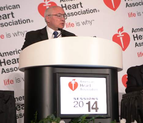
“While this relationship may have residual confounding, we believe that it shows ED time is an important indicator of the coordination of care,” Dr. Matthew Sherwood said at the American Heart Association scientific sessions.
The American College of Cardiology Foundation/AHA guidelines recommend the use of regional systems of care for STEMI patients and set a system goal for first medical contact to device time of 90 minutes or less for patients presenting directly to a percutaneous intervention–capable hospital and 120 minutes or less for those transferred from a non-PCI hospital to a PCI facility.
Only half of patients are treated within guideline goals, largely because of delays related to fragmentation of the health care system and lack of coordination between EMS agencies and acute care hospitals, said Dr. Sherwood, an interventional cardiology fellow with Duke University in Durham, N.C. For example, only 17% of EMS agencies can preactivate cardiac catheter labs in all receiving hospitals.
The STEMI Accelerator study sought to improve STEMI care between 484 hospitals and 1,253 EMS agencies in 16 cities, including New York, St. Louis, Pittsburgh, and Houston, representing roughly 10% of the U.S. population.
Leaders of emergency medical services, ED physicians, and interventional cardiologists were brought together in each region to identify areas for improvement and establish a regional coordinator who would implement a feedback system. They also developed a uniform protocol for preactivation for STEMI by EMS providers and coordinated a plan for rapid transfer of all STEMI patients to appropriate facilities.
At least 70% of PCI centers were also required to enroll in the National Cardiovascular Data Registry–Action GWTG registry for data collection, Dr. Sherwood said.
The Mission Lifeline: STEMI Systems Accelerator operations manual was also distributed at the national level to assist regions in developing their own, more individualized protocols. In New York City, that involved 16 PCI hospitals in downtown Manhattan developing a plan for STEMI patients to bypass the ED and go directly to the catheter lab – a move that has paid off in improved time to device delivery, he said.
The analysis included 23,809 patients with STEMI treated from the third quarter of 2012 to the first quarter of 2014. Of these, 11,765 presented directly to a PCI center via EMS, 6,502 presented to a PCI center via their own transportation, and 5,542 transferred from a non–PCI capable facility for further care. Their median age was 60 years, 29% were female, and 16% had no insurance.
For all three patient groups, there was a modest, but significant improvement from baseline to Q1 2014 in the percentage of patients meeting first medical contact to device time goals, including: direct-presenting patients (62% vs. 65%; P = .025), direct-presenting patients transported by EMS (54% vs. 59%; P = .0046), and patients requiring hospital transfer (50% vs. 53%; P = .007), Dr. Sherwood reported.
While the overall study showed modest improvements, there was a significant amount of variation in regional time and performance improvements. For example, among patients presenting directly to a PCI hospital, some regions showed up to 20% increases in the proportion of patients treated within guideline goals, he said.
Unadjusted in-hospital mortality also declined over the study period for participating regions, compared with national rates, suggesting that regional STEMI programs can provide important improvements to public health, Dr. Sherwood concluded.
Speaking as a practitioner in one of the contributing hospitals, panelist Dr. Roxana Mehran of Mount Sinai Hospital in New York, said that the STEMI program was invaluable in expanding their focus beyond door to balloon time.
“This particular program was extremely helpful for us in not just the timing of getting the patient, but also on the post discharge, which is also a part of this program,” she said. “I must tell you it had a huge impact, and we saw our numbers change dramatically just over the few quarters we saw patients.”
The Medicines Company, AstraZeneca, Philips Healthcare, and Abiomed sponsored the study. Dr. Sherwood reported no relevant financial relationships.
AT THE AHA SCIENTIFIC SESSIONS
Key clinical point: Organizing regional STEMI care results in significant improvements in the percentage of patients achieving national reperfusion goals.
Major finding: For all three patient groups, there was a modest but significant improvement from baseline to study end in the percentage of patients meeting first medical contact to device time goals.
Data source: Mission Lifeline: STEMI Accelerator, an observational study in 23,809 patients with STEMI.
Disclosures: The Medicines Company, AstraZeneca, Philips Healthcare, and Abiomed sponsored the study. Dr. Sherwood reported no relevant financial relationships.
Losartan fails in hypertrophic cardiomyopathy
CHICAGO – A year of losartan had no effect on left ventricular mass in patients with overt hypertrophic cardiomyopathy, compared with placebo, in the INHERIT trial.
The angiotensin receptor blocker did not alter any secondary endpoint, including maximal wall thickness, left ventricular fibrosis, diastolic function, and exercise capacity.
There also was no significant change in symptoms or resting left ventricular outflow tract (LVOT) gradient, Dr. Anna Axelsson reported at the American Heart Association scientific sessions.
Current therapies for hypertrophic cardiomyopathy (HCM) are palliative for symptoms only.
Losartan is known to reduce heart wall thickening in patients with hypertension, but is only used with caution in patients with HCM because of a suspicion of deleterious side effects.
INHERIT was initiated based on promising effects with angiotensin receptor blockade in animal and human studies, said Dr. Axelsson of Rigshospitalet, University of Copenhagen, and Harvard Medical School, Boston.
Losartan (Cozaar) has been shown to prevent the development of hypertrophy and fibrosis in HCM mice when started in the prehypertrophic phase. Candesartan (Atacand) use in humans was associated with regression of LV hypertrophy and improvement in LV function and exercise capacity in a small pilot study (J. Mol. Diagn. 2009;11:35-41).
INHERIT (Inhibition of the Renin Angiotensin System With Losartan in Patients With Hypertrophic Cardiomyopathy) evenly allocated 133 adults with overt HCM to daily losartan 100 mg or placebo for 12 months. LV mass was 108 g/m2 in the placebo group and 105 g/m2 in the losartan group, and 83% of patients had fibrosis.
Of the 124 patients completing the study, 93% were compliant (> 80%,) as assessed by pill count. Corroborating this was a significant decrease in blood pressure in the losartan group compared with the placebo group (P value = .001), Dr. Axelsson said.
At 12 months, both groups had a small decrease from baseline in LV mass, as assessed by magnetic resonance imaging or computed tomography, but the difference between groups was not statistically significant or clinically relevant (mean difference 1 g/m2), she said.
Adverse events were few, and comparable between the losartan and control groups. Sudden cardiac death occurred in two patients on placebo. One patient in each group had worsening of New York Heart Association class. Two controls and one losartan-treated patient had increases in LVOT gradient of at least 10 mm Hg. One patient discontinued losartan because of angioedema and two discontinued because of unspecified symptoms leading to withdrawal of consent.
“The observed safety suggests that losartan may be used for other indications in patients with obstructive physiology,” Dr. Axelsson said.
When asked whether clinicians can conclude that it’s safe to give patients with obstructive physiology a vasodilator, Dr. Axelsson said, “perhaps it shouldn’t be considered such a contraindication anymore, but it should still be done cautiously.”
Invited discussant Dr. Euan Ashley of Stanford (Calif.) University, said that only 15% of patients in the trial had an LVOT gradient of more than 30 mm Hg at baseline, “so to try and extrapolate that vasodilators are safe in older hypertrophics may be going too far.”
Finally, Dr. Axelsson said the overall results do not “close the door” to treatment with angiotensin receptor blockers in HCM, and that future trials may determine whether ARBs can prevent development of disease in preclinical or earlier stages of HCM.
The ongoing VANISH trial is evaluating valsartan (Diovan) in younger patients with early sarcomeric HCM. Also, the Liberty-HCM study will evaluate the effects of the investigational late sodium channel inhibitor, GS-6615, in patients aged 18-65 years, with symptomatic HCM.
INHERIT was funded by the Danish Heart Foundation and several other Danish research organizations. Dr. Axelsson holds stock with AstraZeneca.
AT THE AHA SCIENTIFIC SESSIONS
Key clinical point: Losartan 100 mg daily did not alter LV mass or any secondary endpoint in patients with hypertrophic cardiomyopathy.
Major finding: At 12 months, the change in LV mass from baseline was similar between patients receiving losartan and those on placebo.
Data source: INHERIT, a double-blind, randomized, placebo controlled trial of 133 patients with hypertrophic cardiomyopathy.
Disclosures: The Danish Heart Foundation and several other Danish research organizations funded the study. Dr. Axelsson holds stock with AstraZeneca.
Oxygen may make STEMI worse
CHICAGO – The global practice of giving oxygen to patients having a heart attack may cause more harm than good, results from the AVOID study showed.
Patients who were not hypoxic and received oxygen for ST-segment elevation MI (STEMI) had larger myocardial infarct size, as measured during the first 3 days of hospitalization using cardiac enzymes.
The oxygen arm had a statistically significant 25% increase in creatine kinase, compared with the no-oxygen arm, whether measured as geometric mean peak (1,948 U/L vs. 1,543 U/L) or median peak (2,073 U/L vs. 1,727 U/L), Dr. Stub said.
Cardiac troponin I levels were nonsignificantly higher with oxygen therapy (geometric mean peak, 57.4 mcg/L vs. 48 mcg/L; median peak, 65.7 mcg/L vs. 62.1 mcg/L).
When the preferred treatment approach cardiac magnetic resonance imaging was applied in about a third of patients at 6 months’ follow-up, the median infarct size remained significantly larger, at 20.3 g, in those given oxygen therapy, than in those who did not receive such therapy, whose median infarct size was 13.1 g, Dr. Dion Stub said at the American Heart Association scientific sessions.
“The primary endpoint of infarct size was significantly less without oxygen. That’s an astounding finding and really one that I think will cause many cardiologists to take note and perhaps step back,” said invited discussant Dr. Karl Kern, the Gordon A. Ewy Distinguished Endowed Chair of Cardiovascular Medicine, University of Arizona, Tucson.
“On the other hand, it’s important to realize that is a surrogate endpoint. That is not a mortality or outcome endpoint and the way that this was measured with biomarkers was admirable, but perhaps not today the most accurate. What is accurate was cardiac MR scanning, and the data held up at 6 months as well,” he added.
Although the study was not powered for clinical outcomes, patients receiving oxygen, compared with no oxygen, also had significantly more recurrent MI, at 5.5% and 0.9%, respectively, and major arrhythmia, at 40.4% and 31.4%, at discharge.
Survival at discharge was similar with oxygen versus no oxygen (1.8% vs. 4.5%), Dr. Stub, an interventional cardiologist at St. Paul’s Hospital, Vancouver, B.C., and a researcher at the Baker IDI Heart & Diabetes Institute, Melbourne, reported. .
Oxygen therapy has been used for more than a century in the initial treatment of patients with suspected MI, although there is limited evidence suggesting such therapy is beneficial in patients without hypoxia. A growing body of evidence, however, suggests that even 15 minutes of oxygen can reduce coronary blood flow, increase the production of oxygen-free radicals, and disturb microcirculation, all of which can contribute to reperfusion injury during MI, he said.
The 638 Australian patients in the AVOID (Air Versus Oxygen in ST-Elevation Myocardial Infarction) study were randomized, before hospitalization, by paramedics to oxygen administered via face mask at a flow rate of 8 L/min until patients were stabilized on the ward or to no oxygen, unless oxygen saturation fell below 94%.
Dr. Kern and others pointed out that the oxygen level provided to study participants exceeds the 2-4 L typically given to MI patients in the United States, particularly those who are nonhypoxic.
All parties agreed there is an urgent need for an adequately powered randomized trial to evaluate the effectiveness of oxygen therapy in MI, a conclusion also reached by a recent Cochrane review of the topic (Cochrane Syst. Rev. 2013 August;8:CD007160).
Dr. Jason Lazar, FCCP, comments: This prospective randomized study from Australia of 638 patients presenting with ST segment elevation myocardial infarction challenges the decades long practice of adminstering routine supplemental oxygen in such patients. Oxygen therapy was associated with larger infarct size. The study findings underscore the importance of evidence-based medicine and will undoubtedly spark additional studies to address this issue.
Dr. Lazar is with the Division of Cardiology at the State University of New York Downstate Medical Center in Brooklyn, NY.
CHICAGO – The global practice of giving oxygen to patients having a heart attack may cause more harm than good, results from the AVOID study showed.
Patients who were not hypoxic and received oxygen for ST-segment elevation MI (STEMI) had larger myocardial infarct size, as measured during the first 3 days of hospitalization using cardiac enzymes.
The oxygen arm had a statistically significant 25% increase in creatine kinase, compared with the no-oxygen arm, whether measured as geometric mean peak (1,948 U/L vs. 1,543 U/L) or median peak (2,073 U/L vs. 1,727 U/L), Dr. Stub said.
Cardiac troponin I levels were nonsignificantly higher with oxygen therapy (geometric mean peak, 57.4 mcg/L vs. 48 mcg/L; median peak, 65.7 mcg/L vs. 62.1 mcg/L).
When the preferred treatment approach cardiac magnetic resonance imaging was applied in about a third of patients at 6 months’ follow-up, the median infarct size remained significantly larger, at 20.3 g, in those given oxygen therapy, than in those who did not receive such therapy, whose median infarct size was 13.1 g, Dr. Dion Stub said at the American Heart Association scientific sessions.
“The primary endpoint of infarct size was significantly less without oxygen. That’s an astounding finding and really one that I think will cause many cardiologists to take note and perhaps step back,” said invited discussant Dr. Karl Kern, the Gordon A. Ewy Distinguished Endowed Chair of Cardiovascular Medicine, University of Arizona, Tucson.
“On the other hand, it’s important to realize that is a surrogate endpoint. That is not a mortality or outcome endpoint and the way that this was measured with biomarkers was admirable, but perhaps not today the most accurate. What is accurate was cardiac MR scanning, and the data held up at 6 months as well,” he added.
Although the study was not powered for clinical outcomes, patients receiving oxygen, compared with no oxygen, also had significantly more recurrent MI, at 5.5% and 0.9%, respectively, and major arrhythmia, at 40.4% and 31.4%, at discharge.
Survival at discharge was similar with oxygen versus no oxygen (1.8% vs. 4.5%), Dr. Stub, an interventional cardiologist at St. Paul’s Hospital, Vancouver, B.C., and a researcher at the Baker IDI Heart & Diabetes Institute, Melbourne, reported. .
Oxygen therapy has been used for more than a century in the initial treatment of patients with suspected MI, although there is limited evidence suggesting such therapy is beneficial in patients without hypoxia. A growing body of evidence, however, suggests that even 15 minutes of oxygen can reduce coronary blood flow, increase the production of oxygen-free radicals, and disturb microcirculation, all of which can contribute to reperfusion injury during MI, he said.
The 638 Australian patients in the AVOID (Air Versus Oxygen in ST-Elevation Myocardial Infarction) study were randomized, before hospitalization, by paramedics to oxygen administered via face mask at a flow rate of 8 L/min until patients were stabilized on the ward or to no oxygen, unless oxygen saturation fell below 94%.
Dr. Kern and others pointed out that the oxygen level provided to study participants exceeds the 2-4 L typically given to MI patients in the United States, particularly those who are nonhypoxic.
All parties agreed there is an urgent need for an adequately powered randomized trial to evaluate the effectiveness of oxygen therapy in MI, a conclusion also reached by a recent Cochrane review of the topic (Cochrane Syst. Rev. 2013 August;8:CD007160).
AT THE AHA SCIENTIFIC SESSIONS
Key clinical point: Supplemental oxygen therapy in normoxic patients with STEMI was associated with larger infarct size and more recurrent MIs and major cardiac arrhythmias.
Major finding: Median infarct size on cardiac MRI at 6 months was 203 g in those given oxygen, compared with 3.1 g in those not given oxygen (P = .04).
Data source: A randomized trial in 638 patients with STEMI.
Disclosures: AVOID was funded by the Alfred Hospital Foundation, FALCK Foundation, and Paramedics Australia. Dr. Stub and his coauthors reported having no financial disclosures. Dr. Kern reported serving on the science advisory boards of PhysioControl and Zoll Medical.
CT Screening Not Useful in High-risk Diabetes Patients
CHICAGO – Optimal guideline-directed medical therapy appears more important than routine coronary CT screening in preventing death and cardiac events in patients with diabetes at high risk for asymptomatic coronary artery disease, the FACTOR-64 trial suggests.
At an average follow-up of 4 years, routine coronary computed tomography angiography (CCTA) failed to significantly reduce the primary composite endpoint of all-cause mortality, nonfatal myocardial infarction, or hospitalization for unstable angina, compared with medical management in the intent-to-treat analysis (6.2% vs. 7.6%, respectively; hazard ratio, 0.80; P = .38).
Event rates were also similar between groups in the as-treated analysis (5.6% vs. 7.9%; HR, 0.69; P = .16), study author Dr. Joseph Brent Muhlestein reported at the American Heart Association scientific sessions.
These findings do not support CCTA screening in this population,” he said.
CCTA screening, however, did reveal coronary artery disease in 70% of patients, prompting a recommendation for additional diagnostics and/or more aggressive management of risk factors. This resulted in modest but significant improvements in lipid subfractions and blood pressure levels in the CCTA group and coronary revascularization procedures in 5.8%, Dr. Muhlestein, with the Intermountain Heart Institute in Murray, Utah, said.
FACTOR-64 randomized 900 patients with type 1 or 2 diabetes for at least 3-5 years and without CAD symptoms to 64-slice CCTA screening or standard guideline-based optimal diabetes care including a target hemoglobin A1c level of less than 7.0%, LDL cholesterol level below 100 mg/dL, and systolic BP less than 130 mm Hg. Based on CCTA findings, recommendations were made for standard or aggressive therapy (HbA1c below 6.0%, LDL cholesterol under 70 mg/dL, HDL cholesterol greater than 50 mg/dL in women or above 40 mg/dL in men, triglycerides below 150 mg/dL, and systolic BP under 120 mm Hg) or aggressive therapy with invasive angiography. All patients were recruited from the Intermountain Healthcare system in Utah. Their mean age was 61 years.
The secondary composite endpoint of cardiovascular death, nonfatal myocardial infarction, or hospitalization for unstable angina was similar between the CCTA and control groups analyzed by intention to treat (4.6% vs. 5.1%, respectively; HR, 0.89; P = .70) and as-treated (4.1% vs. 5.6%; HR, 0.72; P = .30), according to the results, published online simultaneously (JAMA 2014 Nov. 17 [doi: 10.1001/jama.2014.15825]).
Continued on next page >>
The overall annual event rates in both the control and intervention groups were low, at less than 2% per year. This may be attributed to the excellent medical management received by all patients, as evidenced by baseline levels near or exceeding system targets for HbA1c, LDL cholesterol, and systolic BP, Dr. Muhlestein said.
In an accompanying editorial (JAMA 2014 Nov. 17 [doi: 10.1001/jama.2014.15958]), Dr. Raymond Gibbons of the Mayo Clinic in Rochester, Minn., agreed that the findings are likely explained by the excellent baseline medical therapy.
“The data in this study suggest that Intermountain Healthcare has set a new published standard for what is achievable in patients with diabetes with respect to blood pressure control and lipid-lowering therapy and that, when therapy is applied this effectively, patients with diabetes are no longer at high risk for major cardiovascular events,” Dr. Gibbons wrote.
Invited discussant Dr. Pamela Douglas, professor of medicine at Duke University in Durham, N.C., said the key message is that the study was tested in a highly managed population, but that nationally less than 10% of diabetes patients are at goal for cardiovascular protection.
Dr. Douglas said CCTA screening may still have a limited role in asymptomatic patients if used to help patients adhere to treatment strategies that can improve outcomes. “This may be a bit roundabout, but that’s the one time,” she added.
Dr. Douglas also called for improved risk-prediction tools to identify even higher-risk individuals, observing that events were not confined to those with obstructive disease.
“We need to be looking for nonobstructive disease, which was in fact the hypothesis of this trial,” she said. “So perhaps we need to look beyond ischemia, beyond atherosclerosis to vulnerable plaque ... and cell targets.”
During the roundtable discussion of the study, panelists suggested that a coronary calcium score could play a role in identifying these higher-risk patients, and that telling a patient they have a calcium score of 800 may also get even the most stubborn diabetes patient to actually take their medications.
The study was supported by the Intermountain Research and Medical Foundation, Intermountain Heart Institute, and grants from Toshiba and Bracco. The study authors reported having no financial disclosures. Dr. Gibbons reported serving as a consultant for Lantheus Medical Imaging. Dr. Douglas reported no conflicts.
AT THE AHA SCIENTIFIC SESSIONS
CT screening not useful in high-risk diabetes patients
CHICAGO – Optimal guideline-directed medical therapy appears more important than routine coronary CT screening in preventing death and cardiac events in patients with diabetes at high risk for asymptomatic coronary artery disease, the FACTOR-64 trial suggests.
At an average follow-up of 4 years, routine coronary computed tomography angiography (CCTA) failed to significantly reduce the primary composite endpoint of all-cause mortality, nonfatal myocardial infarction, or hospitalization for unstable angina, compared with medical management in the intent-to-treat analysis (6.2% vs. 7.6%, respectively; hazard ratio, 0.80; P = .38).
Event rates were also similar between groups in the as-treated analysis (5.6% vs. 7.9%; HR, 0.69; P = .16), study author Dr. Joseph Brent Muhlestein reported at the American Heart Association scientific sessions.
These findings do not support CCTA screening in this population,” he said.
CCTA screening, however, did reveal coronary artery disease in 70% of patients, prompting a recommendation for additional diagnostics and/or more aggressive management of risk factors. This resulted in modest but significant improvements in lipid subfractions and blood pressure levels in the CCTA group and coronary revascularization procedures in 5.8%, Dr. Muhlestein, with the Intermountain Heart Institute in Murray, Utah, said.
FACTOR-64 randomized 900 patients with type 1 or 2 diabetes for at least 3-5 years and without CAD symptoms to 64-slice CCTA screening or standard guideline-based optimal diabetes care including a target hemoglobin A1c level of less than 7.0%, LDL cholesterol level below 100 mg/dL, and systolic BP less than 130 mm Hg. Based on CCTA findings, recommendations were made for standard or aggressive therapy (HbA1c below 6.0%, LDL cholesterol under 70 mg/dL, HDL cholesterol greater than 50 mg/dL in women or above 40 mg/dL in men, triglycerides below 150 mg/dL, and systolic BP under 120 mm Hg) or aggressive therapy with invasive angiography. All patients were recruited from the Intermountain Healthcare system in Utah. Their mean age was 61 years.
The secondary composite endpoint of cardiovascular death, nonfatal myocardial infarction, or hospitalization for unstable angina was similar between the CCTA and control groups analyzed by intention to treat (4.6% vs. 5.1%, respectively; HR, 0.89; P = .70) and as-treated (4.1% vs. 5.6%; HR, 0.72; P = .30), according to the results, published online simultaneously (JAMA 2014 Nov. 17 [doi: 10.1001/jama.2014.15825]).
The overall annual event rates in both the control and intervention groups were low, at less than 2% per year. This may be attributed to the excellent medical management received by all patients, as evidenced by baseline levels near or exceeding system targets for HbA1c, LDL cholesterol, and systolic BP, Dr. Muhlestein said.
In an accompanying editorial (JAMA 2014 Nov. 17 [doi: 10.1001/jama.2014.15958]), Dr. Raymond Gibbons of the Mayo Clinic in Rochester, Minn., agreed that the findings are likely explained by the excellent baseline medical therapy.
“The data in this study suggest that Intermountain Healthcare has set a new published standard for what is achievable in patients with diabetes with respect to blood pressure control and lipid-lowering therapy and that, when therapy is applied this effectively, patients with diabetes are no longer at high risk for major cardiovascular events,” Dr. Gibbons wrote.
Invited discussant Dr. Pamela Douglas, professor of medicine at Duke University in Durham, N.C., said the key message is that the study was tested in a highly managed population, but that nationally less than 10% of diabetes patients are at goal for cardiovascular protection.
Dr. Douglas said CCTA screening may still have a limited role in asymptomatic patients if used to help patients adhere to treatment strategies that can improve outcomes. “This may be a bit roundabout, but that’s the one time,” she added.
Dr. Douglas also called for improved risk-prediction tools to identify even higher-risk individuals, observing that events were not confined to those with obstructive disease.
“We need to be looking for nonobstructive disease, which was in fact the hypothesis of this trial,” she said. “So perhaps we need to look beyond ischemia, beyond atherosclerosis to vulnerable plaque ... and cell targets.”
During the roundtable discussion of the study, panelists suggested that a coronary calcium score could play a role in identifying these higher-risk patients, and that telling a patient they have a calcium score of 800 may also get even the most stubborn diabetes patient to actually take their medications.
The study was supported by the Intermountain Research and Medical Foundation, Intermountain Heart Institute, and grants from Toshiba and Bracco. The study authors reported having no financial disclosures. Dr. Gibbons reported serving as a consultant for Lantheus Medical Imaging. Dr. Douglas reported no conflicts.
AT THE AHA SCIENTIFIC SESSIONS
Key clinical point: CCTA screening did not reduce the composite endpoint, compared with optimal guideline-directed diabetes care.
Major finding: The primary composite of all-cause mortality, nonfatal myocardial infarction, or hospitalization for unstable angina occurred in 6.2% of patients with CCTA screening and 7.6% of patients with medical management in the intent-to-treat analysis (HR, 0.80; P = .38).
Data source: Randomized trial in 900 patients with type 1 or 2 diabetes without symptoms of coronary artery disease.
Disclosures: The study was supported by the Intermountain Research and Medical Foundation, Intermountain Heart Institute, and grants from Toshiba and Bracco. The study authors reported having no financial disclosures.
VIDEO: Study reignites dental antibiotic prophylaxis controversy
CHICAGO – The first guidelines recommending antibiotic prophylaxis for invasive dental procedures were issued in 1955, and controversy has gone hand in hand with each revision that has called for shorter treatment duration and fewer eligible patients.
A study presented at the American Heart Association scientific sessions adds to that controversy – and has prompted the United Kingdom’s National Institute for Health and Care Excellence to immediately review its 2008 guidelines.
Those guidelines recommend that antibiotics should not be prescribed to prevent infective endocarditis (IE) for people undergoing dental procedures or procedures in the upper and lower gastrointestinal tract, genitourinary tract, and upper and lower respiratory tract.
Five years post NICE, the new study found that antibiotic prophylaxis prescribing fell almost 90% in the United Kingdom, from 10,900 prescriptions per month to 1,307 per month in the last 6 months of the study, reported Dr. Mark Dayer of Taunton and Somerset NHS Trust, Somerset, England. The study was simultaneously published in the Lancet (2014 Nov. 18[doi:10.1016/S0140-6736(14)62007-9]).
In a video interview, study coauthor Dr. Martin Thornhill of the University of Sheffield, England, and AHA President-Elect Dr. Mark Creager, director of the vascular center at Brigham and Women’s Hospital, Boston, talked about the findings, their potential limitations, and whether it’s time for clinicians to change their approach to antibiotic prophylaxis.
The study was funded by the National Institutes of Dental and Cranofacial Research, Heart Research–UK, and Simplyhealth. Dr. Thornhill and Dr. Creager reported no conflicting interests.
AT THE AHA SCIENTIFIC SESSIONS
Fatigue checks assist in preventing wrong-site surgery
EDINBURGH – It’s 4 o’clock in the afternoon on a long Mohs surgery day, and you’ve got another patient with innumerable stages plus reconstructions to do.
Performing a fatigue check that gives nursing and biomedical staff the opportunity to admit they’re too tired may be the best next step to prevent wrong-site surgery, Dr. Colin Fleming, president of the British Society for Dermatological Surgery (BSDS), posited at the 15th World Congress on Cancers of the Skin.
“When we introduced this, it gave great power to the staff, whom we as doctors were asking to work hard on our behalf,” he said. “It’s been a really valuable tool in increasing safety at the end of a long operating day.”
The fatigue check is separate from the oft-recommended preprocedural surgical pause. Though helpful, research has shown that the surgical pause failed to prevent wrong-site surgery in 10% to a third of cases, said Dr. Fleming, a consultant dermatologist and Mohs surgeon at Ninewells Hospital, Dundee, Scotland.
A study on the usefulness of proposed methods for correct biopsy site identification in cutaneous surgery with the Delphi consensus method was recently published (JAMA Dermatol. 2014;150:550-8), but “I’m not convinced it really tells us anything,” he remarked.
Dr. Fleming suggested coupling the surgical pause with a site check, and he emphasized the value of good quality preoperative photographs of the index lesion. Today’s ubiquitous “selfie” may even have a role, as patients themselves often misidentify the biopsy site.
In addition, dermatology departments should have a protocol incorporating a variety of safety features, he said.
However, only 54% of surgeons said they have a written protocol in place to identify the correct surgical site, based on data from a recent survey of BSDS members conducted by Dr. Fleming and his colleagues.
More than a half of respondents (60/114) acknowledged it was “sometimes” difficult to locate the surgical site, with 47 respondents saying it was difficult to locate the surgical site in 1%-10% of cases and 13 admitting it was difficult to do so in 10%-25% of cases.
The face, scalp, and back were reported as the most challenging areas in which to locate the exact surgical site, Dr. Fleming said at the meeting, sponsored by the Skin Cancer Foundation.
When asked what steps they take in their local written protocols to avoid wrong site surgery, 82 respondents said they check with the patient, 66 use drawings/templates with the site marked, 50 use a mirror or photographs, and 39 respondents double-check with a colleague.
Overall, slightly less than 40% of respondents recalled having no patient in the past 5 years who underwent wrong-site surgery. Approximately 28% had 1 wrong-site surgery patient, 28% had 2-3 patients, 3% had 4-5 patients, and 1% had more than 10 such patients.
“Wrong-site surgery in U.K. dermatology departments is infrequent,” Dr. Fleming concluded, adding that only a small proportion led to formal complaints or a medicolegal case.
The response rate to the web-based survey was 37.5% (115/306 members); 32 respondents were Mohs surgeons, 75 were non-Mohs surgeons, 68 worked at a teaching hospital, and 110 had a regular surgical list of between 500 and 2,000 procedures per year that they were responsible for or did themselves.
AT THE WCCS 2014
Aspirin fails to protect elderly at-risk patients from cardiac events
CHICAGO– Daily low-dose aspirin did not prevent atherosclerotic events in high-risk, elderly Japanese patients in the Japanese Primary Prevention Project.
After a median follow-up of 5 years, the composite primary outcome of cardiovascular death, nonfatal stroke, and nonfatal myocardial infarction occurred in 2.77% of patients with hypertension, dyslipidemia, or diabetes on aspirin and 2.96% of those not on aspirin, a nonsignificant difference.
Subgroup analyses did not identify significant differences between study groups, Dr. Kazuyuki Shimada reported at the American Heart Association scientific sessions.The study was stopped prematurely because the number of primary events was insufficient for the study to reach statistical power.
Daily low-dose aspirin, compared with no aspirin, however, significantly reduced the rate of nonfatal myocardial infarction (0.30% vs. 0.58%; HR, 0.53; P = .02) and transient ischemic attack (0.26% vs. 0.49%; HR, 0.57; P = .04).
However, these benefits must be weighed against an 85% increased risk of serious extracranial hemorrhage in patients on daily aspirin (0.86% vs. 0.51%; HR, 1.85, P = .004), Dr. Shimada of Shin-Oyama City Hospital, Tochigi, Japan, said.
Prespecified gastrointestinal adverse events, including stomach/abdominal pain, gastroduodenal ulcer, reflux esophagitis, and gastrointestinal hemorrhage, were also increased in patients on aspirin, according to results of the late-breaking study, simultaneously published online in JAMA (doi:10.1001/jama.2014.15690).
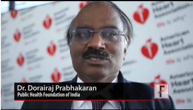
Invited discussant Dr. Dorairaj Prabhakaran of the Public Health Foundation of India in Delhi said the negative results do not spell the “end of the road” for aspirin in primary prevention, but emphasize the need to use an individualized, stepwise risk-benefit approach to aspirin therapy.
“The benefit is very unlikely in very low-risk populations such as those with less than 1% [cardiovascular] events per year,” he said. “There would be a role in special groups such as younger populations, lower-income countries, but these are not evaluated well.”
During the discussion following the study presentation, panelists raised concerns about the development of polypills, most of which are for secondary prevention but typically contain aspirin. Other panelists said the study provides a sobering reminder of the risks faced by patients who put themselves on a daily aspirin regimen without consulting a physician.
The Japanese Primary Prevention Project (JPPP) evenly randomized 14,658 patients, aged at least 60 years, with hypertension, dyslipidemia, or diabetes to enteric aspirin 100 mg or no aspirin. A total of 194 patients were excluded because of protocol violations, study withdrawal, or failing to meet inclusion criteria, leaving 7,220 patients in the aspirin group and 7,244 in the no-aspirin group for the modified intention-to-treat population.
In addition to the lower-than-expected total event rate in both the aspirin and no-aspirin groups (193 vs. 207), the use of statins in both arms could have contributed to the negative results, Dr. Shimada reported. Aspirin adherence also fell from 89% in year 1 to only 76% in year 5, while aspirin use in the no-aspirin group increased from 1.5% to 9.8%.
Further analyses are planned to determine whether aspirin is beneficial in select subgroups or in the prevention of cancer.
Dr. J. Michael Gaziano of Brigham and Women’s Hospital in Boston commented in an accompanying JAMA editorial (doi:10.1001/jama.2014.16047) that information from three ongoing primary prevention aspirin trials in patients at higher-than-average risk “will prove helpful for clinical decision making involving the role of aspirin for primary prevention.”
Those trials include the ASCEND study of aspirin 100 mg with or without omega-3 fatty acids in patients at least 40 years old with diabetes, the ARRIVE trial in middle-aged and older patients at moderate risk of cardiovascular disease, and the ASPREE study in the elderly older than 65 years.
JPPP was sponsored by the Japanese Ministry of Health, Labor, and Welfare, and the Waksman Foundation of Japan. Bayer Yakuhin provided the aspirin. Dr. Shimada reported honorarium from MSD, Shionogi, Takeda, Daiichi-Sankyo, and Dainihon-Sumitomo, and serving as a consultant/advisory board member for Omron. Dr. Prabhakaran reported no relevant financial disclosures. Dr. Gaziano reported serving on the executive committee of the ARRIVE trial and as a consultant for and receiving speaking honoraria from Bayer.
AT THE AHA SCIENTIFIC SESSIONS
Key clinical point: Low-dose aspirin may not prevent adverse cardiovascular outcomes in patients with atherosclerotic risk factors.
Major finding: The cumulative rate of the combined outcome of cardiovascular death, nonfatal stroke, and nonfatal myocardial infarction was 2.77% with aspirin and 2.96% with no aspirin, a nonsignificant difference.
Data source: JPPP, a randomized, open-label parallel-group trial in 14,658 elderly Japanese patients with atherosclerotic risk factors.
Disclosures: JPPP was sponsored by the Japanese Ministry of Health, Labor, and Welfare, and the Waksman Foundation of Japan. Bayer Yakuhin provided the aspirin. Dr. Shimada reported honorarium from MSD, Shionogi, Takeda, Daiichi-Sankyo, and Dainihon-Sumitomo, and serving as a consultant/advisory board member for Omron.
VIDEO: End of the road for aspirin in primary prevention?
CHICAGO – Once-daily, low-dose aspirin failed to reduce the combined outcome of cardiovascular death, nonfatal stroke, and nonfatal MI in elderly Japanese patients with atherosclerotic risk factors in the JPPP study.
The cumulative rate of the composite primary outcome was 2.77% with 100 mg/day of aspirin and 2.96% with no aspirin (HR, 0.94; P = .54), Dr. Kazuyuki Shimada reported at the American Heart Association scientific sessions.
However, patients randomized to aspirin did have reductions of 47% and 43% in the individual endpoints of nonfatal MI and transient ischemic attack, respectively.
These benefits had to be weighed against an 85% increase in serious extracranial hemorrhage in those on aspirin, according to results of the Japanese Primary Prevention Project (JPPP), simultaneously published online in JAMA (doi:10.1001/jama.2014.15690).
How generalizable are these results, and is this the end of the road for aspirin in primary prevention? We asked several experts, including invited discussant Dr. Dorairaj Prabhakaran of the Public Health Foundation of India, Dr. Karol Watson of the UCLA Center for Cholesterol and Lipid Management, and Dr. Donald Lloyd-Jonesof Northwestern University in Chicago.
The study was sponsored by the Japanese Ministry of Health, Labor, and Welfare, and the Waksman Foundation of Japan. Bayer Yakuhin provided the aspirin. Dr. Shimada reported honorarium from MSD, Shionogi, Takeda, Daiichi-Sankyo, and Dainippon-Sumitomo, and serving as a consultant/advisory board member for Omron.
Dr. Prabhakaran and Dr. Jones reported no conflicting interests. Dr. Watson reported participating in the clinical trials adjudication committee for Merck.
FROM THE AHA SCIENTIFIC SESSIONS
Nivolumab monotherapy reaches advanced squamous NSCLC
CHICAGO – Nivolumab monotherapy resulted in a median overall survival of 8.2 months and a 1-year survival rate of 41% in advanced, heavily pretreated squamous cell non–small cell lung cancer in a phase II study.
Advanced, treatment-refractory squamous cell non–small cell lung cancer (NSCLC) is an area of high unmet need with no effective therapeutic options and this cohort was “truly” treatment refractory, study author Dr. Suresh Ramalingam said at the 2014 Chicago Multidisciplinary Symposium in Thoracic Oncology.
Of the 117 patients, 65% had received at least three prior systemic regimens, 61% had progressive disease as their best response to the most recent therapy, and 76% came into the study within 3 months of completing their most recent therapy.
The patients had stage IIIB/IV disease and were given nivolumab 3 mg/kg IV every 2 weeks until progressive disease or unacceptable toxicity. Their median age was 65 years (range 37-87 years).
The study’s primary end point of objective response rate by independent radiology review was 15% (17/117 patients), including eight confirmed partial responses in patients with rapid progression on prior therapy, Dr. Ramalingam, director of medical oncology and the lung cancer program, Winship Cancer Institute of Emory University, Atlanta, said.
The median duration of response has not been reached and 76% of responses are ongoing. In all, 24% of patients received subsequent therapy, excluding subsequent immunotherapy.
Median progression-free survival (PFS) was 2 months and the 1-year PFS rate was 20%, he said.
An exploratory biomarker analysis of 76 evaluable samples showed nivolumab was clinically active in patients whose tumors were positive as well as negative for programmed death-ligand 1 expression.
Dr. Ramalingam highlighted a 73-year-old former smoker with stage IIIB disease and active brain metastasis at study entry who had stable disease after treatment with nivolumab that was ongoing at 68 weeks including a response in his brain lesions, despite having no prior central nervous system-directed therapy, and failing prior radiotherapy and three prior systemic regimens (cisplatin[Platinol]/gemcitabine [Gemzar], docetaxel [Taxotere], and vinorelbine [Navelbine]).
Overall, nivolumab was well tolerated, as shown by the fact that 85% of patients received at least 90% of their planned dose intensity, he said.
The safety profile was consistent with prior nivolumab studies, with 12% of patients discontinuing treatment because of toxicity. Two treatment-related deaths (one hypoxic pneumonia and one ischemic stroke) occurred in patients with multiple comorbidities and concurrent progressive disease.
A poster also presented at the meeting (Ab. 170, Gettinger, S.N., et al.) showed an objective response rate of 17% among 54 treatment-refractory patients given second-line or later nivolumab 1 mg/kg, 3 mg/kg, or 10 mg/kg every 2 weeks for squamous cell NSCLC, among other solid tumors. The median PFS was 3.8 months, 1-year PFS 27%, median overall survival 9.2 months, and 1-year overall survival 41%.
A phase III trial is also ongoing comparing overall survival of nivolumab vs. docetaxel in advanced squamous cell NSCLC after failure of prior platinum-based chemotherapy, Dr. Ramalingam said.
FROM A THORACIC ONCOLOGY SYMPOSIUM
Key clinical point: Nivolumab monotherapy shows clinically meaningful efficacy in patients with advanced, heavily pretreated squamous cell NSCLC, an area of high unmet need.
Major finding: Median overall survival was 8.2 months and 1-year survival was 41%.
Data source: Phase II study in 117 patients with advanced, treatment-refractory squamous cell NSCLC.
Disclosures: The study was sponsored by Bristol-Myers Squibb and Ono Pharmaceuticals, developers of nivolumab. Dr. Ramalingam reported research funding from Bristol-Myers Squibb and a consultant/advisory relationship with Amgen, AstraZeneca, Aveo, Boehringer Ingelheim, Celgene, Genentech, Lilly, and Novartis.
