User login
Vaginal delivery found safe for monoamniotic-monochorionic twins
WASHINGTON – Monoamniotic-monochorionic twins can be safely delivered vaginally, with low rates of adverse fetal or maternal outcomes.
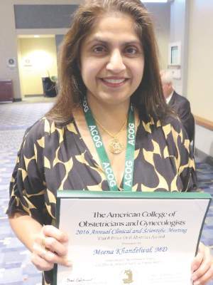
Despite seeing cord entanglement in almost every case, a small retrospective study found no safety signals that support pre-emptive cesarean deliveries for this class of twins, Dr. Meena Khandelwal said at the annual meeting of the American College of Obstetricians and Gynecologists. In fact, vaginal delivery was associated with a significantly lower rate of intracranial hemorrhage, which occurred in 27% of the cesarean-delivered infants and none of the infants delivered vaginally.

Dr. Khandelwal and her colleagues won third prize for the study among the 2016 ACOG research awards.
In 2014, an ACOG technical bulletin advised cesarean delivery for all monoamniotic twins, a recommendation in line with those in Canada and France. But the ACOG advice was based on level C evidence, said Dr. Khandelwal of Cooper University Hospital, Camden, N.J.
“These recommendations are fear based, not fact based,” she said. “It is felt that the risk of entrapment is too great. But cord entanglement is seen in virtually all of these cases, and a large study recently found that it contributed nothing to morbidity or mortality in the fetuses.”
That study, published in 2013, reviewed 114 monoamniotic twin pregnancies (228 fetuses). There were 26 perinatal fetal deaths, only two of which were related to cord entanglement. The author found no significant difference in mortality between the fetuses with entanglement (82) and those without (Ultrasound Obstet Gynecol. 2013 Feb;41[2]:131-5).
Dr. Khandelwal conducted her own retrospective study of 29 sets of monoamniotic-monochorionic twins that were born vaginally or by cesarean delivery at two tertiary care centers in New Jersey, from 1997 to 2014. Her cohort comprised 10 pregnancies at Thomas Jefferson Hospital, Philadelphia, which only offers cesarean delivery for these cases, and 19 at Cooper University Hospital, which offers the option of vaginal delivery if the first twin is in cephalic position, the fetal heart rate is reactive at the onset of labor, and there are no contraindications to vaginal delivery.
Cooper also manages these pregnancies on an outpatient basis with vaginal delivery offered from 32 to 36 weeks. Thomas Jefferson admits them to the antepartum unit from 24 to 28 weeks and offers cesarean delivery from 32 to36 weeks.
The mean age of the women in the series was 29 years; median parity was one. Almost half of the women having a planned cesarean had a history of a prior cesarean; none of the women who planned a vaginal delivery had a prior cesarean delivery.
Of the 19 women offered vaginal delivery, 14 accepted. Of these, 10 (71%) delivered both twins vaginally. The mean time between the twin deliveries was 3 minutes. Cesarean delivery was necessary for four neonates in the planned vaginal delivery group: three due to concerning heart rate tracings and one because of a failed internal version of a transverse presentation of a second twin.
Three fetuses died before birth – one in the planned vaginal delivery group (7%) and two in the planned cesarean delivery group (13%). There was one post partum hemorrhage, which occurred in the planned cesarean group. There were no cases of chorioamnionitis. Birth weight was similar (about 1,800 grams). Cords were entangled in 100% of the vaginal delivery group and 93% of the cesarean group. Apgar at 5 minutes was 6.6 in the vaginal delivery group and 8.3 in the cesarean group.
Respiratory distress syndrome was significantly more common among the planned cesarean group (86% vs. 65%). There were eight cases of intracerebral hemorrhage in that group and none in the planned vaginal delivery group (28% vs. 0%). These were all grade 1 or 2 bleeds. There were no neonatal deaths.
The length of stay was shorter in the planned vaginal delivery group, but not significantly so (median 18 vs. 25 days).
“This is a small study but it does add valuable data on the safety of vaginal delivery in [monoamniotic-monochorionic] twins,” Dr. Khandelwal said. “Vaginal delivery can be considered a safe option in tertiary care centers.
She reported having no financial disclosures.
WASHINGTON – Monoamniotic-monochorionic twins can be safely delivered vaginally, with low rates of adverse fetal or maternal outcomes.
Despite seeing cord entanglement in almost every case, a small retrospective study found no safety signals that support pre-emptive cesarean deliveries for this class of twins, Dr. Meena Khandelwal said at the annual meeting of the American College of Obstetricians and Gynecologists. In fact, vaginal delivery was associated with a significantly lower rate of intracranial hemorrhage, which occurred in 27% of the cesarean-delivered infants and none of the infants delivered vaginally.
Dr. Khandelwal and her colleagues won third prize for the study among the 2016 ACOG research awards.
In 2014, an ACOG technical bulletin advised cesarean delivery for all monoamniotic twins, a recommendation in line with those in Canada and France. But the ACOG advice was based on level C evidence, said Dr. Khandelwal of Cooper University Hospital, Camden, N.J.
“These recommendations are fear based, not fact based,” she said. “It is felt that the risk of entrapment is too great. But cord entanglement is seen in virtually all of these cases, and a large study recently found that it contributed nothing to morbidity or mortality in the fetuses.”
That study, published in 2013, reviewed 114 monoamniotic twin pregnancies (228 fetuses). There were 26 perinatal fetal deaths, only two of which were related to cord entanglement. The author found no significant difference in mortality between the fetuses with entanglement (82) and those without (Ultrasound Obstet Gynecol. 2013 Feb;41[2]:131-5).
Dr. Khandelwal conducted her own retrospective study of 29 sets of monoamniotic-monochorionic twins that were born vaginally or by cesarean delivery at two tertiary care centers in New Jersey, from 1997 to 2014. Her cohort comprised 10 pregnancies at Thomas Jefferson Hospital, Philadelphia, which only offers cesarean delivery for these cases, and 19 at Cooper University Hospital, which offers the option of vaginal delivery if the first twin is in cephalic position, the fetal heart rate is reactive at the onset of labor, and there are no contraindications to vaginal delivery.
Cooper also manages these pregnancies on an outpatient basis with vaginal delivery offered from 32 to 36 weeks. Thomas Jefferson admits them to the antepartum unit from 24 to 28 weeks and offers cesarean delivery from 32 to36 weeks.
The mean age of the women in the series was 29 years; median parity was one. Almost half of the women having a planned cesarean had a history of a prior cesarean; none of the women who planned a vaginal delivery had a prior cesarean delivery.
Of the 19 women offered vaginal delivery, 14 accepted. Of these, 10 (71%) delivered both twins vaginally. The mean time between the twin deliveries was 3 minutes. Cesarean delivery was necessary for four neonates in the planned vaginal delivery group: three due to concerning heart rate tracings and one because of a failed internal version of a transverse presentation of a second twin.
Three fetuses died before birth – one in the planned vaginal delivery group (7%) and two in the planned cesarean delivery group (13%). There was one post partum hemorrhage, which occurred in the planned cesarean group. There were no cases of chorioamnionitis. Birth weight was similar (about 1,800 grams). Cords were entangled in 100% of the vaginal delivery group and 93% of the cesarean group. Apgar at 5 minutes was 6.6 in the vaginal delivery group and 8.3 in the cesarean group.
Respiratory distress syndrome was significantly more common among the planned cesarean group (86% vs. 65%). There were eight cases of intracerebral hemorrhage in that group and none in the planned vaginal delivery group (28% vs. 0%). These were all grade 1 or 2 bleeds. There were no neonatal deaths.
The length of stay was shorter in the planned vaginal delivery group, but not significantly so (median 18 vs. 25 days).
“This is a small study but it does add valuable data on the safety of vaginal delivery in [monoamniotic-monochorionic] twins,” Dr. Khandelwal said. “Vaginal delivery can be considered a safe option in tertiary care centers.
She reported having no financial disclosures.
AT ACOG 2016
Key clinical point: Vaginal delivery is a safe option for monoamniotic-monochorionic twins.
Major finding: Successful vaginal delivery of both twins occurred in 71% of those who attempted it.
Data source: A retrospective study comprising 29 sets of monoamniotic-monochorionic twins.
Disclosures: Dr. Khandelwal reported having no financial disclosures.
Ibuprofen plus acetaminophen works well for postop Mohs pain
EXPERT ANALYSIS FROM THE ACMS ANNUAL MEETING
ORLANDO – An alternating schedule of ibuprofen and acetaminophen every 3 hours is an excellent method of managing postoperative pain associated with Mohs surgery, especially if the initial dose is taken at the start of the procedure.
By starting with ibuprofen, the regimen capitalizes on the drug’s anti-inflammatory component to reduce overall postoperative analgesic requirement, Dr. Bryan Carroll said at the annual meeting of the American College of Mohs Surgery.

“This combination has even been shown to be superior to narcotics, both alone and in combination,” said Dr. Carroll, director of dermatologic surgery at Eastern Virginia Medical School, Norfolk. “This finding has been reinforced in the Mohs literature,” he added, citing a study that found the combination of acetaminophen and ibuprofen was more effective than acetaminophen alone or acetaminophen with codeine in controlling pain after Mohs surgery and reconstruction (Dermatol Surg. 2011 Jul;37[7]:1007-13).
Alternately layering the analgesics allows both to build to a maximum concentration in the blood without any nadirs where pain can get a foothold, an important concept in pain management, Dr. Carroll said.
And having two medications on board allows simultaneous targeting of different portions of the pain signaling pathway, he added. Ibuprofen works at the points of transduction and transmission, while acetaminophen works at the points of transmission and perception.
Dr. Carroll’s regimen starts at the time of surgery, when patients receive 400 mg ibuprofen. Three hours later, they receive 1 gram of acetaminophen; this dose should be adjusted for patients older than 60 years, who should not get more than 3 grams in 24 hours, and for those with liver failure, who should be limited to 2 grams over 24 hours.
This alternating dose is repeated every 3 hours. By the time of discharge, most patients have had at least two doses. This schedule is usually sufficient for patients at moderate to high risk of uncontrolled pain, who can then manage their discomfort with either drug the next day, he said.
Patients at higher risk of uncontrolled pain can use the regimen for the first day, and then titrate off according to their comfort. Some of these patients, however, may benefit from oxycodone, with the addition of laxative and an antiemetic, he noted.
The layering technique provides consistent postoperative pain relief that’s effective for most patients – even those who undergo substantial reconstruction, Dr. Carroll said in an interview. “This schedule is sufficient for all of our procedures, including larger reconstructions such as forehead flaps and cervicofacial rotation flaps. But additional interventions are indicated for patients with a high risk of uncontrolled pain. It’s the patient, not the procedure, which determines need for escalation.”
Teasing out those patients who may need more assertive pain management should be done in a preoperative assessment, Dr. Carroll said. A patient’s expectations of pain and history of chronic pain are some of the biggest factors in predicting a patient who will have uncontrolled pain.
“The experience of pain in Mohs surgery has limited studies,” he said. “Only a handful of investigations have looked at predictors that could help us plan. But of these, two things do stand out: a patient’s expectation of pain and a patient’s history of chronic pain.”
Surprisingly, he said, studies have determined that even a modestly elevated expectation of pain is enough to tip patients into a high-risk category. “If a patient predicted that his pain would be a 4 on a 1-10 scale, that was correlated with a lack of pain control during the operative experience. Maybe we’d expect this correlation if the expectation was an 8 or a 10, but a 4 was surprising. If a patient has even that amount of concern, I start thinking about additional interventions I can provide to maximize comfort.”
A patient’s past experience with pain is also a very large factor in how that person will experience postoperative pain. “Chronic pain does correlate with uncontrolled pain during surgery. I always ask about it. And this talk also helps drive your conversation about what you will be doing to keep them comfortable.”
That chat should include an explanation of how chronic and acute pain differ, Dr. Carroll said. “Chronic and acute pain involve different pathways and need different interventions. If the patient expresses fear, saying something like, ‘Tylenol is like water to me,’ believe him. It is like water for chronic pain. But you can also tell that patient that chronic pain is different from acute pain, and that acetaminophen will be a part of successfully managing it.”
Dr. Carroll had no financial disclosures.
EXPERT ANALYSIS FROM THE ACMS ANNUAL MEETING
ORLANDO – An alternating schedule of ibuprofen and acetaminophen every 3 hours is an excellent method of managing postoperative pain associated with Mohs surgery, especially if the initial dose is taken at the start of the procedure.
By starting with ibuprofen, the regimen capitalizes on the drug’s anti-inflammatory component to reduce overall postoperative analgesic requirement, Dr. Bryan Carroll said at the annual meeting of the American College of Mohs Surgery.
“This combination has even been shown to be superior to narcotics, both alone and in combination,” said Dr. Carroll, director of dermatologic surgery at Eastern Virginia Medical School, Norfolk. “This finding has been reinforced in the Mohs literature,” he added, citing a study that found the combination of acetaminophen and ibuprofen was more effective than acetaminophen alone or acetaminophen with codeine in controlling pain after Mohs surgery and reconstruction (Dermatol Surg. 2011 Jul;37[7]:1007-13).
Alternately layering the analgesics allows both to build to a maximum concentration in the blood without any nadirs where pain can get a foothold, an important concept in pain management, Dr. Carroll said.
And having two medications on board allows simultaneous targeting of different portions of the pain signaling pathway, he added. Ibuprofen works at the points of transduction and transmission, while acetaminophen works at the points of transmission and perception.
Dr. Carroll’s regimen starts at the time of surgery, when patients receive 400 mg ibuprofen. Three hours later, they receive 1 gram of acetaminophen; this dose should be adjusted for patients older than 60 years, who should not get more than 3 grams in 24 hours, and for those with liver failure, who should be limited to 2 grams over 24 hours.
This alternating dose is repeated every 3 hours. By the time of discharge, most patients have had at least two doses. This schedule is usually sufficient for patients at moderate to high risk of uncontrolled pain, who can then manage their discomfort with either drug the next day, he said.
Patients at higher risk of uncontrolled pain can use the regimen for the first day, and then titrate off according to their comfort. Some of these patients, however, may benefit from oxycodone, with the addition of laxative and an antiemetic, he noted.
The layering technique provides consistent postoperative pain relief that’s effective for most patients – even those who undergo substantial reconstruction, Dr. Carroll said in an interview. “This schedule is sufficient for all of our procedures, including larger reconstructions such as forehead flaps and cervicofacial rotation flaps. But additional interventions are indicated for patients with a high risk of uncontrolled pain. It’s the patient, not the procedure, which determines need for escalation.”
Teasing out those patients who may need more assertive pain management should be done in a preoperative assessment, Dr. Carroll said. A patient’s expectations of pain and history of chronic pain are some of the biggest factors in predicting a patient who will have uncontrolled pain.
“The experience of pain in Mohs surgery has limited studies,” he said. “Only a handful of investigations have looked at predictors that could help us plan. But of these, two things do stand out: a patient’s expectation of pain and a patient’s history of chronic pain.”
Surprisingly, he said, studies have determined that even a modestly elevated expectation of pain is enough to tip patients into a high-risk category. “If a patient predicted that his pain would be a 4 on a 1-10 scale, that was correlated with a lack of pain control during the operative experience. Maybe we’d expect this correlation if the expectation was an 8 or a 10, but a 4 was surprising. If a patient has even that amount of concern, I start thinking about additional interventions I can provide to maximize comfort.”
A patient’s past experience with pain is also a very large factor in how that person will experience postoperative pain. “Chronic pain does correlate with uncontrolled pain during surgery. I always ask about it. And this talk also helps drive your conversation about what you will be doing to keep them comfortable.”
That chat should include an explanation of how chronic and acute pain differ, Dr. Carroll said. “Chronic and acute pain involve different pathways and need different interventions. If the patient expresses fear, saying something like, ‘Tylenol is like water to me,’ believe him. It is like water for chronic pain. But you can also tell that patient that chronic pain is different from acute pain, and that acetaminophen will be a part of successfully managing it.”
Dr. Carroll had no financial disclosures.

Sentinel node biopsies may be useful in head and neck squamous cell carcinoma
ORLANDO – Sentinel node biopsies may be a useful staging tool for patients with cutaneous squamous cell carcinomas of the head and neck.
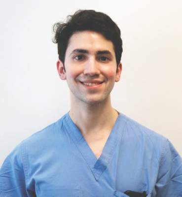
These patients – especially those with compromised immune systems – appear to be at sufficiently high risk of metastasis to justify the procedure, Dr. Jonathan Lopez said at the annual meeting of the American College of Mohs Surgery.
“We found that sentinel lymph node biopsy in our clinic had a 91% negative predictive value for local recurrence, nodal recurrence, and disease-specific death. It provides valuable prognostic information for patients at increased risk of nodal metastasis,” said Dr. Lopez, a dermatology resident at the Mayo Clinic, Rochester, Minn.

He and his associates conducted a chart review of 24 patients treated at the Mayo Clinic from 2000 to 2014 for a cutaneous squamous cell carcinoma (SCC) of the head or neck. Of these, 11 patients were immunosuppressed. Five had undergone a kidney transplant and three a lung transplant. One patient had Hodgkin’s lymphoma, one had cutaneous lymphocytic leukemia, and one, metastatic urothelial carcinoma.
Before sentinel node biopsy, eight patients had a wide local excision; 12 were treated with Mohs micrographic surgery only; and four had a Mohs procedure followed by resection for better margins.
The biopsies identified two patients with nodal disease, but failed to identify a third who had it, Dr. Lopez said.
Patient No. 1 had a primary SCC on the nasal tip that was stage 2, according to the American Joint Committee on Cancer (AJCC) staging system, and 2b according to the Brigham and Women’s Hospital (BWH) system. He had undergone a prior double lung transplant and his lymph node dissection showed no nodal metastasis. He declined radiotherapy and died within 2 months of the biopsy, of unclear causes that were not related to his skin cancer.
Patient No. 2 had a primary lesion on the right cheek, and a history of kidney transplant. His cancer was stage 2 by the AJCC system and 2b by the BWH system. His lymph node dissection of the right parotid and neck was negative. At last follow-up of 3.5 years, he was cancer free. However, Dr. Lopez noted, the patient died at 4 years’ follow-up of unknown causes.
The final patient had a primary lesion on the right conchal bowl. It was a stage 2 cancer by the AJCC system and 2a by the BWH system. His sentinel node biopsy was negative. However, the otolaryngologist who performed the biopsy also took seven superficial parotid nodes and one of those was positive. This patient had no recurrence at the last visit, 1.5 years after the biopsy.
The sentinel node biopsies were negative in the 21 other patients. Of these, 14 had no evidence of recurrence at a mean of 3 years’ follow-up after the sentinel lymph node biopsy. Two developed local recurrence and two others, both of whom had a history of multiple squamous cell carcinomas, developed nodal spread and died of metastatic disease. Three have died of causes unrelated to their cancer.
Dr. Lopez had no financial disclosures.
ORLANDO – Sentinel node biopsies may be a useful staging tool for patients with cutaneous squamous cell carcinomas of the head and neck.
These patients – especially those with compromised immune systems – appear to be at sufficiently high risk of metastasis to justify the procedure, Dr. Jonathan Lopez said at the annual meeting of the American College of Mohs Surgery.
“We found that sentinel lymph node biopsy in our clinic had a 91% negative predictive value for local recurrence, nodal recurrence, and disease-specific death. It provides valuable prognostic information for patients at increased risk of nodal metastasis,” said Dr. Lopez, a dermatology resident at the Mayo Clinic, Rochester, Minn.
He and his associates conducted a chart review of 24 patients treated at the Mayo Clinic from 2000 to 2014 for a cutaneous squamous cell carcinoma (SCC) of the head or neck. Of these, 11 patients were immunosuppressed. Five had undergone a kidney transplant and three a lung transplant. One patient had Hodgkin’s lymphoma, one had cutaneous lymphocytic leukemia, and one, metastatic urothelial carcinoma.
Before sentinel node biopsy, eight patients had a wide local excision; 12 were treated with Mohs micrographic surgery only; and four had a Mohs procedure followed by resection for better margins.
The biopsies identified two patients with nodal disease, but failed to identify a third who had it, Dr. Lopez said.
Patient No. 1 had a primary SCC on the nasal tip that was stage 2, according to the American Joint Committee on Cancer (AJCC) staging system, and 2b according to the Brigham and Women’s Hospital (BWH) system. He had undergone a prior double lung transplant and his lymph node dissection showed no nodal metastasis. He declined radiotherapy and died within 2 months of the biopsy, of unclear causes that were not related to his skin cancer.
Patient No. 2 had a primary lesion on the right cheek, and a history of kidney transplant. His cancer was stage 2 by the AJCC system and 2b by the BWH system. His lymph node dissection of the right parotid and neck was negative. At last follow-up of 3.5 years, he was cancer free. However, Dr. Lopez noted, the patient died at 4 years’ follow-up of unknown causes.
The final patient had a primary lesion on the right conchal bowl. It was a stage 2 cancer by the AJCC system and 2a by the BWH system. His sentinel node biopsy was negative. However, the otolaryngologist who performed the biopsy also took seven superficial parotid nodes and one of those was positive. This patient had no recurrence at the last visit, 1.5 years after the biopsy.
The sentinel node biopsies were negative in the 21 other patients. Of these, 14 had no evidence of recurrence at a mean of 3 years’ follow-up after the sentinel lymph node biopsy. Two developed local recurrence and two others, both of whom had a history of multiple squamous cell carcinomas, developed nodal spread and died of metastatic disease. Three have died of causes unrelated to their cancer.
Dr. Lopez had no financial disclosures.
AT THE ACMS ANNUAL MEETING
Key clinical point: Sentinel node biopsies identified nodal spread in some patients with cutaneous SCC of the head and neck
Major finding: The procedure had a 91% negative predictive value for nodal spread and disease-specific death.
Data source: The retrospective chart review comprised of 24 patients, treated at the Mayo Clinic for cutaneous SCC of the head and neck from 2000 to 2014.
Disclosures: Dr. Lopez had no financial disclosures.

Matrilin-2 protein distinguished BCCs from benign tumors in study
ORLANDO – Matrilin-2 – a matrix protein found in peritumoral stroma – reliably distinguished invasive basal cell carcinoma from the often difficult-to-distinguish basaloid follicular hamartoma (BFH), in a study that evaluated the protein as a marker in this setting.
The protein marked 41 of 42 cancers and none of the hamartomas, Dr. Renato Goreshi reported at the annual meeting of the American College of Mohs Surgery. The one cancer it failed to identify was a superficial basal cell tumor – a finding that makes sense, since dermal fibroblasts appear to secrete matrilin-2 as a response to invasive skin tumors, said Dr. Goreshi of the Roger Williams Cancer Center, Providence, R.I.

Mohs surgery typically employs hematoxylin and eosin staining to delineate tumor boundary. But, Dr. Goreshi said, that stain doesn’t always reliably differentiate adnexal tumors from basal cell carcinomas. “Basaloid follicular hamartoma can be particularly difficult to distinguish from basal cell carcinoma,” he said.
BFH typically presents as individual or linearly arranged, small skin-colored to brown papules or plaques, or as multiple lesions in a generalized distribution on the face, scalp, and occasionally, the trunk (Arch Pathol Lab Med. 2010 Aug;134[8]:1215-9). These are often stable for many years. The differential diagnosis includes basal cell carcinoma and trichoepithelioma.
BFH sometimes occurs near a BCC, although there are no data on how often this happens.
Dr. Goreshi cited a 2007 case report of a young woman that illustrates this problem. The patient presented with a basal cell carcinoma on the side of her nose. The adjacent BFH was unrecognized, however. She underwent a multiple-stage Mohs that was unnecessarily extended because tumor margins included sections of the BFH.
“The lesion was interpreted as malignancy by both the Mohs surgeon and the dermatopathologist, but was later determined to have been a hamartoma. This highlights the importance of finding an effective marker,” Dr. Goreshi said.
He and his fellowship director, Dr. Satori Iwamoto, chief of Mohs micrographic surgery at Roger Williams, looked for a reliable way to differentiate these tumors, capitalizing on the invasive nature of BCC. The peritumoral stroma plays a role in tumor growth and invasion. It involves fibroblasts, inflammatory and endothelial cells, and extracellular matrix proteins. Matrilin-2, which is involved in the formation of filamentous networks, was a promising candidate and the initial investigations looked good, said Dr. Goreshi said.
Their confirmatory study comprised 42 BCC and seven BFH sections that were obtained during Mohs surgery. All were stained for matrilin-2 and scored for location and intensity of staining by two reviewers. The investigators also conducted flow cytometry to determine the source of the protein.
The BCC set consisted of 11 morpheaform/infiltrative BCCs, 25 nodular BCCs, and 6 superficial BCCs. With the exception of one superficial lesion, all of these stained positive for matrilin-2 in the peritumoral stroma. None of the BFH sections stained positive for the protein, however. Flow cytometry determined that the protein was coming from dermal fibroblasts in the stroma.
This is actually a key point, Dr. Goreshi noted. “Matrilin-2 is not acting as a conventional tumor marker would, but as a marker of invasion.”
This was again played out in the variation of staining intensity in the tumor subtypes. It was most intense around the infiltrative subtypes. There was also adnexal staining, but it was significantly less than what was seen in the peritumoral stroma. There was virtually no staining in or around the hamartoma.
Staining was not as intense around the superficial BCC subtypes. In fact, it was not significantly different from what was seen in the adnexal structures. Again, however, there was no staining in or around the hamartoma.
“Now we are looking at the staining patterns of other lesions, including melanoma and squamous cell carcinoma, and trying to figure out why the dermal fibroblasts are secreting matrilin-2,” Dr. Goreshi said.
The study was the winner of the 2016 Theodore Tromovitch Award, presented for original research conducted by a fellow-in-training during his or her year of training.
Neither Dr. Goreshi nor Dr. Iwamoto had any relevant financial disclosures.
ORLANDO – Matrilin-2 – a matrix protein found in peritumoral stroma – reliably distinguished invasive basal cell carcinoma from the often difficult-to-distinguish basaloid follicular hamartoma (BFH), in a study that evaluated the protein as a marker in this setting.
The protein marked 41 of 42 cancers and none of the hamartomas, Dr. Renato Goreshi reported at the annual meeting of the American College of Mohs Surgery. The one cancer it failed to identify was a superficial basal cell tumor – a finding that makes sense, since dermal fibroblasts appear to secrete matrilin-2 as a response to invasive skin tumors, said Dr. Goreshi of the Roger Williams Cancer Center, Providence, R.I.
Mohs surgery typically employs hematoxylin and eosin staining to delineate tumor boundary. But, Dr. Goreshi said, that stain doesn’t always reliably differentiate adnexal tumors from basal cell carcinomas. “Basaloid follicular hamartoma can be particularly difficult to distinguish from basal cell carcinoma,” he said.
BFH typically presents as individual or linearly arranged, small skin-colored to brown papules or plaques, or as multiple lesions in a generalized distribution on the face, scalp, and occasionally, the trunk (Arch Pathol Lab Med. 2010 Aug;134[8]:1215-9). These are often stable for many years. The differential diagnosis includes basal cell carcinoma and trichoepithelioma.
BFH sometimes occurs near a BCC, although there are no data on how often this happens.
Dr. Goreshi cited a 2007 case report of a young woman that illustrates this problem. The patient presented with a basal cell carcinoma on the side of her nose. The adjacent BFH was unrecognized, however. She underwent a multiple-stage Mohs that was unnecessarily extended because tumor margins included sections of the BFH.
“The lesion was interpreted as malignancy by both the Mohs surgeon and the dermatopathologist, but was later determined to have been a hamartoma. This highlights the importance of finding an effective marker,” Dr. Goreshi said.
He and his fellowship director, Dr. Satori Iwamoto, chief of Mohs micrographic surgery at Roger Williams, looked for a reliable way to differentiate these tumors, capitalizing on the invasive nature of BCC. The peritumoral stroma plays a role in tumor growth and invasion. It involves fibroblasts, inflammatory and endothelial cells, and extracellular matrix proteins. Matrilin-2, which is involved in the formation of filamentous networks, was a promising candidate and the initial investigations looked good, said Dr. Goreshi said.
Their confirmatory study comprised 42 BCC and seven BFH sections that were obtained during Mohs surgery. All were stained for matrilin-2 and scored for location and intensity of staining by two reviewers. The investigators also conducted flow cytometry to determine the source of the protein.
The BCC set consisted of 11 morpheaform/infiltrative BCCs, 25 nodular BCCs, and 6 superficial BCCs. With the exception of one superficial lesion, all of these stained positive for matrilin-2 in the peritumoral stroma. None of the BFH sections stained positive for the protein, however. Flow cytometry determined that the protein was coming from dermal fibroblasts in the stroma.
This is actually a key point, Dr. Goreshi noted. “Matrilin-2 is not acting as a conventional tumor marker would, but as a marker of invasion.”
This was again played out in the variation of staining intensity in the tumor subtypes. It was most intense around the infiltrative subtypes. There was also adnexal staining, but it was significantly less than what was seen in the peritumoral stroma. There was virtually no staining in or around the hamartoma.
Staining was not as intense around the superficial BCC subtypes. In fact, it was not significantly different from what was seen in the adnexal structures. Again, however, there was no staining in or around the hamartoma.
“Now we are looking at the staining patterns of other lesions, including melanoma and squamous cell carcinoma, and trying to figure out why the dermal fibroblasts are secreting matrilin-2,” Dr. Goreshi said.
The study was the winner of the 2016 Theodore Tromovitch Award, presented for original research conducted by a fellow-in-training during his or her year of training.
Neither Dr. Goreshi nor Dr. Iwamoto had any relevant financial disclosures.
AT THE ACMS ANNUAL MEETING
Key clinical point: Matrilin-2 is the first marker of tumor invasion to be used in skin cancers.
Major finding: The protein bound to 41 of 42 BCCs, and to none of the hamartoma lesions studied, reliably distinguishing the two.
Data source: 42 frozen section BCCs and seven basaloid follicular hamartomas.
Disclosures: Neither Dr. Goreshi nor Dr. Iwamoto had any relevant financial disclosures.
Patients: Intraperitoneal chemotherapy ‘worth it’ for ovarian cancer
WASHINGTON – Intraperitoneal chemotherapy may be unpleasant and interfere with daily life, but the large majority of women who received it for ovarian cancer agreed that it was the right choice for them.
A small survey of women who completed up to six cycles of the treatment found that most women did experience side effects, including fatigue, pain, and gastrointestinal issues. Despite those problems, more than 80% said they felt the regimen was “worth it,” and more than 90% said they would recommend it to another woman.
“Outpatient administration of chemotherapy appears to be feasible with acceptable toxicities and high completion rates,” Dr. Kristin Gotimer said at the annual meeting of the American College of Obstetricians and Gynecologists. “Toxicities were troublesome, and they did definitely affect quality of life; but almost none of our patients regretted being treated – even the ones who experienced recurrent disease.”
Dr. Gotimer of the Winthrop University Hospital, Mineola, N.Y., discussed a retrospective study of 98 women who underwent intraperitoneal chemotherapy for a gynecologic cancer from 2006 to 2014. Mean age of the patients was 58 years. Most women (71%) had ovarian cancer, and most cancer (70%) was stage IIIC or higher. All patients underwent cytoreductive surgery before starting chemotherapy.
Almost all women were prescribed six cycles of the regimen, which consisted of IV paclitaxel on day 1, intraperitoneal cisplatin on day 2, and intraperitoneal paclitaxel on day 8. Overall, 73% of patients completed their prescribed treatment. Among those prescribed six cycles, 71% completed all of them. Of the 26 patients who discontinued, 12 did so because of port complications, and 14 did so because of toxicities.
The most commonly reported side effects were gastrointestinal effects, fatigue, and neuropathy. Grade 3/4 toxicities were most often fatigue, neuropathy, and pain; those occurred in about 6% of patients per cycle. There were four cases of neutropenic fever; two of those resulted in treatment delays.
Seven patients had to change treatment, Dr. Gotimer said. Six switched from intraperitoneal cisplatin to intraperitoneal carboplatin, and one patient switched from IV paclitaxel to IV Abraxane. Toxicities were more likely to appear in later cycles. “The probability of starting each cycle was lower if the patient had experienced a severe toxicity in the prior cycle,” Dr. Gotimer explained.
A subset of patients (48) completed a survey about the treatment at a mean of 31 months after treatment. Of those, 92% had completed all their prescribed cycles. Disease had recurred in 21%. The three-domain survey queried the mental, physical, and social impact of the treatment.
On the physical health domain, half the group reported fatigue associated with the treatment. Others complained of pain (40%), gastrointestinal effects (37%), “chemo brain” cognitive dysfunction (29%), and alopecia (25%). A few patients experienced infections and dermatologic problems (less than 10% each).
In the mental health domain, patients most often noted stress (25%), anxiety (21%), and depression (15%) during treatment. They also said the therapy interfered with their social health, affecting work attendance (27%), housework (29%), and social interactions (19%). It also imposed a substantial time commitment, 17% of patients noted.
Despite those issues, 83% of patients endorsed intraperitoneal chemotherapy as “worth it,” and almost 90% said they would recommend it to a friend or family member considering it. Only about 5% of patients said they regretted using the treatment, although Dr. Gotimer didn’t specify what those regrets were.
Despite its small size and retrospective nature, the survey offers some clinically useful information, Dr. Gotimer noted. “We can identify modifiable side effects and develop specific interventions aimed at increasing tolerability.”
The results could also be used to “improve physician and patient understanding of realistic short- and long-term expectations to improve patient counseling,” she added.
Dr. Gotimer had no financial declarations.
On Twitter @Alz_Gal
AT ACOG 2016
Key clinical point: Despite its challenges, most women who used intraperitoneal chemotherapy for ovarian cancer said it was a good option for them.
Major finding: More than 80% of women said the regimen was “worth it,” and more than 90% said they would recommend it to another woman.
Data source: The retrospective study comprised 98 women, 48 of whom completed a survey about their treatment.
Disclosures: Dr. Gotimer had no financial disclosures.
Tamoxifen cuts bleeding associated with etonogestrel contraceptive implant
WASHINGTON – A 1-week course of tamoxifen significantly reduced unscheduled bleeding in women using an etonogestrel contraceptive implant, compared with placebo.
The selective estrogen reuptake modifier cut bleeding days by half, compared with placebo, and in some women induced at least 1 month of amenorrhea, Dr. Katharine Simmons said at the annual meeting of the American College of Obstetricians and Gynecologists.
There were no real downsides to using the drug, added Dr. Simmons, an ob.gyn. in Atlanta. There were no significant differences in adverse events between the two treatment groups.
The 6-month study randomized 56 women to 10 mg tamoxifen twice daily or placebo for 7 days. Women were instructed to begin treatment on the third day of any period of unscheduled bleeding. They could use the drug once each month, for up to three cycles during the study period. Every day, the women had to complete a short bleeding diary. This was administered by a daily text message, which asked them to rate the strength of any bleeding over the last 24 hours, and whether or not they had taken the study drug on that day.
The women were young (mean age 25 years), and most were white (about 80%). More than 60% were nulliparous. They had been using the implant for a mean of 275 days. Upon randomization, those in the tamoxifen group reported more unscheduled bleeding days than did those in the placebo group (mean 23 vs. 20 per 30 days). Ten women in the tamoxifen group reported bleeding almost every day of the prior month.
Thirty days after taking the study drug, bleeding days were significantly reduced in the tamoxifen group, compared with the placebo group. Four of 28 women taking the drug experienced complete amenorrhea; 9 reported 5 days of bleeding. The tamoxifen group reported a median of 6 bleeding days after treatment, compared with 12 days in the placebo group.
The effect was sustained, Dr. Simmons said, with a median of 30 days before bleeding resumed in the tamoxifen group, compared with 8 days in the placebo group. Women taking the drug reported significantly greater levels of satisfaction than did those taking placebo. They also were less likely to discontinue the treatment (18% vs. 36%).
There were no significant differences in side effects. Headache was the most common, with 12 women in each group reporting it. Mood changes occurred in 7 taking tamoxifen and in 12 women taking placebo. Hot flashes were slightly more common in the tamoxifen group (6 vs. 4). Reports of nausea, weight gain, and fluid retention were similar.
Dr. Simmons conducted the study during her time at Oregon Health & Science University in Portland. She had no financial disclosures.
On Twitter @Alz_Gal
AT ACOG 2016
Key clinical point: Tamoxifen reduced bleeding days in most and induced extended amenorrhea in some women.
Major finding: Those taking the drug reported a median of 6 bleeding days afterward, compared with 12 in those taking placebo.
Data source: A study randomizing 56 women to 10 mg tamoxifen twice daily for 7 days or a 7-day course of placebo.
Disclosures: Dr. Simmons had no financial disclosures.
Delays in Receiving Zika Test Results Reported
WASHINGTON – The Centers for Disease Control and Prevention is recommending blood or urine testing for all pregnant women with possible Zika exposure through travel or because they live in an endemic area, regardless of whether they are symptomatic, Dr. Denise J. Jamieson said at the annual meeting of the American College of Obstetricians and Gynecologists.
The video associated with this article is no longer available on this site. Please view all of our videos on the MDedge YouTube channel
Couples interested in conceiving who live in or have traveled to areas of active transmission should be alert for any signs of infection, no matter how slight; if they notice any, they should be tested for exposure before attempting conception, Dr. Jamieson, a medical officer with the CDC’s division of reproductive medicine, said in a video interview.
Unfortunately, she said at an update on the Zika situation, labs that process Zika blood and urine samples are backed up, and test results are being “unacceptably delayed.”
During a discussion period, some physicians in the audience complained of waiting up to 6 weeks for results. One said a local health department official told him that the CDC had a backlog of “a million tests.”
“We do not have a 1-million test backup,” Dr. Jamieson said. “But in some places there are still unacceptably long delays. This is not a test you can just check off on a form and send to a lab.”
Many of the tests are being funneled to the CDC’s Division of Vector-Borne Diseases in Ft. Collins, Colo.; that facility is experiencing a long turnaround time. Other samples are being handled in private labs with CDC contracts, but Dr. Jamieson said there’s an urgent need to expand the testing to many more commercial labs. There is no concrete plan for how or when this expansion will happen, however, she added.
Dr. Jamieson also discussed the CDC’s recommendation for managing pregnant women who have a positive serology or urine. These women should have serial ultrasounds every 3-4 weeks to track fetal brain development. Amniocentesis can positively confirm fetal exposure. A maternal-fetal medicine specialist should be on board if the fetus tests positive or if any concerning signs appear on imaging. Newborns can also be tested for exposure, as can placental and cord tissue.
On May 16, the World Health Organization released a comprehensive document describing a management algorithm for pregnant women who live in or travel to Zika-endemic areas.
Women who haven’t experienced signs and symptoms of an infection can proceed with routine care, and, if possible, an ultrasound before 18 weeks’ gestation and another at 28-30 weeks.
Women who have had signs of an infection should be tested for exposure. If negative, routine care with an early ultrasound and one at 28-30 weeks is indicated. If the results are positive, or if an early scan is concerning, patients should have a diagnostic ultrasound and amniocentesis, with referral to specialty care.
In the case of confirmed maternal exposure but normal early ultrasound, serial scans are indicated every month until delivery.
As a federal employee, Dr. Jamieson has no financial disclosures.
EXPERT ANALYSIS FROM ACOG 2016

VIDEO: Delays in receiving Zika test results reported
WASHINGTON – The Centers for Disease Control and Prevention is recommending blood or urine testing for all pregnant women with possible Zika exposure through travel or because they live in an endemic area, regardless of whether they are symptomatic, Dr. Denise J. Jamieson said at the annual meeting of the American College of Obstetricians and Gynecologists.
The video associated with this article is no longer available on this site. Please view all of our videos on the MDedge YouTube channel
Couples interested in conceiving who live in or have traveled to areas of active transmission should be alert for any signs of infection, no matter how slight; if they notice any, they should be tested for exposure before attempting conception, Dr. Jamieson, a medical officer with the CDC’s division of reproductive medicine, said in a video interview.
Unfortunately, she said at an update on the Zika situation, labs that process Zika blood and urine samples are backed up, and test results are being “unacceptably delayed.”
During a discussion period, some physicians in the audience complained of waiting up to 6 weeks for results. One said a local health department official told him that the CDC had a backlog of “a million tests.”
“We do not have a 1-million test backup,” Dr. Jamieson said. “But in some places there are still unacceptably long delays. This is not a test you can just check off on a form and send to a lab.”
Many of the tests are being funneled to the CDC’s Division of Vector-Borne Diseases in Ft. Collins, Colo.; that facility is experiencing a long turnaround time. Other samples are being handled in private labs with CDC contracts, but Dr. Jamieson said there’s an urgent need to expand the testing to many more commercial labs. There is no concrete plan for how or when this expansion will happen, however, she added.
Dr. Jamieson also discussed the CDC’s recommendation for managing pregnant women who have a positive serology or urine. These women should have serial ultrasounds every 3-4 weeks to track fetal brain development. Amniocentesis can positively confirm fetal exposure. A maternal-fetal medicine specialist should be on board if the fetus tests positive or if any concerning signs appear on imaging. Newborns can also be tested for exposure, as can placental and cord tissue.
On May 16, the World Health Organization released a comprehensive document describing a management algorithm for pregnant women who live in or travel to Zika-endemic areas.
Women who haven’t experienced signs and symptoms of an infection can proceed with routine care, and, if possible, an ultrasound before 18 weeks’ gestation and another at 28-30 weeks.
Women who have had signs of an infection should be tested for exposure. If negative, routine care with an early ultrasound and one at 28-30 weeks is indicated. If the results are positive, or if an early scan is concerning, patients should have a diagnostic ultrasound and amniocentesis, with referral to specialty care.
In the case of confirmed maternal exposure but normal early ultrasound, serial scans are indicated every month until delivery.
As a federal employee, Dr. Jamieson has no financial disclosures.
EXPERT ANALYSIS FROM ACOG 2016

Improper transfusion technique linked to Michigan maternal hemorrhage deaths
WASHINGTON – Half of the pregnancy-associated hemorrhage deaths in Michigan were deemed preventable, and most of those occurred in women who received no fresh frozen plasma during their transfusion.
Only 3 women in the 46-case series received the generally accepted one-to-one ratio of fresh frozen plasma to packed red cells, Kathryn De La Rosa said at the annual meeting of the American College of Obstetricians and Gynecologists.

The data, gleaned from state records, were not detailed enough to show with absolute certainty that the failure to follow transfusion protocol was lethal, said Ms. De La Rosa, a medical student at the University of Michigan, Ann Arbor.
“I can’t say in good faith that this is why they died,” she said. “I don’t think we have the ability to say, if these women had gotten fresh frozen plasma, they would be here today. What this does show is that this is an area that needs improvement. It’s something we should be recognizing and dealing with.”
The study plumbed records of all pregnancy-associated deaths examined by the State of Michigan Maternal Mortality review committee from 1998 to 2011. Of these, 46 were due to hemorrhage. As part of the adjudication process, the board reviews each case and determines whether or not it was a preventable death; 23 were deemed preventable.
The most common cause of preventable hemorrhage death was postpartum hemorrhage, occurring in seven cases. Other causes included placental abruption, abortion-related, amniotic fluid embolism, antepartum hemorrhage, uterine rupture, ectopic rupture, preeclampsia, and sepsis.
Most of the women who succumbed to a preventable death were younger than 30 years (13); 7 were aged 30-34 years and the remainder were 35 years or older. There was no relationship between preventable death and race or mode of delivery, Ms. De La Rosa noted.
When she examined details of each patient’s transfusion protocol, it was immediately apparent that the protocol of a one-to-one or higher ratio of fresh frozen plasma to packed red cells was not being applied.
“These women got an average of 10 units of cells but not the proper ratio of fresh frozen plasma. In fact, 17 of our 23 preventable deaths didn’t get any fresh frozen plasma at all,” she said. “Only 3 of the 46 cases conformed to that protocol; of these, one death was considered preventable.”
She conducted a more in-depth analysis of the five postpartum hemorrhage cases that occurred in a hospital (two occurred outside a hospital). These women underwent a large variety of interventions, including uterotonics, oxytocin, bedside interventions like laceration repair, fundal massage and manual extraction of uterine contents, and surgery.
But there were concerns about the adequate application of best transfusion practices, Ms. De La Rosa said. None of the women were at a facility that used a massive transfusion protocol, and none were transfused aggressively with the proper plasma/red cell ratio.
Michigan has recently joined a national project to improve maternal health outcomes, including those related to hemorrhage, she said. The Alliance for Innovation on Maternal Health (AIM) is a partnership of organizations dedicated to reducing severe maternal morbidity by 100,000 events and maternal mortality by 1,000 deaths by 2018. Michigan’s goal is to cut maternal mortality in half by 2017.
In addition to obstetric hemorrhage, the group is developing other bundles on hypertension/preeclampsia, venous thromboembolism, and reduction of primary cesarean section.
Ms. De La Rosa reported having no financial disclosures.
On Twitter @Alz_Gal
WASHINGTON – Half of the pregnancy-associated hemorrhage deaths in Michigan were deemed preventable, and most of those occurred in women who received no fresh frozen plasma during their transfusion.
Only 3 women in the 46-case series received the generally accepted one-to-one ratio of fresh frozen plasma to packed red cells, Kathryn De La Rosa said at the annual meeting of the American College of Obstetricians and Gynecologists.
The data, gleaned from state records, were not detailed enough to show with absolute certainty that the failure to follow transfusion protocol was lethal, said Ms. De La Rosa, a medical student at the University of Michigan, Ann Arbor.
“I can’t say in good faith that this is why they died,” she said. “I don’t think we have the ability to say, if these women had gotten fresh frozen plasma, they would be here today. What this does show is that this is an area that needs improvement. It’s something we should be recognizing and dealing with.”
The study plumbed records of all pregnancy-associated deaths examined by the State of Michigan Maternal Mortality review committee from 1998 to 2011. Of these, 46 were due to hemorrhage. As part of the adjudication process, the board reviews each case and determines whether or not it was a preventable death; 23 were deemed preventable.
The most common cause of preventable hemorrhage death was postpartum hemorrhage, occurring in seven cases. Other causes included placental abruption, abortion-related, amniotic fluid embolism, antepartum hemorrhage, uterine rupture, ectopic rupture, preeclampsia, and sepsis.
Most of the women who succumbed to a preventable death were younger than 30 years (13); 7 were aged 30-34 years and the remainder were 35 years or older. There was no relationship between preventable death and race or mode of delivery, Ms. De La Rosa noted.
When she examined details of each patient’s transfusion protocol, it was immediately apparent that the protocol of a one-to-one or higher ratio of fresh frozen plasma to packed red cells was not being applied.
“These women got an average of 10 units of cells but not the proper ratio of fresh frozen plasma. In fact, 17 of our 23 preventable deaths didn’t get any fresh frozen plasma at all,” she said. “Only 3 of the 46 cases conformed to that protocol; of these, one death was considered preventable.”
She conducted a more in-depth analysis of the five postpartum hemorrhage cases that occurred in a hospital (two occurred outside a hospital). These women underwent a large variety of interventions, including uterotonics, oxytocin, bedside interventions like laceration repair, fundal massage and manual extraction of uterine contents, and surgery.
But there were concerns about the adequate application of best transfusion practices, Ms. De La Rosa said. None of the women were at a facility that used a massive transfusion protocol, and none were transfused aggressively with the proper plasma/red cell ratio.
Michigan has recently joined a national project to improve maternal health outcomes, including those related to hemorrhage, she said. The Alliance for Innovation on Maternal Health (AIM) is a partnership of organizations dedicated to reducing severe maternal morbidity by 100,000 events and maternal mortality by 1,000 deaths by 2018. Michigan’s goal is to cut maternal mortality in half by 2017.
In addition to obstetric hemorrhage, the group is developing other bundles on hypertension/preeclampsia, venous thromboembolism, and reduction of primary cesarean section.
Ms. De La Rosa reported having no financial disclosures.
On Twitter @Alz_Gal
AT ACOG 2016
Key clinical point: Transfusing packed red cells without fresh frozen plasma contributed to preventable maternal hemorrhage deaths in Michigan.
Major finding: Of 23 preventable bleeding deaths, 17 (74%) received no fresh frozen plasma, despite receiving an average of 10 units of packed red cells.
Data source: The review comprised 46 maternal hemorrhage deaths that occurred from 1998-2011.
Disclosures: Kathryn De La Rosa reported having no financial disclosures.
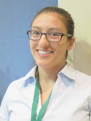
Mohs surgeons, dermatopathologists nearly always agree on frozen section analysis
ORLANDO – Mohs fellowship–trained surgeons are highly skilled at reading frozen section slides obtained during surgery, agreeing with the dermatopathologist’s assessment more than 99% of the time, according to a review of more than 4,000 cases.
Among 4,145 slides in five datasets, only 28 were discordant. The concordance of surgeons’ and dermatopathologists’ assessments was quite consistent, ranging from 95%-of 99.7%.
“There is absolutely no detriment to our interpretation of these frozen section slides,” Dr. James Highsmith said at the annual meeting of the American College of Mohs Surgery. “These numbers give credence as to why we have these good cure rates. We can say with 99% accuracy that yes, we are removing these tumors from our patients.”
Dr. Highsmith, a Mohs surgeon in Birmingham, Ala., reviewed 10 years of his own data (1,720 cases), as well as four published cohorts comprising another 2,425 cases. All of the studies examined the concordance of frozen section assessments between fellowship-trained Mohs surgeons and the dermatopathologists working with them. All of the series reported concordance, discordance, sensitivity, and specificity.
His own database is the largest one yet compiled, spanning the longest period of time. Of 1,720 cases, there were eight discrepancies: one in which the surgeon identified tumor that was not supported by dermatopathology, and seven in which the surgeon missed tumor that dermatopathology confirmed was present. The concordance rate was 99.5%. This worked out to a 99.4% sensitivity and 99.8% specificity, Dr. Highsmith said.
Results were similar with the other four studies.
The largest was a 2009 retrospective review of 1,156 slides collected over 10 years; they were part of a preexisting randomized, blinded study. Of these, 32 slides (2.8%) were discordant between the Mohs surgeon and the dermatopathologist, for a 99.7% concordance rate (J Am Acad Dermatol. 2009 Jan;[1]: 94-8).
The second largest cohort was published in 1989. It comprised 1,000 slides that were evaluated by the Mohs surgeon and a general pathologist; disputed slides were read by two other Mohs surgeons, two pathologists, and a dermatopathologist. The overall concordance rate was 98.9% (J Am Acad Dermatol. 1989;20[4]:670-4).
In 2013, a Mohs surgeon published a set of 170 slides that had been randomly selected from his cases as part of a quality assurance audit. There was one discordant interpretation, for an overall concordance rate of 99.4% (Dermatol Surg. 2013;39[11]:1648-52).
The smallest study, published in 2011, reviewed 99 slides assessed by a single Mohs surgeon. The concordance rate with dermatopathology was 95%.
An overall analysis of these four cohorts determined a sensitivity of 99.4%, specificity of 99.2% and negative predictive value of 99.2%, Dr. Highsmith said.
“When a fellowship-trained Mohs surgeon looks at a slide and says there’s no more tumor present, then there’s a greater-than-99% chance that is accurate,” he said.
Dr. Highsmith had no financial disclosures.
AT THE ACMS ANNUAL MEETING
Key clinical point: Fellowship-trained Mohs surgeons and dermatopathologists almost always agree on slide interpretation.
Major finding: The concordance rate between surgeons and dermatopathologists was over 99% in a pooled review of studies.
Data source: A review of 4,145 frozen section slides analyzed for concordance rates between Mohs surgeons and dermatopathologists.
Disclosures: Dr. James Highsmith had no financial disclosures.