User login
Ectopic pregnancies predicted by easy-to-use risk stratification model
SAN ANTONIO – An easy-to-use risk stratification tool accurately predicted which pregnancies of unknown location were ectopic pregnancies by using a model validated by retrospective chart review.
Reeva Makhijani, MD, and her colleagues built the tool using a composite of risk factors to create a “generalized additive model,” or GAM, in combination with beta HCG levels. They presented the results during a poster session at the annual meeting of the American Society for Reproductive Medicine.
The model showed that a prior history of ectopic pregnancy (EP) (P = .0045), a history of pelvic surgery (P = .397), and a presentation of vaginal bleeding (P = .0003) all significantly increased the risk of EP.
Another statistical measure, the area under the receiver operating curve (AUC), helps estimate the likelihood of EP according to beta-HCG levels. When the initial beta-HCG was considered together with the ratio of the initial beta HCG to the presenting beta-HCG, the AUC was 0.889. For the initial beta-HCG level alone, the AUC was 0.793, while for the ratio alone, the AUC was 0.88. Higher AUC figures indicate more predictive power.
Dr. Makhijani, an ob.gyn. resident physician at Brown University, Providence, R.I., and her colleagues have built a prototype of a computer application that calculates risk of EP when the significant risk factors and lab values are entered.
After reviewing the electronic medical records of 800 patients who had pregnancies of unknown location (PUL), in the final analysis Dr. Makhijani and her coauthors included 398 patients whose medical histories allowed assessment of risk factors and whose record included at least two beta-HCG values taken 36-72 hours apart. The investigators also excluded patients with molar pregnancies, ruptured EPs, or who had undergone surgery before a second beta-HCG was obtained.
Of the 398 patients, 40 (10%) were eventually found to have EP, while 168 (42%) had an intrauterine pregnancy, and 190 (48%) were diagnosed with spontaneous abortion.
The patients were about 27 years old on average, and just over half (n = 224) were parous. Vaginal bleeding was a presenting sign in 233 patients, and 284 had abdominal pain. Of those with EP, 34 of 40 had vaginal bleeding, and 25 of 40 had abdominal pain.
In addition to the three factors found to have significant association with EP, the investigators initially considered a number of other patient characteristics, including age, parity, and presentation with abdominal pain. Additional risk factors examined included history of infertility, pelvic inflammatory disease, sexually transmitted disease, intrauterine device placement, and diethylstilbestrol (DES) exposure. None of these were significantly associated with risk of EP.
“Our model can be translated into an easy-to-use risk stratification tool that can accurately predict the risk of EP,” said Dr. Makhijani and her coauthors. “This tool could potentially be used by clinicians and ob.gyn. residencies nationally as [pregnancies of unknown location] are a very common management scenario.”
Dr. Makhijani reported having no disclosures and no outside sources of funding.
[email protected]
On Twitter @karioakes
AT ASRM 2017
Key clinical point:
Major finding: Incorporating initial and serial beta-HCGs yielded an AUC of 0.889 for predicting ectopic pregnancy.
Data source: A retrospective chart review of 398 patients with pregnancy of unknown location.
Disclosures: The presenter reported having no relevant disclosures and no outside sources of funding.
E-visits less likely to generate antibiotic prescriptions for common ailments
MONTREAL – When the same patient was assessed in person and via an electronic visit (e-visit) for several common complaints, a prescription for antibiotics was more likely to be generated from the face-to-face encounter.
In a recent study, if antibiotics were prescribed in one setting, but not the other, the office visit rather than the e-visit was where the antibiotic prescription was written in 73% of cases. Visits for sinus problems and vaginal symptoms made up over 80% of these cases of nonconcordant prescribing.
The study compared the diagnosis and treatment of five common acute conditions in an outpatient and e-visit setting, examining the concordance of both diagnosis and treatment between the two settings for complaints of vaginal irritation or discharge, urinary symptoms, sinus problems, rash, and diarrhea.
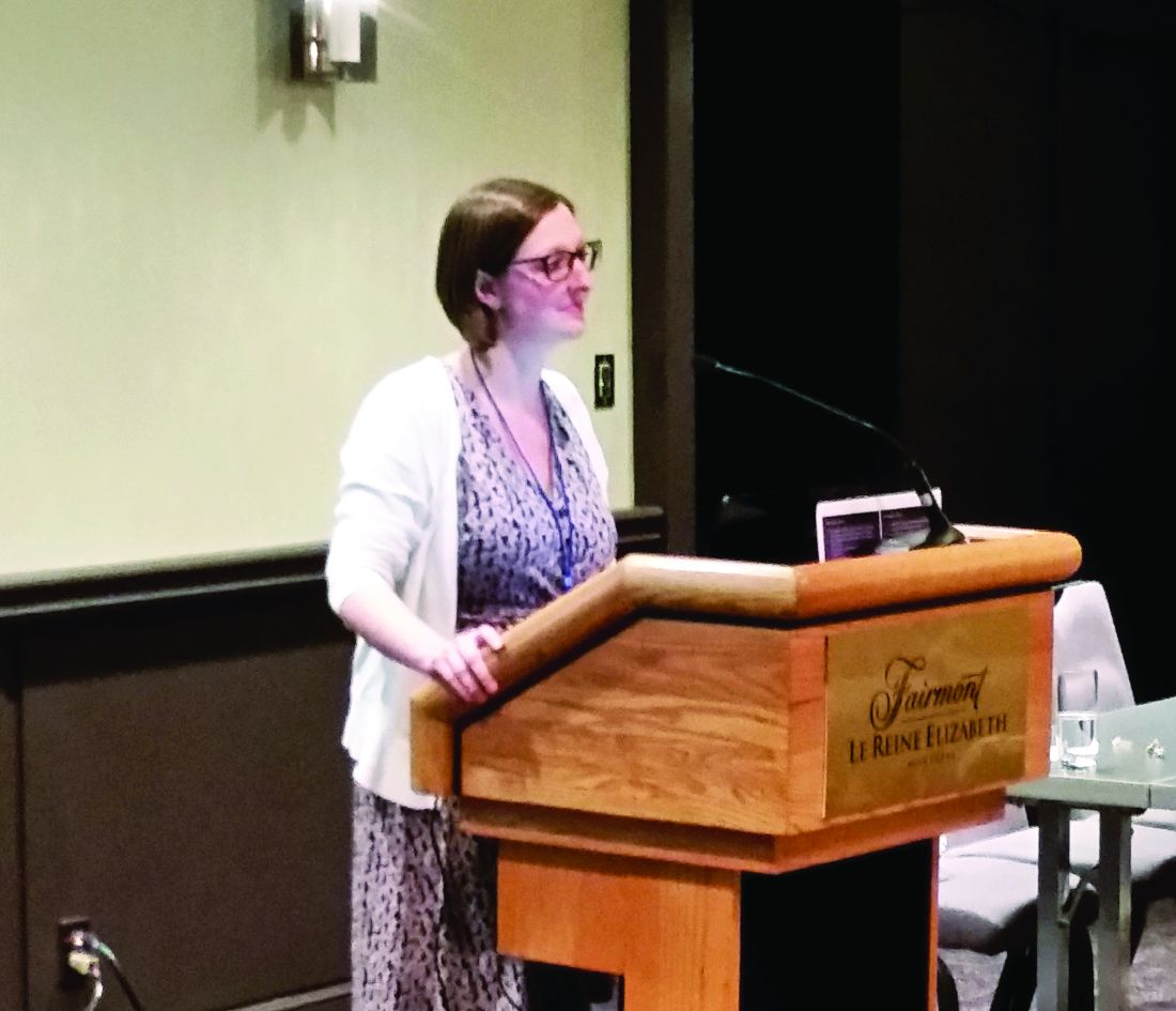
Outcomes tracked included concordance between the office visits and mock e-visits for the diagnosis, whether antibiotics were prescribed, and the general choice of antibiotics. Determinations about concordance were made by a third provider who was not involved with either the in-person visit or the mock e-visit, said Dr. Player, of the department of family medicine at the Medical University of South Carolina, Charleston.
Nonconcordance in treatment could occur either because an antibiotic was prescribed in one setting, but not the other, or because the broad choice of antibiotic class differed between the two settings.
Adult patients who came to the outpatient clinic and agreed to be enrolled in the study also completed the e-visit questionnaires appropriate to their condition before they saw the provider in an office visit. Thus, mock e-visits were created that mirrored the office visit with the e-visit format used in practice.
At a later point in time, the blinded e-visit questionnaires were given to e-visit providers who treated the patients as they would if the questionnaires had been generated in an actual e-visit.
The study generated a total of 142 office visits with accompanying mock e-visits, but 29 were excluded for lack of completeness or inappropriateness for e-visit care. In all, 113 paired visits were evaluated. All but seven patients (94%) were female; slightly more than half (53%) of patients were aged 45 years or older.
About one-third of visits (34%; n = 38) were for vaginal discharge or irritation. Sinus problems were reported by 36 patients (32%). Twenty-five patients (22%) reported urinary problems, while eight patients (7%) reported diarrhea. Six patients (5%) complained of a rash.
In total, 78 visit pairs (69%) were assessed as being concordant. Of the 35 nonconcordant visits, over half (54%) were for sinus problems, 40% were for vaginal discharge or irritation, and 6% were for rash. None of the visits involving urinary problems or diarrhea were assessed as nonconcordant.
Examining the data another way, Dr. Player and his coinvestigators also looked at how many visits involved antibiotic prescribing, and how many of those visits were assessed as nonconcordant. Of the 96 patients (85%) who were prescribed antibiotics, 37 had office and mock e-visits that were assessed as discordant in antibiotic prescribing.
Of these visit pairs, about half (51%) were for sinus problems, and a third (32%) were for vaginal complaints. Urinary complaints made up 11% of the nonconcordant visit pairs where antibiotics were prescribed, and rashes made up the remaining 5%.
Diagnostic concordance was seen in about two-thirds of rash (67%) and vaginal discharge (63%) visit pairs. Concordance of diagnosis for sinus problems occurred in fewer than half (47%) of visit pairs.
Dr. Player said that the investigators excluded visits involving urinary or vaginal complaints that did not have an accompanying urinalysis or vaginal wet mount. This decision was made because the standard of care for both office visits and e-visits requires these laboratory tests for diagnosis, he said.
The study design came with some limitations, said Dr. Player. “Patients self-select for e-visits, and the patients in this study might be different from those in true e-visit encounters,” he said. Also, the diagnosis and treatment of sinus problems, rash, and diarrhea relied on clinical judgment alone in each visit setting. Still, he said, the study supports what many clinicians report anecdotally: Patients want to leave the office knowing that the clinician has “done something” for them, and often, that means walking out with a prescription in hand.
Dr. Player reported no conflicts of interest.
[email protected]
On Twitter @karioakes
AT NAPCRG 2017
Key clinical point:
Major finding: Antibiotics were given in the office but not the e-visit in 73% of cases.
Data source: Prospective study of 113 office visits that were paired with independently assessed e-visits for the same patient and complaint.
Disclosures: Dr. Player reported no conflicts of interest.
December 2017: Click for Credit
Here are 5 articles in the December issue of Clinician Reviews (individual articles are valid for one year from date of publication—expiration dates below):
1. When Is It Really Recurrent Strep Throat?
To take the posttest, go to: http://bit.ly/2lHFh8i
Expires September 21, 2018
2. Revised Bethesda System Resets Thyroid Malignancy Risks
To take the posttest, go to: http://bit.ly/2iSLOvM
Expires August 10, 2018
3. Tips for Avoiding Potentially Dangerous Patients
To take the posttest, go to: http://bit.ly/2lH1Fi7
Expires August 10, 2018
4. Study Findings Support Uncapping MELD Score
To take the posttest, go to: http://bit.ly/2xOA7sI
Expires September 12, 2018
5. 'Motivational Pharmacotherapy' Engages Latino Patients With Depression
To take the posttest, go to: http://bit.ly/2zs2ly4
Expires August 14, 2018
Female family physicians come up short in burnout gender divide
MONTREAL – Female family physicians report more burnout and fewer self-care activities than their male colleagues, according to the results of a survey that found a significant gender divide in physician wellness.
Female respondents to the Texas-wide electronic survey of practicing physicians and residents reported more emotional exhaustion (P = .004) and higher levels of work stress (P = .009) than their male colleagues. Additionally, they reported sleeping less (P = .009), exercising less (P = .001), and eating healthy food less often (P = .005).
In an interview, Dr. Holder said she and her colleagues sent electronic surveys to family physicians, using the Residency Research Network of Texas champion at each of 11 family medicine residency sites. The champion helped with administration to residents, and also engaged community-based physicians in their region to complete the survey.
A total of 324 physicians completed the survey; 54% were female. The female respondents were younger (P = .001) and less likely to be married (P = .019) than the male physicians who responded.
A high level of emotional exhaustion, as assessed by responses on the Maslach Burnout Inventory, was reported by more than a third of physician respondents. Male respondents, however, were more likely to endorse factors protective against burnout, including feeling a higher level of personal accomplishment (P = .004) and having more resilience (P = .002). They also were more likely to participate in activities outside of work (P = .007).
Dr. Holder said she was surprised by the pervasively higher rates of burnout and lower levels of self-care the survey revealed among family physicians in Texas. She would have been less surprised to see the association in female physicians with children, but, she said, unpublished data didn’t bear that association out.
“Creating wellness programs during residency that are aimed at teaching physicians to incorporate self-care into their routines, especially females, may lead to decreased rates of burnout later in physicians’ careers,” said Dr. Holder.
In a discussion during the poster session, Dr. Holder, who is the associate program director for the Baylor Family Medicine residency program in Garland, Tex., said that she herself tries to make room for exercise in a busy schedule and eats so healthily that her residents tease her about it. It’s important to provide positive role modeling for residents, both male and female alike, during their training years, she said.
Dr. Holder reported no relevant financial conflicts of interest.
[email protected]
On Twitter @karioakes
AT NAPCRG 2017
Key clinical point:
Major finding: Female family physicians are more likely to experience emotional exhaustion than their male counterparts (P = .009).
Data source: A survey of 324 family physicians in Texas.
Disclosures: Dr. Holder reported no relevant financial conflicts of interest.
Can increased scope of practice protect against family physician burnout?
MONTREAL – Have restrictions on scope of practice contributed to primary care physician burnout?
Perhaps they have, according to a recent survey that saw less burnout among recently graduated family physicians who retained inpatient or obstetrics work.
Of the 1,167 family practice physicians surveyed, 42% reported that they felt burned out once per week or more, said Amanda Weidner, MPH, of the University of Washington Family Medicine Residency Network in Seattle. However, some physicians were significantly less likely to experience burnout: those whose practice included adult inpatient medicine (P less than .0001), those currently delivering babies (P = .0007), and those who routinely saw their patients at the hospital or in patients’ homes (P = .0016 and P = .02, respectively).
Surveys of family practice physicians have reported varying levels of burnout, ranging from as low as 24% to as high as 65%. “Either way, we can probably agree that even one quarter of physicians saying they’re burned out is too many,” said Ms. Weidner during a presentation at the annual meeting of the National Association of Primary Care Research Groups.
Ms. Weidner and her colleagues polled young family practice physicians about their feelings of burnout and about their practice scope. She said she and her colleagues wondered about the association between those factors because the scope of primary care practice has been narrowing while primary care physicians have been reporting increasing levels of burnout – and at a higher rate than many other specialties.
Their survey, mailed in 2016, was sent to American Board of Family Medicine (ABFM) diplomates who were 2013 graduates of family practice residencies. Overall, 67% responded to the survey. Ms. Weidner and her colleagues used responses from the 78% of respondents who reported providing outpatient continuity care, rather than those who were hospitalists or who practiced in noncontinuity care areas, such as urgent care or emergency medicine.
Participants were asked to indicate how often they felt burned out in their work, with seven choices that ranged from “every day” to “never.” This single question from the Maslach Burnout Inventory, said Ms. Weidner, has been shown to correlate well with the emotional exhaustion domain of the inventory: Any response indicating feeling burned out at least once a week correlates with burnout.
In addition to asking physicians about the scope of their practice and their practice setting, the investigators assessed their work effort by asking about the number of patient encounters per day and whether respondents had after-hours calls and saw patients on weekends and evenings.
Respondents were, on average, about 36 years old, 58.6% were female, and 84.7% held an MD (rather than a DO). Two-thirds were graduates of U.S. medical schools.
Maintaining a broad scope of practice for family physicians can help fulfill the “triple aims” (enhancing patient experience, improving population health, and reducing costs) – now sometimes characterized as the “quadruple aims”with the addition of preventing burnout – of health system performance, said Ms. Weidner. In addition to proven cost reduction, she said, maintaining a broad scope of practice in family practice can enhance the patient experience and improve population health. If increased scope of practice also is associated with improved physician satisfaction, then it will also contribute to the fourth aim, she said.
Questioners in the audience asked Ms. Weidner and her collaborators to dive a little deeper into the data. One audience member remarked that she sees colleagues scheduling themselves for inpatient coverage or an afternoon of endometrial biopsies and other procedures as a strategy to avoid clinic time. She asked whether it’s the practice variety or the reduced number of clinic hours per week that’s driving the lower burnout rates.
Ms. Weidner acknowledged the possibility that working in other practice settings might reduce workload in terms of paperwork and patient communication. However, she pointed out that the number of hours worked per week was not associated with burnout; additionally, she said, both physicians who reported burnout and those who did not saw about the same number of patients per day.
Another audience member wondered whether some of the burnout – and practice restriction – is related to the fact that fewer physicians own their own practices. Maybe, he said, “There’s less of an obligation to be there to take people through all phases of life.”
Some practice restriction may be self-imposed, as may be the case when physicians don’t want the call obligations associated with obstetrics, but some of it is imposed by the system as an attempt to increase clinic throughput, Ms. Weidner said. “This is something for health systems to pay attention to. It’s less of an individual issue and more of a health system issue.”
Ms. Weidner reported that her work was supported by an ABFM Foundation grant while she was a visiting ABFM scholar.
[email protected]
On Twitter @karioakes
AT NAPCRG 2017
Key clinical point:
Major finding: Doing inpatient work and delivering babies was associated with less burnout for recent graduates (P less than .0001 and P = .007).
Data source: A 2016 survey of American Board of Family Medicine (ABFM) diplomates who were 2013 graduates of residency programs; 1,167 physicians’ practices met inclusion criteria.
Disclosures: Ms. Weidner reported that her work was supported by an ABFM Foundation grant while she was a visiting ABFM scholar.
Interprofessional communication demystified with hands-on EHR training
MONTREAL – A hands-on training program designed to improve communication between physicians and allied health professionals, such as physical therapists, produced gains in comfort and confidence to medical students who completed the training.
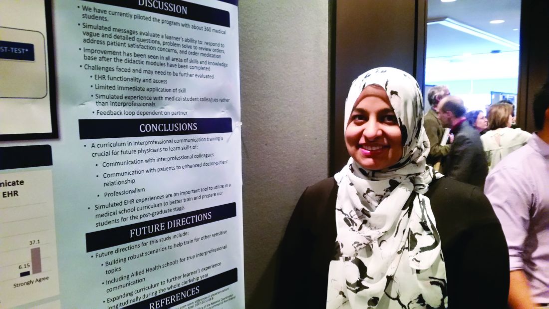
After completing the training, which involved both a didactic and a hands-on component, 92.58% of third-year medical students agreed or strongly agreed that they had the skills needed for effective interprofessional communication in the EHR, up from 43.86% before the training.
The idea for the training began when Zaiba Jetpuri, DO, and her physician colleagues at the University of Texas Southwestern Medical Center, Dallas, noticed that medical students and residents often were unsure how best to communicate with nurses, pharmacists, physical therapists, and other members of the healthcare team via the EHR.
Knowing who has what role in an interdisciplinary setting is far from obvious, and knowing how to strike the right tone in EHR communications was a challenge for trainees, Dr. Jetpuri said in an interview during her poster presentation at annual meeting of the North American Primary Care Research Groups.
To address such uncertainties, which can impact both quality and continuity of care, Dr. Jetpuri and her colleagues devised an intervention aimed at third-year medical students who were doing family medicine clerkships. The intervention has been given to about 360 students thus far.
The intervention consisted of two components. The first, a didactic module, delivered information about the importance of professionalism and teamwork and also defined interprofessional roles.
The second phase of the training had students using the “sandbox” of Epic, UT Southwestern’s EHR, to practice responding to hypothetical messages from a patient, a nurse, a pharmacist, and a physical therapist. One message would arrive in the medical student’s EHR inbox each week, and the student would have the week to craft a response.
Dr. Jetpuri said that she and her colleagues worked hard to make the scenarios as realistic as possible: The lengthy patient email included several questions about the safety of taking supplements or herbal remedies. They also tried to make sure that the nuts and bolts of interprofessional communication were covered so that, for example, medical students would end a module knowing what should be included when writing orders for physical therapy and how to place the order correctly.
The responses then went through peer evaluation according to a rubric constructed by the investigators. The training and evaluation were designed to make sure that students acquired a better understanding of the mechanics of EHR interprofessional communication and of the soft skills needed for collegial and professional communication.
Dr. Jetpuri said that she and her colleagues would like to extend this realistic educational tool through the clerkship year for better continuity. They also are working on some technical aspects of the EHR to make communication easier for medical students. Furthermore, they are in the process of validating the peer evaluation process by having instructors use the rubric to duplicate the students’ evaluation. She said, though, that both trainees and faculty see the value in early, realistic experience using the electronic record in a multidisciplinary team.
“Simulated EHR experiences are an important tool to utilize in a medical school curriculum to better train and prepare our students for the postgraduate stage,” wrote Dr. Jetpuri and her colleagues.
Dr. Jetpuri reported that she has no conflicts of interest.
[email protected]
On Twitter @karioakes
AT NAPCRG 2017
Key clinical point:
Major finding: After the training, 93% of students felt they had the skills needed for effective interprofessional communication via the EHR.
Data source: Summary data from pilot involving about 360 third-year medical students completing family medicine clerkships.
Disclosures: Dr. Jetpuri reported no outside sources of funding and had no relevant disclosures.
Universal paternal Rh screening is cost effective in IVF
SAN ANTONIO – Implementing universal paternal Rh screening would be a cost-effective safety strategy among patients receiving in vitro fertilization, according to a model that used up-to-date data and accounted for ethnic variations in the prevalence of the Rh (D) antibody.
Using a universal Rh screening strategy for semen donors to Rh (D) negative women undergoing in vitro fertilization would result in a cost savings of $11.01 per patient, or $1,120,000 per 100,000 Rh negative IVF pregnancies, according to Pietro Bortoletto, MD, and his coauthors. Their findings were presented during a poster session at the annual meeting of the American Society for Reproductive Medicine.
If paternal Rh factor status is unknown, vaginal bleeding during pregnancy prompts maternal administration of anti-D immune globulin if the mother is Rh (D) negative to prevent hemolytic disease of the fetus and newborn. However, wrote Dr. Bortoletto and his colleagues, “in the IVF population, where paternity status is presumed to be certain, the Rh (D) status of the male partner can be used to triage Rh (D) negative women to more appropriate administration of anti-D globulin.”
To see whether a universal paternal Rh screening strategy would be cost effective, Dr. Bortoletto, a resident physician in obstetrics and gynecology at Harvard Medical School and Brigham and Women’s Hospital, both in Boston, and his collaborators constructed a decision tree to estimate cost savings. The model compared a universal paternal-screening strategy for Rh (D) negative women undergoing IVF with the current standard of practice, which does not involve routine Rh (D) screening for the sperm donor.
In constructing the model, the investigators drew on published data showing that first trimester bleeding is more common in women who undergo IVF: It occurs in one-third of these women, compared with about 20% of the general pregnant population.
They also established probability estimates of pregnancy loss before 20 weeks, with and without first trimester bleeding (0.34 and 0.18, respectively); third trimester bleeding, with and without first trimester bleeding (0.05 and 0.02); and trauma in pregnancy (0.08). They estimated the overall probability of the pregnancy producing an Rh positive neonate at 0.62.
An additional factor that Dr. Bortoletto and his collaborators took into account was the variable prevalence of Rh factor by ethnicity; in the United States, it’s most common in white men and least common in Asian men, with intermediate prevalence in African American and Hispanic men.
When paternal ethnicity was included in the analysis, savings were greatest with white sperm donors, at $1,889,000 per 100,000 Rh negative IVF pregnancies. The lower prevalence of Rh factor in Asian men meant that the strategy was not cost effective in this population since it would cost a net $2,323,000 per 100,000 Rh negative IVF pregnancies.
“A targeted screening approach, by paternal ethnicity, may be a targeted strategy for cost reduction,” wrote Dr. Bortoletto and his colleagues.
Figures for cost estimates were drawn from data from the Centers for Medicare & Medicaid (CMS) using 2017 dollars. The cost for tests to determine blood type and Rh status ranged from $6 to $11, so the investigators set the cost estimate of $8.20. The cost for an antibody screen was estimated at $5.25 (range, $4-$7).
The cost for a 300 mcg dose of anti-D immune globulin was estimated at $93.93 (range, $79-$109), and administration costs were $27.04 (range, $25-$28). Kleihauer-Betke testing to determine the amount of fetal blood in maternal circulation was pinned at $10.61 (range, $8-$14).
Even when the lowest end of the cost range of anti-D immune globulin was used, a universal screening model would still realize a cost savings of $820,000 per 100,000 Rh negative IVF pregnancies. When Rh screening cost was set at $11 – at the high end of the range – “the strategy still remained favorable at $981,000,” wrote Dr. Bortoletto and his colleagues.
“Universal paternal Rh screening provides a cost saving intervention by preventing nonindicated and costly administration of anti-D immune globulin in the IVF population presenting with bleeding or trauma in pregnancy,” they wrote.
Dr. Bortoletto reported no outside sources of funding and no conflicts of interest.
[email protected]
On Twitter @karioakes
At ASRM 2017
Key clinical point:
Major finding: Universal screening would save $1,120,000 per 100,000 Rh negative IVF pregnancies.
Data source: Decision tree incorporating Rh (D) prevalence, bleeding risk, and cost data.
Disclosures: Dr. Bortoletto reported having no relevant disclosures and no outside funding.
FDA clears first-ever neurostimulation device for opioid withdrawal symptoms
The Food and Drug Administration has cleared a medical device that applies electrical stimulation to cranial nerves in order to reduce opioid withdrawal symptoms.
The clearance was granted amid the opioid crisis, which is killing 175 Americans each day, according to the recent report by The President’s Commission on Combating Drug Addiction and the Opioid Crisis. Currently, opioid addiction is treatable by three approved medications, said FDA Commissioner Scott Gottlieb, MD, in a press statement announcing the agency’s decision to permit marketing of the NSS-2 Bridge device.
“While we continue to pursue better medicines for the treatment of opioid use disorder, we also need to look to devices that can assist in this therapy,” Dr. Gottlieb said.
The NSS-2 Bridge device was cleared as an acupuncture aid in 2014. The new use of the device to reduce the symptoms of opioid withdrawal required a new clearance, which was granted based on a single, one-arm clinical study. In the study, 73 patients who were experiencing physical withdrawal from opioids used the neurostimulation device. They reported an initial mean score of 20.1 on the Clinical Opiate Withdrawal Score (COWS). All patients using the device had a reduction of at least 31% on the COWS within 30 minutes of beginning use of the NSS-2 Bridge. A total of 88% of participating patients transitioned to medication-assisted treatment after 5 days of using the device. Additional medications used to treat specific symptoms, such as nausea and vomiting, were permitted during the trial.
The physician-placed battery-powered device sits behind the ear and uses three percutaneous electrode arrays and one single-point needle to provide neurostimulation. The electrode placement is assisted with a transillumination technique, and also is based on known neuroanatomic landmarks for branches of cranial nerves V, VII, and IX, along with branches of the occipital nerve (Clin Med Diagnostics. 2015;5[4]:70-9).
The single-use device is designed to be used for up to 5 days during acute opioid withdrawal and is contraindicated for patients with hemophilia, patients with cardiac pacemakers, or those diagnosed with psoriasis vulgaris. The NSS-2 Bridge device requires a prescription and was cleared through the de novo premarket review pathway. This, said the FDA in the press statement, is “a regulatory pathway for some low- to moderate-risk devices that are novel and for which there is no legally marketed predicate device to which the device can claim substantial equivalence.”
The NSS-2 Bridge device will be marketed by Innovative Health Solutions.
[email protected]
On Twitter @karioakes
The better mammogram: Experts explore sensitivity of new modalities
PHILADELPHIA – Is it time to think about “the better mammogram” as the new standard of care? Can nuclear medicine provide a cost-effective workaround for imaging of women with dense breasts? According to two leading breast imaging researchers,
“Digital breast tomosynthesis is the new kid on the block for screening,” said Emily F. Conant, MD, professor of radiology and chief of breast imaging at the University of Pennsylvania, Philadelphia. “It’s becoming the new standard of care in mammography,” she said, speaking during a plenary session at the annual meeting of the North American Menopause Society.
Digital breast tomosynthesis (DBT) can involve simultaneous acquisition of a conventional 2D mammogram along with a series of images to create a 3D image. Another protocol, which delivers a lower radiation dose, produces a “synthetic” 2D reconstruction of 3D mammography.*
In addition to making visible tumors that otherwise might be obscured by the overlay of dense breast tissue, DBT can help reduce the recall rate, with the 3D images providing immediate clarification at the initial appointment. Studies show that the recall rate can go down by up to 31%, Dr. Conant said.
DBT has been shown to increase detection of invasive cancers, but it does not pick up more ductal carcinoma in situ, Dr. Conant said. This fact helps address the problem of overdiagnosis of small tumors that might regress. Overall, cancer detection is reported to increase by up to 53% with DBT, Dr. Conant said.
When primarily retrospective American studies are taken together with smaller prospective European studies, “the improvement in outcomes achieved with DBT directly addresses the major concerns regarding screening for breast cancer with mammography,” she said.
However, so far the studies have not offered DBT routinely to all comers. Since 2011, DBT has been offered to every woman screened at the University of Pennsylvania, at no additional cost. This created “a sort of natural experiment – there was no bias as to who got it.” Three consecutive years’ worth of outcomes have now been analyzed, Dr. Conant said.
Patient-level data from the University of Pennsylvania experience show statistically significant reductions in recall rate from diagnostic mammography alone. Also, researchers saw a steady increase in the rate of cancers detected per 1,000 patients, from 4.6 with digital mammography alone, to 6.1 by year three of DBT (JAMA Oncol. 2016 Jun 1;2[6]:737-43). This reflected the institutional learning curve with DBT, Dr. Conant said.
She said that the data also showed “a promising trend down in false negatives,” with an early reduction in cancers that were missed by DBT. Time is needed for mature cancer registry data to bear out these early trends, she added.
Other recent data show that DBT has promise to improve detection rates in a population of great interest – younger women, where there are often too many false positives and not enough cancers found, Dr. Conant said. If the risk-benefit ratio for DBT continues to play out as the data pile up, “I would strongly suggest that we should be doing screening in the 40s,” she said.
An important caveat, noted Dr. Conant, is that whether tomosynthesis is used or not, mammography captures anatomy, not physiology, and very dense breast tissue may still obscure a tumor, even when the tomographic slices are peeled back.
Though “DBT is ‘the better mammogram,’ additional outcome data are needed,” she said, including studies that compare modalities, include subgroup analyses, and better delineate the effect of cancer biology.
Molecular breast imaging
Another imaging modality uses nuclear medicine to capture the physiologic changes that accompany cancer. Molecular breast imaging (MBI), or scintimammography, can help “unveil the reservoir of hidden cancers in dense breasts,” said Deborah J. Rhodes, MD, professor of medicine at the Mayo Clinic, Rochester, Minn.
Dr. Rhodes – along with Michael O’Connor, PhD, Connie Hruska, PhD, Katie Hunt, MD, and Amy Conners, MD, her collaborators at the Mayo Clinic – uses a specialized array of gamma cameras to detect uptake of an injected radionuclide that’s preferentially avid for tumor tissue. This technique can unmask smaller tumors not seen on mammogram because it’s not impeded by having to “see” through dense breast tissue.
The radiation dose for an MBI study is a bit more than for DBT, but less than a coronary calcium score scan. The cost is about one-tenth that of breast magnetic resonance imaging (MRI), and interpretation is relatively straightforward, said Dr. Rhodes, who also presented data at the North American Menopause Society plenary.
“The traditional measure of mammography’s performance inflates its effectiveness,” especially in dense breast tissue, said Dr. Rhodes. “What is the sensitivity of mammography in the dense breast? It depends on what you measure it against.”
When cancers detected by MRI or MBI are added, the sensitivity of mammography drops from the 86.9% reported by the Breast Cancer Surveillance Consortium to 21%-31%, according to several published studies.
In one study, Dr. Rhodes and her Mayo colleagues found that the diagnostic yield per 1,000 patients with dense breasts by mammogram alone was 1.9 cancers. When MBI was added, that figure jumped to 8.8 cancers per 1,000 patients, an incremental gain of 363%.
“Tumor size matters profoundly,” she added. “If a tumor is detected above 2 cm, long-term survival drops below 50%.”
That contrasts with the better-than-80% long-term survival rate seen for those with sub-centimeter tumors, even in node-positive disease. “Only a third of tumors are detected when they are less than 1 cm” with regular screening mammography, Dr. Rhodes said.
However, in 2016 the U.S. Preventive Services Task Force concluded that the current evidence was insufficient to assess whether adjunctive screening for breast cancer using breast ultrasonography, MRI, DBT, or other methods should be used in women with dense breasts. The USPSTF noted that there weren’t studies that addressed the effect of supplemental screening on breast cancer morbidity or mortality.
The problem is that it can take 20 years or more to demonstrate mortality reduction, meaning that “no other imaging modality can compete” with mammography when this yardstick is used, Dr. Rhodes said. “This insistence on a mortality endpoint before we change practice” is impeding progress in screening, she said.
The American College of Obstetricians and Gynecologists “does not recommend adjunctive tests to screening mammography in women with dense breasts who are asymptomatic and have no additional risk factors.” However, the organization “strongly supports additional research to identify more effective screening methods” that will improve outcomes and minimize false positives in women with dense breasts.
Though DBT is becoming more widely available, MBI is still primarily used in research centers. Both Dr. Conant and Dr. Rhodes acknowledged that since these techniques are not required to be covered by insurance, payment – and patient access – may vary. Both physicians said their home institutions have worked hard to keep costs down for their studies.
Dr. Conant is consultant or advisory board member for Hologic. Dr. Rhodes reported having no conflicts of interest.
*Correction, 11/15/2017: An earlier version of this story misstated the synthetic mammography protocol.
[email protected]
On Twitter @karioakes
EXPERT ANALYSIS FROM NAMS 2017
Young female hematologic cancer survivors have increased infertility risk
SAN ANTONIO – Young women who were survivors of hematologic cancer were more likely to have a diagnosis of infertility than cancer-free women, according to a large population-based study.
Using Ontario, Canada, universal health care databases, Maria Velez, MD, and her colleagues compared young female hematologic cancer survivors with age-matched women who were cancer-free, finding that 20.4% of the cancer survivors and 15% of the cancer-free women had an infertility diagnosis (P less than .001).
The matched cohort study used the Ontario Cancer Registry and identified 1,226 women aged 16-34 years who had been recurrence free for at least 5 years after a hematologic malignancy such that it captured cancer diagnoses made between 1992 and 2005. Each of these women was matched with four randomly selected, cancer-free women (n = 4,293) by the investigators, who took each woman’s age, location and socioeconomic status into account.
Then, the Ontario Health Insurance Plan database was queried to see which women in each group had claims billed under a diagnosis of infertility, denoted by ICD-9 code 628. Dr. Velez said that, for the survivor group, the investigators began tallying infertility diagnoses a full year after treatment was completed.
Pooling all types of hematologic cancer and adjusting for socioeconomic status, the overall relative risk for infertility was 1.35 for hematologic cancer survivors (95% confidence interval, 1.19-1.54; P less than .001).*
Dr. Velez and her colleagues also compared relative risk by type of hematologic cancer. The relative risk for infertility was 1.35 for survivors of non-Hodgkin lymphoma (n = 371); 1.30 for Hodgkin lymphoma (n = 731); and 1.71 for leukemia (n = 124). These were all statistically significant elevations in RR.
In the survivor group, the mean age at cancer diagnosis was 25.7 years, and patients were followed for a median 16.2 years. The mean age of infertility diagnosis for cancer survivors – 33 years – was not significantly different from that of the cancer-free group (32.8 years).
Dr. Velez and her colleagues also examined whether parity at the time of diagnosis was a factor. Cancer survivors who were nulliparous had a pooled relative risk of 1.35 for infertility, compared with the cancer-free women (P less than .001)*. A significantly elevated relative risk was seen for each individual cancer, except for leukemia. Dr. Velez said that this was likely a statistical artifact of the relatively small number of women who had this diagnosis.
The relative risk of an infertility diagnosis for women who were parous at the time of diagnosis was 1.21, a nonsignificant difference (95% CI, 0.80-1.83; P = .37). No individual diagnosis in this group carried a significantly elevated relative risk for infertility.
It’s difficult to know why parity might make a difference in risk of an infertility diagnosis, Dr. Velez said. There might be “nonbiologic” reasons, such as a difference in motivation to seek care for infertility or in desire for pregnancy, she said.
Strengths of the study included the large sample size and the population-based cohort design. The study was the first to use the ICD-9 code of infertility in cancer research, Dr. Velez said. Also, the relatively recent study period meant that patients received more modern cancer treatment regimens, making the data more relevant than some older Scandinavian studies that reached back into the 1960s, said Dr. Velez of the department of obstetrics and gynecology, in the division of reproductive endocrinology and infertility at Queen’s University, Kingston, Ont.
The study did not track the treatment regimen patients received, so it does not shed light on which chemotherapy regimens might be less gonadotoxic over time. The results are a call to include “the effect of cancer treatment on ovarian reserve as a secondary outcome” in clinical trials for cancer therapies, Dr. Velez said.
The study was conducted through the Institute for Clinical Evaluative Sciences and funded by the Faculty of Health Sciences at Queen's University. Dr. Velez reported that she has no financial disclosures.
[email protected]
On Twitter @karioakes
*Correction 11/14/17: An earlier version of this article misstated the P values.
AT ASRM 2017
Key clinical point:
Major finding: Young women who survived hematologic cancer had a 20.4% risk of infertility, compared with 15% among cancer-free controls (P less than .001). The overall relative risk for infertility among hematologic cancer survivors was 1.35.
Data source: Prospective, age-matched cohort study of 1,226 cancer survivors and 4,293 cancer-free controls.
Disclosures: Dr. Velez reported that she had no disclosures. The Institute for Clinical Evaluative Services in Toronto funded the study.


