User login
Doug Brunk is a San Diego-based award-winning reporter who began covering health care in 1991. Before joining the company, he wrote for the health sciences division of Columbia University and was an associate editor at Contemporary Long Term Care magazine when it won a Jesse H. Neal Award. His work has been syndicated by the Los Angeles Times and he is the author of two books related to the University of Kentucky Wildcats men's basketball program. Doug has a master’s degree in magazine journalism from the S.I. Newhouse School of Public Communications at Syracuse University. Follow him on Twitter @dougbrunk.
Customized airway stents show promise in feasibility trial
SAN DIEGO – for whom conventional stents were not suitable or failed, results from a small study demonstrated.
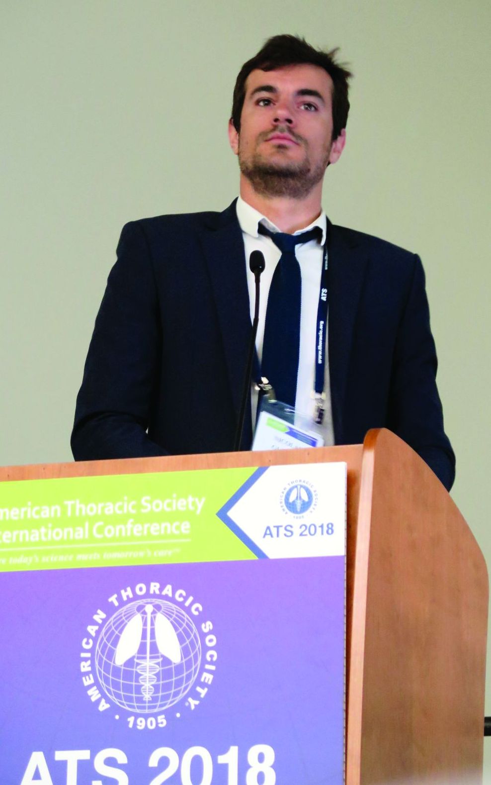
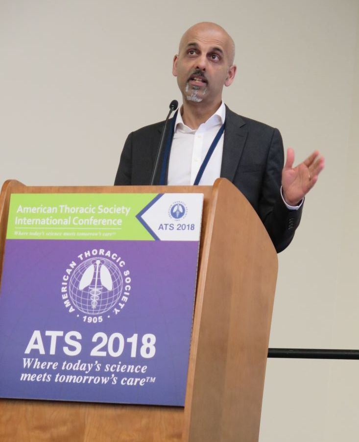
“Anatomically complex airway stenosis remains a challenging situation,” lead study author Nicolas Guibert, MD, said at an international conference of the American Thoracic Society. “Conventional devices are either not suited or may result in a significant complication rate, including poor clinical tolerance, migration, or granulation tissue reaction due to lack of congruence.”
Dr. Guibert reported results from eight patients. Of these, three had posttransplant complex airway stenoses involving the bronchus intermedius. Each improved after placement of the customized stents. For example, one patient with vanishing bronchus intermedius syndrome experienced improvements in NYHA dyspnea score from 3 to 1, the VQ11 score from 22 to 11/55, and forced expiratory volume in 1 second (FEV1) from 70% to 107%. The stent was removed after 3 months. Meanwhile, a patient with localized malacia and stenosis of the right main bronchus experienced improvements in NYHA dyspnea score from 3 to 1, VQ11 score from 27 to 15/55, and FEV1 from 70% to 102%. That person’s stent is still in place with no complications. Another patient with localized malacia and stenosis of the bronchus intermedius experienced improvements in FEV1 from 84% to 100%. That person’s device was removed after 3 months, with no residual stenosis.
A fourth patient underwent stent placement for localized malacia (cartilage ring rupture). That person experienced improvements in NYHA dyspnea score from 3 to 1, VQ11 from 23 to 15/55, and FEV1 from 66% to 92%, and peak flow from 49% to 82%. The device is still in place with no complications. A fifth patient received stent placement for extensive tracheobronchomalacia, but it had imperfect congruence and was removed after 3 months because it caused intense cough.
One patient with post-tracheotomy stenosis experienced improvements in NYHA dyspnea score from 3 to 0, VQ11 from 29 to 12/55, and peak flow from 45% to 81%. That person’s device is still in place, Dr. Guibert said. Two other patients treated for post-tracheotomy experienced stent migration (conventional stents also migrated in these two cases), despite good bronchoscopic congruence after placement.
“Tracheal diseases result in suboptimal congruence, probably due to higher respiratory variation,” Dr. Guibert said. “These devices need to be studied in less selected populations and the technology has to be improved.” He reported having no financial disclosures.
SOURCE: Guibert N et al. ATS 2018, Abstract 4433.
REPORTING FROM ATS 2018
Key clinical point: Customized, 3-D airway stents have the potential for improving tolerance and decreasing the complication rate.
Major finding: Congruence and outcomes tended to be better in stenoses involving the bronchial level (three of three, no complications).
Study details: A feasibility study of eight patients with nonmalignant, anatomically complex, and symptomatic stenosis for which conventional stents were not suitable.
Disclosures: Dr. Guibert reported having no financial disclosures.
Source: Guibert N et al. ATS 2018, Abstract 4433.
App found to improve quality of life for families of premature infants
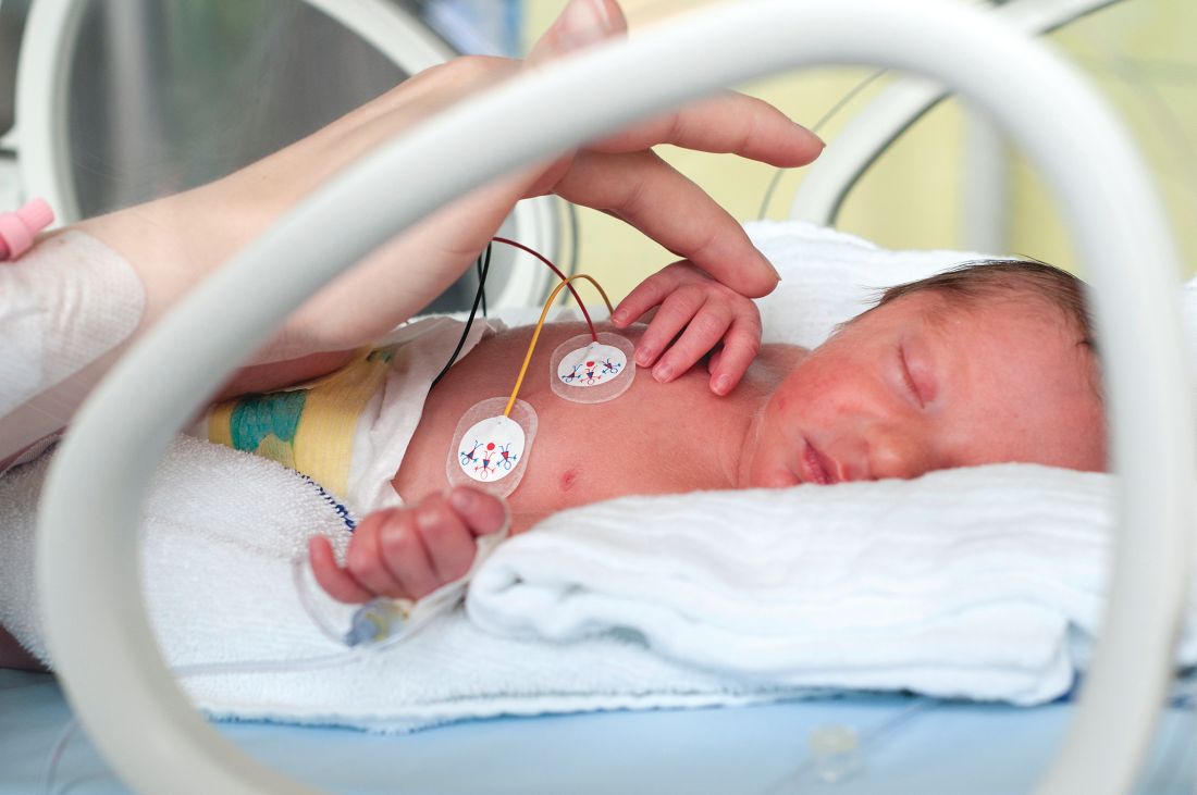
TORONTO – Significant improvement in quality of life was observed in neonatal ICU families using the PreeMe+You app, preliminary results from a two-center study showed.
“NICU time is stressful,” one of the study authors, Abigail Whitney, said at the Pediatric Academic Societies annual meeting. “With the birth of a preterm infant, parents are often quickly transitioned into the role of becoming a parent much sooner and in much different circumstances than they might have anticipated. Parents have reported feelings of isolation, alienation, and insecurity in the parental role while in the NICU. Studies have shown that interventions that engage parents in their infant’s progress can decrease parental stress and anxiety, increase positive parent-infant interaction, and even reduce the infant’s length of stay. Also, with advancing technology there has been a push to find ways to use mobile technology to help parents balance engaging with their infant with the rest of their busy lives.”
In a study overseen by PreeMe+You’s chief medical expert, Bree Andrews, MD, MPH, Ms. Whitney and her associates administered the app to 48 families at either the University of Chicago Medicine Comer Children’s Hospital NICU or the Evanston Hospital NICU to assess readiness for using mobile technologies at the bedside. All families were recommended by a child life specialist who identified families who might be interested in using something like PreeMe+You. They excluded any families that were currently involved with child and family services, those with an infant younger than 7 days old, those whose child required escalation of care or upcoming surgeries, and those whose infant was over 37 weeks’ gestation.
First, the researchers briefed NICU staff about the study at charge nurse meetings, faculty meetings, and daily huddles for 2 weeks before first enrollment. “We did this knowing that parents might go to their nurses or doctors about how to answer specific questions within the app, or maybe want to learn more about a certain topic they learned from PreeMe+You,” Ms. Whitney said.
Data measurements included the PreeMe+You composite survey, which pulled questions from the Fragile Infant Parent Readiness Evaluation (FIPRE) and the NICU Parent Risk Evaluation and Engagement Model and Instrument (PREEMI). “We also included additional questions about technology use and capacity, as well as the PedsQL [Pediatric Quality of Life Inventory] Family Impact Module to assess parental quality of life throughout the study,” she said.
Over a period of 9 months, the researchers collected 153 quality of life measurements from 48 families. Of these, 48 occurred at enrollment, 23 occurred less than 1 week after enrollment, 30 occurred 1-2 weeks after enrollment, 28 occurred 3-4 weeks after enrollment, and 24 occurred 4 weeks or more after enrollment. By study closure, the researchers had follow-up data on 44 of the 48 families. The average gestational age at birth was 29.3 weeks, the average day of life at enrollment was 25.4, and the average birth weight was 1,280 grams.
On the app’s composite survey, 14.6% “agreed” and 79.2% “strongly agreed” that they were currently using a smart phone or tablet to look for information about preemies/NICU on the Internet, and about half “agreed” or “strongly agreed” (27.1% and 33.3%, respectively) that they spent more than 30 minutes per week looking up information about their NICU baby online. Nearly all families “agreed” or “strongly agreed” (14.6% and 85.4%) that they had a smart phone or tablet for Internet use in the NICU, and nearly all “agreed” or “strongly agreed” (33.3% and 62.5%) that having an app at the NICU bedside/home would be helpful. “This showed us that families were ready to use technology and interested in something like PreeMe+You at the bedside,” Ms. Whitney said.
At the time of study enrollment, 12 were in the purple stage, 8 were in the blue stage, 19 infants were in the orange stage, and 9 were in the yellow stage. Ms. Whitney reported that based on the PedsQL Family Impact Module, 35 of the 44 families showed increased quality of life functionality after participating in the study. This change was significant, with a P value of .001. Improvements were seen in the measure’s eight domains (physical, emotional, social, cognitive, communication, worry, daily activities, and family relationship functionality). “We saw increases across all of the domains based on how long the parents had been using the app,” Ms. Whitney said. “We found the biggest increase in quality of life in families of babies born less than 25 weeks’ gestational age, those born 25-26 weeks gestational age, those born 27-28 weeks gestational age, and those born 33-37 weeks gestational age. We are encouraged to see some of these quality of life changes in some of the earliest-born gestation babies because these are presumably the families that would have the longest time to go in the NICU and could benefit the most from using an app like PreeMe+You.”
She acknowledged certain limitations of the study, including the fact that it was conducted in two NICUs, “and we definitely need more comparisons to look at the natural trajectory of quality of life changes while families are in the NICU. Also, all of the families enrolled in our study had access to a research team that checked in with them weekly. In the real world, PreeMe+You would probably be self-guided.” Going forward, PreeMe+You plans to include additional features to give parents more self-guidance, making it easier for them to interact and partner with their baby’s medical team.
Funding for the study was provided by the Bucksbaum Institute for Clinical Excellence. Ms. Whitney was supported by a grant from the National Institute of Diabetes and Digestive and Kidney Diseases.
REPORTING FROM PAS 2018
Key clinical point:
Major finding: In all, 35 of the 44 families showed increased quality of life functionality, based on the PedsQL Family Impact Module (P = .001).
Study details: A two-center study of 44 families with premature infants intended to assess readiness for using mobile technologies at the bedside.
Disclosures: Funding for the study was provided by the Bucksbaum Institute for Clinical Excellence. Ms. Whitney was supported by a grant from the National Institute of Diabetes and Digestive and Kidney Diseases.
Trio of blood biomarkers elevated in children with LRTIs
TORONTO – While C-reactive protein, procalcitonin, and proadrenomedullin are associated with development of severe clinical outcomes in children with lower respiratory tract infections, proadrenomedullin is most strongly associated with disease severity, preliminary results from a prospective cohort study showed.
“Despite the fact that pneumonia guidelines call the site of care decision the most important decision in the management of pediatric pneumonia, no validated risk stratification tools exist for pediatric lower respiratory tract infections (LRTI),” lead study author Todd A. Florin, MD, said at the annual Pediatric Academic Societies meeting. “Biomarkers offer an objective means of classifying disease severity and clinical outcomes.”
PCT is a precursor of calcitonin secreted by the thyroid, lung, and intestine in response to bacterial infections. It also has been shown to be associated with adverse outcomes and mortality in adults, with results generally suggesting that it is a stronger predictor of severity than CRP. “There is limited data on the association of CRP or PCT with severe outcomes in children with LRTIs,” Dr. Florin noted. “One recent U.S. study of 532 children did demonstrate an association of elevated PCT with ICU admission, chest drainage, and hospital length of stay in children with [community-acquired pneumonia] CAP.”
ProADM, meanwhile, is a vasodilatory peptide with antimicrobial and anti-inflammatory functions synthesized during severe infections. It has a half-life of several hours and has been shown to be associated with disease severity in adults with LRTI. Recent studies have shown that it has improved prognostication over WBC, CRP, and PCT. “In two small studies of children with pneumonia, proADM levels were significantly elevated in children with complicated pneumonia, compared to those with uncomplicated pneumonia,” Dr. Florin said. “Although all three of these markers demonstrate promise in predicting severe outcomes in adults with LRTIs, very few studies have examined their association with disease severity in pediatric disease. Therefore, the aim of the current analysis was to determine the association between blood biomarkers and disease severity in children who present to the ED with lower respiratory tract infections.”
In a study known as Catalyzing Ambulatory Research in Pneumonia Etiology and Diagnostic Innovations in Emergency Medicine (CARPE DIEM), he and his associates performed a prospective cohort analysis of children with suspected CAP who were admitted to the Cincinnati Children’s Hospital ED between July 2012 and December 2017. They limited the analysis to children aged 3 months to 18 years with signs and symptoms of an LRTI, and all eligible patients were required to have a chest radiograph ordered for suspicion of CAP. They excluded children hospitalized within 14 days prior to the index ED visit, immunodeficient or immunosuppressed children, those with a history of aspiration or aspiration pneumonia, and those who weighed less than 5 kg because of blood drawing maximums. Biomarkers were measured only in children with focal findings on chest x-ray in the ED. The primary outcome was disease severity: mild (defined as discharged home), moderate (defined as hospitalized, but not severe) and severe (defined as having an ICU length of stay of greater than 48 hours, chest drainage, severe sepsis, noninvasive positive pressure ventilation, intubation, vasoactive infusions, or death). Biomarkers were obtained at the time of presentation to the ED, prior to the occurrence of clinical outcomes.
Over a period of 4.5 years, the researchers enrolled 1,142 patients. Of these, 478 had focal findings on chest x-ray and blood obtained. The median age of these 478 children was 4.4 years, 52% were male, and 82% had all three biomarkers performed. Specifically, 456 had CRP and PCT performed, while 358 had proADM performed. “Not every child had every marker performed due to challenges in obtaining sufficient blood for all three biomarkers in some children,” Dr. Florin explained.
Preliminary data that Dr. Florin presented at PAS found that the median CRP, PCT, and proADM did not differ by gender, race, ethnicity, or insurance status. “In addition, there were not significant differences in the distribution of disease severity by biomarker performed, with approximately 27% of patients being classified as mild, 66% as moderate, and 7% as severe,” he said.
The median CRP was 2.4 ng/mL in those with mild disease, 2.5 ng/mL in those with moderate disease, and 6.25 ng/mL in those with severe disease, with the difference between the two subclasses of nonsevere disease and moderate disease and severe disease reaching statistical significance (P = .002). The median PCT was 0.16 ng/mL in those with mild disease, 0.26 ng/mL in those with moderate disease, and 0.49 ng/mL in those with severe disease, with the difference between the two subclasses of nonsevere disease and moderate disease and severe disease reaching statistical significance (P = .047). Meanwhile, the median proADM was 0.53 ng/mL in those with mild disease, 0.59 ng/mL in those with moderate disease, and 0.81 ng/mL in those with severe disease, with the difference between the two subclasses of nonsevere disease and moderate disease and severe disease also reaching statistical significance (P less than .0001).
Next, the researchers performed logistic regression of each biomarker individually and in combination. They found that and had the best ability to discriminate those developing severe vs. nonsevere disease (area under the receiving operating curve of 0.72, vs. 0.67 and 0.60, respectively). When CRP and PCT markers were combined with proADM, they were no longer associated with severe disease, while a strong association with proADM remained significant.
Dr. Florin acknowledged certain limitations of the study, including the fact that requiring collection of blood samples may have resulted in an enrollment bias toward patients receiving phlebotomy or IV line placement in the ED. “In addition, the children in the moderate-severity group are likely more heterogeneous than the other two severity groups,” he said. “Finally, given that this is a single-center study, we had a relatively small number of outcomes for some of the individual severity measures, which may have limited power and precision.”
He concluded his presentation by saying that he is “cautiously optimistic” about the study results. “As is the case in many biomarker studies, I do not anticipate that any single biomarker will be the magic bullet for predicting disease severity in pediatric CAP,” Dr. Florin said. “It will likely be a combination of clinical factors and several biomarkers that will achieve optimal prognostic ability. That said, our results suggest that similar to adult studies, proADM appears to have the strongest association with severe disease, compared with CRP and PCT. Combinations of biomarkers did not perform better than proADM alone. With the advent of rapid point-of-care diagnostics, these markers may have a role in management and site-of-care decisions for children with LRTI.”
The study received funding support from the Gerber Foundation, the National Institute of Allergy and Infectious Diseases, and Cincinnati Children’s Hospital Medical Center. Dr. Florin reported having no financial disclosures.
AT PAS 18
Key clinical point: Blood biomarkers such as C-reactive protein (CRP), procalcitonin (PCT), and proadrenomedullin (proADM) may have a role in management and site-of-care decisions for children with LRTIs.
Major finding: The proADM alone was associated with the largest odds for severe disease (OR 13.1), compared with CRP alone (OR 1.6) and PCT alone (OR 1.4).
Study details: Preliminary results from prospective cohort analysis of 478 children with suspected community-acquired pneumonia who were admitted to the Cincinnati Children’s Hospital ED.
Disclosures: The study received funding support from the Gerber Foundation, the National Institute of Allergy and Infectious Diseases, and Cincinnati Children’s Hospital Medical Center. Dr. Florin reported having no financial disclosures.
AZD8871 delivered significant bronchodilation in two-week study
SAN DIEGO – , results from a phase 2a trial showed.
AZD8871 is a long-acting, bifunctional bronchodilator that combines a muscarinic antagonist and a beta-2 adrenoceptor agonist. “There are some interesting avenues that you can explore with such a molecule,” one of the study authors, Dave Singh, MD, said at an international conference of the American Thoracic Society. “First, theoretically, as a single molecule you will be able to deposit both the active ingredients to the same site in the lung. On a more practical note, if you want to add something else to a dual bronchodilator, which is essentially what AZD8871 is, this provides a platform. Perhaps that’s the most interesting use of this type of approach.”
Single doses of AZD8871 (400 mcg and 1,800 mcg) administered in COPD patients demonstrated sustained bronchodilation over 36 hours. In a study presented at the 2017 meeting of the European Respiratory Society, Dr. Singh and his associates found that AZD8871 1,800 mcg showed greater bronchodilation than both indacaterol and tiotropium for peak and trough FEV1.
For the current study, researchers at one site in the United Kingdom and one site in Germany conducted a phase 2 randomized, double-blind, placebo-controlled trial of AZD8871 in 42 patients aged 40-80 years with moderate to severe reversible COPD. Patients were randomized to receive repeated once-daily doses of AZD8871 100 mcg, 600 mcg, or placebo via a dry powder inhaler device for 14 days. Between-treatment washout periods were 28-35 days. “We keep the patients in-house on day one and day 14 of each treatment period, and we measure lung function over 24 hours,” said Dr. Singh, professor of clinical pharmacology and respiratory medicine at the University of Manchester, United Kingdom. “Patients were allowed to continue any pre-existing steroid therapy, but at the end of screening they had to withdraw any long-acting bronchodilator therapy.”
The primary efficacy endpoint was change from baseline trough FEV1 on day 15. Secondary endpoints included change from baseline in peak FEV1, total score of breathlessness, cough, sputum scale questionnaire, and rescue medication use.
At baseline, the mean age of the 42 patients was 64 years, and 67% were male. Their mean FEV1 was about 58% predicted, and their FEV1 absolute reversibility was a mean of 379 mL, “which is rather high,” he said.
Of the 42 randomized patients, 31 completed all three treatments. Both doses of AZD8871 had a positive, dose-dependent effect on FEV1, compared with placebo, and both doses demonstrated an onset of action within 15 minutes. On day 15, least square mean change from baseline differences in trough FEV1 for AZD8871 100 mcg and 600 mcg versus placebo were 161 mL and 260 mL, respectively.
A similar association was observed with peak FEV1, which between baseline and day 14 increased by 380 mL at the 100 mcg dose and by 420 mL at the 600 mcg dose, compared with placebo. Sustained bronchodilation was observed over 24 hours on both day 1 and day 14.
Statistically significant COPD symptom improvements, measured by breathlessness, cough and sputum scale (BCSS), were observed for AZD8871 600 mcg on day 8 (P=0.002) and day 14 (P less than 0.001), compared with placebo.
In addition, substantial symptomatic improvements were observed for AZD8871 600 mcg on D14 versus placebo (least square mean of -1.16). Similar results were observed for individual domains of the BCSS. “When you separate out the different components of the scale, most of this is driven by the change in breathlessness,” he said. “We were surprised that we could capture this in such a small number of patients.”
On days 1-8 and days 9-14, the researchers observed a statistically significant improvement in change from baseline rescue medication use for AZD8871 600 mcg (P less than 0.001) and 100 mcg (P=0.029 and P=0.012, respectively), compared with placebo.
The most common adverse events for patients in all three treatment groups were headache (21.4%) and worsening of COPD-related symptoms (14.3%). No dose-dependence was observed with any adverse event, including serious adverse events and/or those leading to discontinuation.
AstraZeneca, the developer of AZD8871, sponsored the study. Dr. Singh reported being a consultant to and receiving research support from AstraZeneca and numerous other pharmaceutical companies.
SOURCE: Singh, D., et al, Abstract 7708, ATS 2018.
REPORTING FROM ATS 2018
Key clinical point: Once daily doses of AZD8871 100 mcg and 600 mcg elicited significant and clinically relevant differences in trough FEV1, compared with placebo.
Major finding: On day 15, least square mean change from baseline differences in trough FEV1 for AZD8871 100 mcg and 600 mcg versus placebo were 161 mL and 260 mL, respectively.
Study details: A phase 2a trial of 42 patients aged 40-80 years with moderate to severe reversible COPD.
Disclosures: AstraZeneca sponsored the study. Dr. Singh reported financial affiliations with AstraZeneca and numerous other pharmaceutical companies.Source: Singh, D., et al. Abstract 7708, ATS 2018.
COPD patient subset gains no benefit from low-dose theophylline
, results from a large trial funded by the United Kingdom found.
“Globally, theophylline was used for decades as a bronchodilator,” one of the study authors, David B. Price, MB BChir, said at an international conference of the American Thoracic Society. “The problem is theophylline has a narrow therapeutic index, it requires some blood monitoring, and it has been replaced by more effective inhaled bronchodilators. However, there has been a lot of discussion about whether low-dose theophylline has anti-inflammatory effects on its own and whether it increases sensitivity to inhaled steroids in COPD.”
According to the 2018 Global Initiative for Chronic Obstructive Lung Disease (GOLD) guidelines, there is “limited and contradictory evidence regarding the effect of low-dose theophylline on exacerbation rates,” and its clinical relevance has “not yet been fully established.” Dr. Price, a professor of primary care respiratory medicine at the University of Aberdeen, United Kingdom, and his associates hypothesized that the addition of low-dose theophylline to inhaled steroid therapy in COPD would reduce the risk of moderate to severe COPD exacerbations after one year of treatment. “If it worked, it would be wonderful; it would save the National Health Service a fortune,” he said.
In a government-funded trial known as Theophylline With Inhaled Corticosteroids (TWICS), people aged 40 years and older with COPD on a drug regimen including inhaled corticosteroids with a history of at least two exacerbations treated with antibiotics and/or oral corticosteroids in the previous year were recruited in 121 U.K. primary and secondary care sites from January 2014 through August 2016. They were randomized to receive low-dose theophylline or placebo for one year. Theophylline dose (200 mg once/twice a day) was determined by ideal body weight and smoking status. Primary outcome was the number of participant-reported exacerbations in the one year treatment period treated with antibiotics and/or oral corticosteroids. Participants were assessed six and 12 months after randomization. The study was powered to detect a 15% reduction in exacerbations and aimed to recruit 1,424 participants.
In all, 1,578 people were randomized: 791 to theophylline and 787 to placebo. Of these, primary outcome data were available for 98% of participants: 772 in the theophylline group and 764 in the placebo group, which amounted to 1,489 person-years of follow-up data. The mean age of patients was 68 years, 54% were male, 32% currently smoked, 80% were using inhaled corticosteroids/long-acting beta 2-agonists/long-acting muscarinic agents, and their mean FEV1 was 51.7% predicted.
Slightly more than one-quarter of study participants (26%) ceased study medication. Dr. Price said that this was balanced between the theophylline and placebo groups and mitigated by over-recruitment and a high rate of follow-up.
He reported that there were 3,430 moderate to severe exacerbations: 1,727 in the theophylline group and 1,703 in the placebo group. The mean number of exacerbations in participants allocated to theophylline and placebo groups were essentially the same: 2.24 vs. 2.23. However, there were a fewer number of exacerbations that required hospitalization in the theophylline group, compared with the placebo groups (0.17 vs. 0.24, for an adjusted rate ratio of 0.72). Dr. Price was quick to point out that this finding applied to a relatively small number of study participants, about 3% overall.
“How you interpret this, I don’t know,” he said. “Our conclusion is that in the broad population there is no benefit [of low-dose theophylline], but maybe someone might want to study its use in frequent exacerbation patients who are getting hospitalized.”
The study was funded by the National Institute for Health Research (NIHR), United Kingdom. Dr. Price reported having no financial disclosures.
SOURCE: Price, D., et al, Abstract 7709, ATS 2018.
AT ATS 2018
Key clinical point: Among COPD patients at high risk of exacerbation, adding low-dose oral theophylline to a drug regimen that includes an inhaled corticosteroid provides no overall clinical benefit.
Major finding: The number of exacerbations was 2.24 in participants allocated to theophylline and 2.23 for participants allocated to placebo.
Study details: A trial of 1,578 people with COPD and a history of at least two exacerbations in the previous year who were randomized to receive low-dose theophylline or placebo for one year.
Disclosures: The study was funded by National Institute for Health Research (NIHR), United Kingdom. Dr. Price reported having no financial disclosures.
Source: Price, D., et al. Abstract 7709, ATS 2018.
Simple tool improves inpatient influenza vaccination rates
TORONTO –
“When we looked at the immunization status of children in New York City, we found that one of the vaccines most commonly missed was influenza vaccine, especially from 2011 through 2014,” one of the study authors, Anmol Goyal, MD, of SUNY Downstate Medical Center, Brooklyn, N.Y., said in an interview at the Pediatric Academic Societies meeting.
“Given this year’s epidemic of influenza and the increasing deaths, we decided to look back on interventions we had done in the past to see if any can be reimplemented to help improve the vaccination status for these children,” he said. “The national goal is 80%, but if we look at the recent trend, even though we have been able to improve vaccination status, it is still below the national goal.” For example, he said, according to New York Department of Health data, the 2012-2013 influenza vaccination rates in New York City were 65% among children 6 months to 5 years old, 47% among those 5-8 years old, and 31% among those 9-18 years old, which were well below the national goal.
In an effort to improve influenza vaccine access, lead author Stephan Kohlhoff, MD, a pediatric infectious disease specialist at the medical center, and his associates, implemented a simple vaccine screening tool to use in the inpatient setting as an opportunity to improve vaccination rates among children in New York City. It consisted of nursing staff assessing the patient’s influenza immunization status on admission and conducting source verification using the citywide immunization registry, or with vaccine cards brought by parents or guardians during admission. Influenza vaccine was administered as a standing order before discharge, unless refused by the parents or guardians. The study population comprised 602 patients between the ages of 6 months and 21 years who were admitted to the inpatient unit during 2 months of the influenza season (November and December) from 2011 to 2013.
Dr. Goyal, a second-year pediatric resident at the medical center, reported that the influenza vaccination status on admission was positive in only 31% of children in 2011, 30% in 2012, and 34% in 2013. The vaccine screening tool was implemented in 64% of admitted children in 2012 and 70% in 2013. Following implementation, the researchers observed a 5% increase in immunization rates in 2012 and an 11% increase in 2013, with an overall increase of 8% over 2 years (P less than .001). He was quick to point out that the influenza rate could have been improved by an additional 22% had 77% of patients not refused vaccination.
“Unfortunately, as our primary objective was to assess the utility of our screening tool in improving inpatient immunization status, we had very limited data points toward refusal of vaccine,” Dr. Goyal said. “Some of the reasons for refusal that were gathered during screening included preferred vaccination by their primary care provider after discharge. Or, maybe they don’t want the vaccine because they feel that the vaccine will make their kids sick. We don’t have enough data to point to any particular reason. This study provides information on acceptance rate of inpatient immunization, which may be useful for implementing additional educational initiatives to overcome potential barriers and help us reach our national goal.”
The researchers reported having no financial disclosures.
AT PAS 18
Key clinical point: The inpatient setting can be used to successfully improve influenza vaccine rates.
Major finding: Following implementation of a simple inpatient vaccine screening tool, a 5% increase in immunization rates occurred in 2012 and an 11% increase occurred in 2013.
Study details: A review of 602 patients between the ages of 6 months and 21 years who were admitted to the inpatient unit during 2 months of the influenza season (November and December) from 2011 to 2013.
Disclosures: The researchers reported having no financial disclosures.
Simple bedside tool effectively detected sepsis in the ED
SAN DIEGO – The product of compared with the quick Sequential Organ Failure Assessment prompt, a small, single-center study showed.
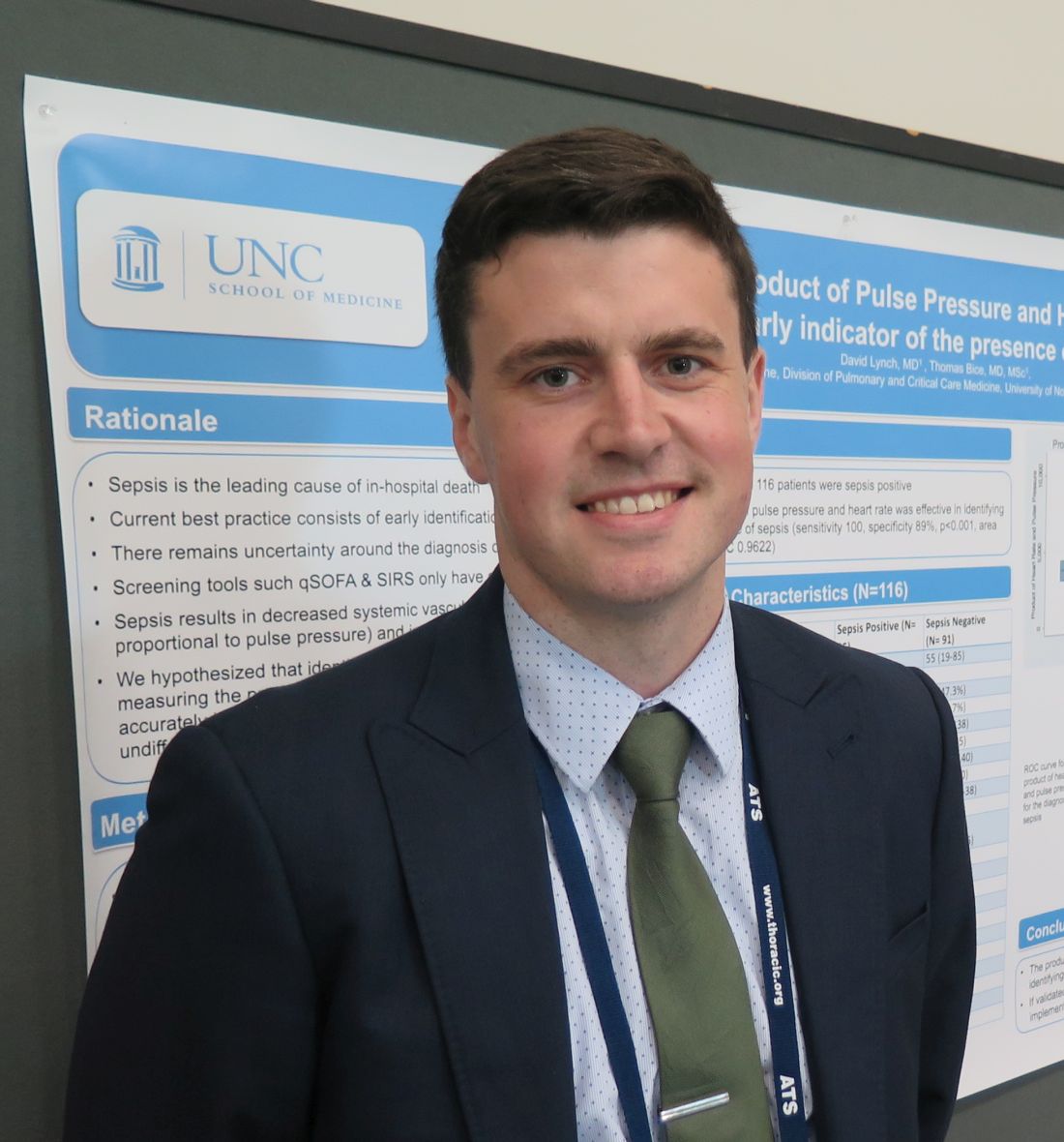
“We know a lot about the pathophysiology of sepsis, but we don’t have great ways of identifying septic patients at an early stage,” lead study author David Lynch, MD, said in an interview at an international conference of the American Thoracic Society.
He noted that screening tools such as the quick Sequential Organ Failure Assessment and Systemic Inflammatory Response Syndrome criteria have a sensitivity of about 70% in detecting sepsis. “Over the last 10-15 years we’ve been able to find ways of improving outcomes in patients whom we confirm are septic with early antibiotics and fluids,” said Dr. Lynch, a second-year resident in the division of pulmonary and critical care medicine within the department of medicine at the University of North Carolina at Chapel Hill. “We know that in sepsis, systemic vascular resistance is decreased and cardiac output is increased. We tried to come up with a way of estimating cardiac output at the bedside by multiplying heart rate with pulse pressure, with the pulse pressure being the surrogate for stroke volume, which you can measure easily.”
In a cross-sectional, observational study, Dr. Lynch, senior author Thomas Bice, MD, and their associates reviewed the records of 116 patients who were admitted directly to the University of North Carolina’s medical ICU (MICU) from the UNC ED between Jan. 5, 2016, and June 30, 2017. The primary outcome of interest was culture-positive sepsis, and the primary exposure was the product of pulse pressure and heart rate. Patients were determined to be positive for sepsis if an infection was suspected (such as if blood cultures were drawn and antibiotics were started), the admitting physician suspected sepsis, and cultures were subsequently positive.
The average age of all patients was 53 years, 51% were female, the mortality rate was 12%, and the median length of stay was 4 days. A total of 25 of the 116 patients (22%) were positive for sepsis. The researchers observed that the pulse pressure multiplied by the heart rate was significantly higher in the culture-positive sepsis group, compared with controls (6,710 vs. 3,741, respectively; P less than .001).
Dr. Lynch and his associates found that, as a continuous variable, pulse pressure multiplied by the heart rate accurately classified 90% of sepsis cases (area under the receiver operator curve, 0.96; P less than .001). When using 5,000 as a cutoff, pulse pressure multiplied by the heart rate had a sensitivity of 100% and a specificity of 89% in detecting culture-positive sepsis. “We were surprised by how high the sensitivity was,” Dr. Lynch said. “The question is, will this translate to a larger cohort? And, would this be transferable to all patients in the ED, as opposed to the sicker patients who are going to the MICU?”
He and his associates plan to confirm the study’s results in a broader population of patients. “We don’t yet understand at what point in time this would be most applicable,” he added. “We looked at the first set of vitals when they came into the ED. We’d like to know if that applies to the second, third and fourth set of vitals, and whether it would be most useful to have an average of those.”
The study was supported in part by a grant from the National Institutes of Health. Dr. Lynch reported having no financial disclosures.
REPORTING FROM ATS 2018
Key clinical point: A simple calculation of pulse pressure multiplied by heart rate at ED triage could lead to earlier implementation of lifesaving interventions in septic patients.
Major finding: When using 5,000 as a cutoff, pulse pressure multiplied by heart rate had a sensitivity of 100% and a specificity of 89% in detecting culture-positive sepsis.
Study details: A cross-sectional, observational study of 116 medical ICU patients who were admitted directly from an ED.
Disclosures: The study was supported in part by a grant from the National Institutes of Health. Dr. Lynch reported having no financial disclosures.
In-hospital mortality predictors eyed in pneumonia patient subset
SAN DIEGO –, results from a large retrospective cohort study found.
In a poster abstract presented at an international conference of the American Thoracic Society, researchers led by Thomas P. Lodise Jr., PharmD, noted that ventilator-associated pneumonia is one of the most common hospital-acquired infections in intensive care units and affected an estimated 9%-27% of all intubated patients. “While data are readily available surrounding mortality associated with VAP, scant data are available on outcomes associated with any type of pneumonia requiring intubation and mechanical ventilation (MV) caused by gram-negative organisms,” they wrote.
In an effort to describe mortality rates and associated risk factors for intubated and MV patients diagnosed with gram-negative pneumonia, Dr. Lodise of the Albany (N.Y.) College of Pharmacy and Health Sciences and his associates conducted a retrospective cohort study of data from the Healthcare Cost and Utilization Project (HCUP) National Readmission Database (NRD). HCUP is the largest source of hospital care data in the United States, accounting for 49.3% of the total U.S. resident population and 49.1% of U.S. hospitalizations. The researchers included patients at least 18 years of age who were hospitalized with a primary or secondary diagnosis of gram-negative pneumonia between Feb. 1, 2013, and Nov. 30, 2013. They excluded index hospitalizations with a primary or secondary diagnosis of viral pneumonia, fungal pneumonia, atypical organisms, gram-positive bacterial pneumonia, or pneumonia occurring secondary to an infectious disease. They examined mortality rates descriptively and modeled them via adjusted multivariate logistic regression to evaluate the impact of baseline characteristics and comorbidities on risk of mortality. All analyses incorporated sample weights to increase generalizability and allow for extrapolation to the entire U.S. population.
A total of 32,683 patients met all study criteria. Of these, 2,323 (7.1%) had a primary diagnosis and 30,360 (92.9%) had a secondary diagnosis for gram-negative pneumonia. Their mean age was 64 years, and 61.1% were male. In all, 7,928 patients (24.3%) died during hospitalization. Multivariate analysis revealed that patients with concomitant sepsis had the highest risk of mortality (odds ratio, 2.60), followed by patients aged 65 years and older (OR, 1.88) and those with any prior hospitalization within 30 days (OR, 1.34). Comorbidities upon admission with highest risk of mortality included cancer (OR, 2.45), liver disease (OR, 1.91), AIDS/HIV (OR, 1.59), renal disease (OR, 1.33), and congestive heart failure (OR, 1.15). Diabetes was found to have a decreased risk of mortality, with an OR of 0.80. “However, a majority of patients with diabetes had no complications; thus, these patients may be representative of a less severe patient population,” Dr. Lodise and his associates noted in the poster.
They acknowledged certain limitations of the study, including the potential for coding errors. They also pointed out the HCUP NRD does not contain treatment-specific information, drugs administered or treatment patterns during hospitalization, the number of days patients spent in the ICU, or the number of days on ventilation, “which can influence outcomes in pneumonia patients.” In addition, the study did not attempt to determine cause of death. “Death may have been due to combinations of factors separate from pneumonia,” they wrote.
Bayer Healthcare Pharmaceuticals funded the study. Dr. Lodise reported having no financial disclosures.
[email protected]
SOURCE: Lodise T. et al. ATS 2018, Poster 272.
AT ATS 2018
Key clinical point: Before this analysis, mortality and associated risk factors for intubated or mechanically ventilated patients diagnosed with gram-negative pneumonia were poorly understood.
Major finding: Among hospitalized, intubated or mechanically ventilated patients with gram-negative pneumonia, 24.3% died during their hospital stay.
Study details: A retrospective cohort study of data from 32,683 patients who were hospitalized with a primary or secondary diagnosis of gram-negative pneumonia.
Disclosures: Bayer Healthcare Pharmaceuticals funded the study. Dr. Lodise reported having no financial disclosures.
Source: Lodise T. et al. ATS 2018, Poster 272.
Four-meter gait speed predicts mortality in IPF
SAN DIEGO – Among patients with idiopathic pulmonary fibrosis (IPF), an improvement in 4-meter gait speed with pulmonary rehabilitation is an independent predictor of all-cause mortality at 1 year, suggest results from a multicenter study presented at an international conference of the American Thoracic Society.
In all, 11% of patients died within 1 year of completing pulmonary rehabilitation.
“Mortality is an attractive endpoint in IPF clinical research but requires large sample sizes and long follow-up duration, making clinical trials expensive and challenging to undertake,” lead study author Claire M. Nolan, MSc, said at the conference.
“Consequently, there is much interest in surrogate endpoints of mortality. In the elderly population, a lot of work has been done on performance measures, in particular the 4-meter gait test. It’s a simple test to do from the assessor’s perspective, because you just need a 4-meter corridor and a stopwatch. From the patient’s perspective, they only have to walk at their usual speed, making it feasible in most settings.”
The study by Ms. Nolan, a National Institute for Health Research fellow, and her associates, involved recruiting 90 IPF patients referred to three outpatient pulmonary rehabilitation programs in London. All patients underwent the following assessments before and after 8 weeks of pulmonary rehabilitation: spirometry; Medical Research Council dyspnea score; anthropometry; 4-meter gait speed; incremental shuttle walk test, and King’s Brief Interstitial Lung Disease questionnaire. Ms. Nolan, a respiratory physiotherapist with the Harefield Pulmonary Rehabilitation and Muscle Research Group, Royal Brompton and Harefield NHS Foundation Trust, Harefield, London, and her associates drew from national databases to obtain data on all-cause mortality 1 year following pulmonary rehabilitation.
“We also identified a cutpoint, so if patients improved their walking speed by 0.009 meters per second or above, that was associated with a longer survival time at 1 year (area under the curve of 0.76, for sensitivity of 69.6% and a specificity of 70%; P less than 0.01),” she said.* “Among patients who achieved that cutpoint or exceeded it, only 5% of them died in the 1-year follow-up period, compared with 23% in the group that didn’t achieve that cutpoint. That’s quite a big difference, but this requires external validation in another population.”
To determine the 4-meter gait speed change cut-off that best discriminated between patients who died and survived, the investigators plotted receiver operating characteristic curves. For validation, they conducted a Kaplan-Meier analysis to assess time to death, with significance assessed via the log-rank test. Finally, they used a multivariate Cox proportional hazards model to characterize the relationship between 4-meter gait speed change and all-cause mortality, adjusting for independent predictors of mortality (age, previous respiratory hospitalizations in the past year, forced vital capacity percent predicted) and baseline 4-meter gait speed.
At baseline, 70% of the 90 patients were male, mean age was 74 years, mean forced vital capacity was 72.8% predicted, and mean Medical Research Council dyspnea score was 3. In addition, mean body mass index was 27.2 kg/m2, mean 4-meter gait speed was 0.92 meters per second, mean incremental shuttle walk test measurement was 271 meters, and mean King’s Brief Interstitial Lung Disease total score was 56.4. Following 8 weeks of pulmonary rehabilitation, the patients’ 4-meter gait speed improved significantly by a mean of 0.15 meters per second (P less than .001). All other variables also improved significantly, with the exception of forced vital capacity.
In an interview, Ms. Nolan characterized the results as “one piece of the puzzle in answering whether 4-meter gait speed is a useful test for clinicians and researchers. It needs to be taken in the context of 4-meter gait speed in other populations as well as with what we’re finding in patients with IPF. We know that this test is reliable, valid, and responsive to treatment. Now we know that it has predictive capacity as well.”
During her presentation, she cited potential reasons why change in gait speed is associated with survival. “Firstly, gait speed has been described as a clinical indicator of multisystem well-being and the ‘sixth vital sign,’ ”she said. “Walking ability and speed rely on multiple factors and the integration of many systems, cardiovascular and otherwise. We know that pulmonary rehab has multiple benefits and improves these systems, and it’s plausible that change in gait speed may be a surrogate marker for, say, improvement in exercise capacity or health status. But the precise mechanism requires verification.”
Ms. Nolan acknowledged certain limitations of the study, including the fact that contemporaneous measurement of full lung function testing and pulmonary hypertension diagnosis were not available at the time of the study. “Therefore, we were unable to account for [diffusing capacity of the lung for carbon monoxide] and pulmonary hypertension diagnosis,” she said. “Secondly, we were unable to identify the precise cause of death from the national database of harm and care records, but this corroborates previous data which suggest that it’s difficult to reliably discern if a death is IPF- or non-IPF related. Lastly, we know that the benefits of pulmonary rehab experienced by IPF patients tend to wane after 6 months. It would be interesting to compare the short-term improvements in gait speed that we observed to more sustained improvements, to identify whether this impacts prognostability.”
National Institute for Health Research funded the study. Ms. Nolan reported having no financial disclosures.
*Correction, 5/23/18: An earlier version of this article misstated the 4-meter gait speed cutoff point.
SOURCE: Nolan CM et al. ATS 2018, Abstract A2456.
AT ATS 2018
Key clinical point: Change in 4-meter gait speed following pulmonary rehabilitation is an independent predictor of all-cause mortality at 1 year in patients with IPF.
Major finding: IPF patients who improved their 4-meter walking speed by 0.009 meters per second or more were more likely to be alive 1 year following pulmonary rehabilitation, compared with those who did not reach that cut point (P less than .01).
Study details: A multicenter study of 90 IPF patients who underwent 8 weeks of pulmonary rehabilitation.
Disclosures: The National Institute for Health Research funded the study. Ms. Nolan reported having no financial disclosures.
Source: Nolan, CM et al. ATS 2018, Abstract A2456.
Most COPD patients on triple therapy can withdraw steroids
SAN DIEGO – In patients on long-term triple therapy and up to one exacerbation in the previous year, the withdrawal of inhaled corticosteroids (ICS) led to a small decrease in lung function that was not clinically important, with no associated difference in the rates of chronic obstructive pulmonary disease (COPD) exacerbations, dyspnea or as-needed bronchodilator use.
Those are key findings from the SUNSET trial, a 26-week, randomized, double-blind, parallel-group multicenter study to assess the efficacy and safety of the switch from long-term triple therapy to indacaterol/glycopyrronium (IND/GLY, 110/50 mcg once daily) or continuation of triple therapy with tiotropium 18 mcg once daily and salmeterol/fluticasone propionate fixed-dose combination 50/500 mcg b.i.d.
Dr. Chapman, director of the asthma and airways clinic at University Health Network, Toronto, noted that relatively few patients with COPD benefit from inhaled steroids. “Given the risk of adverse events (pneumonia, osteoporosis, etc.), we’d rather not give them when they’re not needed,” he said. “Inhaled steroids seem to play only one role in COPD: They tend to reduce exacerbations in the exacerbation-prone COPD patient. That’s about 20% of the COPD population. Despite this, a great many patients end up on triple therapy [long-acting bronchodilators/long-acting muscarinic antagonist (LABA/LAMA) and ICS] needlessly.”
For the study, Dr. Chapman and his associates enrolled 1,053 patients with moderate to severe COPD who’d had no more than one exacerbation in the previous year who had used triple therapy for at least 6 months prior to study inclusion. The primary endpoint of the study was noninferiority on change from baseline in postdose trough forced expiratory volume in 1 second (FEV1) (with a noninferiority margin of –50 mL) after 26 weeks. Exacerbations, health-related quality of life as measured by the St. George’s Respiratory Questionnaire (SGRQ-C), and breathlessness as measures by the Transition Dyspnea Index also were evaluated. Of the 1,053 patients, 527 were randomized to IND/GLY and 526 to triple therapy. Their mean age was 65 years and their mean postbronchodilator FEV1 was 1.6 L.
The researchers found that ICS withdrawal led to a mean reduction in trough FEV1 of –26 mL, which exceeded the noninferiority margin. This difference between treatments on trough FEV1 was driven by the subset of patients with high blood eosinophil counts at baseline (a mean of –68 mL for patients with at least 300 cells/mcL and a mean of –13 mL for patients with fewer than 300 cells/mcL). The two treatments showed similar annualized rates of moderate/severe COPD exacerbations (rate ratio, 1.08) and all (mild/moderate/severe) exacerbations (RR, 1.07). ICS withdrawal led to a small difference in SGRQ-C (1.4 U on week 26), but no differences in Transition Dyspnea Index or use of rescue medication over 26 weeks. Safety and tolerability were balanced across the two treatment groups.
“Although we found no overall increase in exacerbations with ‘de-escalation,’ there were, of course, exacerbations that occurred during the trial,” Dr. Chapman said. “We found that they tended to occur in the minority of patients who had elevated blood eosinophil counts, especially if the counts were elevated persistently (at screening and randomization). The relevant cut-point was blood eosinophil counts above 300/UL. If exacerbations did occur in this easily identifiable subpopulation, they tended to occur early, in the first month after de-escalation. This gives physicians a simply way to identify a population they might exercise caution and a period when careful monitoring is useful.”
He acknowledged certain limitations of the study, including its 6-month duration, which is shorter than most exacerbation studies. “But by recruiting at multiple sites in multiple countries and across seasons, we don’t think this was an importation limitation,” he said. “Of course, like most investigators, I can always think of things I wish I had tracked. My personal hunch is that FeNO [exhaled nitric oxide levels] might offer some useful information but that will be a hunch to explore in another study.”
SUNSET was sponsored by Novartis. Dr. Chapman disclosed that he has received fees for research, consulting and lectures from Novartis, as well as from several other pharmaceutical companies.
[email protected]
SOURCE: Chapman K et al. ATS 2018 Abstract A1009.
REPORTING FROM ATS 2018
Key clinical point: Switching from triple therapy to indacaterol/glycopyrronium (IND/GLY) is effective in COPD patients and avoids long-term exposure to inhaled corticosteroids.
Major finding: ICS withdrawal led to a mean reduction in trough FEV1 of –26 mL, which exceeded the noninferiority margin.
Study details: A randomized trial of COPD patients in which 527 were randomized to IND/GLY and 526 to triple therapy.
Disclosures: SUNSET was sponsored by Novartis. Dr. Chapman disclosed that he has received fees for research, consulting, and lectures from Novartis, as well as from several other pharmaceutical companies.
Source: Chapman K et al. ATS 2018. Abstract A1009.
