User login
Doug Brunk is a San Diego-based award-winning reporter who began covering health care in 1991. Before joining the company, he wrote for the health sciences division of Columbia University and was an associate editor at Contemporary Long Term Care magazine when it won a Jesse H. Neal Award. His work has been syndicated by the Los Angeles Times and he is the author of two books related to the University of Kentucky Wildcats men's basketball program. Doug has a master’s degree in magazine journalism from the S.I. Newhouse School of Public Communications at Syracuse University. Follow him on Twitter @dougbrunk.
Canagliflozin after metabolic surgery may aid weight loss, reduce glucose levels
LOS ANGELES – Patients who took the sodium-glucose cotransporter-2 inhibitor canagliflozin after undergoing metabolic surgery experienced reductions in blood glucose, body mass index, and truncal body fat, results from a small pilot study have shown.
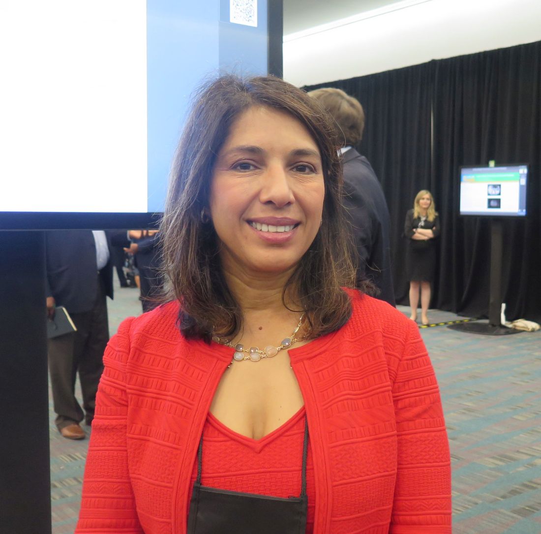
“We hypothesized that canagliflozin would be a good choice for these patients, because these drugs work independently of insulin,” the study’s principal investigator, Sangeeta R. Kashyap, MD, said in an interview at the annual scientific and clinical congress of the American Association of Clinical Endocrinologists. “They help promote weight loss and improve blood pressure. [After] bariatric surgery, patients have an issue with weight regain, and sometimes their diabetes comes back.”
In what she said is the first prospective, randomized, controlled trial of its kind, Dr. Kashyap, an endocrinologist at the Cleveland Clinic, and her colleagues enrolled 11 women and 5 men with type 2 diabetes who had undergone Roux-en-Y gastric bypass or sleeve gastrectomy to study the effects of canagliflozin on clinical parameters over a period of 6 months. At baseline, the patients’ mean body mass index was 39.2 kg/m2 and their mean hemoglobin A1c level was 7.4%. The researchers used maximum likelihood estimation in a linear mixed-effect model to deduce differences between the treatment and placebo groups. Patients randomized to the study drug were assigned a 6-month course of canagliflozin, starting on 100 mg for 2 weeks titrated up to 300 mg daily.
At 6 months, fasting glucose was significantly reduced in the canagliflozin group, compared with baseline (from 163 to 122 mg/dL; P = .007), but it rose in the placebo group (from 164 to 192 mg/dL), a between-group difference that fell short of statistical significance (P = .12). In addition, C-reactive protein in the treatment group fell from 8.9 mg/L to 3.9 mg/L, but rose from 1.6 mg/L to 4.7 mg/L in the placebo group, a between-group difference that trended toward significance (P = .07).
During the 6-month study period, the mean BMI fell from 39.6 kg/m2 to 38 kg/m2 in the canagliflozin group but increased from 38 to 41 in the placebo group, a between-group difference that reached statistical significance (P = .014). Mean changes in body fat (a reduction of 1.82%), truncal fat (a reduction of 2.67%), and android fat (a reduction of 3%) also reached statistical significance in the treatment group, compared with the placebo group. Reductions in adiponectin, leptin, and high–molecular weight adiponectin did not reach statistical significance.
“I think these drugs have a place in post–bariatric surgery care,” Dr. Kashyap said. “Canagliflozin after metabolic surgery improved weight-loss outcomes and blood sugar levels. It also improved abdominal fat levels, and in this way might even lower cardiovascular disease risk in these patients.”
She acknowledged the study’s small sample size and single-center design as limitations. “It was very difficult to recruit patients for this study,” she said. “Not many patients have recurrent diabetes after bariatric surgery.”
Janssen provided funding to Dr. Kashyap for the trial.
LOS ANGELES – Patients who took the sodium-glucose cotransporter-2 inhibitor canagliflozin after undergoing metabolic surgery experienced reductions in blood glucose, body mass index, and truncal body fat, results from a small pilot study have shown.
“We hypothesized that canagliflozin would be a good choice for these patients, because these drugs work independently of insulin,” the study’s principal investigator, Sangeeta R. Kashyap, MD, said in an interview at the annual scientific and clinical congress of the American Association of Clinical Endocrinologists. “They help promote weight loss and improve blood pressure. [After] bariatric surgery, patients have an issue with weight regain, and sometimes their diabetes comes back.”
In what she said is the first prospective, randomized, controlled trial of its kind, Dr. Kashyap, an endocrinologist at the Cleveland Clinic, and her colleagues enrolled 11 women and 5 men with type 2 diabetes who had undergone Roux-en-Y gastric bypass or sleeve gastrectomy to study the effects of canagliflozin on clinical parameters over a period of 6 months. At baseline, the patients’ mean body mass index was 39.2 kg/m2 and their mean hemoglobin A1c level was 7.4%. The researchers used maximum likelihood estimation in a linear mixed-effect model to deduce differences between the treatment and placebo groups. Patients randomized to the study drug were assigned a 6-month course of canagliflozin, starting on 100 mg for 2 weeks titrated up to 300 mg daily.
At 6 months, fasting glucose was significantly reduced in the canagliflozin group, compared with baseline (from 163 to 122 mg/dL; P = .007), but it rose in the placebo group (from 164 to 192 mg/dL), a between-group difference that fell short of statistical significance (P = .12). In addition, C-reactive protein in the treatment group fell from 8.9 mg/L to 3.9 mg/L, but rose from 1.6 mg/L to 4.7 mg/L in the placebo group, a between-group difference that trended toward significance (P = .07).
During the 6-month study period, the mean BMI fell from 39.6 kg/m2 to 38 kg/m2 in the canagliflozin group but increased from 38 to 41 in the placebo group, a between-group difference that reached statistical significance (P = .014). Mean changes in body fat (a reduction of 1.82%), truncal fat (a reduction of 2.67%), and android fat (a reduction of 3%) also reached statistical significance in the treatment group, compared with the placebo group. Reductions in adiponectin, leptin, and high–molecular weight adiponectin did not reach statistical significance.
“I think these drugs have a place in post–bariatric surgery care,” Dr. Kashyap said. “Canagliflozin after metabolic surgery improved weight-loss outcomes and blood sugar levels. It also improved abdominal fat levels, and in this way might even lower cardiovascular disease risk in these patients.”
She acknowledged the study’s small sample size and single-center design as limitations. “It was very difficult to recruit patients for this study,” she said. “Not many patients have recurrent diabetes after bariatric surgery.”
Janssen provided funding to Dr. Kashyap for the trial.
REPORTING FROM AACE 2019
FMT explored as a potential weight loss treatment
“Currently, fecal microbiota transplantation [FMT] can only be performed clinically in the US to treat recurrent Clostridium difficile infection,” lead study author Jessica R. Allegretti, MD, MPH, said during a media briefing in advance of the annual Digestive Disease Week.

“However, there is ongoing research to find out whether FMT works for other health conditions such as obesity, a condition which affects millions of people worldwide. [It’s] a condition that also increases the risk of developing many other illnesses, including diabetes, heart disease, and certain cancers. In my clinical practice, we regularly see patients who have not yet developed some of these other conditions related to obesity, but really do have difficulty losing weight. Through our research we wanted to focus on a population we call the medically uncompromised obese, and understand if FMT might be a viable treatment option for them,” she said.
Dr. Allegretti, director of the fecal microbiota transplant program at Brigham and Women’s Hospital, Boston, conducted a parallel study of 22 patients with a body mass index of 35 kg/m2 or higher who were metabolically healthy – defined as having no type 2 diabetes, nonalcoholic steatohepatitis (NASH), or metabolic syndrome. They randomized study participants 1:1 to receive 30 FMT capsules followed by two doses of 12 capsules over a 12-week period, or identical placebo capsules. A single healthy lean donor with a BMI of 17 kg/m2 was used.
The researchers assessed patients with a mixed meal tolerance test at baseline, week 6, and week 12 post-FMT, at which biomarkers GLP-1 and leptin were measured. Stool was collected at baseline and at one, four, six, eight, and 12 weeks post-FMT. The primary outcomes were safety and change in the area under the curve for GLP-1 at 12 weeks compared to baseline. Secondary endpoints include gut microbiome profiles and diversity as well as bile acid profiles at 12 weeks post FMT. Additional endpoints include a decrease in BMI and waist circumference at week 12. Standard stool microbiome and bile acid analysis was performed.
The mean age of the patients at baseline was 43 years, and their mean BMI was 41 kg/m2. Between baseline and week 12, the research observed no increase of GLP-1 in either group, while the change in leptin revealed an increase in the placebo group only (P less than .001). At week 12, no early changes in BMI were noted in either group (P = .51). No serious adverse events occurred in either arm.
Dr. Allegretti and her colleagues observed global signals of donor community engraftment following FMT, including an increase in alpha diversity and increased similarity to stool samples from the FMT donor – trends that were not observed in the placebo arm. In addition, bile acid analysis suggested a sustained decrease in taurocholic acid in the FMT arm, comparable with the donor – an effect that was not seen in the placebo arm. “We know that what leads to the germination of C. diff. spores is brought out by bile acids,” she said. “That’s one of the critical components of pathogenesis in that disease. My group has been able to show that after FMT, you regain bile acid homeostasis.”
Dr. Allegretti concluded her remarks by noting that the current study “adds an encouraging first step in trying to understand the role that the gut microbiome is playing in the pathogenesis of medically uncompromised obese patients. As a next step, we plan to seek more sensitive measures of GLP-1, as well as conduct additional research into varied doses of FMT capsules, as well as potentially investigating other microbial pathways to better understand the role the microbiome is playing in obesity.”
Somerville, Mass.–based Finch Therapeutics provided funding for the research. Dr. Allegretti reported having no financial disclosures.
SOURCE: Allegretti J et al. DDW 2019. Abstract 621.
“Currently, fecal microbiota transplantation [FMT] can only be performed clinically in the US to treat recurrent Clostridium difficile infection,” lead study author Jessica R. Allegretti, MD, MPH, said during a media briefing in advance of the annual Digestive Disease Week.
“However, there is ongoing research to find out whether FMT works for other health conditions such as obesity, a condition which affects millions of people worldwide. [It’s] a condition that also increases the risk of developing many other illnesses, including diabetes, heart disease, and certain cancers. In my clinical practice, we regularly see patients who have not yet developed some of these other conditions related to obesity, but really do have difficulty losing weight. Through our research we wanted to focus on a population we call the medically uncompromised obese, and understand if FMT might be a viable treatment option for them,” she said.
Dr. Allegretti, director of the fecal microbiota transplant program at Brigham and Women’s Hospital, Boston, conducted a parallel study of 22 patients with a body mass index of 35 kg/m2 or higher who were metabolically healthy – defined as having no type 2 diabetes, nonalcoholic steatohepatitis (NASH), or metabolic syndrome. They randomized study participants 1:1 to receive 30 FMT capsules followed by two doses of 12 capsules over a 12-week period, or identical placebo capsules. A single healthy lean donor with a BMI of 17 kg/m2 was used.
The researchers assessed patients with a mixed meal tolerance test at baseline, week 6, and week 12 post-FMT, at which biomarkers GLP-1 and leptin were measured. Stool was collected at baseline and at one, four, six, eight, and 12 weeks post-FMT. The primary outcomes were safety and change in the area under the curve for GLP-1 at 12 weeks compared to baseline. Secondary endpoints include gut microbiome profiles and diversity as well as bile acid profiles at 12 weeks post FMT. Additional endpoints include a decrease in BMI and waist circumference at week 12. Standard stool microbiome and bile acid analysis was performed.
The mean age of the patients at baseline was 43 years, and their mean BMI was 41 kg/m2. Between baseline and week 12, the research observed no increase of GLP-1 in either group, while the change in leptin revealed an increase in the placebo group only (P less than .001). At week 12, no early changes in BMI were noted in either group (P = .51). No serious adverse events occurred in either arm.
Dr. Allegretti and her colleagues observed global signals of donor community engraftment following FMT, including an increase in alpha diversity and increased similarity to stool samples from the FMT donor – trends that were not observed in the placebo arm. In addition, bile acid analysis suggested a sustained decrease in taurocholic acid in the FMT arm, comparable with the donor – an effect that was not seen in the placebo arm. “We know that what leads to the germination of C. diff. spores is brought out by bile acids,” she said. “That’s one of the critical components of pathogenesis in that disease. My group has been able to show that after FMT, you regain bile acid homeostasis.”
Dr. Allegretti concluded her remarks by noting that the current study “adds an encouraging first step in trying to understand the role that the gut microbiome is playing in the pathogenesis of medically uncompromised obese patients. As a next step, we plan to seek more sensitive measures of GLP-1, as well as conduct additional research into varied doses of FMT capsules, as well as potentially investigating other microbial pathways to better understand the role the microbiome is playing in obesity.”
Somerville, Mass.–based Finch Therapeutics provided funding for the research. Dr. Allegretti reported having no financial disclosures.
SOURCE: Allegretti J et al. DDW 2019. Abstract 621.
REPORTING FROM DDW 2019
Key clinical point: Using capsules filled with fecal matter from a lean donor, researchers successfully changed some of the composition of the gut microbiota of patients with obesity.
Major finding: Following fecal microbiota transplantation, researchers detected a decrease in a specific bile acid and alterations in stool samples that showed increased similarity to those of the lean donor.
Study details: A randomized, placebo-controlled trial of 22 healthy obese patients.
Disclosures: Somerville, Mass.–based Finch Therapeutics provided funding for the research. Dr. Allegretti reported having no financial disclosures.
Source: Allegretti J et al. DDW 2019. Abstract 621.
Age may be a driver of therapeutic testosterone level in transgender men
LOS ANGELES – The dose of testosterone required to maintain a therapeutic level in transgender men is not correlated with body mass index but does decrease with age, results from a single-center study have shown.
“At this point, there are limited data regarding the dosing of testosterone in relation to age and BMI, which is what makes our work unique,” one of the study authors, Sushmitha Echt, MD, said in an interview following the annual scientific and clinical congress of the American Association of Clinical Endocrinologists.

“Although we have guidelines regarding initiating hormonal therapy for transgender patients, such as the Endocrine Society’s Clinical Practice Guidelines from 2017, we do not have much data regarding the optimal dosing of testosterone in transgender men. We hope that with more research, we will have more data to drive our treatment decisions for our transgender patients.”
Dr. Echt, an endocrinology fellow at North Shore University Hospital, Manhassett, NY, and her colleague, Aren Skolnick, DO, retrospectively evaluated 40 transgender men who were treated at the medical center between July 1, 2014 and July 1, 2018. They performed univariate and multivariate mixed-model analyses to determine the relationship between testosterone dose, age, and body mass index (BMI), and Cox regression analysis to determine the relationship between time to development of each physical attribute, testosterone dose, and route of administration.
The patients ranged in age from 18 to 54 years, and their mean baseline age was 25 years. At the time of their first visit, 32 of the patients were on intramuscular testosterone, four were on subcutaneous testosterone, and five were on the transdermal form.
By the end of the study, 28 patients remained on intramuscular testosterone, six were on the subcutaneous form, five were on the transdermal form, and one had transitioned to testosterone pellets. The majority of patients (37) became therapeutic during the course of the study, and the average therapeutic testosterone level was 551.7 ng/dL.
During an average follow-up time of one year, the researchers observed no correlation between testosterone dose and BMI. However, they found a negative correlation between testosterone dose and age, with or without adjustment for BMI (P = .016 and P = .020, respectively). Adjusted for BMI, the dose decreased by 2.0 mg for every one-year increase in age.
A subgroup analysis of the new patients again revealed a negative correlation between testosterone dose and age, with or without adjustment for BMI (P = .013 and P = .019, respectively). Adjusted for BMI, the dose decreased by 2.5 mg for every one-year increase in age.
Subgroup analysis of the patients already on testosterone therapy revealed no association between testosterone dose, age, and BMI. Among the new patients, no association was observed between time to development of each physical attribute and testosterone dose or route of administration. Among the new patients, 73% reported hair growth (mean time, 89 days), deepening of voice (51 days), and cessation of menses (136 days), and 59% reported clitoromegaly (51 days).
“The finding that surprised us the most is that the testosterone dose needed to maintain a therapeutic testosterone level in transgender men is not related to BMI,” Dr. Echt said. “Some medications are dosed based on body weight, and it was surprising to us that testosterone dosing was not related to weight in kilograms or BMI. Although the findings from our project suggest that the testosterone dose needed for hormonal therapy for transgender men may decrease with older age, further research and larger studies are needed in this field to help guide management of transgender patients.”
Dr. Echt acknowledged certain limitations of the study, including its small sample size. “At this point, our population of transgender patients has been increasing in our practice,” she said. “In the future, we would like to build upon our study by including more patients in our retrospective analysis and then stratify the results by mode of administration of testosterone.”
The researchers reported having no financial disclosures.
LOS ANGELES – The dose of testosterone required to maintain a therapeutic level in transgender men is not correlated with body mass index but does decrease with age, results from a single-center study have shown.
“At this point, there are limited data regarding the dosing of testosterone in relation to age and BMI, which is what makes our work unique,” one of the study authors, Sushmitha Echt, MD, said in an interview following the annual scientific and clinical congress of the American Association of Clinical Endocrinologists.
“Although we have guidelines regarding initiating hormonal therapy for transgender patients, such as the Endocrine Society’s Clinical Practice Guidelines from 2017, we do not have much data regarding the optimal dosing of testosterone in transgender men. We hope that with more research, we will have more data to drive our treatment decisions for our transgender patients.”
Dr. Echt, an endocrinology fellow at North Shore University Hospital, Manhassett, NY, and her colleague, Aren Skolnick, DO, retrospectively evaluated 40 transgender men who were treated at the medical center between July 1, 2014 and July 1, 2018. They performed univariate and multivariate mixed-model analyses to determine the relationship between testosterone dose, age, and body mass index (BMI), and Cox regression analysis to determine the relationship between time to development of each physical attribute, testosterone dose, and route of administration.
The patients ranged in age from 18 to 54 years, and their mean baseline age was 25 years. At the time of their first visit, 32 of the patients were on intramuscular testosterone, four were on subcutaneous testosterone, and five were on the transdermal form.
By the end of the study, 28 patients remained on intramuscular testosterone, six were on the subcutaneous form, five were on the transdermal form, and one had transitioned to testosterone pellets. The majority of patients (37) became therapeutic during the course of the study, and the average therapeutic testosterone level was 551.7 ng/dL.
During an average follow-up time of one year, the researchers observed no correlation between testosterone dose and BMI. However, they found a negative correlation between testosterone dose and age, with or without adjustment for BMI (P = .016 and P = .020, respectively). Adjusted for BMI, the dose decreased by 2.0 mg for every one-year increase in age.
A subgroup analysis of the new patients again revealed a negative correlation between testosterone dose and age, with or without adjustment for BMI (P = .013 and P = .019, respectively). Adjusted for BMI, the dose decreased by 2.5 mg for every one-year increase in age.
Subgroup analysis of the patients already on testosterone therapy revealed no association between testosterone dose, age, and BMI. Among the new patients, no association was observed between time to development of each physical attribute and testosterone dose or route of administration. Among the new patients, 73% reported hair growth (mean time, 89 days), deepening of voice (51 days), and cessation of menses (136 days), and 59% reported clitoromegaly (51 days).
“The finding that surprised us the most is that the testosterone dose needed to maintain a therapeutic testosterone level in transgender men is not related to BMI,” Dr. Echt said. “Some medications are dosed based on body weight, and it was surprising to us that testosterone dosing was not related to weight in kilograms or BMI. Although the findings from our project suggest that the testosterone dose needed for hormonal therapy for transgender men may decrease with older age, further research and larger studies are needed in this field to help guide management of transgender patients.”
Dr. Echt acknowledged certain limitations of the study, including its small sample size. “At this point, our population of transgender patients has been increasing in our practice,” she said. “In the future, we would like to build upon our study by including more patients in our retrospective analysis and then stratify the results by mode of administration of testosterone.”
The researchers reported having no financial disclosures.
REPORTING FROM AACE 2019
Key clinical point:
Major finding: Adjusted for BMI, the dose of testosterone decreased by 2.0 mg for every one-year increase in age.
Study details: A retrospective analysis of 40 transgender men.
Disclosures: The researchers reported having no financial conflicts.
Subclinical hypothyroidism may be associated with increased cancer risks
LOS ANGELES – There is no consistent evidence to suggest that subclinical hypothyroidism is linked to an increased risk of incident breast, prostate, or colon cancer. The condition, however, may be linked to an increased risk of thyroid malignancy and to an increased risk of overall cancer mortality.

These findings come from the first systematic review to examine the effect of subclinical hypothyroidism on the risk of incident cancer and cancer mortality.
“Subclinical hypothyroidism is very prevalent,” lead study author Oriana Yu, MD, MSc, said in an interview at the annual scientific and clinical congress of the American Association of Clinical Endocrinologists. “It affects up to 10% of people, yet at this time we are uncertain as to whether or not [we should] treat them. Most studies have focused on cardiovascular outcomes, because we know that thyroid hormone is very important in lipid metabolism.”
Dr. Yu, an endocrinologist at McGill University, Montreal, and her colleagues searched the Ovid MEDLINE database for articles published from the date of its inception until Nov. 13, 2017, and combined words related to thyroid function and cancer in their search. They limited the analysis to randomized clinical trials and cohort and case-control studies in which the thyroid dysfunction chronologically preceded the cancer incidence or mortality by at least one year.
Of the 180 records screened, 51 full-text articles were assessed for eligibility. Of those, nine met the criteria for systematic review – seven related to cancer risk and two to cancer mortality. The studies were deemed to be of good to medium quality, but six of the nine were cohort studies, the rest case-control studies. “We had hoped to do a systematic review and meta-analysis, but there was high heterogeneity in the studies, especially in terms of different risk measures and outcomes reported,” Dr. Yu said. “As a result, we were not able to perform a meta-analysis.”
The researchers found that the studies had inconsistent findings when it came to the impact of subclinical hypothyroidism on the risk of breast, prostate, and colon cancer. For example, one prospective cohort study of 2,738 patients found no association between subclinical hypothyroidism and the risk of breast cancer (odds ratio, 1.9; 95% confidence interval, 0.8-4.9; Thyroid. 2005;15[11]:1253-9). Women with breast cancer, however, were more likely to have thyroid autoantibodies.
Meanwhile, a case-control study of 1,201 men found that those with elevated TSH levels had a decreased risk of prostate cancer (OR, 0.71; 95% CI, 0.47-1.06; PLoS One. 2012 Oct 30. doi: 10.1371/journal.pone.0047730). “That was a bit surprising to us,” Dr. Yu said. In addition, a nested case-control study of 103,044 patients found that subclinical hypothyroidism was linked to an increased risk for colon cancer (OR, 1.16; 95% CI, 1.08-1.24; J Natl Cancer Inst. 2015 Apr 8. doi: 10.1093/jnci/djv084).
Of the two studies that focused on cancer mortality, one retrospective cohort analysis of 4,735 patients showed that treatment of subclinical hypothyroidism was associated with a decreased risk of cancer mortality in those aged 40-70 years (hazard ratio, 0.59; 95% CI, 0.21-0.99; Arch Intern Med. 2012;172[10]:811-7). The other study, a retrospective cohort analysis of 115,746 patients, found that subclinical hypothyroidism was associated with an increased risk of cancer mortality (relative risk, 1.51; 95% CI, 1.06-2.15; PLoS One. 2015 Apr 1. doi: 10.1371/journal.pone.0122955).
“We need to interpret the cancer-related mortality findings with caution,” Dr. Yu said. “There’s concern about whether patients who are treated might be [generally] healthier or were less frail, compared with those who were not treated. Although these studies adjusted for a number of confounders, it may be difficult to measure and adjust for [those two] factors. That might explain the findings in the two studies on cancer-related mortality.”
Dr. Yu and two coauthors have received salary awards from the Fonds de recherche du Québec–Santé.
LOS ANGELES – There is no consistent evidence to suggest that subclinical hypothyroidism is linked to an increased risk of incident breast, prostate, or colon cancer. The condition, however, may be linked to an increased risk of thyroid malignancy and to an increased risk of overall cancer mortality.
These findings come from the first systematic review to examine the effect of subclinical hypothyroidism on the risk of incident cancer and cancer mortality.
“Subclinical hypothyroidism is very prevalent,” lead study author Oriana Yu, MD, MSc, said in an interview at the annual scientific and clinical congress of the American Association of Clinical Endocrinologists. “It affects up to 10% of people, yet at this time we are uncertain as to whether or not [we should] treat them. Most studies have focused on cardiovascular outcomes, because we know that thyroid hormone is very important in lipid metabolism.”
Dr. Yu, an endocrinologist at McGill University, Montreal, and her colleagues searched the Ovid MEDLINE database for articles published from the date of its inception until Nov. 13, 2017, and combined words related to thyroid function and cancer in their search. They limited the analysis to randomized clinical trials and cohort and case-control studies in which the thyroid dysfunction chronologically preceded the cancer incidence or mortality by at least one year.
Of the 180 records screened, 51 full-text articles were assessed for eligibility. Of those, nine met the criteria for systematic review – seven related to cancer risk and two to cancer mortality. The studies were deemed to be of good to medium quality, but six of the nine were cohort studies, the rest case-control studies. “We had hoped to do a systematic review and meta-analysis, but there was high heterogeneity in the studies, especially in terms of different risk measures and outcomes reported,” Dr. Yu said. “As a result, we were not able to perform a meta-analysis.”
The researchers found that the studies had inconsistent findings when it came to the impact of subclinical hypothyroidism on the risk of breast, prostate, and colon cancer. For example, one prospective cohort study of 2,738 patients found no association between subclinical hypothyroidism and the risk of breast cancer (odds ratio, 1.9; 95% confidence interval, 0.8-4.9; Thyroid. 2005;15[11]:1253-9). Women with breast cancer, however, were more likely to have thyroid autoantibodies.
Meanwhile, a case-control study of 1,201 men found that those with elevated TSH levels had a decreased risk of prostate cancer (OR, 0.71; 95% CI, 0.47-1.06; PLoS One. 2012 Oct 30. doi: 10.1371/journal.pone.0047730). “That was a bit surprising to us,” Dr. Yu said. In addition, a nested case-control study of 103,044 patients found that subclinical hypothyroidism was linked to an increased risk for colon cancer (OR, 1.16; 95% CI, 1.08-1.24; J Natl Cancer Inst. 2015 Apr 8. doi: 10.1093/jnci/djv084).
Of the two studies that focused on cancer mortality, one retrospective cohort analysis of 4,735 patients showed that treatment of subclinical hypothyroidism was associated with a decreased risk of cancer mortality in those aged 40-70 years (hazard ratio, 0.59; 95% CI, 0.21-0.99; Arch Intern Med. 2012;172[10]:811-7). The other study, a retrospective cohort analysis of 115,746 patients, found that subclinical hypothyroidism was associated with an increased risk of cancer mortality (relative risk, 1.51; 95% CI, 1.06-2.15; PLoS One. 2015 Apr 1. doi: 10.1371/journal.pone.0122955).
“We need to interpret the cancer-related mortality findings with caution,” Dr. Yu said. “There’s concern about whether patients who are treated might be [generally] healthier or were less frail, compared with those who were not treated. Although these studies adjusted for a number of confounders, it may be difficult to measure and adjust for [those two] factors. That might explain the findings in the two studies on cancer-related mortality.”
Dr. Yu and two coauthors have received salary awards from the Fonds de recherche du Québec–Santé.
REPORTING FROM AACE 2019
Key clinical point: Further studies are needed to assess the association between untreated subclinical hypothyroidism and the risks of different types of cancer.
Major finding:
Study details: A systematic review of nine studies.
Disclosures: Dr. Yu and two coauthors have received salary awards from the Fonds de recherche du Québec–Santé.
Appendectomy linked to increased risk of subsequent Parkinson’s
.

“One of the factors that’s seen in the brains of patients with Parkinson’s disease is accumulation of an abnormal protein known as alpha-synuclein,” one of the study authors, Gregory S. Cooper, MD, said during a media briefing in advance of the annual Digestive Disease Week. “It’s released by damaged nerve cells in the brain. Not only is alpha-synuclein found in the brain of patients with Parkinson’s disease; it’s also found in the GI tract. It’s thought that its accumulation in the GI tract occurs prior to the development of its accumulation in the brain.”
This has prompted scientists around the world to evaluate the GI tract, including the appendix, for evidence about the pathophysiology and onset of Parkinson’s disease, said Dr. Cooper, professor of medicine, oncology, and population and quantitative health sciences at Case Western Reserve University, Cleveland. “It’s thought that, potentially, in the presence of inflammation, [molecules] of this protein are released from damaged nerves in the gut and then are transported to the brain, where they accumulate,” he said. “Or, it could be that the appendix is a storage place for this protein and gets released at the time of appendectomy.”
To investigate if appendectomy increases the risk of Parkinson’s disease, Dr. Cooper and colleagues drew from the Explorys database, which contains EHRs from 26 integrated U.S. health care systems. They limited their search to patients who underwent appendectomies and those who were diagnosed with Parkinson’s disease based on Systematized Nomenclature of Medicine–Clinical Terms. The researchers chose a washout period of 6 months to the development of Parkinson’s disease after appendectomy, and compared the prevalence of Parkinson’s disease in the general population to those with appendectomies.
Of the 62,218,050 records in the database, Dr. Cooper and colleagues identified 488,190 patients who underwent appendectomies. In all, 4,470 cases of Parkinson’s disease were observed in patients with appendectomies, and 177,230 cases of Parkinson’s disease in patients without appendectomies. The overall relative risk of developing Parkinson’s disease in patients after appendectomies was 3.19 (95% confidence interval, 3.10-3.28; P less than .0001), compared with those who did not undergo the procedure. The relative risk was higher in patients aged 18-64 years (RR, 4.27; 95% CI, 3.99-4.57; P less than .0001), compared with those 65 years and older (RR, 2.20; 95% CI, 2.13-2.27; P less than .0001). “We know that Parkinson’s disease is more common in the elderly,” Dr. Cooper said. “But at virtually all ages, the prevalence of Parkinson’s disease was higher in patients who had an appendectomy, compared to those without an appendectomy.”
The overall relative risk of developing Parkinson’s disease in patients after appendectomies was slightly higher in females (RR, 3.86; 95% CI, 3.71-4.02; P less than .0001), compared with males (RR, 2.67; 95% CI, 2.56-2.79; P less than .0001). The researchers also observed a similar effect of appendectomy by race. The overall relative risk of developing Parkinson’s disease in patients after appendectomy was slightly higher in African Americans (RR, 3.11; 95% CI, 2.69-3.58; P less than .0001), compared with Asians (RR, 2.73; 95% CI, 2.19-3.41; P less than .0001), and whites (RR, 2.55; 95% CI, 2.48-2.63; P less than .0001).
“If these data get borne out, it may question the role of doing a discretionary appendectomy in a patient who’s having surgery for another reason,” Dr. Cooper said. “Our research does show a clear relationship between appendectomy and Parkinson’s disease. However, at this point, it’s only an association. As a next step, we’d like to conduct additional research to confirm this connection and better understand the mechanisms involved.”
He pointed out that, because of the nature of the Explorys database, he and his colleagues were unable to determine the length of time following appendectomy to the development of Parkinson’s disease.
The study’s lead author was Mohammed Z. Sheriff, MD, also of Case Western Reserve University, Cleveland. The researchers reported having no financial disclosures.
SOURCE: Sheriff MZ et al. DDW 2019, Abstract 739.
.
“One of the factors that’s seen in the brains of patients with Parkinson’s disease is accumulation of an abnormal protein known as alpha-synuclein,” one of the study authors, Gregory S. Cooper, MD, said during a media briefing in advance of the annual Digestive Disease Week. “It’s released by damaged nerve cells in the brain. Not only is alpha-synuclein found in the brain of patients with Parkinson’s disease; it’s also found in the GI tract. It’s thought that its accumulation in the GI tract occurs prior to the development of its accumulation in the brain.”
This has prompted scientists around the world to evaluate the GI tract, including the appendix, for evidence about the pathophysiology and onset of Parkinson’s disease, said Dr. Cooper, professor of medicine, oncology, and population and quantitative health sciences at Case Western Reserve University, Cleveland. “It’s thought that, potentially, in the presence of inflammation, [molecules] of this protein are released from damaged nerves in the gut and then are transported to the brain, where they accumulate,” he said. “Or, it could be that the appendix is a storage place for this protein and gets released at the time of appendectomy.”
To investigate if appendectomy increases the risk of Parkinson’s disease, Dr. Cooper and colleagues drew from the Explorys database, which contains EHRs from 26 integrated U.S. health care systems. They limited their search to patients who underwent appendectomies and those who were diagnosed with Parkinson’s disease based on Systematized Nomenclature of Medicine–Clinical Terms. The researchers chose a washout period of 6 months to the development of Parkinson’s disease after appendectomy, and compared the prevalence of Parkinson’s disease in the general population to those with appendectomies.
Of the 62,218,050 records in the database, Dr. Cooper and colleagues identified 488,190 patients who underwent appendectomies. In all, 4,470 cases of Parkinson’s disease were observed in patients with appendectomies, and 177,230 cases of Parkinson’s disease in patients without appendectomies. The overall relative risk of developing Parkinson’s disease in patients after appendectomies was 3.19 (95% confidence interval, 3.10-3.28; P less than .0001), compared with those who did not undergo the procedure. The relative risk was higher in patients aged 18-64 years (RR, 4.27; 95% CI, 3.99-4.57; P less than .0001), compared with those 65 years and older (RR, 2.20; 95% CI, 2.13-2.27; P less than .0001). “We know that Parkinson’s disease is more common in the elderly,” Dr. Cooper said. “But at virtually all ages, the prevalence of Parkinson’s disease was higher in patients who had an appendectomy, compared to those without an appendectomy.”
The overall relative risk of developing Parkinson’s disease in patients after appendectomies was slightly higher in females (RR, 3.86; 95% CI, 3.71-4.02; P less than .0001), compared with males (RR, 2.67; 95% CI, 2.56-2.79; P less than .0001). The researchers also observed a similar effect of appendectomy by race. The overall relative risk of developing Parkinson’s disease in patients after appendectomy was slightly higher in African Americans (RR, 3.11; 95% CI, 2.69-3.58; P less than .0001), compared with Asians (RR, 2.73; 95% CI, 2.19-3.41; P less than .0001), and whites (RR, 2.55; 95% CI, 2.48-2.63; P less than .0001).
“If these data get borne out, it may question the role of doing a discretionary appendectomy in a patient who’s having surgery for another reason,” Dr. Cooper said. “Our research does show a clear relationship between appendectomy and Parkinson’s disease. However, at this point, it’s only an association. As a next step, we’d like to conduct additional research to confirm this connection and better understand the mechanisms involved.”
He pointed out that, because of the nature of the Explorys database, he and his colleagues were unable to determine the length of time following appendectomy to the development of Parkinson’s disease.
The study’s lead author was Mohammed Z. Sheriff, MD, also of Case Western Reserve University, Cleveland. The researchers reported having no financial disclosures.
SOURCE: Sheriff MZ et al. DDW 2019, Abstract 739.
REPORTING FROM DDW 2019
Key clinical point: Appendectomy appears to increase the risk of Parkinson’s disease.
Major finding: The overall relative risk of developing Parkinson’s disease in patients after appendectomy was 3.19 (95% CI, 3.10-3.28; P less than .0001), compared with those who did not undergo the procedure.
Study details: A population-based study of more than 62 million medical records from a national database.
Disclosures: The researchers reported having no financial disclosures.
Source: Sheriff MZ et al. DDW 2019, Abstract 739.
Simple initiative boosted proportion of residents who screen for obesity
LOS ANGELES – After a simple educational initiative was implemented, the proportion of internal medicine residents who discussed the topic of overweight and obesity with patients improved from 17% to 69%, results from a single center have demonstrated.

“Everybody knows about the obesity epidemic, but nobody’s talking about it,” Hassan Mehmood, MD, said in an interview at the annual scientific and clinical congress of the American Association of Clinical Endocrinologists. “They’re not doing enough to treat obesity. The United States Preventive Services Task Force [USPSTF] recommends screening all adults over the age of 18 years for obesity.”
In an effort to determine how often internal medicine residents are discussing obesity with patients, Dr. Mehmood and colleagues retrospectively reviewed the medical charts of 301 adults with a body mass index of 30 kg/m2 or greater who were seen at Temple University/Conemaugh Memorial Medical Center in Johnstown, Pa., between May and June of 2018. They recorded the total number of problems addressed, type of visit, whether obesity was discussed, and the resources used by the residents and attendings for management.
Between July and December of 2018, residents received education through lectures and conferences on obesity screening and management tools. The educational initiative included placement of posters in the clinic about obesity, and all physicians were encouraged to schedule separate visits to discuss the topic with patients. To evaluate the effects of the initiative, the researchers collected data on 255 adults with a BMI of 30 or greater who were seen in the clinic between May and June of 2018.
The mean age of patients in the study sample was between 40 and 50 years, 61% were women, and 91% of the office visits were for follow-up. The patients’ average BMI was 38, they had an average of two comorbidities, and residents most often addressed five diagnoses. From preintervention to postintervention, the researchers observed a statistically significant improvement in the frequency with which residents addressed general health maintenance with patients (from 62% to 83%; P less than .0005) and obesity (from 17% to 69%; P less than .0005). The discussions around obesity included talking about lifestyle modification, medication management, or bariatric surgical intervention.
Before the intervention, many residents reported not being aware of the USPSTF recommendations to screen all patients aged 18 years or older for obesity. They also felt pinched for time during office visits.
“They have only 30 minutes for treatment of chronic problems, so they didn’t find time to talk about obesity,” said Dr. Mehmood, who is a third-year resident at the medical center. “If residents don’t find time for talking about obesity, they can ask patients to return for a separate visit to talk about obesity and give management options to patients. Patients should know what their BMI is so that they can discuss it with their physician.”
He acknowledged that few patients are comfortable talking about their weight, and he suggested starting the conversation by asking “How do you feel about your weight?” during office visits. “That is the best question you can ask,” he said. “[It] helps open the conversation.”
Dr Mehmood reported having no financial disclosures.
LOS ANGELES – After a simple educational initiative was implemented, the proportion of internal medicine residents who discussed the topic of overweight and obesity with patients improved from 17% to 69%, results from a single center have demonstrated.
“Everybody knows about the obesity epidemic, but nobody’s talking about it,” Hassan Mehmood, MD, said in an interview at the annual scientific and clinical congress of the American Association of Clinical Endocrinologists. “They’re not doing enough to treat obesity. The United States Preventive Services Task Force [USPSTF] recommends screening all adults over the age of 18 years for obesity.”
In an effort to determine how often internal medicine residents are discussing obesity with patients, Dr. Mehmood and colleagues retrospectively reviewed the medical charts of 301 adults with a body mass index of 30 kg/m2 or greater who were seen at Temple University/Conemaugh Memorial Medical Center in Johnstown, Pa., between May and June of 2018. They recorded the total number of problems addressed, type of visit, whether obesity was discussed, and the resources used by the residents and attendings for management.
Between July and December of 2018, residents received education through lectures and conferences on obesity screening and management tools. The educational initiative included placement of posters in the clinic about obesity, and all physicians were encouraged to schedule separate visits to discuss the topic with patients. To evaluate the effects of the initiative, the researchers collected data on 255 adults with a BMI of 30 or greater who were seen in the clinic between May and June of 2018.
The mean age of patients in the study sample was between 40 and 50 years, 61% were women, and 91% of the office visits were for follow-up. The patients’ average BMI was 38, they had an average of two comorbidities, and residents most often addressed five diagnoses. From preintervention to postintervention, the researchers observed a statistically significant improvement in the frequency with which residents addressed general health maintenance with patients (from 62% to 83%; P less than .0005) and obesity (from 17% to 69%; P less than .0005). The discussions around obesity included talking about lifestyle modification, medication management, or bariatric surgical intervention.
Before the intervention, many residents reported not being aware of the USPSTF recommendations to screen all patients aged 18 years or older for obesity. They also felt pinched for time during office visits.
“They have only 30 minutes for treatment of chronic problems, so they didn’t find time to talk about obesity,” said Dr. Mehmood, who is a third-year resident at the medical center. “If residents don’t find time for talking about obesity, they can ask patients to return for a separate visit to talk about obesity and give management options to patients. Patients should know what their BMI is so that they can discuss it with their physician.”
He acknowledged that few patients are comfortable talking about their weight, and he suggested starting the conversation by asking “How do you feel about your weight?” during office visits. “That is the best question you can ask,” he said. “[It] helps open the conversation.”
Dr Mehmood reported having no financial disclosures.
REPORTING FROM AACE 2019
Key clinical point:
Major finding: From preintervention to postintervention, the researchers observed a statistically significant improvement in the frequency with which residents addressed obesity with patients (from 17% to 69%; P less than .0005).
Study details: A retrospective analysis of 301 patient visits before and 255 after implementation of an educational initiative.
Disclosures: Dr. Mehmood reported having no financial disclosures.
Endo fellows report lack of confidence in managing some after-hour calls
LOS ANGELES – When it comes to handling after-hours phone calls from patients, endocrinology fellows generally have less confidence in their decision making than their supervising faculty have in them, as measured by faculty’s reported agreement with fellows’ management plans. In addition, their confidence varies widely depending on the reason for call, results from a small, single-center study suggest.

“The research on training in telephone medicine is sparse,” lead study author Ramya Punati, MD, said in an interview in advance of the annual scientific and clinical congress of the American Association of Clinical Endocrinologists. “Our field relies on blood sugars and patient monitoring at home. We get a large volume of such calls, so while supervising faculty are always available for questions overnight, I think it would be useful to have a formal curriculum or feedback process in place at all major medical centers for fellows to learn how to appropriately manage outpatient calls.”
In October 2018, 6 endocrinology fellows and 13 endocrinology faculty members at the Hospital of the University of Pennsylvania, Philadelphia, completed baseline surveys. Fellows were asked how often they felt confident on after-hours calls managing specific conditions, such as problems with insulin pumps, positive ketones, and hyperglycemia and hypoglycemia. Faculty were asked how often they agreed with the fellows’ management of the same conditions.
One month later, the researchers implemented a two-part intervention. Part one consisted of a 1-hour didactic session for the six fellows about how to handle common outpatient diabetes-related calls. Part two involved a process by which faculty gave feedback through the EMR on decisions that fellows make on after-hours calls. Assistance provided in real time by supervising faculty was not measured.
In April 2019, the researchers used the same surveys to reassess the fellows’ and the faculty’s confidence. In addition, “pre- and postintervention, fellows were asked two questions to reflect on the perceived utility of taking outpatient calls,” said Dr. Punati, who is an endocrinology fellow at the Hospital of the University of Pennsylvania. “One was, ‘Do you feel that taking outpatient calls allows you to impact patient care in a meaningful way?’ The other was, ‘Do you feel that taking after-hours calls is educational for you?’ ”
At baseline, all fellows reported that they rarely received feedback from outpatient faculty on decisions they make on after-hours calls. Following implementation of the intervention, most fellows reported a slight increase in the feedback received. Pre- and postintervention, most fellows said that they were able to affect care in a meaningful way by handling after-hours calls, but felt that taking such calls had more limited educational value.
She and her colleagues found that faculty confidence in after-hour decision making by endocrine fellows is generally higher than fellows’ reported confidence in the same domains. At baseline, both faculty and fellows had highest confidence in the ability of fellows to manage calls related to fluctuations in blood glucose, and both groups had lowest ratings in management of insulin pump problems and diabetic ketoacidosis. After the intervention, both fellows and faculty had increased confidence in the ability of fellows to manage all types of calls, compared with baseline.
“It’s hard to say how much of the improvement is due to the intervention versus the passage of time and experience,” Dr. Punati said. “By the nature of taking more calls and getting more experience, fellows are going to get more confident as the year goes on.”
Fellows’ responses about their confidence in handling after-hours calls and their perception of the educational value of that responsibility highlight the need for a more formalized curriculum in telephone medicine and feedback process, she said.
Dr. Punati recommends a combination of didactic instruction early in endocrinology fellowship training and ongoing feedback for decisions that fellows make during after-hours calls.
“That process of feedback is the only way fellows will improve their knowledge and their confidence in handling these calls,” she said. In fact, as a result of the study, the university’s endocrinology fellowship program has already implemented changes in the academic curriculum to enhance the fellows’ educational experience and provide them with tools they need to confidently manage these calls.
Dr. Punati reported having no financial disclosures.
LOS ANGELES – When it comes to handling after-hours phone calls from patients, endocrinology fellows generally have less confidence in their decision making than their supervising faculty have in them, as measured by faculty’s reported agreement with fellows’ management plans. In addition, their confidence varies widely depending on the reason for call, results from a small, single-center study suggest.
“The research on training in telephone medicine is sparse,” lead study author Ramya Punati, MD, said in an interview in advance of the annual scientific and clinical congress of the American Association of Clinical Endocrinologists. “Our field relies on blood sugars and patient monitoring at home. We get a large volume of such calls, so while supervising faculty are always available for questions overnight, I think it would be useful to have a formal curriculum or feedback process in place at all major medical centers for fellows to learn how to appropriately manage outpatient calls.”
In October 2018, 6 endocrinology fellows and 13 endocrinology faculty members at the Hospital of the University of Pennsylvania, Philadelphia, completed baseline surveys. Fellows were asked how often they felt confident on after-hours calls managing specific conditions, such as problems with insulin pumps, positive ketones, and hyperglycemia and hypoglycemia. Faculty were asked how often they agreed with the fellows’ management of the same conditions.
One month later, the researchers implemented a two-part intervention. Part one consisted of a 1-hour didactic session for the six fellows about how to handle common outpatient diabetes-related calls. Part two involved a process by which faculty gave feedback through the EMR on decisions that fellows make on after-hours calls. Assistance provided in real time by supervising faculty was not measured.
In April 2019, the researchers used the same surveys to reassess the fellows’ and the faculty’s confidence. In addition, “pre- and postintervention, fellows were asked two questions to reflect on the perceived utility of taking outpatient calls,” said Dr. Punati, who is an endocrinology fellow at the Hospital of the University of Pennsylvania. “One was, ‘Do you feel that taking outpatient calls allows you to impact patient care in a meaningful way?’ The other was, ‘Do you feel that taking after-hours calls is educational for you?’ ”
At baseline, all fellows reported that they rarely received feedback from outpatient faculty on decisions they make on after-hours calls. Following implementation of the intervention, most fellows reported a slight increase in the feedback received. Pre- and postintervention, most fellows said that they were able to affect care in a meaningful way by handling after-hours calls, but felt that taking such calls had more limited educational value.
She and her colleagues found that faculty confidence in after-hour decision making by endocrine fellows is generally higher than fellows’ reported confidence in the same domains. At baseline, both faculty and fellows had highest confidence in the ability of fellows to manage calls related to fluctuations in blood glucose, and both groups had lowest ratings in management of insulin pump problems and diabetic ketoacidosis. After the intervention, both fellows and faculty had increased confidence in the ability of fellows to manage all types of calls, compared with baseline.
“It’s hard to say how much of the improvement is due to the intervention versus the passage of time and experience,” Dr. Punati said. “By the nature of taking more calls and getting more experience, fellows are going to get more confident as the year goes on.”
Fellows’ responses about their confidence in handling after-hours calls and their perception of the educational value of that responsibility highlight the need for a more formalized curriculum in telephone medicine and feedback process, she said.
Dr. Punati recommends a combination of didactic instruction early in endocrinology fellowship training and ongoing feedback for decisions that fellows make during after-hours calls.
“That process of feedback is the only way fellows will improve their knowledge and their confidence in handling these calls,” she said. In fact, as a result of the study, the university’s endocrinology fellowship program has already implemented changes in the academic curriculum to enhance the fellows’ educational experience and provide them with tools they need to confidently manage these calls.
Dr. Punati reported having no financial disclosures.
REPORTING FROM AACE 2019
Key clinical point:
Major finding: Before and after intervention, most fellows said that they were able to affect care in a meaningful way by handling after-hours calls, but felt that taking such calls had more limited educational value.
Study details: A survey of 6 endocrinology fellows and 13 endocrinology faculty members at the Hospital of the University of Pennsylvania, Philadelphia.
Disclosures: Dr. Punati reported having no financial disclosures.
Type 2 diabetes bumps up short-term risk for bone fracture
LOS ANGELES – results from a large community-based study have shown.
“Osteoporotic fractures are a significant public health burden, causing high morbidity, mortality, and associated health care costs,” Elizabeth J. Samelson, PhD, said in an interview in advance of the annual scientific and clinical congress of the American Association of Clinical Endocrinologists. “Risk of fractures are higher in patients with type 2 diabetes. Further, outcomes are worse in type 2 diabetes patients, with greater frequency of complications following a fracture.”
Given the projected increase in type 2 diabetes in the U.S. population, Dr. Samelson, an associate scientist at the Marcus Institute for Aging Research at Hebrew SeniorLife and Harvard Medical School, Boston, and colleagues set out to evaluate the short- and long-term risks of bone fractures associated with the disease. They drew from 2,105 women and 1,130 men who participated in the Framingham Original and Offspring Cohorts and whose baseline osteoporosis visit was around 1990. Type 2 diabetes was defined as having a fasting plasma glucose of greater than 125 mg/dL or being on treatment for the disease. Incident fractures excluded finger, toe, skull, face, and pathologic fractures, and the researchers used repeated measures analyses to estimate hazard ratios for the association between type 2 diabetes, type 2 diabetes medication use, and type 2 diabetes duration and incident fracture, adjusted for age, sex, height, and weight.
The mean age of the study participants was 67 years, and the mean follow-up was 9 years. The prevalence of type 2 diabetes in women and men was 7% and 13%, respectively, and 63% and 51% of those were on medication for the disease. The mean duration of diabetes was 8 years.
Dr. Samelson and colleagues found that the cumulative incidence of fracture was 37% in women with type 2 diabetes and 30% in those without the disease. Meanwhile, the cumulative incidence of fracture was 11% in men with type 2 diabetes and 16% in those without the disease. The researchers also found that type 2 diabetes was associated with 1-year fracture risk in women (hazard ratio, 2.23), but not in men.
In the entire study population, longer duration of type 2 diabetes increased the 2-year fracture risk (HR, 1.28), as did the use of any type 2 diabetes medication (HR, 1.70). The researchers observed no statistically significant differences between type 2 diabetes and long-term incidence of fracture.
“Previous studies have contributed to understanding the higher incidence of fractures and worse outcomes in type 2 diabetes, [but] the current study demonstrated that patients [with type 2 diabetes] have 50% to 100% higher short-term [1- to 2-year] risk of fracture independent of clinical risk factors, whereas long-term [10-year] risk of fracture was similar in [patients with] type 2 diabetes and those who do not have [the disease],” Dr. Samelson said. “The current study has some inherent limitations of observational studies, including a lack of definitive determination of causality and that the results are not generalizable to patients with similar demographics. The study, however, is robust in the availability of detailed clinical information, which allows for control of multiple confounding variables.”
Dr. Samelson reported having no financial disclosures. Coauthors Setareh Williams, PhD, and Rich Weiss, MD, are employees and shareholders of Radius Health Inc.
REPORTING FROM AACE 2019
VIDEO: Physicians fall short on adequate sleep, consumption of fruits and vegetables
LOS ANGELES – Physicians appear to meet Centers for Disease Control and Prevention guidelines for exercise, but they fall short when it comes to getting enough sleep and consuming an adequate amount of fruits and vegetables.
Those are key findings from a survey of 20 Tennessee-based physicians from a variety of medical specialties whom Deepti G. Bulchandani, MD, presented at the annual scientific & clinical congress of the American Association of Clinical Endocrinologists.
Inspired by her daughter, Eesha Nachnani, Dr. Bulchandani, an endocrinologist with Saint Thomas Medical Partners in Hendersonville, Tenn., created a survey in which physicians were asked about their nutritional habits, as well as how much sleep and exercise they were getting. The duo found that only half of the survey respondents were eating at least 1.5-2 cups of fruit and 2-3 cups of vegetables a day, as recommended by the CDC, and only half were consuming less than 2,300 mg of sodium per day. They also found that only one in four physicians were sleeping more than 7 hours a day on a regular basis. The good news? All respondents met the recommended CDC guidelines for exercise.
“What was most neglected was sleep,” Dr. Bulchandani said. “[Electronic medical reports are] taking a lot of time. We do have [work hours] protection for residents, but physicians don’t have rules that are set for them. I think that is taking a toll.”
Dr. Bulchandani reported having no financial disclosures.
REPORTING FROM AACE 2019

Older women with ESRD face higher mortality, compared with male counterparts
LOS ANGELES – In patients with end-stage renal disease, women older than 50 years have a significantly higher mortality, compared with their male counterparts, results from an analysis of national data showed.
“The racial and ethnic disparities in the prevalence, treatment, risks, and outcomes of [hypertension] in patients with CKD [chronic kidney disease], are well recognized,” the study’s senior author, Ricardo Correa, MD, said in an interview in advance of the annual scientific and clinical congress of the American Association of Clinical Endocrinologists. “Whites have better control of blood pressure, compared with Hispanics or African Americans with CKD, for example. On the other hand, gender differences in the outcome of blood pressure control and mortality across the different CKD stages have been very poorly studied, with conflicting results.”
The importance of gender difference has been mostly the focus in cardiovascular diseases, he continued, with compelling data revealing a higher incidence in men than in women of similar age, and a menopause-associated increase in cardiovascular disease in women.
“Whether the same can be said for hypertension, remains to be elucidated,” said Dr. Correa, an endocrinologist who directs the diabetes and metabolism fellowship at the University of Arizona in Phoenix.
In what he said is the first study of its kind, Dr. Correa and his colleagues set out to determine if gender in the U.S. population and menopausal age affect the inpatient survival rate in hypertensive patients across different stages of CKD. They drew from the 2005-2012 National Inpatient Sample to identify 2,121,750 hospitalized hypertensive patients and compared a number of factors between men and women, including crude mortality and mortality per CKD stage, menopausal age, length of stay, and total hospital charges.
Of the 2,121,750 patients, 1,092,931 (52%) were men and 1,028,819 (48%) were women; their mean age was 65 years. Among women, 32% had stage 3 CKD, 15% had stage 4 disease, 3% had stage 5 CKD, and 54% had end-stage renal disease (ESRD). Among men, 33% had stage 3 CKD, 13% had stage 4 disease, 3% had stage 5 CKD, and 51% had ESRD. The researchers observed that in-hospital crude mortality was significantly higher for men, compared with a matched group of women at CKD stages 3 and 4 (3.09% vs. 3.29% for CDK 3; P less than .0001 and 4.05% vs. 4.36% for CDK 4; P = .0004), yet was nonsignificant among those with ESRD (4.68% vs. 4.83%; P = .45).
When the researchers factored in menopausal age, they found that women with stage 3, 4, or 5 CKD who were aged 50 years or younger had a mortality rate similar to that of men with same stage disease, whereas women older than 50 years with ESRD had a significantly higher mortality, compared with their male counterparts, especially those of Asian, African American, and Hispanic descent (P less than .001, compared with those of white, non-Hispanic descent).
“One could hypothesize that cardiac remodeling in hemodialysis women may be different than that in hemodialysis men to the extent that it affects mortality,” Dr. Correa said. “However, it is unclear if the survival benefit for dialysis men is owing to the possibility of a selection bias or not. Dialysis women may not be receiving equal access to cardiovascular procedures or surgical interventions (arteriovenous fistula, for example) or women may not be offered adequate hemodialysis to the same extent as men are. Further investigations regarding sex-based differences in dialysis treatment are required.”
He acknowledged certain limitations of the study, including its observational design. “We lacked detailed information regarding the cause of death, dialysis efficiency, types of dialysis accesses, and left ventricular hypertrophy measurements. We did not account for transitions between different hemodialysis modalities [and] we do not have information about distances or traveling time to dialysis units.”
The study’s first author was Kelvin Tran, MD. The researchers reported having no financial disclosures.
REPORTING FROM AACE 2019
Key clinical point: .
Major finding: Women older than 50 years with end-stage renal disease had significantly higher mortality, compared with their male counterparts, especially those of Asian, African American, and Hispanic descent (P less than .001 vs. those of white, non-Hispanic descent).
Study details: An observational study of more than 2 million hypertensive patients from the Nationwide Inpatient Sample.
Disclosures: Dr. Correa reported having no financial disclosures.