User login
Doug Brunk is a San Diego-based award-winning reporter who began covering health care in 1991. Before joining the company, he wrote for the health sciences division of Columbia University and was an associate editor at Contemporary Long Term Care magazine when it won a Jesse H. Neal Award. His work has been syndicated by the Los Angeles Times and he is the author of two books related to the University of Kentucky Wildcats men's basketball program. Doug has a master’s degree in magazine journalism from the S.I. Newhouse School of Public Communications at Syracuse University. Follow him on Twitter @dougbrunk.
Psychiatrist recounts haunting ordeal with an anonymous stalker
Looking back on his experience of being stalked by a former patient for nearly 1 year, William J. Newman, MD, regrets not reaching out to colleagues about the patient boundary violations earlier than he did.

“My mindset was: ‘Maybe I did something wrong that created this,’ ” Dr. Newman, professor and interim chair of psychiatry at Saint Louis University, said during an annual psychopharmacology update held by the Nevada Psychiatric Association. “That’s a common theme among victims of stalking, being kind of embarrassed and not wanting to share it with other people.”
Dr. Newman’s ordeal began in August 2014, when the first of several threatening emails messages were sent to his account at the University of California, Davis, where he held a faculty post and worked on the teaching service at the Sacramento Mental Health Treatment Center, a county hospital that serves mainly uninsured or underinsured populations. The messages always contained a nonspecific email recipient name and the first wasn’t terribly worrisome, Dr. Newman said. It basically read (profanities excluded): “What is wrong with you? Leave me alone. All I want is some privacy.”
About 3 months later, he received another message in a similar writing pattern, but the name of the sender was “god devil,” which raised a red flag to him. “Once you start to get religious concepts, people are compelled to commit acts when they believe they’re doing so beyond the laws of the land and are doing so for a religious purpose,” said Dr. Newman, who is immediate past president of the American Academy of Psychiatry and the Law.
The content of the message contained the first name of a coworker and phrasing inferring suicide, which gave Dr. Newman a hint that it was someone he had cared for at the mental health treatment center, “but as anybody who has worked on a busy inpatient service can tell you, you encounter several suicidal patients, and this didn’t really narrow it down,” he said. “This told me the person had presented after a suicide attempt. In some ways, that made me a little more concerned, because because they don’t really worry about the consequences of being shot by law enforcement or dying in an attack.”
Dr. Newman contacted the university’s information technology team, which was able to trace all messages to an IP address from a computer located at a downtown branch of the public library, which had surveillance video. Armed with this information, he contacted the Sacramento Police Department to see if they would help. He had “what I can only describe as an unsatisfying and somewhat condescending conversation with an officer, who said: ‘Sir, we can’t just go around asking people questions without knowing they did something wrong. There’s nothing we’re going to do.’ ”
Between November 2014 and May 2015, Dr. Newman continued to receive periodic messages from the individual of varied length and intensity.
“Some messages were more disorganized and difficult to follow, while others were very intense and pointed about my imminent death,” he recalled. “I started to ignore these messages as much as possible, tried to put my head in the sand and move forward.”
However, one phrase contained in a message read “you won’t even recognize me,” which gave Dr. Newman pause. “It highlighted the idea that because I don’t know who this is, they could walk up to me on the sidewalk, and I would have no idea, which in its own right is somewhat terrorizing.”
At this point, he contacted the police again, telling them he was fearful for his imminent safety. He also met with his department chair and administrators, who helped Dr. Newman develop a plan to enter and exit the hospital at different times. Then, in May 2015, the stalker sent Dr. Newman another email message threatening not only his life, but the lives of his colleagues at the hospital.
“This was viewed as a terroristic threat, because [it inferred that] other people were going to be shot other than just me,” he said.
After this, Dr. Newman’s administrators contacted the police about the threat, who identified the individual through video surveillance footage at the public library and began to search for him. It was a patient who had been on testosterone and previously had sent similar messages to another mental health provider in town and wound up showing up at that person’s office with a loaded firearm.
“At that point, the police were called to the scene, picked the individual up, and took him to a local emergency room where he was placed on an involuntary 5150 psychiatric hold,” he said. “It was frustrating to me that this was very much minimized and kind of put to the side.”
Once he learned the stalker’s name, Dr. Newman had no recollection of the individual. “The patient had presented after a carbon monoxide overdose, had been sent to a local emergency room and came to my service,” he said. “It was a very nonconfrontational hospitalization, nothing out of the ordinary.”
At this point, the stalker was still at large, so Dr. Newman wrote farewell notes to his wife, children, and loved ones, “just in case,” he said. “I had those tucked away. That wasn’t an overly pleasant experience.” He also lived away from his family outside of Sacramento while police searched for his stalker.
In late May 2015, police located and arrested the individual, and Dr. Newman began a series of conversations with the District Attorney’s office. “They told me there were seven terroristic threat charges that had been levied. They said they were taking this very seriously and [that the case] would be going to trial.” About 1 year later, after Dr. Newman’s move to Missouri, the District Attorney indicated that there would be a court trial and that Dr. Newman would be asked to serve as a fact witness. “I gave them all the information I had, talked to investigators, and the process was moving along for about a year to the point that they had an anticipated trial date,” he said. About 1 year later, he received an automated phone message which stated that the individual had been released from jail. He called the District Attorney to ask what happened.
“He said the judge didn’t really want to deal with this [case] anymore, and accepted a plea with time served and released him,” Dr. Newman said. “That was the outcome of the situation.”
According to a 1997 study of 100 stalking victims, 94% made major lifestyle changes after their ordeal, 82% modified usual activities, 73% increased security measures, 70% curtailed social outings, 53% decreased/stopped work or school, and 39% relocated. “You do change a lot of what you do and how you do it in your life when you’ve had this experience, especially when it’s been a chronic experience for months or years,” said Dr. Newman, who is also medical director of adult psychiatric inpatient services for Saint Louis University. “To this day I get antsy any time I think about the story or prepare to talk about it. It remains uncomfortable even 6 years later, even without an ongoing direct threat at this point.”
The physiological impact of chronic stalking also takes its toll. The body releases adrenaline and cortisol as part of the fight or flight response, while chronic stress “is when you feel an increased stress response and have adrenaline and cortisol elevated for an extended period of time,” he said. “There are negative impacts in terms of increased inflammation in the body and in the brain. I have spoken to several professionals who have been stalked by former patients. Commonly, they have been diagnosed in the period after that with an autoimmune illness or a cancer. Less than a year after my stalking situation ended, I was diagnosed with a metastatic cancer and had to start chemotherapy. I would not at all be surprised that those things are highly related to one another.”
When patient boundary violations start to become problematic or worrisome, Dr. Newman advised reaching out to colleagues and law enforcement for help. “Don’t let it go on insidiously for an extended period of time,” he said. “I think that was the biggest lesson I learned.”
He reported having no financial disclosures.
Looking back on his experience of being stalked by a former patient for nearly 1 year, William J. Newman, MD, regrets not reaching out to colleagues about the patient boundary violations earlier than he did.
“My mindset was: ‘Maybe I did something wrong that created this,’ ” Dr. Newman, professor and interim chair of psychiatry at Saint Louis University, said during an annual psychopharmacology update held by the Nevada Psychiatric Association. “That’s a common theme among victims of stalking, being kind of embarrassed and not wanting to share it with other people.”
Dr. Newman’s ordeal began in August 2014, when the first of several threatening emails messages were sent to his account at the University of California, Davis, where he held a faculty post and worked on the teaching service at the Sacramento Mental Health Treatment Center, a county hospital that serves mainly uninsured or underinsured populations. The messages always contained a nonspecific email recipient name and the first wasn’t terribly worrisome, Dr. Newman said. It basically read (profanities excluded): “What is wrong with you? Leave me alone. All I want is some privacy.”
About 3 months later, he received another message in a similar writing pattern, but the name of the sender was “god devil,” which raised a red flag to him. “Once you start to get religious concepts, people are compelled to commit acts when they believe they’re doing so beyond the laws of the land and are doing so for a religious purpose,” said Dr. Newman, who is immediate past president of the American Academy of Psychiatry and the Law.
The content of the message contained the first name of a coworker and phrasing inferring suicide, which gave Dr. Newman a hint that it was someone he had cared for at the mental health treatment center, “but as anybody who has worked on a busy inpatient service can tell you, you encounter several suicidal patients, and this didn’t really narrow it down,” he said. “This told me the person had presented after a suicide attempt. In some ways, that made me a little more concerned, because because they don’t really worry about the consequences of being shot by law enforcement or dying in an attack.”
Dr. Newman contacted the university’s information technology team, which was able to trace all messages to an IP address from a computer located at a downtown branch of the public library, which had surveillance video. Armed with this information, he contacted the Sacramento Police Department to see if they would help. He had “what I can only describe as an unsatisfying and somewhat condescending conversation with an officer, who said: ‘Sir, we can’t just go around asking people questions without knowing they did something wrong. There’s nothing we’re going to do.’ ”
Between November 2014 and May 2015, Dr. Newman continued to receive periodic messages from the individual of varied length and intensity.
“Some messages were more disorganized and difficult to follow, while others were very intense and pointed about my imminent death,” he recalled. “I started to ignore these messages as much as possible, tried to put my head in the sand and move forward.”
However, one phrase contained in a message read “you won’t even recognize me,” which gave Dr. Newman pause. “It highlighted the idea that because I don’t know who this is, they could walk up to me on the sidewalk, and I would have no idea, which in its own right is somewhat terrorizing.”
At this point, he contacted the police again, telling them he was fearful for his imminent safety. He also met with his department chair and administrators, who helped Dr. Newman develop a plan to enter and exit the hospital at different times. Then, in May 2015, the stalker sent Dr. Newman another email message threatening not only his life, but the lives of his colleagues at the hospital.
“This was viewed as a terroristic threat, because [it inferred that] other people were going to be shot other than just me,” he said.
After this, Dr. Newman’s administrators contacted the police about the threat, who identified the individual through video surveillance footage at the public library and began to search for him. It was a patient who had been on testosterone and previously had sent similar messages to another mental health provider in town and wound up showing up at that person’s office with a loaded firearm.
“At that point, the police were called to the scene, picked the individual up, and took him to a local emergency room where he was placed on an involuntary 5150 psychiatric hold,” he said. “It was frustrating to me that this was very much minimized and kind of put to the side.”
Once he learned the stalker’s name, Dr. Newman had no recollection of the individual. “The patient had presented after a carbon monoxide overdose, had been sent to a local emergency room and came to my service,” he said. “It was a very nonconfrontational hospitalization, nothing out of the ordinary.”
At this point, the stalker was still at large, so Dr. Newman wrote farewell notes to his wife, children, and loved ones, “just in case,” he said. “I had those tucked away. That wasn’t an overly pleasant experience.” He also lived away from his family outside of Sacramento while police searched for his stalker.
In late May 2015, police located and arrested the individual, and Dr. Newman began a series of conversations with the District Attorney’s office. “They told me there were seven terroristic threat charges that had been levied. They said they were taking this very seriously and [that the case] would be going to trial.” About 1 year later, after Dr. Newman’s move to Missouri, the District Attorney indicated that there would be a court trial and that Dr. Newman would be asked to serve as a fact witness. “I gave them all the information I had, talked to investigators, and the process was moving along for about a year to the point that they had an anticipated trial date,” he said. About 1 year later, he received an automated phone message which stated that the individual had been released from jail. He called the District Attorney to ask what happened.
“He said the judge didn’t really want to deal with this [case] anymore, and accepted a plea with time served and released him,” Dr. Newman said. “That was the outcome of the situation.”
According to a 1997 study of 100 stalking victims, 94% made major lifestyle changes after their ordeal, 82% modified usual activities, 73% increased security measures, 70% curtailed social outings, 53% decreased/stopped work or school, and 39% relocated. “You do change a lot of what you do and how you do it in your life when you’ve had this experience, especially when it’s been a chronic experience for months or years,” said Dr. Newman, who is also medical director of adult psychiatric inpatient services for Saint Louis University. “To this day I get antsy any time I think about the story or prepare to talk about it. It remains uncomfortable even 6 years later, even without an ongoing direct threat at this point.”
The physiological impact of chronic stalking also takes its toll. The body releases adrenaline and cortisol as part of the fight or flight response, while chronic stress “is when you feel an increased stress response and have adrenaline and cortisol elevated for an extended period of time,” he said. “There are negative impacts in terms of increased inflammation in the body and in the brain. I have spoken to several professionals who have been stalked by former patients. Commonly, they have been diagnosed in the period after that with an autoimmune illness or a cancer. Less than a year after my stalking situation ended, I was diagnosed with a metastatic cancer and had to start chemotherapy. I would not at all be surprised that those things are highly related to one another.”
When patient boundary violations start to become problematic or worrisome, Dr. Newman advised reaching out to colleagues and law enforcement for help. “Don’t let it go on insidiously for an extended period of time,” he said. “I think that was the biggest lesson I learned.”
He reported having no financial disclosures.
FROM NPA 2021
Expert calls for paradigm shift in lab monitoring of some dermatology drugs
From time to time, Joslyn Kirby, MD, asks other physicians about their experience with certain medications used in dermatology, especially when something new hits the market.
“Sometimes I get an answer like, ‘The last time I used that medicine, my patient needed a liver transplant,’ ” Dr. Kirby, associate professor of dermatology, Penn State University, Hershey, said during the Orlando Dermatology Aesthetic and Clinical Conference. “It’s typically a story of something rare, uncommon, and awful. The challenge with an anecdote is that for all its power, it has a lower level of evidence. But it sticks with us and influences us more than a better level of evidence because it’s a situation and a story that we might relate to.”

Dr. Kirby said that when she thinks about managing side effects from drugs used in dermatology, it usually relates to something common and low-risk such as sore, dry skin with isotretinoin use. In contrast, if there is an uncommon but serious side effect, then mitigation rather than management is key. “I want to mitigate the risk – meaning warn my patient about it or be careful about how I select my patients when it is a serious side effect that happens infrequently,” she said. “The worst combination is a frequent and severe side effect. That is something we should avoid, for sure.”
Isotretinoin
But another aspect of prescribing a new drug for patients can be less clear-cut, Dr. Kirby continued, such as the rationale for routine lab monitoring. She began by discussing one of her male patients with moderate to severe acne. After he failed oral antibiotics and topical retinoids, she recommended isotretinoin, which carries a risk of hypertriglyceridemia-associated pancreatitis. “Early in my career, I was getting a lot of monthly labs in patients on this drug that were totally normal and not influencing my practice,” Dr. Kirby recalled. “We’ve seen studies coming out on isotretinoin lab monitoring, showing us that we can keep our patients safe and that we really don’t need to be checking labs as often, because lab changes are infrequent.”
In one of those studies, researchers evaluated 1,863 patients treated with isotretinoin for acne between Jan. 1, 2008, and June 30, 2017 (J Am Acad Dermatol. 2020 Jan;82[1]:72-9).Over time, fewer than 1% of patients screened developed grade 3 or greater triglyceride testing abnormalities, while fewer than 0.5% developed liver function testing (LFT) abnormalities. Authors of a separate systematic review concluded that for patients on isotretinoin therapy without elevated baseline triglycerides, or risk thereof, monitoring triglycerides is of little value (Br J Dermatol. 2017 Oct;177[4]:960-6). Of the 25 patients in the analysis who developed pancreatitis on isotretinoin, only 3 had elevated triglycerides at baseline.
“I was taught that I need to check triglycerides frequently due to the risk of pancreatitis developing with isotretinoin use,” Dr. Kirby said. “Lipid changes on therapy are expected, but they tend to peak early, meaning the first 3 months of treatment when we’re ramping up from a starting dose to a maintenance dose. It’s rare for somebody to be a late bloomer, meaning that they have totally normal labs in the first 3 months and then suddenly develop an abnormality. People are either going to demonstrate an abnormality early or not have one at all.”
When Dr. Kirby starts patients on isotretinoin, she orders baseline LFTs and a lipid panel and repeats them 60 days later. “If everything is fine or only mildly high, we don’t do more testing, only a review of systems,” she said. “This is valuable to our patients because fear of needles and fainting peak during adolescence.”
Spironolactone
The clinical use of regularly monitoring potassium levels in young women taking spironolactone for acne has also been questioned. The drug has been linked to an increased risk for hyperkalemia, but the prevalence is unclear. “I got a lot of normal potassium levels in these patients [when] I was in training and I really questioned, ‘Why am I doing this? What is the rationale?’ ” Dr. Kirby said.
In a study that informed her own practice, researchers reviewed the rate of hyperkalemia in 974 healthy young women taking spironolactone for acne or for an endocrine disorder with associated acne between Dec. 1, 2000, and March 31, 2014 (JAMA Dermatol. 2015 Sep;151[9]:941-4). Of the total of 1,802 serum potassium measurements taken during treatment, 13 (0.72%) were mildly elevated levels and none of the patients had a potassium level above 5.5 mEq/L. Retesting within 1 to 3 weeks in 6 of 13 patients with elevated levels found that potassium levels were normal. “The recommendation for spironolactone in healthy women is not to check the potassium level,” Dr. Kirby said, adding that she does counsel patients about the risk of breast tenderness (which can occur 5% to 40% of the time) and spotting (which can occur in 10% to 20% of patients). Gynecomastia can occur in 10% to 30% of men, which is one of the reasons she does not use spironolactone in male patients.
TB testing and biologics
Whether or not to test for TB in patients with psoriasis taking biologic therapies represents another conundrum, she continued. Patients taking biologics are at risk of reactivation of latent TB infection, but in her experience, package inserts contain language like “perform TB testing at baseline, then periodically,” or “use at baseline, then with active TB symptoms,” and “after treatment is discontinued.”
“What the inserts didn’t recommend was to perform TB testing every year, which is what my routine had been,” Dr. Kirby said. “In the United States, thankfully we don’t have a lot of TB.” In a study that informed her own practice, researchers at a single academic medical center retrospectively reviewed the TB seroconversion rate among 316 patients treated with second-generation biologics (J Am Acad Dermatol. 2020 Oct 1;S0190-9622[20]32676-1. doi: 10.1016/j.jaad.2020.09.075). It found that only six patients (2%) converted and had a positive TB test later during treatment with the biologic. “Of these six people, all had grown up outside the U.S., had traveled outside of the U.S., or were in a group living situation,” said Dr. Kirby, who was not affiliated with the study.
“This informs our rationale for how we can do this testing. If insurance requires it every year, fine. But if they don’t, I ask patients about travel, about their living situation, and how they’re feeling. If everything’s going great, I don’t order TB testing. I do favor the interferon-gamma release assays because they’re a lot more effective than PPDs [purified protein derivative skin tests]. Also, PPDs are difficult for patients who have a low rate of returning to have that test read.”
Terbinafine for onychomycosis
Dr. Kirby also discussed the rationale for ordering regular LFTs in patients taking terbinafine for onychomycosis. “There is a risk of drug-induced liver injury from taking terbinafine, but it’s rare,” she said. “Can we be thoughtful about which patients we expose?”
Evidence suggests that patients with hyperkeratosis greater than 2 mm, with nail matrix involvement, with 50% or more of the nail involved, or having concomitant peripheral vascular disease and diabetes are recalcitrant to treatment with terbinafine
(J Am Acad Dermatol. 2019 Apr;80[4]:853-67). “If we can frame this risk, then we can frame it for our patients,” she said. “We’re more likely to cause liver injury with an antibiotic. When it comes to an oral antifungal, itraconazole is more likely than terbinafine to cause liver injury. The rate of liver injury with terbinafine is only about 2 out of 100,000. It’s five times more likely with itraconazole and 21 times more likely with Augmentin.”
She recommends obtaining a baseline LFT in patients starting terbinafine therapy “to make sure their liver is normal from the start.” In addition, she advised, “let them know that there is a TB seroconversion risk of about 1 in 50,000 people, and that if it happens there would be symptomatic changes. They would maybe notice pruritus and have a darkening in their urine, and they’d have some flu-like symptoms, which would mean stop the drug and get some care.”
Dr. Kirby emphasized that a patient’s propensity for developing drug-induced liver injury from terbinafine use is not predictable from LFT monitoring. “What you’re more likely to find is an asymptomatic LFT rise in about 1% of people,” she said.
She disclosed that she has received honoraria from AbbVie, ChemoCentryx, Incyte, Janssen, Novartis, and UCB Pharma.
From time to time, Joslyn Kirby, MD, asks other physicians about their experience with certain medications used in dermatology, especially when something new hits the market.
“Sometimes I get an answer like, ‘The last time I used that medicine, my patient needed a liver transplant,’ ” Dr. Kirby, associate professor of dermatology, Penn State University, Hershey, said during the Orlando Dermatology Aesthetic and Clinical Conference. “It’s typically a story of something rare, uncommon, and awful. The challenge with an anecdote is that for all its power, it has a lower level of evidence. But it sticks with us and influences us more than a better level of evidence because it’s a situation and a story that we might relate to.”
Dr. Kirby said that when she thinks about managing side effects from drugs used in dermatology, it usually relates to something common and low-risk such as sore, dry skin with isotretinoin use. In contrast, if there is an uncommon but serious side effect, then mitigation rather than management is key. “I want to mitigate the risk – meaning warn my patient about it or be careful about how I select my patients when it is a serious side effect that happens infrequently,” she said. “The worst combination is a frequent and severe side effect. That is something we should avoid, for sure.”
Isotretinoin
But another aspect of prescribing a new drug for patients can be less clear-cut, Dr. Kirby continued, such as the rationale for routine lab monitoring. She began by discussing one of her male patients with moderate to severe acne. After he failed oral antibiotics and topical retinoids, she recommended isotretinoin, which carries a risk of hypertriglyceridemia-associated pancreatitis. “Early in my career, I was getting a lot of monthly labs in patients on this drug that were totally normal and not influencing my practice,” Dr. Kirby recalled. “We’ve seen studies coming out on isotretinoin lab monitoring, showing us that we can keep our patients safe and that we really don’t need to be checking labs as often, because lab changes are infrequent.”
In one of those studies, researchers evaluated 1,863 patients treated with isotretinoin for acne between Jan. 1, 2008, and June 30, 2017 (J Am Acad Dermatol. 2020 Jan;82[1]:72-9).Over time, fewer than 1% of patients screened developed grade 3 or greater triglyceride testing abnormalities, while fewer than 0.5% developed liver function testing (LFT) abnormalities. Authors of a separate systematic review concluded that for patients on isotretinoin therapy without elevated baseline triglycerides, or risk thereof, monitoring triglycerides is of little value (Br J Dermatol. 2017 Oct;177[4]:960-6). Of the 25 patients in the analysis who developed pancreatitis on isotretinoin, only 3 had elevated triglycerides at baseline.
“I was taught that I need to check triglycerides frequently due to the risk of pancreatitis developing with isotretinoin use,” Dr. Kirby said. “Lipid changes on therapy are expected, but they tend to peak early, meaning the first 3 months of treatment when we’re ramping up from a starting dose to a maintenance dose. It’s rare for somebody to be a late bloomer, meaning that they have totally normal labs in the first 3 months and then suddenly develop an abnormality. People are either going to demonstrate an abnormality early or not have one at all.”
When Dr. Kirby starts patients on isotretinoin, she orders baseline LFTs and a lipid panel and repeats them 60 days later. “If everything is fine or only mildly high, we don’t do more testing, only a review of systems,” she said. “This is valuable to our patients because fear of needles and fainting peak during adolescence.”
Spironolactone
The clinical use of regularly monitoring potassium levels in young women taking spironolactone for acne has also been questioned. The drug has been linked to an increased risk for hyperkalemia, but the prevalence is unclear. “I got a lot of normal potassium levels in these patients [when] I was in training and I really questioned, ‘Why am I doing this? What is the rationale?’ ” Dr. Kirby said.
In a study that informed her own practice, researchers reviewed the rate of hyperkalemia in 974 healthy young women taking spironolactone for acne or for an endocrine disorder with associated acne between Dec. 1, 2000, and March 31, 2014 (JAMA Dermatol. 2015 Sep;151[9]:941-4). Of the total of 1,802 serum potassium measurements taken during treatment, 13 (0.72%) were mildly elevated levels and none of the patients had a potassium level above 5.5 mEq/L. Retesting within 1 to 3 weeks in 6 of 13 patients with elevated levels found that potassium levels were normal. “The recommendation for spironolactone in healthy women is not to check the potassium level,” Dr. Kirby said, adding that she does counsel patients about the risk of breast tenderness (which can occur 5% to 40% of the time) and spotting (which can occur in 10% to 20% of patients). Gynecomastia can occur in 10% to 30% of men, which is one of the reasons she does not use spironolactone in male patients.
TB testing and biologics
Whether or not to test for TB in patients with psoriasis taking biologic therapies represents another conundrum, she continued. Patients taking biologics are at risk of reactivation of latent TB infection, but in her experience, package inserts contain language like “perform TB testing at baseline, then periodically,” or “use at baseline, then with active TB symptoms,” and “after treatment is discontinued.”
“What the inserts didn’t recommend was to perform TB testing every year, which is what my routine had been,” Dr. Kirby said. “In the United States, thankfully we don’t have a lot of TB.” In a study that informed her own practice, researchers at a single academic medical center retrospectively reviewed the TB seroconversion rate among 316 patients treated with second-generation biologics (J Am Acad Dermatol. 2020 Oct 1;S0190-9622[20]32676-1. doi: 10.1016/j.jaad.2020.09.075). It found that only six patients (2%) converted and had a positive TB test later during treatment with the biologic. “Of these six people, all had grown up outside the U.S., had traveled outside of the U.S., or were in a group living situation,” said Dr. Kirby, who was not affiliated with the study.
“This informs our rationale for how we can do this testing. If insurance requires it every year, fine. But if they don’t, I ask patients about travel, about their living situation, and how they’re feeling. If everything’s going great, I don’t order TB testing. I do favor the interferon-gamma release assays because they’re a lot more effective than PPDs [purified protein derivative skin tests]. Also, PPDs are difficult for patients who have a low rate of returning to have that test read.”
Terbinafine for onychomycosis
Dr. Kirby also discussed the rationale for ordering regular LFTs in patients taking terbinafine for onychomycosis. “There is a risk of drug-induced liver injury from taking terbinafine, but it’s rare,” she said. “Can we be thoughtful about which patients we expose?”
Evidence suggests that patients with hyperkeratosis greater than 2 mm, with nail matrix involvement, with 50% or more of the nail involved, or having concomitant peripheral vascular disease and diabetes are recalcitrant to treatment with terbinafine
(J Am Acad Dermatol. 2019 Apr;80[4]:853-67). “If we can frame this risk, then we can frame it for our patients,” she said. “We’re more likely to cause liver injury with an antibiotic. When it comes to an oral antifungal, itraconazole is more likely than terbinafine to cause liver injury. The rate of liver injury with terbinafine is only about 2 out of 100,000. It’s five times more likely with itraconazole and 21 times more likely with Augmentin.”
She recommends obtaining a baseline LFT in patients starting terbinafine therapy “to make sure their liver is normal from the start.” In addition, she advised, “let them know that there is a TB seroconversion risk of about 1 in 50,000 people, and that if it happens there would be symptomatic changes. They would maybe notice pruritus and have a darkening in their urine, and they’d have some flu-like symptoms, which would mean stop the drug and get some care.”
Dr. Kirby emphasized that a patient’s propensity for developing drug-induced liver injury from terbinafine use is not predictable from LFT monitoring. “What you’re more likely to find is an asymptomatic LFT rise in about 1% of people,” she said.
She disclosed that she has received honoraria from AbbVie, ChemoCentryx, Incyte, Janssen, Novartis, and UCB Pharma.
FROM ODAC 2021
Expert shares hyperhidrosis treatment pearls
Even though over-the-counter topical antiperspirants are a common go-to treatment for primary axillary hyperhidrosis, a large survey commissioned by the International Hyperhidrosis Society showed that, while OTC aluminum products are the most recommended, they offer the least satisfaction to patients.

Of the 1,985 survey respondents who self-identified as having excessive sweating, those who received treatment were most satisfied with injections and least satisfied with prescription and OTC antiperspirants and liposuction. “It’s important to recognize that, while these are not invasive, they’re simple, you need to keep up with it, and they’re really not that effective for primary hyperhidrosis,” Adam Friedman, MD, said during the virtual Orlando Dermatology Aesthetic and Clinical Conference.
A major development came in 2018, when the Food and Drug Administration approved topical glycopyrronium tosylate for the treatment of primary axillary hyperhidrosis in adults and in children as young as age 9. It marked the first topical anticholinergic approved for the condition. Results from the pivotal phase 2 ATMOS-1 and ATMOS-2 randomized, controlled trials found that, after 4 weeks of daily use, 53%-66% of patients reported a 4-point improvement or greater on the ASDD item 2, which is defined as the worst sweating they experienced in a 24-hour period on an 11-point scale.
“Patients want to know: How quickly am I going to see improvement? The answer to this can be central to treatment compliance,” said Dr. Friedman, professor and interim chair of dermatology at the George Washington University, Washington. “We have data showing that 23%-29% of patients using glycopyrronium tosylate met that primary outcome within 1 week of use. So, you can tell patients: ‘Help is on the way. You may see a response relatively soon.’ ”
The most common adverse events in the two trials were dry mouth, which affected 24% of patients, followed by mydriasis (7%), and oropharyngeal pain (6%). He advises patients to apply it once at night. “I tell my patients make this the last thing you do during your nighttime routine,” said Dr. Friedman, who coauthored a case-based clinical algorithm for approaching primary hyperhidrosis patients.

“Open it up, one swipe to the right [underarm], flip it over, one wipe of the left [underarm], toss the towelette, and wash your hands thoroughly. You don’t need to remove axillary hair or occlude the area. I tell them they may find some improvement within one week of daily use, but I give realistic expectations, usually 2-3 weeks. Tell them about the potential for side effects, which certainly can happen,” he said.
Investigators are evaluating how this product could be delivered to other body sites. Dr. Friedman said that he uses glycopyrronium tosylate off label for palmar and plantar hyperhidrosis. He advises patients to rub their hands or feet the cloth until it dries, toss the towelette, apply an occlusive agent like Aquaphor followed by gloves/socks for at least an hour, and then wash their hands or feet. “If they can keep the gloves or socks on overnight, that’s fine, but that’s very rare,” Dr. Friedman added.
“Typically, an hour or 2 of occlusive covering will get the product in where it needs to be. The upside of this product is that it’s noninvasive, there’s minimal irritation, it’s effective, and FDA approved. On the downside, it’s a long-term therapy. This is forever, so cost can be an issue, and you have to think about the anticholinergic effects as well.”
Iontophoresis is a first-line treatment for moderate to severe palmar and plantar hyperhidrosis. It’s also effective for mild hyperhidrosis with limited side effects, but it’s cumbersome, he said, requiring thrice-weekly treatment of each palm or sole for approximately 30 minutes to a controlled electric current at 15-20 mA with tap water.
There are no systemic agents approved for hyperhidrosis, only case reports or small case series. For now, the two commonly used anticholinergics are glycopyrrolate and oxybutynin. Glycopyrrolate comes in 1- and 2-mg capsules. “You can break the tablets easily and it’s pretty cheap, with an estimated cost of 2 mg/day at $756 per year,” Dr. Friedman said. “I typically start patients on 1 mg twice per day for a week, then ask how they’re doing. If they notice improvement, have minimal side effects but think they can do better, then I increase it by 1 mg and reassess. I give them autonomy, and at most, want them to max out at 6 mg per day. There is an oral solution for kids, which can make this a little more accessible.”
He prescribes oxybutynin infrequently but considers it effective. “Most patients respond to 5- to 10-mg/day dosing, but doses up to 15 or 20 mg daily may be required,” he noted.
For persistent flushing with hyperhidrosis, Dr. Friedman typically recommends treatment with clonidine. “I start patients pretty low, sometimes 0.05 mg twice per day.”
For patients who sweat because of social phobias and performance anxiety, he typically recommends treatment with a beta-adrenergic blocker. “These are highly lipophilic, so I advise patients not to take them with food,” he said. “The peak concentration is 1-1.5 hours. Usually, I start at 10 mg and I have people do a test run at home. I also take a baseline blood pressure in the office to make sure they’re not hypotensive.” The use of beta-adrenergic blockers is contraindicated in patients with bradycardia, atrioventricular block, and asthma. They can also exacerbate psoriasis.
On Sept. 20, 2020, Brickell Biotech announced the approval of sofpironium bromide gel, 5%, in Japan for the treatment of primary axillary hyperhidrosis. Sofpironium bromide is an analog of glycopyrrolate “that gets metabolized very quickly in order to limit systemic absorption of the active agent and therefore mitigate side effects,” Dr. Friedman said.
A recently published Japanese study found that 54% of patients with primary axillary hyperhidrosis who received sofpironium bromide experienced a 1- or 2-point improvement on the Hyperhidrosis Disease Severity Scale and a 50% or greater reduction in gravimetric sweat production from baseline to week 6 of treatment, compared with 36% of patients in the control group (P = .003). According to Dr. Friedman, a 15% formulation of this product is being studied in the United States, “but the experience in Japan with the 5% formulation should give us some real-world information about this product,” he said. “Out of the gate, we’re going to know something about how it’s being used.”
Dr. Friedman reported that he serves as a consultant and/or advisor to numerous pharmaceutical companies, including some that produce cannabinoids. He is also a speaker for Regeneron, Abbvie, Novartis, LRP, Dermira, and Brickel Biotech, and has received grants from Pfizer, the Dermatology Foundation, Almirall, and Janssen.
Even though over-the-counter topical antiperspirants are a common go-to treatment for primary axillary hyperhidrosis, a large survey commissioned by the International Hyperhidrosis Society showed that, while OTC aluminum products are the most recommended, they offer the least satisfaction to patients.
Of the 1,985 survey respondents who self-identified as having excessive sweating, those who received treatment were most satisfied with injections and least satisfied with prescription and OTC antiperspirants and liposuction. “It’s important to recognize that, while these are not invasive, they’re simple, you need to keep up with it, and they’re really not that effective for primary hyperhidrosis,” Adam Friedman, MD, said during the virtual Orlando Dermatology Aesthetic and Clinical Conference.
A major development came in 2018, when the Food and Drug Administration approved topical glycopyrronium tosylate for the treatment of primary axillary hyperhidrosis in adults and in children as young as age 9. It marked the first topical anticholinergic approved for the condition. Results from the pivotal phase 2 ATMOS-1 and ATMOS-2 randomized, controlled trials found that, after 4 weeks of daily use, 53%-66% of patients reported a 4-point improvement or greater on the ASDD item 2, which is defined as the worst sweating they experienced in a 24-hour period on an 11-point scale.
“Patients want to know: How quickly am I going to see improvement? The answer to this can be central to treatment compliance,” said Dr. Friedman, professor and interim chair of dermatology at the George Washington University, Washington. “We have data showing that 23%-29% of patients using glycopyrronium tosylate met that primary outcome within 1 week of use. So, you can tell patients: ‘Help is on the way. You may see a response relatively soon.’ ”
The most common adverse events in the two trials were dry mouth, which affected 24% of patients, followed by mydriasis (7%), and oropharyngeal pain (6%). He advises patients to apply it once at night. “I tell my patients make this the last thing you do during your nighttime routine,” said Dr. Friedman, who coauthored a case-based clinical algorithm for approaching primary hyperhidrosis patients.
“Open it up, one swipe to the right [underarm], flip it over, one wipe of the left [underarm], toss the towelette, and wash your hands thoroughly. You don’t need to remove axillary hair or occlude the area. I tell them they may find some improvement within one week of daily use, but I give realistic expectations, usually 2-3 weeks. Tell them about the potential for side effects, which certainly can happen,” he said.
Investigators are evaluating how this product could be delivered to other body sites. Dr. Friedman said that he uses glycopyrronium tosylate off label for palmar and plantar hyperhidrosis. He advises patients to rub their hands or feet the cloth until it dries, toss the towelette, apply an occlusive agent like Aquaphor followed by gloves/socks for at least an hour, and then wash their hands or feet. “If they can keep the gloves or socks on overnight, that’s fine, but that’s very rare,” Dr. Friedman added.
“Typically, an hour or 2 of occlusive covering will get the product in where it needs to be. The upside of this product is that it’s noninvasive, there’s minimal irritation, it’s effective, and FDA approved. On the downside, it’s a long-term therapy. This is forever, so cost can be an issue, and you have to think about the anticholinergic effects as well.”
Iontophoresis is a first-line treatment for moderate to severe palmar and plantar hyperhidrosis. It’s also effective for mild hyperhidrosis with limited side effects, but it’s cumbersome, he said, requiring thrice-weekly treatment of each palm or sole for approximately 30 minutes to a controlled electric current at 15-20 mA with tap water.
There are no systemic agents approved for hyperhidrosis, only case reports or small case series. For now, the two commonly used anticholinergics are glycopyrrolate and oxybutynin. Glycopyrrolate comes in 1- and 2-mg capsules. “You can break the tablets easily and it’s pretty cheap, with an estimated cost of 2 mg/day at $756 per year,” Dr. Friedman said. “I typically start patients on 1 mg twice per day for a week, then ask how they’re doing. If they notice improvement, have minimal side effects but think they can do better, then I increase it by 1 mg and reassess. I give them autonomy, and at most, want them to max out at 6 mg per day. There is an oral solution for kids, which can make this a little more accessible.”
He prescribes oxybutynin infrequently but considers it effective. “Most patients respond to 5- to 10-mg/day dosing, but doses up to 15 or 20 mg daily may be required,” he noted.
For persistent flushing with hyperhidrosis, Dr. Friedman typically recommends treatment with clonidine. “I start patients pretty low, sometimes 0.05 mg twice per day.”
For patients who sweat because of social phobias and performance anxiety, he typically recommends treatment with a beta-adrenergic blocker. “These are highly lipophilic, so I advise patients not to take them with food,” he said. “The peak concentration is 1-1.5 hours. Usually, I start at 10 mg and I have people do a test run at home. I also take a baseline blood pressure in the office to make sure they’re not hypotensive.” The use of beta-adrenergic blockers is contraindicated in patients with bradycardia, atrioventricular block, and asthma. They can also exacerbate psoriasis.
On Sept. 20, 2020, Brickell Biotech announced the approval of sofpironium bromide gel, 5%, in Japan for the treatment of primary axillary hyperhidrosis. Sofpironium bromide is an analog of glycopyrrolate “that gets metabolized very quickly in order to limit systemic absorption of the active agent and therefore mitigate side effects,” Dr. Friedman said.
A recently published Japanese study found that 54% of patients with primary axillary hyperhidrosis who received sofpironium bromide experienced a 1- or 2-point improvement on the Hyperhidrosis Disease Severity Scale and a 50% or greater reduction in gravimetric sweat production from baseline to week 6 of treatment, compared with 36% of patients in the control group (P = .003). According to Dr. Friedman, a 15% formulation of this product is being studied in the United States, “but the experience in Japan with the 5% formulation should give us some real-world information about this product,” he said. “Out of the gate, we’re going to know something about how it’s being used.”
Dr. Friedman reported that he serves as a consultant and/or advisor to numerous pharmaceutical companies, including some that produce cannabinoids. He is also a speaker for Regeneron, Abbvie, Novartis, LRP, Dermira, and Brickel Biotech, and has received grants from Pfizer, the Dermatology Foundation, Almirall, and Janssen.
FROM ODAC 2021
How to choose the best aesthetic devices when launching your career
When a new body contouring device hit the market a few years ago, Nazanin Saedi, MD, had an opportunity to become the first Philadelphia area dermatologist to add the technology to her practice.

“I thought about it, but it didn’t make sense because it wasn’t something important to my patient population,” Dr. Saedi, who directs the Jefferson Laser Surgery and Cosmetic Dermatology Center in Philadelphia, said during the Orlando Dermatology Aesthetic and Clinical Conference. “If I’m not going to have the patient demand and make money from it, then it just doesn’t make sense.”
That experience illustrates one of many pearls of advice that Dr. Saedi shared during . “Include additional questions in new patient intake forms or online forms to get a sense of what your patient population is interested in,” she advised. “It’s important to understand that before you start to offer new services. Don’t just depend on social media to inform you of the latest trends and what people are doing across the country, because if you purchase something that is very popular on social media for people in New York or L.A., that might not be the best for your practice.”
According to market trends from the American Society for Dermatologic Surgery, 3.5 million laser-, light-, and energy-based procedures were performed in 2018. The top five were for wrinkles (809,166), sun damage (786,856), facial redness (612,367), excess hair (385,466), and melasma (226,007). “Considering this data, when you start a practice, do you buy something for wrinkles or for sun damage right away?” Dr. Saedi asked. “Maybe, but you really need to gauge the market that you practice in. You also want to consider your own skill set and what other dermatologists in your area are offering. If you don’t want to do aggressive procedures, then purchasing a fractional CO2 laser might not be the best device to start off with. If you are not comfortable dealing with those patients, and potential infections and scarring, then that’s not the right treatment for you. You have to reflect on and identify what you’re comfortable learning and doing and managing.”
Taking time to investigate the services offered by dermatologists and med spas within a few miles of your practice can help you avoid redundancy. “Learn the techniques and the small nuances that will give you a little bit of finesse and make you an expert, to set you apart from other practices,” said Dr. Saedi, who coauthored a chapter in the book, “The Business of Dermatology” (New York: Thieme Medical Publishers, 2020). “I always recommend treating your staff and members of your family, to understand how you can tweak treatments to get the most out of them. Once you treat your staff, they are walking advertisements for what you do. They can also counsel patients, walking them through the healing process after a procedure, so they can know what to expect.”
Appropriate planning and preparation can help avoid acquiring the wrong device, she continued. This includes patient demand, scheduling availability, office space, overhead costs, and the level of staff training. She recommends buying one device at a time and clearing profitability from that device before purchasing another, “because it can be a burden on your practice to have multiple devices all at once,” she said. “You also have to think about the hidden costs – the maintenance and the service contracts. That can exceed $10,000 per year, so consider that when you’re looking to purchase a new device.”
Most people buy laser-, light-, and energy-based devices, but renting for a stretch can help you test the waters without a significant long-term investment. “It might not be the newest laser, but it can help you gauge how much of demand you have for that service to see if you have the patient base to make that larger step of purchasing the device,” she said. “If you buy a new device, make sure that it’s not a counterfeit and that you still have a company service contract. There are many third-party companies selling pre-owned laser aesthetics. Make sure you’re getting the authentic device and that there is some kind of a service contract with the actual manufacturer so they can help fix it when things break down.”
When Dr. Saedi counsels residents about purchasing devices, she typically recommends these five categories in order of preference: vascular, pigment, hair, resurfacing, and body contouring/skin tightening. “If you can cover vascular, pigment, and some kind of textural improvement, you can treat about 90% of aesthetic patients who come through your door,” she said. “Sure, there are some who may want skin tightening that you may not be able to offer with laser resurfacing, but you’re going to be able capture a high patient population by offering these services,” she added. That is why a lot of people end up getting a platform with attachable handpieces, “where you can have one system that is able to offer many different services right off the bat.”
She advised factoring in the amount of time it takes for a procedure and how much time it will take up in a certain room. “That will affect your revenue as well. Are you going to delegate this, or is this something you will do on your own? Take that into account.”
Above all, don’t rush your device purchase. “Some laser company sales representatives may pressure you at the end of a quarter by saying, ‘This is the best deal I’m going to offer you. You’re never going to get a deal like this ever again,’ ” she said. “I advise people to do multiple demos so you’re not just doing a demo for a day and seeing one or two patients. Treat the same patients again a month later. Do multiple demos so that you can feel comfortable. Talk to dermatologists who have the device, who have real experience with it, so you can have the most amount of information moving forward.”
Dr. Saedi reported that she has received equipment from Alma, Aerolase, Cartessa, and Cynosure. She is a consultant to and/or an advisory board member for those companies, as well as for Alastin.
When a new body contouring device hit the market a few years ago, Nazanin Saedi, MD, had an opportunity to become the first Philadelphia area dermatologist to add the technology to her practice.
“I thought about it, but it didn’t make sense because it wasn’t something important to my patient population,” Dr. Saedi, who directs the Jefferson Laser Surgery and Cosmetic Dermatology Center in Philadelphia, said during the Orlando Dermatology Aesthetic and Clinical Conference. “If I’m not going to have the patient demand and make money from it, then it just doesn’t make sense.”
That experience illustrates one of many pearls of advice that Dr. Saedi shared during . “Include additional questions in new patient intake forms or online forms to get a sense of what your patient population is interested in,” she advised. “It’s important to understand that before you start to offer new services. Don’t just depend on social media to inform you of the latest trends and what people are doing across the country, because if you purchase something that is very popular on social media for people in New York or L.A., that might not be the best for your practice.”
According to market trends from the American Society for Dermatologic Surgery, 3.5 million laser-, light-, and energy-based procedures were performed in 2018. The top five were for wrinkles (809,166), sun damage (786,856), facial redness (612,367), excess hair (385,466), and melasma (226,007). “Considering this data, when you start a practice, do you buy something for wrinkles or for sun damage right away?” Dr. Saedi asked. “Maybe, but you really need to gauge the market that you practice in. You also want to consider your own skill set and what other dermatologists in your area are offering. If you don’t want to do aggressive procedures, then purchasing a fractional CO2 laser might not be the best device to start off with. If you are not comfortable dealing with those patients, and potential infections and scarring, then that’s not the right treatment for you. You have to reflect on and identify what you’re comfortable learning and doing and managing.”
Taking time to investigate the services offered by dermatologists and med spas within a few miles of your practice can help you avoid redundancy. “Learn the techniques and the small nuances that will give you a little bit of finesse and make you an expert, to set you apart from other practices,” said Dr. Saedi, who coauthored a chapter in the book, “The Business of Dermatology” (New York: Thieme Medical Publishers, 2020). “I always recommend treating your staff and members of your family, to understand how you can tweak treatments to get the most out of them. Once you treat your staff, they are walking advertisements for what you do. They can also counsel patients, walking them through the healing process after a procedure, so they can know what to expect.”
Appropriate planning and preparation can help avoid acquiring the wrong device, she continued. This includes patient demand, scheduling availability, office space, overhead costs, and the level of staff training. She recommends buying one device at a time and clearing profitability from that device before purchasing another, “because it can be a burden on your practice to have multiple devices all at once,” she said. “You also have to think about the hidden costs – the maintenance and the service contracts. That can exceed $10,000 per year, so consider that when you’re looking to purchase a new device.”
Most people buy laser-, light-, and energy-based devices, but renting for a stretch can help you test the waters without a significant long-term investment. “It might not be the newest laser, but it can help you gauge how much of demand you have for that service to see if you have the patient base to make that larger step of purchasing the device,” she said. “If you buy a new device, make sure that it’s not a counterfeit and that you still have a company service contract. There are many third-party companies selling pre-owned laser aesthetics. Make sure you’re getting the authentic device and that there is some kind of a service contract with the actual manufacturer so they can help fix it when things break down.”
When Dr. Saedi counsels residents about purchasing devices, she typically recommends these five categories in order of preference: vascular, pigment, hair, resurfacing, and body contouring/skin tightening. “If you can cover vascular, pigment, and some kind of textural improvement, you can treat about 90% of aesthetic patients who come through your door,” she said. “Sure, there are some who may want skin tightening that you may not be able to offer with laser resurfacing, but you’re going to be able capture a high patient population by offering these services,” she added. That is why a lot of people end up getting a platform with attachable handpieces, “where you can have one system that is able to offer many different services right off the bat.”
She advised factoring in the amount of time it takes for a procedure and how much time it will take up in a certain room. “That will affect your revenue as well. Are you going to delegate this, or is this something you will do on your own? Take that into account.”
Above all, don’t rush your device purchase. “Some laser company sales representatives may pressure you at the end of a quarter by saying, ‘This is the best deal I’m going to offer you. You’re never going to get a deal like this ever again,’ ” she said. “I advise people to do multiple demos so you’re not just doing a demo for a day and seeing one or two patients. Treat the same patients again a month later. Do multiple demos so that you can feel comfortable. Talk to dermatologists who have the device, who have real experience with it, so you can have the most amount of information moving forward.”
Dr. Saedi reported that she has received equipment from Alma, Aerolase, Cartessa, and Cynosure. She is a consultant to and/or an advisory board member for those companies, as well as for Alastin.
FROM ODAC 2021
Diagnosing, treating delayed nodules an imperfect science, expert says
When a is no easy task.

“It’s sometime very difficult to distinguish between the two,” Terrence Keaney, MD, said during the Orlando Dermatology Aesthetic and Clinical Conference. “Classically, an early-onset infection presents as a suppurative mass that’s fluctuant and tender. The challenge with delayed-onset infection is that it often does not tend to be fluctuant. It doesn’t resemble the classic infection you see in regular dermatology practice.”
Dr. Keaney, a dermatologist who is founder and director of SkinDC in Arlington, Va., said that the source of delayed infection could stem from inoculation at the time of injection – primarily via the skin microflora. “There are also rare case reports of mycobacterial infections from watered gauze,” which he said is why he does not use watered gauze in his practice. “This risk reinforces the importance of filler hygiene when you’re using dermal fillers. Isopropyl alcohol is often not enough. A lot of practices use chlorhexidine, avoiding its use around the eyes, to reduce the skin flora. Hypochlorous acid is another safe antiseptic for the face. You also want to be very careful with the needle or cannula tip not to touch your glove and to minimize going in and out of the skin so you’re not seeding the filler with bacteria.”
Other potential sources of a delayed infection described in the literature include a dental abscess, pimple popping, and subsequent injections from acupuncture or hyaluronidase.
When patients present with a nonfluctuant delayed nodule that shows no obvious signs of infection, however, the root cause can stump clinicians. “Is this infectious or not?” asked Dr. Keaney, who is also clinical associate faculty in the department of dermatology at George Washington University, Washington. “Is this a focus on chronic inflammation in response to the product, or is this a collection of chronic bacteria, a biofilm too large to be engulfed by a single cell?” A review of the topic found that three risk factors for the development of biofilms include the surface area of product (large boluses of filler), longevity of the product, and inadequate sterilization technique.
Dr. Keaney said that biofilms create an impaired immune system penetration, which boosts their resistance to antibiotics by 1,000-fold. “These bacteria also have a reduced growth rate, an altered microenvironment, and altered gene expression, so it makes it difficult to clear these biofilms.”
To determine if a delayed nodule is infectious or not, performing a biopsy with polymerase chain reaction (PCR) analysis of tissue samples is ideal. “This would amplify the DNA by electrophoresis,” Dr. Keaney continued. “The problem is, it is often difficult to find labs to perform PCR. Also, you’re likely going to have to biopsy someone’s face. The patient is likely already upset that they have a delayed nodule. Ideally, you would want to avoid having to do a punch biopsy of a patient’s lip, tear trough, temple, or chin. The flip side of the coin is, how do you accurately determine if this is a noninfectious delayed nodule? If it is noninfectious, what is the mechanism of action?”
According to Dr. Keaney, short hyaluronic acid (HA) fragments can act as substrates for cell trafficking and can activate macrophages, dendritic cells, and T cells. In an analysis of immune cell response that used in vitro cell-based assays and was presented during a poster session at the 2018 Anti-Aging Medicine World Congress, researchers found no evidence of inflammatory or immune response to HA used for dermal fillers, regardless of size or formulation. However, physiologic degradation of HA to intermediate/small fragments tends to occur 4-5 months after injection.
“The hypothesis is that proinflammatory HA fragments may prime the immune system for an inflammatory response in the setting of a triggering event,” Dr. Keaney said. “The presence of an inflammatory reaction triggers an immune response to the HA fragments. Possible triggers include infections, dental procedures, and immunizations.”
The American Society for Dermatologic Surgery (ASDS) recently published a guidance regarding SARS-CoV-2 mRNA vaccine side effects in dermal filler patients after three patients developed a reaction to the Moderna vaccine, in clinical trials. “One patient, a 29-year-old, had previous angioedema from a flu vaccine, so the question is: Is it truly a delayed nodule or an immunologic reaction to the ingredients in the vaccine?” Dr. Keaney said. Two other patients, a 51-year-old female and a 46-year-old female, developed facial swelling that were believed to be related to a previous filler injection. Both cases resolved.
“Is the COVID vaccine more of an immunologic trigger than other vaccines?” Dr. Keaney asked. “Are we going to see this more frequently? We may. We just don’t know the denominator. We do not know how many patients in the Moderna or Pfizer vaccine studies had been previously treated with dermal fillers. In patients who have had previous filler treatments, I’m still advising them to get the COVID vaccine if they can.”
Dr. Keaney’s algorithm for treating a delayed nodule that is fluctuant starts with culturing any exudate and beginning a course of empiric antibiotic therapy. “If it’s a nonfluctuant delayed nodule where you’re not sure if it’s related to a biofilm or to an immunologic reaction, there are multiple global consensus papers about this challenging condition in the medical literature,” he said. “Among the papers, there is no consensus treatment, even among consensus panels. They often recommend multiple antibiotic regimens when biofilm is the suspected culprit. For a noninfectious delayed nodule, they recommend prednisone or anti-inflammatory medications. If the nodule is recalcitrant to anti-inflammatory treatments, consider adding empiric antibiotic therapy or dissolve the product.”
In other specialties, the No. 1 priority of a biofilm infection is to get rid of the implant. In orthopedics, for example, the surgeon may remove the artificial joint, Dr. Keaney said. “If that delayed nodule is not responding to comprehensive antibiotic therapy or prednisone anti-inflammatories, you may consider dissolving the filler. The challenge is, there is wide variation in the ability of different hyaluronidase [products] and fillers to dissolve. Another concern is that you may make smaller, more immunogenic HA fragments by dissolving the filler.”
One approach for vascular occlusions introduced by Claudio DeLorenzi, MD, a plastic surgeon in private practice in Kitchener, Ontario, is to dissolve dermal fillers with high-dose pulsed hyaluronidase using up to 1,500 IU every hour. “In the U.S., hyaluronidase comes in 150-200-unit sizes,” Dr. Keaney said. “In my practice, it’s not enough to have one bottle of hyaluronidase. You need around 15-20 bottles to be able to treat for a vascular incident, but if you have a delayed nodule you may also have to use high doses of hyaluronidase.”
Dr. Keaney reported that he is a consultant to and/or an advisory board member for several pharmaceutical companies.
When a is no easy task.
“It’s sometime very difficult to distinguish between the two,” Terrence Keaney, MD, said during the Orlando Dermatology Aesthetic and Clinical Conference. “Classically, an early-onset infection presents as a suppurative mass that’s fluctuant and tender. The challenge with delayed-onset infection is that it often does not tend to be fluctuant. It doesn’t resemble the classic infection you see in regular dermatology practice.”
Dr. Keaney, a dermatologist who is founder and director of SkinDC in Arlington, Va., said that the source of delayed infection could stem from inoculation at the time of injection – primarily via the skin microflora. “There are also rare case reports of mycobacterial infections from watered gauze,” which he said is why he does not use watered gauze in his practice. “This risk reinforces the importance of filler hygiene when you’re using dermal fillers. Isopropyl alcohol is often not enough. A lot of practices use chlorhexidine, avoiding its use around the eyes, to reduce the skin flora. Hypochlorous acid is another safe antiseptic for the face. You also want to be very careful with the needle or cannula tip not to touch your glove and to minimize going in and out of the skin so you’re not seeding the filler with bacteria.”
Other potential sources of a delayed infection described in the literature include a dental abscess, pimple popping, and subsequent injections from acupuncture or hyaluronidase.
When patients present with a nonfluctuant delayed nodule that shows no obvious signs of infection, however, the root cause can stump clinicians. “Is this infectious or not?” asked Dr. Keaney, who is also clinical associate faculty in the department of dermatology at George Washington University, Washington. “Is this a focus on chronic inflammation in response to the product, or is this a collection of chronic bacteria, a biofilm too large to be engulfed by a single cell?” A review of the topic found that three risk factors for the development of biofilms include the surface area of product (large boluses of filler), longevity of the product, and inadequate sterilization technique.
Dr. Keaney said that biofilms create an impaired immune system penetration, which boosts their resistance to antibiotics by 1,000-fold. “These bacteria also have a reduced growth rate, an altered microenvironment, and altered gene expression, so it makes it difficult to clear these biofilms.”
To determine if a delayed nodule is infectious or not, performing a biopsy with polymerase chain reaction (PCR) analysis of tissue samples is ideal. “This would amplify the DNA by electrophoresis,” Dr. Keaney continued. “The problem is, it is often difficult to find labs to perform PCR. Also, you’re likely going to have to biopsy someone’s face. The patient is likely already upset that they have a delayed nodule. Ideally, you would want to avoid having to do a punch biopsy of a patient’s lip, tear trough, temple, or chin. The flip side of the coin is, how do you accurately determine if this is a noninfectious delayed nodule? If it is noninfectious, what is the mechanism of action?”
According to Dr. Keaney, short hyaluronic acid (HA) fragments can act as substrates for cell trafficking and can activate macrophages, dendritic cells, and T cells. In an analysis of immune cell response that used in vitro cell-based assays and was presented during a poster session at the 2018 Anti-Aging Medicine World Congress, researchers found no evidence of inflammatory or immune response to HA used for dermal fillers, regardless of size or formulation. However, physiologic degradation of HA to intermediate/small fragments tends to occur 4-5 months after injection.
“The hypothesis is that proinflammatory HA fragments may prime the immune system for an inflammatory response in the setting of a triggering event,” Dr. Keaney said. “The presence of an inflammatory reaction triggers an immune response to the HA fragments. Possible triggers include infections, dental procedures, and immunizations.”
The American Society for Dermatologic Surgery (ASDS) recently published a guidance regarding SARS-CoV-2 mRNA vaccine side effects in dermal filler patients after three patients developed a reaction to the Moderna vaccine, in clinical trials. “One patient, a 29-year-old, had previous angioedema from a flu vaccine, so the question is: Is it truly a delayed nodule or an immunologic reaction to the ingredients in the vaccine?” Dr. Keaney said. Two other patients, a 51-year-old female and a 46-year-old female, developed facial swelling that were believed to be related to a previous filler injection. Both cases resolved.
“Is the COVID vaccine more of an immunologic trigger than other vaccines?” Dr. Keaney asked. “Are we going to see this more frequently? We may. We just don’t know the denominator. We do not know how many patients in the Moderna or Pfizer vaccine studies had been previously treated with dermal fillers. In patients who have had previous filler treatments, I’m still advising them to get the COVID vaccine if they can.”
Dr. Keaney’s algorithm for treating a delayed nodule that is fluctuant starts with culturing any exudate and beginning a course of empiric antibiotic therapy. “If it’s a nonfluctuant delayed nodule where you’re not sure if it’s related to a biofilm or to an immunologic reaction, there are multiple global consensus papers about this challenging condition in the medical literature,” he said. “Among the papers, there is no consensus treatment, even among consensus panels. They often recommend multiple antibiotic regimens when biofilm is the suspected culprit. For a noninfectious delayed nodule, they recommend prednisone or anti-inflammatory medications. If the nodule is recalcitrant to anti-inflammatory treatments, consider adding empiric antibiotic therapy or dissolve the product.”
In other specialties, the No. 1 priority of a biofilm infection is to get rid of the implant. In orthopedics, for example, the surgeon may remove the artificial joint, Dr. Keaney said. “If that delayed nodule is not responding to comprehensive antibiotic therapy or prednisone anti-inflammatories, you may consider dissolving the filler. The challenge is, there is wide variation in the ability of different hyaluronidase [products] and fillers to dissolve. Another concern is that you may make smaller, more immunogenic HA fragments by dissolving the filler.”
One approach for vascular occlusions introduced by Claudio DeLorenzi, MD, a plastic surgeon in private practice in Kitchener, Ontario, is to dissolve dermal fillers with high-dose pulsed hyaluronidase using up to 1,500 IU every hour. “In the U.S., hyaluronidase comes in 150-200-unit sizes,” Dr. Keaney said. “In my practice, it’s not enough to have one bottle of hyaluronidase. You need around 15-20 bottles to be able to treat for a vascular incident, but if you have a delayed nodule you may also have to use high doses of hyaluronidase.”
Dr. Keaney reported that he is a consultant to and/or an advisory board member for several pharmaceutical companies.
FROM ODAC 2021
RAP device cleared for short-term improvement in appearance of cellulite

As described in a press release issued by Soliton, the RAP device emits rapid acoustic pulses (shock waves) that are transmitted through the skin at a rate of up to 100 pulses per second to rupture or “shear” the fibrotic septa. This causes the release of septa, which results in a smoothening of skin dimples. The procedure takes 40-60 minutes to perform.
“This is a novel, noninvasive treatment for cellulite that appears to be safe, with little pain and little downtime,” Mathew M. Avram, MD, JD, director of laser, cosmetics, and dermatologic surgery at Massachusetts General Hospital, Boston, said in an interview. “Further study and experience will determine the efficacy of this device and the optimization of its parameters going forward.”
In clinical trials that were part of the FDA’s 510(k) application process, patients underwent a single, noninvasive treatment that required no anesthesia and caused no unexpected or serious adverse events. The procedure also received strong patient satisfaction ratings, and clinical trial participants rated their average pain score as 2.4 out of 10.
Soliton plans to begin selling the device for both tattoo removal and cellulite treatment in the first half of 2021. “While the technology is broadly the same, the replaceable treatment cartridges [for tattoo removal and cellulite treatment] differ in significant ways,” Dr. Avram said.
Dr. Avram disclosed that he has received consulting fees from Allergan, Merz, Sciton, and Soliton. He also reported having ownership and/or shareholder interest in Cytrellis.
Gene expression profile test helps inform management of high-risk SCC patients
, according to Anna A. Bar, MD.
“The incidence of SCC has been growing rapidly, and the disease-related mortality is actually more than that of melanoma,” Dr. Bar, associate professor of dermatology at Oregon Health & Science University, Portland, said during a virtual forum on cutaneous malignancies jointly presented by Postgraduate Institute for Medicine and Global Academy for Medical Education.
“Like many cancers, SCC management plans are guided by the risk of metastasis. The current staging systems, like NCCN, AJCC, or Brigham and Women’s systems, struggle to provide accurate data of the metastatic potential of an individual’s SCC,” she said. “Furthermore, the predictive accuracy of these systems in SCC is variable, and many patients who have high risk factors do not experience poor outcomes, while others initially classified as having less concerning tumors will go on to have metastatic disease. That is where new gene expression tests come into play.”
Developed by and commercially available from Castle Biosciences, DecisionDx-SCC classifies an individual SCC patient’s tumor into one of the categories: low (class 1), moderate (class 2A), or high (class 2B) biologic risk of metastasis. “We’re hoping that DecisionDx results can help make management decisions within established guidelines,” Dr. Bar said. The test is indicated for patients with high-risk features including tumor size greater than 2 cm; tumor location on the head, neck, hands, genitals, feet, or pretibial surface; immunosuppression; a rapidly growing tumor; a tumor with poorly defined borders; a tumor at the site of prior radiation or chronic inflammation; perineural invasion; poorly defined tumor grade, and a deep tumor beyond the subcutaneous fat.
One validity study and three clinical utility studies of DecisionDx-SCC have been published that include data from more than 1,100 patients (see Curr Med Res Opin. 2020 Aug;36[8]:1301-7; Curr Med Res Opin. 2020 Aug;36[8]:1295-1300, and J Drugs Dermatol. 2019 Oct 1;18[10]:980-4). “This is a work in progress,” said Dr. Bar, director of the university’s Mohs micrographic surgery and cutaneous oncology fellowship.
The test was validated in an another study, which was prospectively designed and used archival tissue from 33 independent academic and community centers, including Oregon Health & Science University. All 420 patients in the clinical validation study had one or more high-risk factors, meeting the definition of high risk by NCCN or Mohs Appropriate Use Criteria (AUC). Their mean age was 71 years, 73% were male, 99% were White, and 25% were immune deficient.
Of the 420 patients, 63 had metastasis, and 86% of metastases were located on the head and neck. About 30% of metastasized lesions had perineural involvement, 27% had invasion beyond subcutaneous fat, and metastasized lesions were about 1 cm wider compared with lesions that were not. The overall metastasis rate at 3 years was 15%, “which is similar to that seen in the medical literature for high-risk populations,” Dr. Bar said.
The median time to metastasis was 0.9 years and the 95th percentile was 2.7 years. “This means that the 3-year horizon for identifying events in this study enabled identification of most patients who eventually experienced metastatic events,” she said. In this cohort, approximately half of the metastatic events occurred around 11 months post diagnosis, which “may provide guidance about the timeline and duration of high-intensity follow-up with frequency of clinical visits and imaging for patients at highest risk within the first year.”
The positive predictive value of the DecisionDx-SCC is 52%, meaning that half of class 2B lesions will metastasize. “This compares favorably when you look at the lower positive predictive value of the other staging systems,” Dr. Bar said. “The negative predictive value is 93%, meaning there are not a lot of false negatives. This also compares favorably to the other staging systems.”
Kaplan-Meier analysis of metastasis-free survival showed strong separation between patients with class 1, class 2A, and class 2B results, Dr. Bar said. While the overall risk of metastasis in this patient cohort was 15%, the risk among those with a class 1 result was less than half of that. “Patients with a class 2A result behave similarly to those with traditional risk factors such as deep invasion and poor differentiation, having about a 20% risk of metastasis,” she said. “The class 2B result identifies the most worrisome SCCs, with a greater than 50% risk of metastasis. While the results distribution from routine clinical testing is not yet known, this large validation study of high-risk SCC revealed that approximately half of the patients were class 1, less than half were class 2A, and about 1 in 18 had a class 2B result.”
On univariate analyses with traditional risk factors and use of the Brigham and Women’s staging system, the hazard ratio (HR) for class 2A lesions was 3.2, “which is similar to deep invasion, poor differentiation, or perineural involvement,” Dr. Bar said. At the same time, the HR for class 2B lesions was 11.6, “so class 2B is the strongest predictor of metastasis. The class 2B HR remained statistically significant in the multivariate analysis and is three times higher than that of the next highest HR in this cohort. For example, a high-risk SCC with deep invasion is already two times more likely to metastasize. Adding a class 2B score would be over 14 times more likely to metastasize than a tumor with a class 1 result.”
DecisionDx-SCC test results can inform management decisions within established guidelines. For example, for a high-risk SCC patient who has a class 1 result, or low risk of metastasis, “you may proceed with surgery and clinical nodal exam, and then follow up a couple of times a year,” Dr. Bar said. “For a high-risk patient with a 2A or moderate risk result, you might proceed with surgical treatment plus consider imaging studies such as ultrasound, CT, PET CT, and consider referral to other specialties.”
For a high-risk patient with a 2B or high risk result, she continued, “you may want to proceed with imaging studies right away in addition to surgery and consider consultation with radiation oncology or medical oncology, as well as more frequent follow-up with nodal exams, because the class 2B patients have been shown to have a greater than 50% risk of metastasis.”
Global Academy for Medical Education and this news organization are owned by the same parent company.
Dr. Bar disclosed that Oregon Health & Science University has received research funding from Castle Biosciences.
FROM THE CUTANEOUS MALIGNANCIES FORUM
Expert offers tips for sorting out pink lesions on dermoscopy
Even in the most experienced hands, .
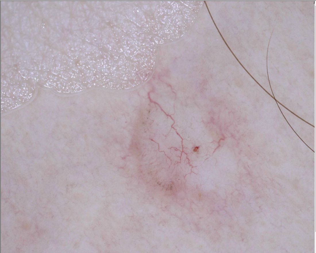
“For me, pink lesions are challenging,” Jennifer A. Stein, MD, PhD, said during the virtual Orlando Dermatology Aesthetic and Clinical Conference. “How can dermoscopy help us distinguish between Spitz nevus, melanoma, clear cell acanthoma, psoriasis, basal cell carcinoma, and squamous cell carcinoma?”

Dr. Stein, professor of dermatology at New York University, offered four tips. First, look for the shiny white perpendicular lines, otherwise known as the chrysalis or crystalline pattern. “You can only see this feature when you’re looking with polarized light,” she said. “This is why you want a dermatoscope that has polarized light, and better yet, one that you’re able to turn on and off, the hybrid kind, because then you can convince yourself that you’re looking at this feature, because it blinks on and off.”
The differential diagnosis for white shiny perpendicular lines includes dermatofibroma/scars (which is most common), Spitz and atypical genital nevi, BCC, and melanoma. “Dermatofibromas sometimes have white circles or rings in the center,” Dr. Stein said. “In BCC, the lines aren’t always perpendicular. Sometimes it’s more of a blotch or strands.”
A second tip for managing a pink lesion on dermoscopy is to look for any brown color. “When you see that combo together you have to worry,” she said. “When you see pigment network on dermoscopy, you have to put melanoma in your differential. If you see shiny white lines in something that is melanocytic, there’s a 98% specificity for melanoma.”
A third tip she offered for managing pink lesions is to check the blood vessels for clues. “For years, I was just naming the vessels based on making the diagnosis and then deciding, ‘that’s a basal cell carcinoma; those must be branching vessels,’ ” said Dr. Stein, who manages NYU’s medical dermatology faculty group practice.
However, blood vessel patterns differ. For example, branching or arborizing vessels are suggestive of BCC. “These vessels are very crisp-looking on dermoscopy,” she said. “They’re all in the same plane of focus and they look like they were drawn in with a fine point marker. That’s different from other blood vessel patterns.” She also pointed out that superficial basal cells have short, fine telangiectasias. “When you put on the polarized light, the clue is the white, shiny structures,” she said.
Dotted vessels, meanwhile, appear on dermoscopy as small red dots aligned perpendicular to the skin surface. The differential includes inflammatory lesions like psoriasis, stasis, and trauma; clear cell acanthoma (characterized by a “string of pearls” arrangement), nevi, and melanoma. “I find dermoscopy most useful in diagnosing SCC – especially squamous cell in situ,” she said. “Important clinical clues suggestive of SCC or melanoma include a solitary lesion, it’s new, it’s growing, and it’s not going away with a topical steroid.”
An additional pattern to be aware of are hairpin vessels, which are looped and feature a sharp bend at one end. These are often seen in seborrheic keratoses. “You can’t count on the hairpin vessels alone, because you can see this in anything keratotic, such as in keratoacanthoma (at the periphery with a yellow keratotic center), warts, SCC, BCC, as well as in dermal nevi and Spitz nevi,” said Dr. Stein, who recommended dermoscopedia.org as resource.
Comma vessels, meanwhile, appear in dermal or compound nevi. She described these as “slightly curved vessels that are much less in focus than branched vessels, because they come in and out of the plane of focus,” she said. “If you put your dermatoscope on top of the nevus and wobble it around you can appreciate the curve. If you look at it from the side, it looks like a curve. If you look at it straight on it will look more like a line. If you look at from the end it will look like a dot.”
Another vessel type she discussed are linear irregular and polymorphous vessels, which she described as “any combination of different types of vessels. We get most worried when we see dotted and linear irregular vessels together. In that case, you worry about melanoma. These can also be seen in nevi and other tumors, such as BCC.”
Dr. Stein’s fourth tip of the presentation was a reminder to consider dermoscopy as one piece of the clinical exam. “Always think about the lesion in context of the rest of the clinical picture and history,” she said. “Don’t get discouraged if it’s hard; just keep practicing. Look for any brown and use your clinical clues to put together to make the right decision.”
She disclosed that NYU receives compensation from MoleSafe for her telemedicine dermoscopic diagnoses.
Even in the most experienced hands, .
“For me, pink lesions are challenging,” Jennifer A. Stein, MD, PhD, said during the virtual Orlando Dermatology Aesthetic and Clinical Conference. “How can dermoscopy help us distinguish between Spitz nevus, melanoma, clear cell acanthoma, psoriasis, basal cell carcinoma, and squamous cell carcinoma?”
Dr. Stein, professor of dermatology at New York University, offered four tips. First, look for the shiny white perpendicular lines, otherwise known as the chrysalis or crystalline pattern. “You can only see this feature when you’re looking with polarized light,” she said. “This is why you want a dermatoscope that has polarized light, and better yet, one that you’re able to turn on and off, the hybrid kind, because then you can convince yourself that you’re looking at this feature, because it blinks on and off.”
The differential diagnosis for white shiny perpendicular lines includes dermatofibroma/scars (which is most common), Spitz and atypical genital nevi, BCC, and melanoma. “Dermatofibromas sometimes have white circles or rings in the center,” Dr. Stein said. “In BCC, the lines aren’t always perpendicular. Sometimes it’s more of a blotch or strands.”
A second tip for managing a pink lesion on dermoscopy is to look for any brown color. “When you see that combo together you have to worry,” she said. “When you see pigment network on dermoscopy, you have to put melanoma in your differential. If you see shiny white lines in something that is melanocytic, there’s a 98% specificity for melanoma.”
A third tip she offered for managing pink lesions is to check the blood vessels for clues. “For years, I was just naming the vessels based on making the diagnosis and then deciding, ‘that’s a basal cell carcinoma; those must be branching vessels,’ ” said Dr. Stein, who manages NYU’s medical dermatology faculty group practice.
However, blood vessel patterns differ. For example, branching or arborizing vessels are suggestive of BCC. “These vessels are very crisp-looking on dermoscopy,” she said. “They’re all in the same plane of focus and they look like they were drawn in with a fine point marker. That’s different from other blood vessel patterns.” She also pointed out that superficial basal cells have short, fine telangiectasias. “When you put on the polarized light, the clue is the white, shiny structures,” she said.
Dotted vessels, meanwhile, appear on dermoscopy as small red dots aligned perpendicular to the skin surface. The differential includes inflammatory lesions like psoriasis, stasis, and trauma; clear cell acanthoma (characterized by a “string of pearls” arrangement), nevi, and melanoma. “I find dermoscopy most useful in diagnosing SCC – especially squamous cell in situ,” she said. “Important clinical clues suggestive of SCC or melanoma include a solitary lesion, it’s new, it’s growing, and it’s not going away with a topical steroid.”
An additional pattern to be aware of are hairpin vessels, which are looped and feature a sharp bend at one end. These are often seen in seborrheic keratoses. “You can’t count on the hairpin vessels alone, because you can see this in anything keratotic, such as in keratoacanthoma (at the periphery with a yellow keratotic center), warts, SCC, BCC, as well as in dermal nevi and Spitz nevi,” said Dr. Stein, who recommended dermoscopedia.org as resource.
Comma vessels, meanwhile, appear in dermal or compound nevi. She described these as “slightly curved vessels that are much less in focus than branched vessels, because they come in and out of the plane of focus,” she said. “If you put your dermatoscope on top of the nevus and wobble it around you can appreciate the curve. If you look at it from the side, it looks like a curve. If you look at it straight on it will look more like a line. If you look at from the end it will look like a dot.”
Another vessel type she discussed are linear irregular and polymorphous vessels, which she described as “any combination of different types of vessels. We get most worried when we see dotted and linear irregular vessels together. In that case, you worry about melanoma. These can also be seen in nevi and other tumors, such as BCC.”
Dr. Stein’s fourth tip of the presentation was a reminder to consider dermoscopy as one piece of the clinical exam. “Always think about the lesion in context of the rest of the clinical picture and history,” she said. “Don’t get discouraged if it’s hard; just keep practicing. Look for any brown and use your clinical clues to put together to make the right decision.”
She disclosed that NYU receives compensation from MoleSafe for her telemedicine dermoscopic diagnoses.
FROM ODAC 2021
Are pediatric and adult dermatitis the same disease?

“Maybe not,” Jonathan I. Silverberg, MD, PhD, MPH, said during the Revolutionizing Atopic Dermatitis symposium.
Dr. Silverberg, director of clinical research in the division of dermatology at George Washington University, Washington, based his comments largely on a review that he and his colleagues carried out to understand how features of atopic dermatitis (AD) vary by region globally as well as by age. They identified 101 studies with sufficient data for meta-analysis and stratified the results by pediatric and adult age groups.
Several signs and symptoms occurred with similar frequency among pediatric and adult patients, including pruritus, xerosis, flexural involvement, extensor involvement, early onset of disease, comorbid atopy, head and neck involvement, and ophthalmic comorbidities. However, adults were found to have more signs of chronic disease, more hand eczema, different patterns of hand eczema, and a stronger relationship of disease activity with emotional factors. Meanwhile, children were found to have more exudative or weeping lesions, more perifollicular eczema, and more pityriasis alba.
Dr. Silverberg showed photos of three adults with varied presentations of extensor involvement, including one “who had a lot of lichenification and thickening of the skin, but over knees where you might think about psoriasis,” he said. “All three of these patients were of Southeast Asian descent. That happens to be a region where this feature was reported much more commonly. It may even tie to some underlying immunopathophysiologic differences of the disease across different patient populations.”
AD signs that occur more commonly in adults than children include lichenification (100% vs. 48%), urticaria (32% vs. 20%), popular lichenoid lesions (46% vs. 8%), Hertoghe’s sign (25% vs. 2%), erythroderma (29% vs. 1%), and nodular prurigo (18% vs. 4%).
Hand eczema features also differ between adults and children, including hand or foot dermatitis (44% vs. 25%), dyshidrosis/pompholyx (21% vs. 3%), knuckle dermatitis (25% vs. 8%), nail involvement (15% vs. 8%), and fissured heels. However, ventral wrist dermatitis was found to be more than twice as common in children, compared with adults (34% vs. 15%).
Other signs of AD were more common in children, compared with adults, including exudative eczema (61% vs. 42%), pityriasis alba (28% vs. 18%), Dennie-Morgan infraorbital folds (47% vs. 36%), seborrheic dermatitis–like lesions (40% vs. 18%), and perifollicular accentuation (37% vs. 21%). “This is such an important sign to wrap your head around and get comfortable assessing,” he said. “I have seen patients who are erythrodermic with follicular eczema who were told that they were crazy and had psychogenic itch, and they should go to a shrink.”
AD triggers can differ between adults and children as well, including course influenced by emotions/environmental factors (72% vs. 32%), worsening itch worse (65% vs. 49%), course influenced by environment (62% vs. 37%), and course influenced by emotions (70% vs. 15%).
According to Dr. Silverberg, emerging research suggests that there may be differences in the immune pathways activated in pediatric versus adult AD. Specifically, more Th17 and interferon-gamma in AD lesions have been observed in children, compared with adults, and more Th22 and Th17 in nonlesional AD have been seen in children, compared with adults. “This leads to a question: Will children respond differently than adults to treatment?” Dr. Silverberg said. “We see that omalizumab doesn’t seem to help much in adults, yet a recent study suggested that it might work reasonably well for children. Dupilumab has different dosing requirements and potentially different responses between the pediatric and adult populations.”
Age differences in AD may also be related to differences in the skin microbiome. In 2016, researchers led by Richard L. Gallo, MD, PhD, professor of dermatology, University of California, San Diego, compared the skin microbiome between adults and children with AD by swabbing the volar forearm and performing 16S rRNA gene sequencing. The study included 59 young children, 13 teenagers, and 56 adults with AD as well as 68 age-matched non-atopic healthy controls. The researchers found a greater abundance of Streptococcus, Granulicatella, Gemella, Rothia, and Haemophilus in young children, compared with adults, while Propionibacterium, Corynebacterium, Staphylococcus, Lactobacillus, Finegoldia, and Anaerococcus were more abundant in adults, compared with children.
Dr. Silverberg reported that he is a consultant to and/or an advisory board member for several pharmaceutical companies. He is also a speaker for Regeneron and Sanofi and has received a grant from Galderma.
FROM REVOLUTIONIZING AD 2020
Bathing now more widely accepted as an eczema treatment strategy
According to Noreen Heer Nicol, PhD, RN, FNP, frustration still exists for patients, families, and health care providers regarding the lack of consensus that routine bathing is good for patients with atopic dermatitis.

During the Revolutionizing Atopic Dermatitis symposium, she said that conflicting and vague guidelines currently exist on the topic.
“This stems from the fact that we just don’t have good studies,” said Dr. Nicol, associate dean and associate professor of nursing at the University of Colorado at Denver, Aurora. “Particularly, we don’t have randomized, controlled trials on the effects of water and bathing. It’s not just parents that are frustrated, but health care providers are as well.”
In an observational analysis, researchers evaluated results from three online surveys of dermatologists, allergists, and immunologists, and primary care physicians regarding routine bathing frequency recommendations for children with AD. It found that PCPs recommended daily bathing less than 50% of the time, while specialists recommended daily bathing more than 50% of the time.
“It seems like the PCPs have embraced that old dermatology notion when bathing was avoided in patients with AD,” Dr. Nicol said. “This lack of consensus on the basic daily care steps in AD management causes a great deal of confusion amongst patients, families, and young health care providers, in particular,” she added.
She believes that this goes back to a century-long debate about the pros and cons of bathing in AD. “We used to say that bathing will dry the skin out if you take a bath or a shower without immediately applying something like a good moisturizer. That’s where the 3-minute rule came along from the National Eczema Association, meaning that bathing hydrates the stratum corneum if you take a bath or a shower and you immediately apply that good moisturizer within 3 minutes to retain that hydration and keep the barrier intact and flexible.”

Dr. Nicol presented a stepwise management model that she has published many times over the years (see Pediatr Nursing 2020;46[2]:92-8 and J Allergy Clin Immunol Pract 2019;7[1]:1-16).
Step 1 consists of basic care, including skin hydration/bathing, application of a daily moisturizer, avoiding irritants, and identifying and addressing specific triggers. “This is the foundation for every step as you go forward,” she explained. Soak and seal has been a mainstay of treatment at National Jewish Health, she noted. “By that, I mean taking a soaking 10-15 minute bath in warm water daily. Gently pat away excess water. Immediately apply skin medications or moisturizer within 3 minutes. Using a gentle fragrance-free, dye-free cleanser to clean skin is also important. Avoid scrubbing.”
A review article on bathing and associated treatments in AD was published in 2017 and includes 144 references to bathing studies. A separate recommendation known as the “AD Yardstick” published by Dr. Nicol’s colleague at National Jewish Health, Mark Boguniewicz, MD, and coauthors, elaborated on the definition of basic skin care for nonlesional AD. Besides recommending the liberal and frequent application of moisturizers, it suggests management with warm baths or showers using nonsoap cleansers, usually once per day, followed by application of a moisturizer, even on clear areas.
“This is now what people are thinking as the basis of skin care in patients with AD,” Dr. Nicol said. “Warm baths and showers don’t look so controversial anymore. This model nicely lays out what we want people to remember. In the past, many times we just skipped that important step of telling people about bathing.”
In a small 2009 study, researchers conducted a quantitative assessment of combination bathing and moisturizing regimens on skin hydration in AD. They found that bathing followed by application of a moisturizer provides modest hydration benefits, though less than that of simply applying moisturizer alone. “That has not been the case for most of us who are bathing advocates,” Dr. Nicol said. “We believe that there is an additional hydration that’s gained from bathing and moisturizers done properly.”
In an earlier retrospective study of 28 patients referred to a tertiary care center for refractory chronic pruritic eruptions, researchers found that a plain-water 20-minute soak followed by smearing of midstrength corticosteroid ointment led to clearing or dramatic improvement of the lesions (Arch Dermatol 2005;14:1556-9). The authors recommended prospective studies to confirm the findings.
In a separate review of medical literature, researchers explored the role of frequent bathing in the treatment of pediatric AD (Ann Allergy Asthma Immunol 2016;117[1]:9-13). They found that the weight of evidence suggests that the frequent soak and smear bathing is preferred to infrequent bathing in the management of AD. Frequent bathing was defined as bathing at least once a day, while infrequent bathing was defined as bathing less than once a day.
“Bleach baths have received much attention in recent years, and have been endorsed by multiple AD guidelines, though not to the same degree as regular bathing,” Dr. Nicol said. “Right now, you can find almost as much literature for this practice as against it. The populations that seem to value from beach baths the most, however, are those with frequent infections, particularly those who are methicillin resistant. Most people recommend a maximum of two to three times per week but only with an active infection. Care must be taken to avoid additional drying or irritation of the skin from bleach.”
Many bleach bath recipes call for adding one-eighth to one-half of a cup of bleach to a tub full or water.
Dr. Nicol disclosed that she has served as an advisory board member for Eli Lilly.
According to Noreen Heer Nicol, PhD, RN, FNP, frustration still exists for patients, families, and health care providers regarding the lack of consensus that routine bathing is good for patients with atopic dermatitis.
During the Revolutionizing Atopic Dermatitis symposium, she said that conflicting and vague guidelines currently exist on the topic.
“This stems from the fact that we just don’t have good studies,” said Dr. Nicol, associate dean and associate professor of nursing at the University of Colorado at Denver, Aurora. “Particularly, we don’t have randomized, controlled trials on the effects of water and bathing. It’s not just parents that are frustrated, but health care providers are as well.”
In an observational analysis, researchers evaluated results from three online surveys of dermatologists, allergists, and immunologists, and primary care physicians regarding routine bathing frequency recommendations for children with AD. It found that PCPs recommended daily bathing less than 50% of the time, while specialists recommended daily bathing more than 50% of the time.
“It seems like the PCPs have embraced that old dermatology notion when bathing was avoided in patients with AD,” Dr. Nicol said. “This lack of consensus on the basic daily care steps in AD management causes a great deal of confusion amongst patients, families, and young health care providers, in particular,” she added.
She believes that this goes back to a century-long debate about the pros and cons of bathing in AD. “We used to say that bathing will dry the skin out if you take a bath or a shower without immediately applying something like a good moisturizer. That’s where the 3-minute rule came along from the National Eczema Association, meaning that bathing hydrates the stratum corneum if you take a bath or a shower and you immediately apply that good moisturizer within 3 minutes to retain that hydration and keep the barrier intact and flexible.”
Dr. Nicol presented a stepwise management model that she has published many times over the years (see Pediatr Nursing 2020;46[2]:92-8 and J Allergy Clin Immunol Pract 2019;7[1]:1-16).
Step 1 consists of basic care, including skin hydration/bathing, application of a daily moisturizer, avoiding irritants, and identifying and addressing specific triggers. “This is the foundation for every step as you go forward,” she explained. Soak and seal has been a mainstay of treatment at National Jewish Health, she noted. “By that, I mean taking a soaking 10-15 minute bath in warm water daily. Gently pat away excess water. Immediately apply skin medications or moisturizer within 3 minutes. Using a gentle fragrance-free, dye-free cleanser to clean skin is also important. Avoid scrubbing.”
A review article on bathing and associated treatments in AD was published in 2017 and includes 144 references to bathing studies. A separate recommendation known as the “AD Yardstick” published by Dr. Nicol’s colleague at National Jewish Health, Mark Boguniewicz, MD, and coauthors, elaborated on the definition of basic skin care for nonlesional AD. Besides recommending the liberal and frequent application of moisturizers, it suggests management with warm baths or showers using nonsoap cleansers, usually once per day, followed by application of a moisturizer, even on clear areas.
“This is now what people are thinking as the basis of skin care in patients with AD,” Dr. Nicol said. “Warm baths and showers don’t look so controversial anymore. This model nicely lays out what we want people to remember. In the past, many times we just skipped that important step of telling people about bathing.”
In a small 2009 study, researchers conducted a quantitative assessment of combination bathing and moisturizing regimens on skin hydration in AD. They found that bathing followed by application of a moisturizer provides modest hydration benefits, though less than that of simply applying moisturizer alone. “That has not been the case for most of us who are bathing advocates,” Dr. Nicol said. “We believe that there is an additional hydration that’s gained from bathing and moisturizers done properly.”
In an earlier retrospective study of 28 patients referred to a tertiary care center for refractory chronic pruritic eruptions, researchers found that a plain-water 20-minute soak followed by smearing of midstrength corticosteroid ointment led to clearing or dramatic improvement of the lesions (Arch Dermatol 2005;14:1556-9). The authors recommended prospective studies to confirm the findings.
In a separate review of medical literature, researchers explored the role of frequent bathing in the treatment of pediatric AD (Ann Allergy Asthma Immunol 2016;117[1]:9-13). They found that the weight of evidence suggests that the frequent soak and smear bathing is preferred to infrequent bathing in the management of AD. Frequent bathing was defined as bathing at least once a day, while infrequent bathing was defined as bathing less than once a day.
“Bleach baths have received much attention in recent years, and have been endorsed by multiple AD guidelines, though not to the same degree as regular bathing,” Dr. Nicol said. “Right now, you can find almost as much literature for this practice as against it. The populations that seem to value from beach baths the most, however, are those with frequent infections, particularly those who are methicillin resistant. Most people recommend a maximum of two to three times per week but only with an active infection. Care must be taken to avoid additional drying or irritation of the skin from bleach.”
Many bleach bath recipes call for adding one-eighth to one-half of a cup of bleach to a tub full or water.
Dr. Nicol disclosed that she has served as an advisory board member for Eli Lilly.
FROM REVOLUTIONIZING AD 2020