User login
Doug Brunk is a San Diego-based award-winning reporter who began covering health care in 1991. Before joining the company, he wrote for the health sciences division of Columbia University and was an associate editor at Contemporary Long Term Care magazine when it won a Jesse H. Neal Award. His work has been syndicated by the Los Angeles Times and he is the author of two books related to the University of Kentucky Wildcats men's basketball program. Doug has a master’s degree in magazine journalism from the S.I. Newhouse School of Public Communications at Syracuse University. Follow him on Twitter @dougbrunk.
Hospital factors play key role in readmission risk after surgery
CORONADO, CALIF. – Variation in readmission risk across hospitals following certain surgical procedures is more attributable to hospital factors than to patient characteristics, results from a large analysis demonstrated.
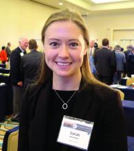
Such is the impact of the care delivery macro environment (CDM), which Sarah A. Brownlee and coauthors defined as a series of complex interactions between patient characteristics and imposed hospital attributes than can impact patient outcomes postoperatively.
The purpose of the current study was to determine the relative contribution of various aspects of the CDM to 1-year readmission risk after surgery. Working with colleagues Anai Kothari, MD, and Paul Kuo MD, in the One:MAP Section of Clinical informatics and Analytics in the department of surgery at Loyola University Medical Center, Ms. Brownlee analyzed the Healthcare Cost and Utilization Project State Inpatient Databases from Florida, New York, and Washington between 2009 and 2013, which were linked to the American Hospital Association Annual Survey from that same time period.
The researchers used smoothed hazard estimates to determine all-cause readmission in the year after surgery, and multilevel survival models with shared frailty to determine the relative impact of hospital versus patient characteristics on the heterogeneity of readmission risk between hospitals. They limited the analysis to patients aged 18 years and older who underwent one the following procedures: abdominal aortic aneurysm repair, pancreatectomy, colectomy, coronary artery bypass graft, and total hip arthroplasty.
Ms. Brownlee reported results from 502,157 patients who underwent surgical procedures at 347 hospitals. The 1-year readmission rate was 23.5%, and ranged from 12% to 36% across procedures. After controlling for procedure, the researchers observed a 7.9% variation in readmission risk between hospitals. Staffing accounted for 9.8% of variance, followed by hospital structural characteristics such as teaching status and clinical programs (7.5%), patient ZIP code (3.8%), hospital perioperative resources such as inpatient rehab (2.9%), hospital volume (2.8%), and patient clinical characteristics (2.1%). The following hospital characteristics were significantly associated with a lower risk of 1-year readmission: high physician/bed ratio (hazard ratio 0.85; P = .00017); transplant status (HR 0.87; P = .022); high-income ZIP code (HR 0.89; P less than .001); high nurse bed/bed ratio (HR 0.90; P = .047), and cancer center designation (HR 0.93; P = .021).
“Compared to patient clinical characteristics, hospital factors such as staffing ratios, perioperative resources, and structural elements account for more variation in postoperative outcomes,” Ms. Brownlee concluded. “However, it’s important to note that in the present study, over 70% of variation in readmission rates is not explained by the covariates that we analyzed. It’s possible that there are other factors we need to consider. That’s where the direction of this research is going. Much of the variation in readmission risk across hospitals cannot be characterized with currently utilized administrative data.”
The National Institutes of Health provided funding for the study. Ms. Brownlee reported having no financial disclosures.
AT WSA 2016
Key clinical point:
Major finding: Staffing accounted for 9.8% of variance in readmission risk between hospitals, followed by hospital structural characteristics such as teaching status and clinical programs (7.5%).
Data source: Results from 502,157 patients who underwent surgical procedures at 347 hospitals in three states.
Disclosures: The National Institutes of Health provided funding for the study. Ms. Brownlee reported having no financial disclosures.
Recovery path complicated for trauma patients with VTE
CORONADO, CALIF. – Patients who develop a venous thromboembolism (VTE) following severe hemorrhage are more susceptible to complications, compared with their counterparts who do not; they also exhibit hypercoagulability and enhanced platelet function at admission, and have delayed recovery of coagulation and platelet function following injury.
Those are the key findings from a secondary analysis of data from the Pragmatic Randomized Optimal Platelet and Plasma Ratio (PROPPR) trial, which randomized 680 severely injured trauma patients from 12 level I trauma centers to receive 1:1:1 or 1:1:2 ratios of plasma to platelets to red blood cells (JAMA 2015;313[5]:471-82). “The prevention of VTE following traumatic injury is an ongoing challenge,” Belinda H. McCully, PhD, said at the annual meeting of the Western Surgical Association. “Despite prophylaxis, about 25% of patients present with VTE, which is associated with higher complications and an increased risk for mortality. Common risk factors for mortality include age, body mass index, extremity injury, and immobility, but the precise mechanisms that contribute to VTE development are not well understood. We do know that the three main factors contributing to thrombosis include static flow, endothelial injury, and hypercoagulability. Clinically, coagulation is the most feasible factor to assess, mainly through the use of conventional coagulation tests, thromboelastography, platelet levels, and platelet function assays.”
Dr. McCully of the division of trauma, critical care, and acute care surgery in the department of surgery at Oregon Health & Science University, Portland, and her associates hypothesized that enhanced, earlier recovery of coagulation function is associated with increased VTE risk in severely injured trauma patients. To test this hypothesis, they conducted a secondary analysis of the PROPPR database, excluding patients who received anticoagulants, to rule out any bias against VTE development, as well as patients who died within 24 hours, to reduce the survival bias. This left 558 patients: 475 who did not develop a VTE, and 83 who did (defined as those who developed deep vein thrombosis or pulmonary embolism). Patient characteristics of interest included age, sex, BMI, mechanism of injury, and injury severity, as well as the transfusion group, the type of blood products given, and the percentage of patients given procoagulants. The investigators also assessed length of stay and complication incidence previously defined by the trial. During the trial, blood samples were taken from admission up to 72 hours and were used to asses both whole blood coagulation using thromboelastography and platelet function using the Multiplate assay.
Dr. McCully reported that VTE patients and non-VTE patients demonstrated similar admission platelet function activity and inhibition of all platelet function parameters at 24 hours (P less than .05). The onset of platelet function recovery was delayed in VTE patients, specifically for arachidonic acid, adenosine-5’-diphosphate, and collagen. Changes in thromboelastography, clot time to initiation, formation, rate of formation, and strength and index of platelet function from admission to 2 hours indicated increasing hypocoagulability (P less than .05) but suppressed clot lysis in both groups. Compared with patients in the non-VTE group, the VTE group had lower mortality (4% vs. 13%) but increased total hospital days (a mean of 30 vs. 16; P less than .05).
Adverse outcomes were also more prevalent in the VTE group, compared with the non-VTE group, and included systemic inflammatory response syndrome (82% vs. 72%), acute kidney injury (36% vs. 26%), infection (61% vs. 31%), sepsis (60% vs. 28%), and pneumonia (34% vs. 19%; P less than 0.05 for all associations). Conversely, regression analysis showed that VTE was associated only with total hospital days (odds ratio, 1.12), while adverse events were similar between the two groups. “From this we can conclude that VTE development following trauma may be attributed to hypercoagulable thromboelastography parameters and enhanced platelet function at admission, and compensatory mechanisms in response to a delayed recovery of coagulation and platelet function,” Dr. McCully said.
She acknowledged certain limitations of the study, including the fact that it was a secondary analysis of prospectively collected data. “We also plan to assess plasma markers of clot strength and fibrinolysis, which is an ongoing process,” she said. “Despite excluding patients that died within 24 hours, there was still a survival bias in the VTE group.”
The PROPPR study was supported by the National Heart, Lung, and Blood Institute and by the Department of Defense. Dr. McCully reported having no relevant financial disclosures.
AT WSA 2016
Key clinical point:
Major finding: Compared with patients in the non-VTE group, the VTE group had lower mortality (4% vs. 13%) but increased total hospital days (a mean of 30 vs. 16; P less than .05).
Data source: A secondary analysis of 558 patients from the Pragmatic Randomized Optimal Platelet and Plasma Ratio (PROPPR) trial, which randomized severely injured trauma patients from 12 level I trauma centers to receive 1:1:1 or 1:1:2 ratios of plasma to platelets to red blood cells.
Disclosures: The PROPPR study was supported by the National Heart, Lung, and Blood Institute and by the Department of Defense. Dr. McCully reported having no relevant financial disclosures.
Chief resident service increased trainees’ confidence and independence
CORONADO, CALIF. – Creation of a surgical chief resident service meant to increase resident autonomy and provide continuity of patient care with appropriate faculty supervision has been successful, results from a small single-center study showed.
“Providing opportunities for autonomy to bolster the development of independence and confidence during surgery residency remains among the most pronounced challenges of the current training paradigm,” Benjamin T. Jarman, MD, said at the annual meeting of the Western Surgical Association. “Prior to 2011, our graduating surgery residents reported a lack of perceived autonomy during their training and a need to improve practice management skills. To be clear, they consistently felt confident in their surgical abilities, but they did not sense that they were routinely engaged in directing all phases of care.”
In an effort to provide chief surgery residents with increased autonomy and full-spectrum continuity of patient care, Dr. Jarman and his associates initiated a chief resident service (CRS) in January of 2011. It was designed as an independent service with call responsibilities, office hours, operative scheduling, procedural coding, and endoscopy time. “We constructed a weekly schedule to be consistent with the practice of a general surgeon in the first year after residency,” Dr. Jarman explained. “We also added administrative time for research, patient coordination, and completion of records. Each class of chief residents was educated about these responsibilities as a group, and individual sit-down sessions occurred before they started the rotation. Expectations were made clear, and the importance of clear communication was stressed. The service was geared to provide excellent exposure to practice management skills.” Members of teaching faculty were assigned to each episode of patient care to meet all supervision guidelines and patients were educated accordingly. “The primary difference of and key to this service is that of patient continuity with the chief resident from preoperative assessment to postoperative care,” he said. “So our faculty had to adapt to the transient role that our residents are accustomed to.”
Dr. Jarman presented results from a study of nine surgeons who completed the CRS between January 2011 and June 2014. Total operative volume during residency was assessed in addition to select procedures for the chief service experience versus the residents’ first year of clinical practice. Residents who pursued fellowship training submitted their operative logs from their first year postfellowship. Graduates were surveyed to assess their current clinical practice, satisfaction with the chief service, and whether they perceived a correlation of the CRS with their clinical practice. Patient evaluations were reviewed as well. The researchers focused on the following procedures for comparison: laparoscopic appendectomy, laparoscopic cholecystectomy, colectomy, ventral/incisional hernia repair, inguinal hernia repair, upper endoscopy, and lower endoscopy.
All nine chief surgery residents completed the chief service and completed case logs. “The first three residents to graduate after implementation of the service spent 2 months each on the rotation, while subsequent graduates spent between 4 and 6 months, depending on how many chiefs we had in a given year,” Dr. Jarman said. The median total case volume was 1,101 during the 5-year residency, 92 during the CRS, and 299 during the first year of practice. When the researchers evaluated overall median case volumes, lower endoscopy volumes were higher during the first year of practice, compared with during the CRS (a median of 71 vs. 10 cases, respectively); otherwise there were similar case volumes across the other procedures selected for evaluation. Next, they determined the mean case volumes by month for the selected general surgical procedures and found similar case volumes with the exception of colectomy, which was more commonly performed during the CRS, compared with during the first year of practice (a mean of 1 vs. 0.4 cases; P=0.016).
All nine graduates completed an electronic survey relaying details about their current practice and degree of satisfaction with the CRS; 100% reported being “very satisfied” with their CRS, and 100% found it “very beneficial” to their practice. In addition, 56% said that their cases on the CRS were “somewhat similar” to their current practice, while 44% said that their cases were “very similar” to their current practice.
Since the inception of the CRS, Dr. Jarman and his associates have made several adjustments to the CRS, including incorporation of endoscopy time, adjusted office hours, the required presence of surgery assistants in the OR, and requiring fourth-year residents to attend the ACS Leadership Conference in preparation for the CRS role. He acknowledged certain limitations of the study, including its small sample size and the fact that its participants had variable clinical experience. “But we’re on the ground running,” Dr. Jarman said of the CRS. “The chief residents are wide-eyed and very engaged in this process, and the impact on their development and respect for all the caveats of independent practice has been significant. The strengths of the service include exposure to practice management skills, whole-spectrum clinical care for a single resident at a time, and operative experience which correlates to that experience of a first-year surgeon.” He reported having no financial disclosures.
AT WSA 2016
Key clinical point:
Major finding: More than half of general surgery residency graduates (56%) said that their cases on the chief resident service were “somewhat similar” to their current practice, while 44% said that their cases were “very similar” to their current practice.
Data source: An study of nine surgeons who completed the chief resident service between January 2011 and June 2014.
Disclosures: Dr. Jarman reported having no financial disclosures.
Discharging select diverticulitis patients from the ED found to be acceptable
CORONADO, CALIF. – Among patients diagnosed with diverticulitis via CT scan in the emergency department who were discharged home, only 13% required a return visit to the hospital, results from a long-term retrospective analysis demonstrated.
“In select patients whose assessment includes a CT scan, discharge to home from the emergency department with treatment for diverticulitis is safe,” study author Anne-Marie Sirany, MD, said at the annual meeting of the Western Surgical Association.
A few years ago, researchers conducted a randomized trial to evaluate the treatment of uncomplicated diverticulitis (Ann Surg. 2014;259[1]:38-44). Patients were diagnosed with diverticulitis in the emergency department and randomized to either hospital admission or outpatient management at home. The investigators found no significant differences between the readmission rates of the inpatient and outpatient groups, but the health care costs were three times lower in the outpatient group. Dr. Sirany and her associates set out to compare the outcomes of patients diagnosed with and treated for diverticulitis in the emergency department who were discharged to home, versus those who were admitted to the hospital. They reviewed the medical records of 240 patients with a primary diagnosis of diverticulitis by CT scan who were evaluated in the emergency department at one of four hospitals and one academic medical center from September 2010 to January 2012. The primary outcome was hospital readmission or return to the emergency department within 30 days, while the secondary outcomes were recurrent diverticulitis or surgical resection for diverticulitis.
The mean age of the 240 patients was 59 years, 45% were men, 22% had a Charlson Comorbidity Index (CCI) of greater than 2, and 7.5% were on steroids or immunosuppressant medications. More than half (62%) were admitted to the hospital, while the remaining 38% were discharged home on oral antibiotics. Compared with patients discharged home, those admitted to the hospital were more likely to be older than age 65 (43% vs. 24%, respectively; P = .003), have a CCI of 2 or greater (28% vs. 13%; P = .007), were more likely to be on immunosuppressant or steroid medications (11% vs. 1%; P = .003), show extraluminal air on CT (30% vs. 7%; P less than .0001), or show abscess on CT (19% vs. 1%; P less than .0001). “Of note: We did not have any patients who had CT scan findings of pneumoperitoneum who were discharged home, and 48% of patients admitted to the hospital had uncomplicated diverticulitis,” she said.
After a median follow-up of 37 months, no significant differences were observed between patients discharged to home and those admitted to the hospital in readmission or return to the emergency department (13% vs. 14%), recurrent diverticulitis (23% in each group), or in colon resection at subsequent encounter (16% vs. 19%). “Among patients discharged to home, only one patient required emergency surgery, and this was 20 months after their index admission,” Dr. Sirany said. “We think that the low rate of readmission in patients discharged home demonstrates that this is a safe approach to management of patients with diverticulitis, when using information from the CT scan.”
Closer analysis of patients who were discharged home revealed that six patients had extraluminal air on CT scan, three of whom returned to the emergency department or were admitted to the hospital. In addition, 11% of those with uncomplicated diverticulitis returned to the emergency department or were admitted to the hospital.
Dr. Sirany acknowledged certain limitations of the study, including its retrospective design, a lack of complete follow-up for all patients, and the fact that it included patients with recurrent diverticulitis. “Despite the limitations, we recommend that young, relatively healthy patients, with uncomplicated findings on CT scan, can be discharged to home and managed as an outpatient,” she said. “In an era where there’s increasing attention to health care costs, we need to think more critically about which patients need to be admitted for management of uncomplicated diverticulitis.” She reported having no financial disclosures.
[email protected]
AT WSA 2016
Key clinical point:
Major finding: After a median follow-up of 37 months, no significant differences were observed between patients discharged to home and those admitted to the hospital in readmission or return to the emergency department (13% vs. 14%, respectively).
Data source: A retrospective review of 240 patients with a primary diagnosis of diverticulitis by CT scan who were evaluated in the emergency department at one of four hospitals and one academic medical center from September 2010 to January 2012.
Disclosures: Dr. Sirany reported having no financial disclosures.
Emergent colon cancer resection does not negatively affect patient outcomes
CORONADO, CALIF. – With the exception of patients that present with perforation, emergent resection of colon cancers does not appear to adversely affect operative outcomes or patient survival, a 3-year analysis of data showed.
At the annual meeting of the Western Surgical Association, Jason W. Smith, MD, said that of the estimated 106,100 new cases of colon cancer each year, 6%-30% of patients have symptoms or late complications related to the disease that require an emergency intervention, often leading to dismal outcomes. “The problem with many existing studies of emergent colon cancer resections is that they tend to throw everybody into one large group, making it difficult to compare some of these patients,” said Dr. Smith, a trauma surgeon in the department of surgery at the University of Louisville (Ky.) School of Medicine. “Our thought was, if we provide an appropriate oncologic resection at the time of our initial management in these patients when they come to the emergency department, can we affect the similar rate of overall outcomes for these patients with regard to their cancer prognosis?”
Of the 117 patients in the emergent group, 35 had a perforation and 82 had an emergent resection. In an unmatched analysis comparing perforation, emergent resection, and elective resection, the patients who presented with a perforation had a much higher Charlson Comorbidity Index (CCI) score and a higher American Society of Anesthesiologists (ASA) class. They tended to be on vasopressors or suffering from inflammatory response related to their perforation, they had lower levels of blood pressure and hemoglobin, and they had much higher rates of 30-day mortality and overall 30-day morbidity, compared with their counterparts in the other two groups. Of the eight deaths that occurred in patients with colon perforation, four were related to sepsis and multiple organ failure, one to respiratory failure/acute respiratory distress syndrome, one to acute MI, one to exacerbation of chronic lung disease, and one to transition to palliative care due to cancer diagnosis. “So the overall predominance of the deaths associated in the first 30 days were related to the inflammatory responses associated with that perforation, not specific to the cancer itself,” Dr. Smith said. At the same time, the ASA and CCI scores were not different between those with morbidity/mortality and those who survived. “So it’s difficult to identify these patients out of the gate,” he said.
When the researchers more closely examined data from patients with a perforation, 27 of 35 (77%) survived at 30 days. Survival at 1, 2, and 3 years was 78%, 57%, and 43%, respectively. “This is a mixture of stage II and stage IV patients, so they’re difficult to compare and difficult to standardize across the board,” Dr. Smith noted. “But what you see is that their survival is not significantly different related to their disease if you discount the inflammatory process. Our initial thought was that for these perforated cancers, what we really need to do is provide the appropriate oncologic resection management [in order to] get the same oncologic outcomes.”
Next, the researchers compared the 82 patients who presented without a perforation but required an emergent operation with 82 of the elective surgery patients, matched for age, gender, the CCI, ASA class, oncology stage, and body mass index. There were no differences between the two groups in terms of R0 resection, the number of lymph nodes sampled, or estimated blood loss. However, compared with patients in the elective resection group, those in the emergent resection group had higher rates of ostomy placement (30% vs. 10%, respectively; P = .01), and a longer hospital length of stay (an average of 18 vs. 12 days; P = .0007). “Most of that difference occurred on the front end of hospital stays,” Dr. Smith said. “Their postoperative days were not significantly different.”
As for long-term outcomes, more than 90% of all patients in both groups received chemotherapy within the first year postprocedure, and overall time to initiation of chemotherapy was not significantly different in the emergent vs. elective groups (6.6 vs. 5.5 weeks, respectively; P = .43). However, patients suffering postsurgical complications had an increased risk of delayed chemotherapy.
In a risk-adjusted analysis, overall survival at 3 years was not different between the emergent and elective operation groups (hazard ratio, 1.1; P = .54). Similarly, disease-free survival was not different at 3 years between the two groups (HR, 1.06; P = .84). Independent predictors of poor long-term outcome included age greater than 70 (HR, 1.45; P less than 0.03); elevated ASA class (HR, 2.99 for class III vs. class I-II; P = .08; and HR, 7.45 for ASA class IV vs. I-II; P = .03); presence of residual disease (HR, 3.08; P less than .001), and advanced cancer stage.
He acknowledged certain limitations of the study, including the fact that it was a blinded retrospective cohort with the potential for unrecognized bias, and that it measured 3-year survival instead of 5-year survival data.
“Emergent resection of nonperforated colon cancers does not appear to adversely affect operative outcomes or patient survival when proper oncologic principles are applied to their initial management,” Dr. Smith concluded. “Outcome differences in patients suffering perforation may correlate with the physiologic derangements associated with the perforation rather than the oncologic disease; thus, every effort should be made to provide an appropriate oncologic operation.” He reported having no financial disclosures.
[email protected]
AT WSA 2016
Key clinical point:
Major finding: In a risk-adjusted analysis, overall survival at 3 years was not different between the emergent and elective operation groups (HR, 1.1; P = .54).
Data source: A retrospective review of 548 elective and emergent colectomies for colon cancer performed at the University of Louisville (Ky.) from 2011 to 2015.
Disclosures: Dr. Smith reported having no financial disclosures.
Fragmented readmission after liver transplant linked to adverse outcomes
CORONADO, CALIF. – Postdischarge surgical care fragmentation significantly increases the risk of both 30-day mortality and subsequent readmission in the first year following orthotopic liver transplantation, results from a study of national data showed.
“In an era of regionalization and centers of excellence, the likelihood for postdischarge fragmentation, defined as readmission to any hospital other than the hospital at which the surgery was performed, is an increasing reality,” Anai N. Kothari, MD, said at the annual meeting of the Western Surgical Association. “In many different surgical subspecialties – major vascular operations, bariatric surgery, oncologic resections – it’s known to be a risk factor for adverse events and poor quality. Postdischarge fragmentation is common, [and related to] as often as one in four readmissions. It increases the risk for short- and long-term morbidity and mortality, decreases survival, and increases cost.”
Dr. Kothari reported results from 2,996 patients with 7,485 readmission encounters at 299 hospitals. Of the 7,485 readmissions, 6,249 (83.5%) were nonfragmented, and 1,236 (16.5%) were fragmented. The mean age of patients was 55 years. There were no significant differences in baseline characteristics between patients with nonfragmented and fragmented admissions in terms of patient age, sex, preoperative and postoperative length of stay, Charlson comorbidity index, and comorbidities, with the exception of renal failure, which was more common among patients in the fragmented admission group.
Compared with the patients in the nonfragmented admission group, those in the fragmented admission group had a greater number of average readmissions per patient (3.3 vs. 2.5, respectively; P less than .0001) and a greater number of average days to readmission (168 vs. 105; P less than .0001). Reasons for readmission differed among the two groups. Patients readmitted to the index transplant center were more likely to have a biliary, hematologic, or neurologic complication, while those in the fragmented admissions group were more likely to be readmitted for things like electrolyte disturbances, respiratory issues, gastrointestinal issues, or hematologic-related issues. There was no difference in overall cost of care between the two groups (an average of $11,621.68 vs. $11.585.39, respectively).
After the investigators adjusted for age, sex, reason for readmission, cost of the index liver transplant, readmission length of stay, number of previous readmissions, and time from transplant, postdischarge fragmentation increased the odds of both 30-day mortality (OR, 1.75) and 30-day readmission (OR, 2.14). “It looks like just having a fragmented readmission is an independent predictor for an adverse event,” Dr. Kothari said.
Significant predictors of adverse events following a fragmented readmission included an increased number of previous readmissions (OR, 1.07) and readmission within 90 days of orthotopic liver transplant (OR, 2.19). “These two factors may be important for guiding providers to say, ‘If you have these things, this patient should likely come back to their index transplant center,’” Dr. Kothari said.
He reported having no relevant financial disclosures.
AT WSA 2016
Key clinical point:
Major finding: After investigators adjusted for numerous variables, postdischarge fragmentation following orthotopic liver transplantation increased the odds of both 30-day mortality (OR, 1.75) and 30-day readmission (OR, 2.14).
Data source: An analysis of data from the Healthcare Cost and Utilization Project State Inpatient Databases for Florida and California between 2006 and 2011 to identify 2,996 patients who underwent orthotopic liver transplantation.
Disclosures: Dr. Kothari reported having no relevant financial disclosures.
Detecting PH in IPF ‘more art than science’
LOS ANGELES – When it comes to the optimal management of pulmonary hypertension (PH) in patients with idiopathic pulmonary fibrosis (IPF), there are more questions than definitive answers, according to Brett E. Fenster, MD, FACC.
“Is PH in IPF a disease marker, an independent treatment target, or both?” he asked attendees at the annual meeting of the American College of Chest Physicians. “I think we have conflicting information about that.”
The prevalence of PH is estimated to be 10% in patients with mild to moderate IPF and tends to progress slowly. Common features of PH in IPF patients include shortness of breath, a greater degree of exertional desaturation, and an increased mortality rate.
Dr. Fenster described the ability to detect PH in IPF patients as “more art than science. A lot of different work has been done to look at different testing to get at the patients that may have PH that is a comorbid disease to their IPF. But a lot of times it comes down to assessing their level of dyspnea proportional to their level of disease. In those patients where we think there is something else going on besides their IPF, we’ll oftentimes get an echocardiogram and try to look at their right heart to see if they have features of PH. If it looks like they do, we will circle back and look at the amount of lung disease they have as characterized by their chest imaging, by their pulmonary function testing, and getting a blood gas. If this patient has findings of right heart enlargement, systolic dysfunction, et cetera, that clues us more into looking at PH and referring them to a center that has expertise in that version of PH.”
According to the most recent European Society of Cardiology/European Respiratory Society guidelines, the diagnosis of chronic obstructive pulmonary disease (COPD)/IPF/combined pulmonary fibrosis and emphysema (CPFE) without PH can be considered when the mean pulmonary artery pressure (mPAP) on right heart catheterization is less than 25 mm Hg. The diagnosis of COPD/IPF/CPFE with PH, based on these same guidelines, can be considered when the mPAP is 25 mm Hg or more. A patient may have COPD/IPF/CPFE with severe PH when his or her mPAP exceeds 35 mm Hg, or is 25 mm Hg or greater in the presence of a low cardiac output, the guidelines says (Eur Heart J. 2016;37:67-119). “That begins to separate out a group that may potentially benefit from targeted PH therapy,” Dr. Fenster said.
Current treatment approaches include long-term oxygen, diuretics, transplant, and pulmonary rehabilitation, but Dr. Fenster said there is sparse data on the optimal treatment approach. A study of sildenafil in IPF known as STEP-IPF failed to increase 6-minute walking test distance but improved diffusion capacity of carbon monoxide, quality of life, and arterial oxygenation (N Engl J Med. 2010;363:620-8). A study evaluating riociguat for idiopathic interstitial pneumonitis PH was discontinued early because of increased risk of death and adverse events. More recently, the drug ambrisentan was found to be ineffective at reducing the rate of IPF progression and was linked to an increase in disease progression events, including a decline in pulmonary function test values, hospitalization, and death, in the ARTEMIS-IPF trial (Ann Intern Med. 2013;158:641-9).
Randomized, placebo-controlled studies of bosentan in IPF give researchers pause for hope, Dr. Fenster said. BUILD-1 demonstrated a trend toward delayed time to death, delayed disease progression, improved quality of life, and no clear worsening of IPF (Am J Respir Crit Care Med. 2008;177[1]:75-81), while BUILD-3 showed a significant improvement in forced vital capacity (FVC) and carbon monoxide diffusing capacity (Am J Respir Crit Care Med. 2011;184[1]:92-9). A more recent trial evaluated IV treprostinil in 15 patients with interstitial lung disease and PH (Thorax 2014;69[2]:123-9). Eight of the patients had IPF. “After 12 weeks of treprostinil therapy, almost all of them experienced some degree of improvement in their walk distance,” said Dr. Fenster, who was not involved with the study. “Perhaps more importantly there were significant improvements in almost all parameters from their right heart catheterizations. So when we think about how to treat these patients, we have to weigh the risks and benefits of what we make potentially worse with our IPF therapy, such as worsening hypoxia, V/Q mismatch, disease progression, and volume overload. On the flip side, we might be improving right heart hemodynamics and RV function, which are prognostic in this disease. What’s the net balance of these things in terms of how it translates into functional capacity, quality of life, hospitalization, and mortality? We don’t know.”
According to Dr. Fenster, current data suggest that future IPF PH research should focus on prostanoid pathways and not on the estrogen-receptor and riociguat pathways to determine effective treatments. “We have numerous studies showing potential harm with IPF PH therapy, so we need to very much wade cautiously into this arena,” he said. “There is a potential role for PH therapy in IPF, but we will likely need to study patients with severe PH and IPF to show benefit. I think that’s where we’ll have the most success.”
Dr. Fenster reported having no relevant financial disclosures.
Indacaterol/glycopyrronium OK as preferred COPD treatment
AT CHEST 2016
LOS ANGELES – Indacaterol/glycopyrronium was superior to salmeterol/fluticasone at reducing the risk and rate of moderate to severe exacerbations in chronic obstructive pulmonary disease (COPD) patients with more than one or zero to one exacerbations in the previous year, results from an indirect comparison showed.
“Acute exacerbations of COPD are associated with accelerated decline in lung function and increased mortality,” Kenneth R. Chapman, MD, said at the annual meeting of the American College of Chest Physicians. “Current GOLD [Global Initiative for Chronic Obstructive Lung Disease] strategy recommends LABA/ICS [long-acting beta-agonist/inhaled corticosteroid] combination, and/or LAMA [long-acting muscarinic antagonist] as the first-line treatment, and LABA/LAMA as an alternative treatment for COPD patients at a high risk of exacerbations.”
In an effort to examine the reduction in moderate or severe exacerbations in COPD patients taking indacaterol/glycopyrronium (a combination of a LABA bronchodilator and a LAMA bronchodilator) or salmeterol/fluticasone (a LABA and inhaled glucocorticoid combination), researchers compared results from the FLAME and LANTERN trials. The FLAME study evaluated the rate and risk of exacerbations with indacaterol/glycopyrronium versus salmeterol/fluticasone in 3,362 moderate to very severe COPD patients with at least one exacerbation in the previous year (N Engl J Med. 2016;374[23]:2222-34).The LANTERN study compared the efficacy and safety of indacaterol/glycopyrronium versus salmeterol/fluticasone in 744 moderate to very severe COPD patients with 0-1 exacerbation in the previous year (Int J Chron Obstruct Pulmon Dis. 2015;10:1015-26).
Dr. Chapman, professor of medicine at the University of Toronto, reported that in the FLAME study, which was 52 weeks long, indacaterol/glycopyrronium significantly reduced the annualized rate of moderate or severe COPD exacerbations in patients who had one or more exacerbation in the previous year (a rate ratio of 0.83; P less than 0.001), which translated in to a clinically meaningful 17% reduction, compared with their counterparts taking salmeterol/fluticasone. In the LANTERN study, which was 26 weeks long, indacaterol/glycopyrronium also significantly reduced the annualized rate of patients who had 0-1 exacerbation in the previous year, compared with those taking salmeterol/fluticasone (RR, 0.69; P = .048).
In FLAME, indacaterol/glycopyrronium significantly delayed the time to first moderate or severe exacerbation, with a clinically meaningful 22% risk reduction, compared with salmeterol/fluticasone (hazard ratio, 0.78; P less than .001). Similar findings were observed in LANTERN; indacaterol/glycopyrronium significantly delayed the time to first moderate or severe exacerbation, with a clinically meaningful 35% risk reduction, compared with salmeterol/fluticasone (HR, 0.65; P less than .028).
“These results suggest that LABA/LAMA combinations such as indacaterol/glycopyrronium can be considered as a preferred treatment option in the management of COPD patients, irrespective of exacerbation history,” Dr. Chapman said.He went on to note that in FLAME, the incidence of pneumonia was 3.2% in the indacaterol/glycopyrronium group, compared with 4.8% in the salmeterol/fluticasone group P = .02). In LANTERN, the incidence of pneumonia was 0.8% in the indacaterol/glycopyrronium group, compared with 2.7% in the salmeterol/fluticasone group. Finally, in FLAME, the incidence of oral candidiasis was 1.2% in the indacaterol/glycopyrronium group, compared with 4.2% in the salmeterol/fluticasone group (P less than .001). In LANTERN, the respective values were 0% and 0.3%.
Dr. Chapman reported having numerous financial disclosures, including receiving consulting fees and research grants from Novartis.
[email protected]
Key clinical point:
Major finding: Indacaterol/glycopyrronium significantly reduced the annualized rate of moderate or severe COPD exacerbations in patients who had one or more exacerbation in the previous year (a rate ratio of 0.83; P less than 0.001), which translated into a clinically meaningful 17% reduction, compared with their counterparts taking salmeterol/fluticasone.
Data source: An indirect comparison of 3,362 patients in the FLAME study and 744 patients in the LANTERN study.
Disclosures: Dr. Chapman reported having numerous financial disclosures, including receiving consulting fees and research grants from Novartis.
Prognostic scores helpful in subset of COPD patients
LOS ANGELES – The Sequential Organ Failure Assessment (SOFA) score and the Glasgow Coma Scale (GCS) are simple, accurate tools for risk stratification of hospitalized patients with acute exacerbation of COPD, results from a single-center study showed.
“Acute exacerbations of chronic obstructive pulmonary disease often require hospitalization, may necessitate mechanical ventilation, and can be fatal,” Mohamed Metwally, MD, FCCP, said in an interview in advance of the annual meeting of the American College of Chest Physicians. “There are currently no validated disease-specific scores that measure the severity of acute exacerbation. Prognostic tools are needed to assess acute exacerbations of chronic obstructive pulmonary disease.”
For the 2-year study, Dr. Metwally and his associates prospectively evaluated 250 critically ill ICU AECOPD patients, mean age 65 years, at Assiut University Hospital between December 2012 and December 2014. The primary outcome was in-hospital mortality while the secondary endpoint was need for intubation and mechanical ventilation. The researchers excluded patients who died less than 24 hours after admission, those with underlying COPD who were admitted with another primary diagnosis such as an accident or a stroke, or for elective hospitalizations such as elective surgery or diagnostic procedures.
Dr. Metwally and his associates collected sociodemographic data, vital signs, and other clinical variables, and collected scores from five tools used to measure mortality prediction: the Acute Physiology and Chronic Health Evaluation (APACHE II), the SOFA score, the Early Warning Score (EWS), the GCS, and the Charlson Comorbidity Index (CCI). To assess performance of the scores, they used area under the receiver operating characteristic curve (AUC) analysis and the Hosmer-Lemeshow goodness-of-fit test for logistic regression.
Of the 250 patients, 43 (17%) died during their hospital stay and 54% required mechanical ventilation. All recorded scores were significantly higher in nonsurvivors, compared with survivors, and the risk of clinical deterioration increased with increasing scores. The discriminatory power of each score varied as measured by AUC analysis. The AUC of APACHE II, SOFA, EWS, GCS, and CCI were 0.79, 0.81, 0.76, 0.69, and 0.68, respectively “and all these models had good calibration in mortality prediction,” Dr. Metwally said. The SOFA score was the best in predicting mortality (its predicted mortality was 16%, compared with the actual mortality of 17%), while the APACHE II score overestimated mortality by at least twofold (46% vs. 17%). In addition, the EWS outperformed the GCS in predicting mortality. “This may be due to EWS containing all vital signs plus level of consciousness,” he said in an interview.
The GCS was found to be the most useful in predicting need for mechanical ventilation, with an AUC of 0.81. The AUCs of APACHE II, SOFA, EWS, and CCI were 0.79, 0.80, 0.73, and 0.61, respectively. All of the scores had good calibration in mortality prediction, Dr. Metwally said, with the exception of SOFA.
As for the APACHE II, Dr. Metwally said that instrument “can be used as a tool to predict both mortality and intubation in a specific group of patients, but with low discriminatory power.” He acknowledged certain limitations of the study, including the fact that it was limited to patients with AECOPD. “Future studies should include any critically ill respiratory patients,” he said.
He reported having no financial disclosures.
AT CHEST 2016
Key clinical point:
Major finding: The Sequential Organ Failure Assessment score was the best in predicting mortality of patients with acute exacerbation of COPD (its predicted mortality was 16%, compared with the actual mortality of 17%).
Data source: A prospective evaluation of 250 critically ill ICU patients hospitalized with acute exacerbation of COPD.
Disclosures: Dr. Metwally reported having no financial disclosures.
Comorbidities common in COPD patients
LOS ANGELES – Comorbidities are common in patients with chronic obstructive pulmonary disease, especially cardiovascular disease, diabetes, anemia, and osteoporosis, results from a single-center analysis showed.
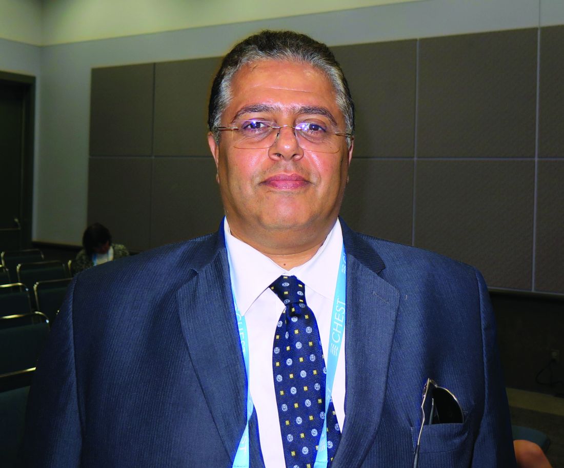
“These affect the course and outcome of COPD, so identification and treatment of these comorbidities is very important,” Hamdy Mohammadien, MD, said in an interview in advance of the annual meeting of the American College of Chest Physicians.
In an effort to estimate the presence of comorbidities in patients with COPD and to assess the relationship of comorbid diseases with age, sex, C-reactive protein, and COPD severity, Dr. Mohammadien and his associates at Sohag (Egypt) University, retrospectively evaluated 400 COPD patients who were at least 40 years of age. Those who presented with bronchial asthma or other lung diseases were excluded from the analysis. The mean age of patients was 62 years, 69% were male, and 36% were current smokers. Their mean FEV1/FVC ratio (forced expiratory volume in 1 second/forced vital capacity) was 48%, and 57% had two or more exacerbations in the previous year.
Dr. Mohammadien reported that all patients had at least one comorbidity. The most common comorbidities were cardiovascular diseases (85%), diabetes (35%), dyslipidemia (23%), osteopenia (11%), anemia (10%), muscle wasting (9%), pneumonia (7%), osteoporosis (6%), GERD (2%), and lung cancer (2%). He also noted that the association between cardiovascular events, dyslipidemia, diabetes, osteoporosis, muscle wasting, and anemia was highly significant in COPD patients aged 60 years and older, in men, and in patients with stage III and IV COPD. In addition, a significant relationship was observed between a positive CRP level and each comorbidity, with the exception of gastroesophageal reflux disease and lung cancer. The three comorbidities with the greatest significance were ischemic heart disease (P = .0001), dyslipidemia (P = .0001), and pneumonia (P = .0003). Finally, frequent exacerbators were significantly more likely to have two or more comorbidities (odds ratio 2; P = .04) and to have more hospitalizations in the past year (P less than .01).
“Comorbidities are common in patients with COPD, and have a significant impact on health status and prognosis, thus justifying the need for a comprehensive and integrating therapeutic approach,” Dr. Mohammadien said at the meeting. “In the management of COPD all these conditions need to be carefully evaluated and treated.”
He acknowledged certain limitations of the study, including its relatively small sample size and the fact that bone density was measured by sonar and not by dual-energy x-ray absorptiometry. Dr. Mohammadien reported having no financial disclosures.
AT CHEST 2016
Key clinical point:
Major finding: The three most common comorbidities in COPD patients were cardiovascular diseases (85%), diabetes (35%), and dyslipidemia (23%).
Data source: A retrospective study of 400 patients with COPD.
Disclosures: Dr. Mohammadien reported having no financial disclosures.
