User login
Damian McNamara is a journalist for Medscape Medical News and MDedge. He worked full-time for MDedge as the Miami Bureau covering a dozen medical specialties during 2001-2012, then as a freelancer for Medscape and MDedge, before being hired on staff by Medscape in 2018. Now the two companies are one. He uses what he learned in school – Damian has a BS in chemistry and an MS in science, health and environmental reporting/journalism. He works out of a home office in Miami, with a 100-pound chocolate lab known to snore under his desk during work hours.
Segmentectomy Supported for Select NSCLC Patients
SAN FRANCISCO – Thoracic surgeons should not shy away from segmentectomy in select patients with NSCLC, an expert advises, because the technique confers specific advantages.
In addition, it is as feasible as lobectomy. "If you can do a lobectomy, you can do a segmentectomy. There is no doubt about it," Dr. Matthew J. Schuchert said at the annual meeting of the American Association for Thoracic Surgery.
He shared patient selection criteria and technique tips based on experience with the more than 800 segmentectomies performed at the University of Pittsburgh Medical Center/UPMC Cancer Institute, where he is a general and thoracic surgeon.
Anatomic segmentectomy accomplishes the fundamental surgical tenets achieved by lobectomy, including R0 resection, adequate margins, and an opportunity for systematic nodal staging in early lung cancer, Dr. Schuchert said.
Lung preservation is another potential benefit of segmentectomy and the procedure is particularly useful for tumors with low malignancy potential where you may not have to take out an entire lobe to gain oncologic control, he said.
Equivalent survival to lobectomy has been demonstrated for stage 1A disease, especially for lesions smaller than 2 cm. In addition, "there may be decreased morbidity and mortality risk, especially among the elderly, a population we are going to be seeing more and more of."
Patient selection is paramount. In addition to the elderly, segmentectomy is particularly suitable for patients with marginal pulmonary function; those with "ground glass opacity" that may have low nodal positivity rates; and those who had prior lobectomy seeking parenchymal preservation.
"If you are contemplating the use of segmentectomy, it all really comes down to evaluation of the case," he said. Preoperative imaging ideally reveals a small tumor (less than 2 cm) in the outer one third of the lung. In addition, tumors should be confined to a discrete segmental boundary. "That’s critical. That’s the ticket for success," he said.
Surgeons can use the same anatomic approach they employ for lobectomy, only direct it at one segment. It is important to know segmental vascular and segmental bronchial anatomy, Dr. Schuchert noted.
"All of the same anatomic concerns, exposure concerns, and dissection concerns and techniques really apply." Segmentectomy can be performed through video-assisted thoracic surgery (VATS) or an open approach; the majority of cases at the University of Pittsburgh are VATS.
"We typically position the camera at about the seventh interspace in the mid-axillary line. Along the same interspace, a little more posteriorly, we will utilize a 10-mm incision for retraction and stapling. The access incision is pretty much the same as it is for a VATS lobectomy, usually somewhere along the line of the inframammary crease, and we place it right over the anterior hilum." This incision is usually around the level of the minor fissure on the right and slightly above the major fissure on the left, he added. Next, a 5-mm incision is made for retraction; it can also be particularly useful during node dissection, Dr. Schuchert said.
Preservation of the remaining lung is always a goal. "If you devitalize the remaining lung or impinge upon the bronchial supply, that patient is going to be doomed to have some perioperative issues." Remember that segmentectomy is a functional operation as well, he pointed out. "We are not just taking things out; what we leave behind still has to work."
Dissection assisted by an energy device is a more recent development in their hands. "We have now utilized energy in well over 100 patients undergoing both segmentectomy and lobectomy," he said.
Another essential goal of segmentectomy is to achieve a margin-to-tumor ratio greater than the size of the tumor itself, he said. As an example, he cited the case of a 71-year-old man with a history of diverticulitis with a pulmonary nodule picked up on an abdominal CT scan. The nodule was 1.7 cm, well confined in the outer third of the lung, and well centered within the basilar segment. Fine-needle aspiration of the nodule revealed adenocarcinoma. "He was considered to be an excellent candidate for segmentectomy. In this case, the margin was about 5 cm for a 1.7-cm tumor."
He and his colleagues published additional details of the segmentectomies they performed between 2002 and 2010 at UPMC in a retrospective study (Ann. Thorac. Surg. 2012:93:1780-7).
He said that he had no disclosures.
Isotretinoin Quells EGFR Inhibitor-Related Rash
ORLANDO – Oral isotretinoin holds potential as a bridge therapy for cancer patients who develop severe rashes during treatment, according to Dr. Milan J. Anadkat.
Dermatologists can play an integral role here. "Most oncologists see these patients first, and most oncologists are not enrolled in the iPledge program," said Dr. Anadkat of the division of dermatology at Washington University in St. Louis.
He noted that all treatment options are off label because there are no Food and Drug Administration–approved agents to treat chemotherapy-related cutaneous toxicities.
Getting patients through rashes that occur within a week or two of beginning targeted chemotherapy is important, as patients who develop the greatest reactions tend to have cancers that respond best to treatment, said Dr. Anadkat at the annual meeting of the Florida Society of Dermatology and Dermatologic Surgery. For this reason, the management of these patients is more complicated than simple drug cessation.
"The problem with just taking them off the drug is that they have cancer. Ultimately, the big goal here is treating the cancer, not avoiding the rash," he said.
Tetracycline or doxycycline can also reduce the severity of the rash, but the timing of administration is important. Better outcomes are associated with prophylaxis that is timed with the initiation of EGFR (epidermal growth factor receptor) inhibitors (Cancer 2008;113:847-53). "Waiting for the rash to appear is not the time to give it. It makes a difference if you give at day 0, not in terms of incidence but in the severity of the rash," he said.
Educate patients that the rash typically appears in an estimated 60%-90% of people within 8-10 days of EGFR inhibitor initiation, with a peak presentation at 2-4 weeks. "Bridging them through with something as effective as isotretinoin is useful," said Dr. Anadkat.
The rash generally appears on the face and upper trunk, but be careful not to confuse the presentation with a photo-exposure phenomenon.
Although some oncologists will describe the skin eruptions as "acnelike," histology will show mixed inflammatory infiltrate and follicular rupture. For this reason, "topical acne medications do very little," Dr. Anadkat said. Also, rule out infection, except when patients present with pustules on their arms, legs, or other non–EGFR receptor areas.
EGFR inhibitors can cause inflammatory alopecia, eyelash trichomegaly, and periungual and nail alterations.
About one in six patients will develop periungual or nail abnormalities, typically on their first finger or toe. These effects can be painful, Dr. Anadkat said. Culture is mandatory to rule out superinfection.
Again, the approach is to get patients through the adverse events with petroleum jelly, high-dose topical steroids, or oral tetracyclines. "Tumor markers are going down; [oncologists] are not going to want to stop chemotherapy for a painful thumb or toe," he said. "I recommend antimicrobial soaks with bleach or vinegar to prevent paronychia superinfection."
Dry, itchy skin is another concern with long-term EGFR inhibitor treatment. Histology shows "stark differences" in the stratum corneum. "This is the No. 1 side effect for patients on long-term EGFR – 3 months or longer; [it is] not a grade 3 or higher toxicity, but it is annoying."
Investigators compared oral minocycline and topical tazarotene prophylaxis in a study of 48 patients with cetuximab associated rash (J. Clin. Oncol. 2007:25:5390-6). Although oral minocycline was associated with reduced lesion counts, topical tazarotene yielded no significant benefit. "Again, this is not acne," Dr. Anadkat said.
He disclosed being a consultant or speaker for AstraZeneca, Bristol-Myers Squibb, Eisai, Genentech, and ImClone regarding strategies for managing skin toxicities from chemotherapy. He has never prescribed chemotherapy agents.
EXPERT ANALYSIS FROM THE ANNUAL MEETING OF THE FLORIDA SOCIETY OF DERMATOLOGY AND DERMATOLOGIC SURGERY
Expert: Reclaim Radiation for Skin Cancer
ORLANDO – Careful patient selection is key to treating nonmelanoma skin cancer patients with superficial radiation therapy, according to Dr. William I. Roth.
"If you pick the right patients for radiation therapy, you can get great results," Dr. Roth said. An elderly patient with a basal cell or squamous cell carcinoma on the face or leg can be a good candidate, for example, especially if there are other comorbidities that make that person less than ideal as a candidate for surgery.
The typical skin cancer patient at Dr. Roth’s practice in Boynton Beach, Fla., is between 75 and 80 years old, and many are very active and play golf or tennis several times a week. They prefer radiation therapy over surgery because it is less likely to limit their activities or impinge on their lifestyle, he said.
Reduced risk of bleeding and infection are other advantages of radiation therapy compared with surgery, Dr. Roth said at the annual meeting of the Florida Society of Dermatology and Dermatologic Surgery.
"Radiation therapy is not going to replace Mohs. [But] I think skin cancer practices need both," said Dr. Roth, a volunteer clinical professor in the department of dermatology at the University of Miami.
Cure rates of up to 95% or more are possible with superficial radiation therapy. "Radiation oncologists in my area are advertising 98% cure rates," Dr. Roth said. "I do still send a lot of patients to radiation oncologists, but I think you get excellent results [within dermatology] as long as you pick the right patient. It’s a great therapy."
Referral is appropriate when a lesion is deep and significantly large, and its depth and width cannot be accurately determined, Dr. Roth said. Patients also should be referred to a specialist if there is any evidence of perineural involvement.
Patients should be advised that some immediate posttreatment hypopigmentation is possible and that it generally improves quickly. The treated area usually looks its worst at about 2 weeks post treatment, Dr. Roth said. Some patients experience late effects, even up to a decade post treatment.
"I always scallop my port in the lead shielding so that 10 years later when the person gets some hypopigmentation it’s not going to be sharply defined," he said.
Four weeks post therapy skin can still appear thick, said Dr. Roth. "I would not rush to rebiopsy these patients, especially on the legs where there is a low turnover."
Although some physicians avoid radiation treatment of basal cell or squamous cell carcinoma on the legs, Dr. Roth has found value to this approach. "The legs are not absolutely a piece of cake, but as you move [treatment] up from the middle of the leg it becomes easier and easier." He added that grafting can be a challenge on the legs and surgical sutures can sometime pull through the skin. "I’m happy to do surgery. But to be honest with you, radiation therapy on the legs generally is less of a hassle for the patient."
However, use caution when treating lesions directly over the anterior tibial surface, he advised. "You have to be more careful ... right over the bone." He encountered two patients treated in this area whose skin healed nicely but they still experienced some sensitivity in their tibia. Dr. Roth suggested less aggressive treatment near the anterior tibia because "bone absorbs radiation to a much greater degree than the skin."
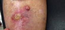
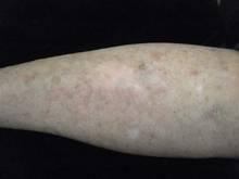
Dr. Roth gave an example of a patient with two well differentiated SCC lesions on her leg that he treated with radiation therapy. Twelve fractions of radiation were delivered with the SRT-100 (Sensus Healthcare, Boca Raton, Fla.). The patient ulcerated and had a fairly brisk response but did not report any pain. Follow-up was conducted at months 1,2 and 5. "From my standpoint, it was a remarkably good response."
Results with the SRT-100 are very reproducible, Dr. Roth said. Energy imparted across the port or opening in the metal shield is consistent. The device delivers a flat field of radiation with a fairly fast drop-off deeper into the skin. The depth of the nonmelanoma skin cancer lesion generally determines the setting, Dr. Roth said.
The control unit is outside the radiation room, thus avoiding the need to work behind a leaded glass window. The control unit counts down duration of treatment and features an emergency stop. "I use a baby monitor to speak with patients during treatment," Dr. Roth said. "I did have one patient who yelled that her eye shields fell off. I hit the emergency button, entered the room, and put the shields back on." The unit holds the reading (pauses therapy) and then allows physicians to continue when ready.
"Radiation therapy has been a part of dermatology basically forever. We have been treating skin cancers since before I was born," Dr. Roth said. "Now that we have some more choices, this should be a part of dermatology again. We should take this back."
Dr. Roth is a consultant and researcher for Sensus, but purchased his SRT-100 at full price.
EXPERT ANALYSIS FROM THE ANNUAL MEETING OF THE FLORIDA SOCIETY OF DERMATOLOGY AND DERMATOLOGIC SURGERY
Multiple Milia Signal Need for Genetic Testing
ORLANDO – Loeys-Dietz syndrome is a rare genetic disease that can be life threatening if not caught early, said Dr. Milan J. Anadkat.
Children with the connective tissue disorder often present with up to 50 or more milia, as well as velvety and translucent skin, easy bruising, varicose veins, and atrophic scars. Craniosynostosis, cleft lip or palate, and ocular hypertelorism are among the potential craniofacial abnormalities.
"If you have a patient less than 10 years old who has these milia and craniofacial features, they need to be genetically tested," said Dr. Anadkat of the division of dermatology at Washington University in St. Louis.
The experience at Washington University includes at least 25 children with Loeys-Dietz syndrome. Exposure to multiple patients with such a relatively rare disorder is an advantage of practicing dermatology at a large academic center, Dr. Anadkat said at the annual meeting of the Florida Society of Dermatology and Dermatologic Surgery.
Loeys-Dietz syndrome, first described in 2005 by Dr. Bart Loeys and Dr. Hal Dietz, affects connective tissue throughout the body. There are different subtypes, receptor mutations, and phenotypes, but what is most remarkable is that 75% of cases present as de novo, spontaneous mutations.
"The importance of knowing about Loeys-Dietz syndrome has to do with the cardiovascular complications," he said. This autosomal dominant genetic disorder is part of the subset of aortic aneurysm syndromes that includes Marfan syndrome.
Aggressive cardiovascular disease, aortic dissection by the teenage years (or "decades earlier than what we see in Marfan syndrome"), and arterial tortuosity are among the serious, extracutaneous manifestations, said Dr. Anadkat. Because arterial tortuosity can occur anywhere in the body (not just around the heart), a total body vascular scan is warranted.
Presence of milia may facilitate an early distinction from Marfan syndrome. Other syndromes with milia to include in the differential diagnosis include Bazex-Dupré-Christol syndrome, Rombo syndrome, and Rasmussen syndrome.
Patients with Loeys-Dietz syndrome also can present with musculoskeletal anomalies such as joint laxity, club foot, pectus deformity, scoliosis, and cervical spine abnormalities. "This could explain how, for decades, we saw patients with Marfanlike abnormalities who did not have a genetic test positive for Marfan syndrome," Dr. Anadkat said.
He and his associates published a report on four unrelated patients with Loeys-Dietz syndrome who developed milia in early childhood and reported increases in their number over time (Arch. Dermatol. 2011:147:223-6). Although more needs to be studied regarding specific mutations and phenotypes, each of these patients had the same TGFBR2 mutation.
More information on the disease is available online at the Loeys-Dietz Foundation website.
Dr. Anadkat said that he had no relevant financial disclosures.
EXPERT ANALYSIS FROM THE ANNUAL MEETING OF THE FLORIDA SOCIETY OF DERMATOLOGY AND DERMATOLOGIC SURGERY
Metabolic Syndrome Spurs CVD Risk in Hispanic Women
MIAMI BEACH – Metabolic syndrome drives the cardiovascular disease risk disparity for young and middle-age Hispanics more than for other women, according to a large community-based study of 6,843 women.
Hispanic women deserve additional attention to mitigate risk factors and intervention to ultimately lessen their cardiometabolic risk, Dr. Fatima Rodriguez said.
Overall, increasing age was associated with a higher prevalence of metabolic syndrome. However, compared with white and black women, Hispanics had the highest rates across all ages in the cross-sectional study. The Hispanic women aged 30-65 years had the greatest disparity in metabolic syndrome rates, suggesting this age group is at particular risk, Dr. Rodriguez said at the annual meeting of the International Society on Hypertension in Blacks.
Dr. Rodriguez and her colleagues assessed data from Sister to Sister: The Women’s Heart Health Foundation collected through free health screenings in 17 U.S. cities. In 2008 and 2009, 18,892 women were screened for obesity (using both body mass index and waist circumference), hypertension, hyperglycemia, and dyslipidemia. These women also completed cardiovascular risk questionnaires. Nearly 7,000 women had complete clinical and demographic data and were studied further.
"This was a very diverse sample," Dr. Rodriguez said. A total 42% self-identified as non-Hispanic white, 37% as black, 13% as Hispanic, and 8% as "other" race or ethnicity.
Overall prevalence of metabolic syndrome in the study was high, at 35%. In addition, "there was a disproportionate burden for Hispanic women and black women," said Dr. Rodriguez, an internal medicine resident at Brigham and Women’s Hospital in Boston. A total 40% of Hispanic women met the criteria for metabolic syndrome, as did 39% of black women, 31% of non-Hispanic white women, and 29% of women who identified as "other."
For Hispanic woman, much of the disparity is driven by abnormal lipid levels. "Many of these women have high triglyceride levels and low HDL levels ... and this disparity was most pronounced in young women," Dr. Rodriguez said. "It is a different pattern than the metabolic syndrome in black women, where it’s largely driven by waist circumference and hypertension."
In addition to assessment of race/ethnicity and age, a third objective of the study was to identify risk-adjusted predictors of metabolic syndrome. The No.1 predictor was Hispanic ethnicity (odds ratio, 1.65), followed by being black (OR, 1.39), compared with non-Hispanic whites. Smoking also was an independent predictor for the syndrome (OR, 1.30), as was increasing age (OR, 1.13).
Dr. Rodriguez and her colleagues used the National Cholesterol Education Program definition (NCEP ATP III) for metabolic syndrome. Women had to meet at least three of the following criteria: waist circumference of at least 35 inches; triglyceride level of at least 150 mg/dL; HDL cholesterol below 50 mg/dL; systolic blood pressure at least 130 mm Hg, or diastolic blood pressure at least 85mm Hg; or pharmacologic treatment for hypertension; or a fasting glucose of at least 110 mg/dL.
Use of cross-sectional data in the current study limits assessment of any causality. Other potential limitations include aggregating all Hispanic women into one group (even though there is a great deal of heterogeneity among Hispanics) and an inability to account for lifestyle or patient level factors (for example, diet or exercise).
A disparity in insurance status was another finding. "One thing that was very interesting in this study was the high rates of lack of insurance for Hispanic women. Alarmingly, almost 65% of these women were uninsured had no insurance whatsoever," Dr. Rodriguez said. "This suggests these women have little access to the health care setting and a population-based approach would be best for primary prevention."
Dr. Rodriguez said that she had no financial disclosures.
AT THE ANNUAL MEETING OF THE INTERNATIONAL SOCIETY ON HYPERTENSION IN BLACKS
Major Finding: A total 40% of Hispanic women met the criteria for metabolic syndrome, as did 39% of black women, 31% of non-Hispanic white women, and 29% of women who identified as "other" in a study of 6,843 women.
Data Source: Cross-sectional study of a diverse group of community-based women screened for cardiovascular risk factors at health fairs in 2008 and 2009.
Disclosures: Dr. Rodriguez said that she had no relevant financial disclosures.
Negative Emotions Could Drive Abnormal BP Pattern in Some Teens
MIAMI BEACH – Nondipping of nighttime blood pressure – a recognized risk factor for hypertension – emerges as early as adolescence, preferentially affects blacks, and is associated with lower socioeconomic status, according to a study.
In addition, negative psychological attributes such as trait anger and interpersonal conflict by day were independent factors for blood pressure nondipping at night, Tanisha I. Burford, Ph.D., said at the annual meeting of the International Society on Hypertension in Blacks.
Dr. Burford recommended asking potentially at-risk adolescent patients about their sleep quality, because those with fragmented or interrupted sleep are less likely to experience a normal, nocturnal restorative decline in blood pressure.
She also suggested that clinicians consider asking at-risk adolescents to wear an ambulatory blood pressure monitor for more-comprehensive, real-time feedback on circadian blood pressures, compared with conventional intermittent clinical readings.
Dr. Burford and her colleagues did just that – they asked 139 black and 106 white healthy adolescents to wear ambulatory blood pressure monitors for 48 consecutive hours.
"Most of what we know about nondipping blood pressure is [from studies] in adults." There is some evidence that blunting of blood pressure decreases at night "emerges early in the life course, during adolescence, and even in some children 9-11 years old," said Dr. Burford, a postdoctoral scholar in the cardiovascular behavioral medicine research program at the University of Pittsburgh.
Study participants used electronic diaries to rate social interactions and any conflict (on a 6-point scale) in the 10 minutes preceding each blood pressure reading. They also completed standard measures of depression (Center for Epidemiologic Studies Depression scale or CES-D); trait anger (State Trait Anxiety Inventory or STAI), and negative affect (Positive and Negative Aspect Schedule or PANAS).
The researchers found a higher ratio of average night to day blood pressures (both systolic and diastolic) among black teenagers who reported higher rates of negative emotions and/or conflict compared to white teenagers in a regression analysis that adjusted for age, sex, and body mass index.
"Most of this is a systolic effect – which makes sense for hypertension – [where] systolic changes are more detrimental in early stages," Dr. Burford said.
Among blacks, the beta value (the interaction between average systolic blood pressure night/day ratio and race) was a significant 0.43 for trait anger; a significant 0.52 for negative affect; and a significant 0.59 for depression.
The interaction between interpersonal stress and race had a nonsignificant trend for an adverse effect on the systolic blood pressure ratio (beta value, 0.38).
"The most fascinating thing is that these negative psychological attributes did [interact with] race," Dr. Burford said. Trait anger, depression, and conflict were only associated with nighttime nondipping of blood pressure among black teens, even though white teens reported higher levels of trait anger. A possible explanation, she added, was that positive attributes were less protective for black teenagers.
Some blacks have a "lower resource capacity," Dr. Burford said, which could include lower levels of self-esteem and less social support, particularly if they live in a stressful environment.
Participants were 14-19 years old (median, 16 years) and part of the Pittsburgh Project Pressure II.
Typically, blood pressure is low during the morning, increases during the day, and then drops at nighttime. Nondipping nighttime blood pressure was defined as less than a 10% decrease vs. daytime pressures. Although blood pressure readings can be highly variable and influenced by multiple factors, this 10% or less cutoff is a reliable predictor of risk, Dr. Burford said.
Dr. Burford had no relevant financial disclosures.
AT THE ANNUAL MEETING OF THE INTERNATIONAL SOCIETY ON HYPERTENSION IN BLACKS
Major Finding: Compared with white Americans, black adolescents who reported negative emotions were at higher risk for nocturnal blood pressure nondipping. Significant interactions were found between race, average systolic blood pressure night/day ratio, and trait anger (beta value, 0.43), negative affect (0.52), and depression (0.59).
Data Source: A comparison of 139 black and 106 white American teenagers who reported negative emotions and wore ambulatory blood pressure monitors for 48 hours.
Disclosures: Dr. Burford reported having no financial disclosures.
Radiofrequency Ablation Frees Majority From Atrial Fibrillation
SAN FRANCISCO – Considerable variation by institution suggests that additional training is needed to standardize radiofrequency ablation of persistent atrial fibrillation in patients undergoing concomitant cardiac surgery, according to a prospective, multicenter study.
"As surgeons we need to take the ‘a fib’ part of the procedure more seriously. There can be a high cure rate," Dr. Ralph J. Damiano Jr. said at the annual meeting of the American Association for Thoracic Surgery.
He and his associates studied 150 consecutive patients with persistent or permanent atrial fibrillation (AF) undergoing irrigated unipolar or bipolar radiofrequency treatment at 15 centers between May 2007 and July 2011. The study protocol included use of the Cox Maze IV lesion set.
Freedom from AF at 6-9 months’ follow-up was a primary efficacy end point of the CURE AF (Concomitant Utilization of Radiofrequency Energy for Atrial Fibrillation) trial; this outcome was achieved by 66% of patients. Just more than half of patients, 53%, were not taking antiarrhythmia medications at this follow-up time, another measure of efficacy.
"The Cox Maze IV procedure performed with irrigated radiofrequency ablation in patients with persistent atrial fibrillation restored the majority of patients to sinus rhythm with a low complication rate," Dr. Damiano said.
There was no statistical difference in efficacy outcomes by AF type. Most of the participants, 75%, had long-standing persistent atrial fibrillation, 22% had persistent AF, and 3% had paroxysmal AF.
The mean patient age was 71 years, 56% were men, and the majority had New York Heart Association (NYHA) class II or III heart failure. The mean duration of AF was 64 months.
The primary safety measure in the study was the major cardiac composite adverse event rate within 30 days. A total 6.6% of participants experienced such an event, although none were device related, Dr. Damiano said. There were no cases of pulmonary vein stenosis, he added. Operative mortality was 4%.
Significant predictors of success, defined as freedom from AF, included shorter duration of the persistent or permanent atrial fibrillation, smaller left atrial diameter, and fewer concomitant cardiac procedures, said Dr. Damiano, a cardiothoracic surgeon at Barnes Jewish Hospital in St. Louis.
As an example, the success rate was 50% when total radiofrequency ablation time was less than 6 minutes. By comparison, success grew to 80% with ablation times of 12 minutes or longer.
Left atrial diameter was the only significant predictor of success that remained on a multivariate analysis. Dr. Damiano and his colleagues found that 69% of patients with a left atrial diameter of 3.0-4.5 cm were free from AF, compared with 36% who had a diameter larger than 6 cm. The overall mean left atrial diameter was 5.2 cm.
There were significant differences in achievement of success among different study centers, including a 33% success rate at one site versus 100% at three other sites, Dr. Damiano said.
"The variability between centers is probably one of the most important findings in this study," study discussant Dr. Niv Add said. He asked: "How would you see moving forward with training and credentialing of surgeons?" Dr. Add is chief of cardiac surgery at Inova Fairfax Hospital, Falls Church, Va.
"We were supposed to perform the exact same procedure in the same way," Dr. Damiano replied. "The surgeons all agreed on the lesion set [but] it’s hard to quantify experience. You can see a huge variation in ablation time, so clearly we were not all performing the same procedure. This variability suggests a need for more effective procedural and device training."
A total 80% of patients had concomitant mitral disease; 58% had heart failure; and 52% had tricuspid disease.
The most common surgical procedure was single valve with or without coronary artery bypass grafting (CABG) in 53%. Double-valve surgery with or without CABG was performed in 30% of patients; CABG in 15%; triple-valve with or without CABG in 1%; and other surgery in 1%.
Intraoperative pulmonary vein isolation was measured using exit block and was achieved for 81% of patients. Radiofrequency ablation was performed using Medtronic’s Cardioblate unipolar or bipolar device. As the device is not yet cleared by the Food and Drug Administration for this indication, such use is considered off label.
Dr. Damiano is a consultant for Medtronic. Medtronic sponsored the trial.
CURE AF, Concomitant Utilization of Radiofrequency Energy for Atrial Fibrillation trial,
SAN FRANCISCO – Considerable variation by institution suggests that additional training is needed to standardize radiofrequency ablation of persistent atrial fibrillation in patients undergoing concomitant cardiac surgery, according to a prospective, multicenter study.
"As surgeons we need to take the ‘a fib’ part of the procedure more seriously. There can be a high cure rate," Dr. Ralph J. Damiano Jr. said at the annual meeting of the American Association for Thoracic Surgery.
He and his associates studied 150 consecutive patients with persistent or permanent atrial fibrillation (AF) undergoing irrigated unipolar or bipolar radiofrequency treatment at 15 centers between May 2007 and July 2011. The study protocol included use of the Cox Maze IV lesion set.
Freedom from AF at 6-9 months’ follow-up was a primary efficacy end point of the CURE AF (Concomitant Utilization of Radiofrequency Energy for Atrial Fibrillation) trial; this outcome was achieved by 66% of patients. Just more than half of patients, 53%, were not taking antiarrhythmia medications at this follow-up time, another measure of efficacy.
"The Cox Maze IV procedure performed with irrigated radiofrequency ablation in patients with persistent atrial fibrillation restored the majority of patients to sinus rhythm with a low complication rate," Dr. Damiano said.
There was no statistical difference in efficacy outcomes by AF type. Most of the participants, 75%, had long-standing persistent atrial fibrillation, 22% had persistent AF, and 3% had paroxysmal AF.
The mean patient age was 71 years, 56% were men, and the majority had New York Heart Association (NYHA) class II or III heart failure. The mean duration of AF was 64 months.
The primary safety measure in the study was the major cardiac composite adverse event rate within 30 days. A total 6.6% of participants experienced such an event, although none were device related, Dr. Damiano said. There were no cases of pulmonary vein stenosis, he added. Operative mortality was 4%.
Significant predictors of success, defined as freedom from AF, included shorter duration of the persistent or permanent atrial fibrillation, smaller left atrial diameter, and fewer concomitant cardiac procedures, said Dr. Damiano, a cardiothoracic surgeon at Barnes Jewish Hospital in St. Louis.
As an example, the success rate was 50% when total radiofrequency ablation time was less than 6 minutes. By comparison, success grew to 80% with ablation times of 12 minutes or longer.
Left atrial diameter was the only significant predictor of success that remained on a multivariate analysis. Dr. Damiano and his colleagues found that 69% of patients with a left atrial diameter of 3.0-4.5 cm were free from AF, compared with 36% who had a diameter larger than 6 cm. The overall mean left atrial diameter was 5.2 cm.
There were significant differences in achievement of success among different study centers, including a 33% success rate at one site versus 100% at three other sites, Dr. Damiano said.
"The variability between centers is probably one of the most important findings in this study," study discussant Dr. Niv Add said. He asked: "How would you see moving forward with training and credentialing of surgeons?" Dr. Add is chief of cardiac surgery at Inova Fairfax Hospital, Falls Church, Va.
"We were supposed to perform the exact same procedure in the same way," Dr. Damiano replied. "The surgeons all agreed on the lesion set [but] it’s hard to quantify experience. You can see a huge variation in ablation time, so clearly we were not all performing the same procedure. This variability suggests a need for more effective procedural and device training."
A total 80% of patients had concomitant mitral disease; 58% had heart failure; and 52% had tricuspid disease.
The most common surgical procedure was single valve with or without coronary artery bypass grafting (CABG) in 53%. Double-valve surgery with or without CABG was performed in 30% of patients; CABG in 15%; triple-valve with or without CABG in 1%; and other surgery in 1%.
Intraoperative pulmonary vein isolation was measured using exit block and was achieved for 81% of patients. Radiofrequency ablation was performed using Medtronic’s Cardioblate unipolar or bipolar device. As the device is not yet cleared by the Food and Drug Administration for this indication, such use is considered off label.
Dr. Damiano is a consultant for Medtronic. Medtronic sponsored the trial.
CURE AF, Concomitant Utilization of Radiofrequency Energy for Atrial Fibrillation trial,
CURE AF, Concomitant Utilization of Radiofrequency Energy for Atrial Fibrillation trial,
AT THE ANNUAL MEETING OF THE AMERICAN ASSOCIATION FOR THORACIC SURGERY
No Consensus on Best Surgery for Neonatal Heart Syndrome
SAN FRANCISCO – There is no consensus among experts on the optimal surgical approach to repair neonatal hypoplastic left heart syndrome, if a series of consecutive talks at the annual meeting of the American Association for Thoracic Surgery is any indication.
Dr. David J. Barron is a proponent of the placement of a stage 1 right ventricle–pulmonary artery (RV-PA) conduit (Circulation 2003;108[suppl. 1]:II155-60); Dr. J. William Gaynor prefers a stage 1 Blalock-Taussig (BT) shunt; and Dr. Mark E. Galantowicz advocates a hybrid stage 1 procedure.
Dr. Emile A. Bacha tied all these strategies together in a differential approach to management of neonates with hypoplastic left heart syndrome. There may be no one answer; local factors such as surgeon experience or medical center volume can impart significant difference on outcomes, Dr. Bacha said. His bias, in general, is to use the BT shunt for aortic stenosis and the RV-PA conduit for aortic atresia, and to reserve the hybrid approach for high-risk patients. Dr. Bacha is director of the congenital and pediatric cardiac surgery at the Morgan Stanley Children’s Hospital of New York–Presbyterian in New York City.
The surgeons provided the following overview:
• Stage 1 RV-PA conduits. "If you have any condition where there are three different ways to do the same operation, [it indicates that] we are still looking for the right way of doing it. What is important is trying to find the right operation for the right patient," said Dr. Barron, a consultant cardiac surgeon at Birmingham (England) Children’s Hospital.
"It’s all about diastole" with the RV-PA conduit, Dr. Barron said. The maintenance of diastolic pressure is a benefit with RV-PA, compared with the classic Norwood shunt, he added. "When you turn off the shunt in the OR, you get dramatic drop with Norwood where both systolic and diastolic drop. With the RV-PA, the systolic pressure drops but the diastolic pressure is maintained. This facilitates "more of cardiac output to systemic circulation, where you want it to be."
"We’re in an era of evidence-based medicine, and it’s not always easy to find class I evidence in congenital heart disease. The strategy sounds good, but can we actually prove it is better?" Dr. Barron asked. He pointed to a multicenter comparison of 549 infants who were randomized to a modified BT or PA-RV shunt; the study revealed a 10% survival advantage for the PV-RA patients at 1 year (N. Engl. J. Med. 2010:362:1980-92).
A disadvantage of the PV-RA shunt was more catheterization lab interventions (41%, vs. 26% for the modified BT shunt). In addition, the transplantation-free survival advantage was no longer significant after 12 months, he said.
• Stage 1 BTshunts. "We really need to focus on how well these children do over the long run," said Dr. Gaynor, attending cardiothoracic surgeon at the Children’s Hospital of Philadelphia (CHOP).
"Most of the benefit of the RV-PA is in the early interstage period," Dr. Gaynor said. He pointed out that transplant-free survival was not statistically different in the New England Journal of Medicine study at a mean of 32 months’ follow-up.
Dr. Bacha noted that with both speakers using the same study to argue their points," it may be time for a new trial."
Dr. Gaynor suggested that he will remain a proponent of the modified BT shunt until sufficient, long-term evidence supports survival and other advantages with the use of the RV-PA. The RV PA may have some advantages for high-risk subgroups, but more data are needed, he said.
Likewise, an examination of stage 1 reconstruction at CHOP with either the RV-PA or a modified BT shunt showed no significant difference on overall survival, Dr. Gaynor said. (Ann. Thorac. Surg. 2005:80:1582-90). Interestingly, timing made a difference: Patients with the modified BT shunt had significantly higher morbidity during the interstage period, but those with an RV-PA conduit demonstrated a trend toward increased death or transplant for heart failure after stage 2 reconstruction.
• Hybrid stage 1 surgery. "I am in favor of hybrid stage 1 for initial palliation for hypoplastic left heart syndrome. Hybrid stage 1 has at least equivalent results to traditional approaches in standard-risk patients," said Dr. Galantowicz, chief of cardiothoracic surgery at Nationwide Children’s Hospital in Columbus, Ohio.
A hybrid stage 1 can effectively bridge a child to recovery and can salvage a child who was not diagnosed at birth, Dr. Galantowicz said.
There is some evidence that a hybrid approach is less costly overall, compared with placement of a modified BT shunt (Ann. Thorac. Surg. 2009;87:1885-92).
"The standard approach is one of the most costly and resource intensive for any of the congenital children we have," Dr. Galantowicz said. "It requires significant resource utilization, even in the modern era."
Ultimately, "it’s really not about which of these procedures is better as all or nothing. It’s which is better for which subcategory of patient," said Dr. Galantowicz.
According to Dr. Bacha, "I think we can all agree there is equipoise between the BT shunt and the RV-PA conduit, and the hybrid procedures are being increasingly employed for high-risk patients."
Dr. Barron, Dr. Gaynor, Dr. Galantowicz, and Dr. Bacha each said they had no relevant financial disclosures.
EXPERT ANALYSIS FROM THE ANNUAL MEETING OF THE AMERICAN ASSOCIATION FOR THORACIC SURGERY
Tips on Cardiovascular Testing Before Cancer Surgery
MIAMI BEACH – When you are called to assess a patient before cancer surgery, how do you know when noninvasive cardiovascular testing is warranted?
Start by asking patients to describe their functional status before they started any treatment to combat their cancer, Dr. Sunil K. Sahai said.
Also assess for any ischemia preoperatively, because its presence might direct a surgeon to prescribe a less cardiotoxic postoperative treatment for your patient, Dr. Sahai said at a meeting on perioperative medicine sponsored by the University of Miami. Occult ischemia might be found if a patient reports shortness of breath during prior chemotherapy administration, he added.
"Everything you’ve heard about perioperative medicine is true for cancer patients, but they are also unique," Dr. Sahai said. The physiologic burden of cancer and its treatment makes preoperative evaluation challenging, but it’s worth doing right to ensure the patient receives the optimal therapy. Also, in some cases, either the patient or surgeon will decide not to proceed with surgery based on your risk assessment, said Dr. Sahai, medical director of the Internal Medicine Perioperative Assessment Center at the University of Texas M.D. Anderson Cancer Center in Houston.
To illustrate some of the challenges, Dr. Sahai described an actual patient, a 60-year-old man referred for preoperative assessment 1 week before a scheduled neck dissection and total laryngectomy. He presented with dysphagia and sore throat. A biopsy revealed postcricoid squamous cell carcinoma. He was otherwise healthy, except for psoriasis and benign prostatic hyperplasia. He had undergone surgery and radiation for nasopharyngeal cancer 15 years earlier. The current physical examination was unremarkable, except for bilateral carotid bruits. Doppler ultrasound findings led to a diagnosis of radiation-induced carotid stenosis with diffuse, bilateral atherosclerosis and greater than 70% stenosis.
Head and neck cancer patients can have double the risk of transient ischemic attack or cerebrovascular accident, compared with a patient with normal pathologic narrowing of the carotid arteries, Dr. Sahai said. This is a controversial area because "data are not clear on what to do."
"We postponed and all discussed with all the providers involved," Dr. Sahai said. A stent was placed in the patient’s right internal carotid artery, and cancer surgery was delayed for 1 month while the patient took clopidogrel and aspirin. "He then went to the operating room on aspirin, and he did well."
Another case, a 70-year-old woman scheduled for a 6-hour cystectomy for bladder cancer, raised issues around preoperative cardiovascular assessment. "She reports fatigue and shortness of breath with exertion on her evening walks," Dr. Sahai said. "Before chemotherapy, she was able to walk eight blocks and up two flights of stairs without stopping. Now she can walk only four blocks and stops to rest between flights." She does not describe typical angina symptoms, he added.
The patient is obese, has diabetes mellitus, and is taking a statin for hyperlipidemia. She does not report any angina symptoms. Her history includes a myocardial infarction 5 years earlier addressed with medical management only.
Cancer can sap a patient’s energy, but the precise etiology in this case was unclear, Dr. Sahai said. Was her shortness of breath related to coronary artery disease, heart failure, pulmonary hypertension, or treatment with cardiotoxic chemotherapy? Should the patient be tested, for example, with an echocardiogram for heart function, stress test for ischemia, or both?
"Because this patient had received cardiotoxic chemotherapy ... we would do a stress echo on this patient," Dr. Sahai said. "In addition, BNP [B-type natriuretic peptide] levels may be helpful to detect cardiomyopathy. I would also optimize cardiac function and heart rate and send her to the operating room with the statin on board."
Patients with no cardiovascular symptoms can generally go to the operating room. If a patient is symptomatic, however, especially if the symptoms are new since cancer therapy was begun, Dr. Sahai said he generally considers further testing and work-up.
Dr. Sahai had no relevant financial disclosures.
EXPERT ANALYSIS FROM A MEETING ON PERIOPERATIVE MEDICINE SPONSORED BY THE UNIVERSITY OF MIAMI
Don't Disregard High-Dose Brachytherapy for Skin Cancer
ORLANDO – Although Mohs surgery remains a mainstay of skin cancer treatment, some patients benefit from targeted brachytherapy of their basal or squamous cell carcinoma lesions, according to Dr. Michael E. Kasper.
Patients who are elderly, infirm, or on blood thinners are good candidates for noninvasive brachytherapy using high-dose, small surface applicators, Dr. Kasper said. This therapeutic strategy also works well for treating lesions in anatomic locations at risk for delayed surgical healing.
"We are treating various small lesions with these surface applicators," Dr. Kasper said. For example, superficial squamous cell carcinoma (SCC) lesions up to 2 cm can be targeted "where we feel comfortable about the visible margins," he said at the annual meeting of the Florida Society of Dermatology and Dermatologic Surgery.
"We also see patients who are tired of [invasive resection] or who are poor candidates for surgery, and that is really the bulk of our patients," Dr. Kasper said. He assured meeting attendees that his goal as a radiation oncologist is not to take skin cancer patients away from dermatologists. "We’re not interested in treating 40- or 50-year-olds and really competing," said Dr. Kasper of Lynn Cancer Institute at Boca Raton (Fla.) Regional Hospital. "We are interested in working with dermatologists and really helping you with those patients who might be neglected or who are at high risk of developing a serious recurrence."
Neglected patients may include nursing home residents who do not get medical attention for their skin cancer in its earlier stages, he said.
Available data point to good local control and cosmesis for a majority of patients, Dr. Kasper said. A typical patient might experience acute effects such as crusting and some mild erythema about 10 days to 2 weeks after brachytherapy of a well differentiated SCC lesion of the lower extremity. More brisk erythema also occurs in about 10%-15% of patients, Dr. Kasper said.
Late hypopigmentation also develops in about 10% of patients, he said. "We are also seeing a few telangiectasias, but it’s fairly mild."
Interpret postradiation therapy biopsies with caution, Dr. Kasper warned. Of the 240 patients treated to date at his institution over about 6.5 years, there were three documented recurrences, including a couple at 3 and 4 months. "While we are counting those as recurrences, they probably aren’t. That’s way too early to biopsy these lesions." Use discretion and ideally wait until you see clear progression prior to performing a postradiation biopsy, he said. "There certainly are false positives that occur due to delayed tumor regression. The cancer cells die when they reproduce and many of these are slow growing tumors. So we would not expect them to all be completely resolved at 3 or 4 months although clinically, on the surface, they can appear that way."
Historically brachytherapy was delivered as a low-dose treatment over a long period of time at many sessions. Low-dose brachytherapy is typically in the 0.4 to 2.0 Gy/hr range, medium dose is greater than 2 and up to 12 Gy/hr, and the high dose exceeds 12 Gy/hr.
Availability of high-dose rate brachytherapy was a "major breakthrough" because patients no longer had to lie in the hospital all weekend to receive treatment. "It was a bit controversial at first, but now there are really good data to show there are radiobiologic reasons why the high dose rate may actually be advantageous in killing cancer cells compared to this low trickle effect that we were using with low-dose regimens."
Dr. Kasper determined that up to 30 sessions of low-dose rate brachytherapy can be a real impediment to patient compliance and worked to design a safe and effective regimen delivered in fewer sessions. "We wanted to be at six treatments and worked backwards." Striking a balance between the dose-response rate and the potential late side effects was another consideration.
A meeting attendee asked about the relative cost of brachytherapy, compared with other treatment modalities. Brachytherapy is generally more expensive than Mohs surgery, Dr. Kasper replied. The avoidance of cancer recurrences will hopefully justify the higher initial costs, he said. He added that brachytherapy costs are coming down, more so in outpatient centers, compared with hospital settings.
Dr. Kasper said he receives consulting fees from Nucletron/Elekta.
EXPERT ANALYSIS FROM THE ANNUAL MEETING OF THE FLORIDA SOCIETY OF DERMATOLOGY AND DERMATOLOGIC SURGERY

