User login
KIT Inhibition Promising for Select Few
ORLANDO – Patients who express a KIT genetic abnormality represent a small minority of those with melanoma, but there is a lot of interest in the development of specific inhibitors, said Dr. Richard D. Carvajal at the annual meeting of the Florida Society of Dermatology and Dermatologic Surgery.
Studies from Dr. Carvajal and other researchers indicate that inhibitors could significantly prolong median progression-free survival for patients who have amplification, overexpression, or a mutation of this genetic driver of melanoma.
In his phase II study of Novartis’ imatinib (Gleevec) for patients with melanoma and KIT alterations, 4 of 25 evaluable patients responded, for an overall durable response rate of 16%. Median progression-free survival was "pretty modest at 3 to 3.5 months," Dr. Carvajal said. Two patients had a complete response to therapy, and another two achieved a partial response (JAMA 2011;305:2327-34).
"So the question is: Can we better select patients likely to respond?" Dr. Carvajal asked at the meeting. "Certainly this is not vemurafenib [Zelboraf], where we get a response in 50% of patients. It could be due to variability in KIT mutations," said Dr. Carvajal, a medical oncologist specializing in melanoma and sarcomas at Memorial Sloan-Kettering Cancer Center in New York.
Only 3% of patients with melanoma harbor a KIT mutation. "You have to have a mutation in KIT; otherwise, the patient is not going to respond to imatinib," he said. The study was preceded by three negative clinical trials in unselected patients (before the mutation was identified).
In another phase II study enrolling only patients with KIT alterations, investigators reported that 10 of 43 patients responded to treatment with imatinib, for a 23% overall response rate (J. Clin. Oncol. 2011;29:2904-9).
Studies are underway with potential KIT inhibitors other than imatinib, including Pfizer’s sunitinib (Sutent), Novartis’ nilotinib (Tasigna), and Bristol-Myers Squibb’s dasatinib (Sprycel).
Sunitinib
Sunitinib is "reasonably promising" for melanoma patients with KIT mutations, said Dr. David Minor, of the California Pacific Center for Melanoma Research and Treatment at the University of California, San Francisco.
He and his colleagues conducted gene sequencing of tumors from 90 patients with advanced melanoma. The findings revealed that 11% featured a KIT mutation (Clin. Cancer Res. 2012:18:1457-63).
Ten of 12 patients treated with sunitinib were evaluable – 4 with KIT mutations and 6 with KIT amplification or overexpression. Although the mutation was a significant predictor of shortened survival time, treatment with sunitinib was associated with complete remission in 1 patient for 15 months and 2 partial responses for 1 and 7 months, he noted. A KIT mutation might be more clinically relevant because there were no complete responses and only one partial response in patients with KIT amplification or overexpression. Dr. Minor is director of inpatient oncology at the California Pacific Medical Center.
Nilotinib
Because only a small percentage of melanoma patients harbor a KIT mutation, conducting large efficacy studies remains a challenge, Dr. Carvajal said. "Indeed, a randomized, phase III trial of nilotinib versus DTIC [dacarbazine] in KIT-mutant melanoma was designed to be the definitive study demonstrating improved outcomes with nilotinib versus DTIC," he said. However, after patient accrual began for the TEAM (Tasigna Efficacy in Advanced Melanoma) trial in 2010, researchers reported difficulty enrolling a sufficient number of patients with metastatic or inoperable melanoma and a KIT mutation. Therefore, they redesigned the study into a single-arm phase II trial of nilotinib alone.
Researchers at the University of Pittsburgh also termed nilotinib a promising agent for melanoma patients with relevant KIT mutations in a review article (Expert Opin. Investig. Drugs 2012;21:861-9).
Dasatinib
Dr. Harriet Kluger and her colleagues at Yale University in New Haven, Conn., reported mixed efficacy for dasatinib in a phase II study (Cancer 2011;117:2202-8).
"At this point I would say that other than in the setting of patients with c-KIT mutations in their tumors, which is still being studied, dasatinib is not a promising agent for this disease," Dr. Kluger said in an interview. She is on the medical oncology faculty at Yale.
She and her colleagues studied 36 evaluable patients with stage 3 or 4 unresectable melanoma. Dasatinib was associated with two partial responses (24 weeks and 64 weeks) and three minor responses. Another patient who initially responded discontinued because of noncompliance. The median progression-free survival was 8 weeks, and the 6-month progression-free survival was 13%. Some activity of dasatinib was observed in a small subset of patients without KIT mutations, suggesting that more research is needed to identify predictive biomarkers for response to this inhibitor.
The KIT gene is located on chromosome 4 and codes for a family of proteins, the receptor tyrosine kinases. KIT function is essential for normal melanocyte genesis and migration. KIT protein signaling also is important for the development of reproductive germ cells, early hematopoietic stem cells, immune mast cells, and interstitial cells of Cajal located in the gastrointestinal tract.
Available data suggest that patients with mutations affecting exons 11 or 13 of KIT may be more likely to achieve clinical benefit with KIT inhibition, Dr. Carvajal said. He added that greater expression of the mutant KIT allele, compared with the wild type, may be a marker of tumors more "addicted" to KIT activation and that may be more susceptible to inhibition.
"This work is still fairly new," he said.
Dr. Carvajal is a consultant to Novartis. Dr. Minor and Dr. Kluger said that they had no relevant disclosures.
EXPERT ANALYSIS FROM THE ANNUAL MEETING OF THE FLORIDA SOCIETY OF DERMATOLOGY AND DERMATOLOGIC SURGERY
Most Psoriasis Patients at Risk for Heart Disease
ORLANDO – A recent study that found a high prevalence of undiagnosed or untreated cardiovascular risk factors among patients with moderate to severe psoriasis reinforces the need for routine screening, said Dr. Jeffrey P. Callen.
Cardiovascular disease risk "seems to be increased in our patients with moderate to severe psoriasis, the kinds of patients who require systemic therapy," said Dr. Callen.
In the study, a total 59% of patients had at least two established cardiovascular risk factors, and 29% had three or more. Importantly, using Framingham risk scores, the investigators found 19% were at high risk for a cardiovascular event (J. Am. Acad. Dermatol. 2012;67:76-85).
Although comorbidities of psoriasis have garnered a lot of research, "what is relatively new is some of our systemic therapies might moderate or lessen these comorbidities and lessen patient risks in the future," said Dr. Callen, chief of the division of dermatology at the University of Louisville (Ky.).
Methotrexate therapy, for example, reduced the incidence of vascular disease in veterans with psoriasis or rheumatoid arthritis (J. Am. Acad. Dermatol. 2005;52:262-7). A lower to moderate cumulative dose appeared to be more beneficial than higher dose methotrexate, he said at the annual meeting of the Florida Society of Dermatology and Dermatologic Surgery.
In a more recent study, tumor necrosis factor (TNF) antagonist therapy improved aortic stiffness among 60 patients with inflammatory arthropathies, including psoriatic arthritis, rheumatoid arthritis, and ankylosing spondylitis (Hypertension 2010;55:333-8). "Those treated had a decrease in aortic stiffness, suggesting there is something we are doing with our systemic therapies," Dr. Callen said. "At least in the short run, this intervention might have some impact on cardiovascular disease."
How much of a concern is aortic stiffness? A comparison of 58 normotensive patients with psoriasis and 36 controls found significantly higher rates of abnormal aortic stiffness in the psoriasis group (Blood Press. 2010:19:351-8).
Another study suggests that treatment with a TNF inhibitor or hydroxychloroquine reduces the risk for new-onset diabetes mellitus among patients with psoriasis or rheumatoid arthritis (JAMA 2011;305:2525-31). The decrease was statistically significant, compared with treatment with other nonbiologic disease-modifying antirheumatic drugs.
"Dermatologists often overlook systemic aspects of skin disease, including psoriasis," said Dr. Callen.
For this reason, Dr. Callen recommends a comprehensive comorbidity monitoring plan, especially for patients with severe psoriasis. Assess blood pressure, heart rate, and body mass index every 2 years, for example. Order a lipid profile and check fasting blood glucose every 5 years (or more frequently in the presence of other risk factors). Ask patients questions about arthritis symptoms regularly, as well. He adapted these recommendations from a report in the British Medical Journal (2010;340:b5666).
Also ask your psoriasis patients about the other medical professionals they consult. "There are patients with severe psoriasis who are not seeing other doctors. I see patients who come in and list me as their primary care physician. I’m not a PCP," he said.
Dr. Callen said he is a consultant to Amgen and a member of the safety monitoring committee for Celgene.
AT THE ANNUAL MEETING OF THE FLORIDA SOCIETY OF DERMATOLOGY AND DERMATOLOGIC SURGERY
Isotretinoin Quells EGFR Inhibitor-Related Rash
ORLANDO – Oral isotretinoin holds potential as a bridge therapy for cancer patients who develop severe rashes during treatment, according to Dr. Milan J. Anadkat.
Dermatologists can play an integral role here. "Most oncologists see these patients first, and most oncologists are not enrolled in the iPledge program," said Dr. Anadkat of the division of dermatology at Washington University in St. Louis.
He noted that all treatment options are off label because there are no Food and Drug Administration–approved agents to treat chemotherapy-related cutaneous toxicities.
Getting patients through rashes that occur within a week or two of beginning targeted chemotherapy is important, as patients who develop the greatest reactions tend to have cancers that respond best to treatment, said Dr. Anadkat at the annual meeting of the Florida Society of Dermatology and Dermatologic Surgery. For this reason, the management of these patients is more complicated than simple drug cessation.
"The problem with just taking them off the drug is that they have cancer. Ultimately, the big goal here is treating the cancer, not avoiding the rash," he said.
Tetracycline or doxycycline can also reduce the severity of the rash, but the timing of administration is important. Better outcomes are associated with prophylaxis that is timed with the initiation of EGFR (epidermal growth factor receptor) inhibitors (Cancer 2008;113:847-53). "Waiting for the rash to appear is not the time to give it. It makes a difference if you give at day 0, not in terms of incidence but in the severity of the rash," he said.
Educate patients that the rash typically appears in an estimated 60%-90% of people within 8-10 days of EGFR inhibitor initiation, with a peak presentation at 2-4 weeks. "Bridging them through with something as effective as isotretinoin is useful," said Dr. Anadkat.
The rash generally appears on the face and upper trunk, but be careful not to confuse the presentation with a photo-exposure phenomenon.
Although some oncologists will describe the skin eruptions as "acnelike," histology will show mixed inflammatory infiltrate and follicular rupture. For this reason, "topical acne medications do very little," Dr. Anadkat said. Also, rule out infection, except when patients present with pustules on their arms, legs, or other non–EGFR receptor areas.
EGFR inhibitors can cause inflammatory alopecia, eyelash trichomegaly, and periungual and nail alterations.
About one in six patients will develop periungual or nail abnormalities, typically on their first finger or toe. These effects can be painful, Dr. Anadkat said. Culture is mandatory to rule out superinfection.
Again, the approach is to get patients through the adverse events with petroleum jelly, high-dose topical steroids, or oral tetracyclines. "Tumor markers are going down; [oncologists] are not going to want to stop chemotherapy for a painful thumb or toe," he said. "I recommend antimicrobial soaks with bleach or vinegar to prevent paronychia superinfection."
Dry, itchy skin is another concern with long-term EGFR inhibitor treatment. Histology shows "stark differences" in the stratum corneum. "This is the No. 1 side effect for patients on long-term EGFR – 3 months or longer; [it is] not a grade 3 or higher toxicity, but it is annoying."
Investigators compared oral minocycline and topical tazarotene prophylaxis in a study of 48 patients with cetuximab associated rash (J. Clin. Oncol. 2007:25:5390-6). Although oral minocycline was associated with reduced lesion counts, topical tazarotene yielded no significant benefit. "Again, this is not acne," Dr. Anadkat said.
He disclosed being a consultant or speaker for AstraZeneca, Bristol-Myers Squibb, Eisai, Genentech, and ImClone regarding strategies for managing skin toxicities from chemotherapy. He has never prescribed chemotherapy agents.
EXPERT ANALYSIS FROM THE ANNUAL MEETING OF THE FLORIDA SOCIETY OF DERMATOLOGY AND DERMATOLOGIC SURGERY
Expert: Reclaim Radiation for Skin Cancer
ORLANDO – Careful patient selection is key to treating nonmelanoma skin cancer patients with superficial radiation therapy, according to Dr. William I. Roth.
"If you pick the right patients for radiation therapy, you can get great results," Dr. Roth said. An elderly patient with a basal cell or squamous cell carcinoma on the face or leg can be a good candidate, for example, especially if there are other comorbidities that make that person less than ideal as a candidate for surgery.
The typical skin cancer patient at Dr. Roth’s practice in Boynton Beach, Fla., is between 75 and 80 years old, and many are very active and play golf or tennis several times a week. They prefer radiation therapy over surgery because it is less likely to limit their activities or impinge on their lifestyle, he said.
Reduced risk of bleeding and infection are other advantages of radiation therapy compared with surgery, Dr. Roth said at the annual meeting of the Florida Society of Dermatology and Dermatologic Surgery.
"Radiation therapy is not going to replace Mohs. [But] I think skin cancer practices need both," said Dr. Roth, a volunteer clinical professor in the department of dermatology at the University of Miami.
Cure rates of up to 95% or more are possible with superficial radiation therapy. "Radiation oncologists in my area are advertising 98% cure rates," Dr. Roth said. "I do still send a lot of patients to radiation oncologists, but I think you get excellent results [within dermatology] as long as you pick the right patient. It’s a great therapy."
Referral is appropriate when a lesion is deep and significantly large, and its depth and width cannot be accurately determined, Dr. Roth said. Patients also should be referred to a specialist if there is any evidence of perineural involvement.
Patients should be advised that some immediate posttreatment hypopigmentation is possible and that it generally improves quickly. The treated area usually looks its worst at about 2 weeks post treatment, Dr. Roth said. Some patients experience late effects, even up to a decade post treatment.
"I always scallop my port in the lead shielding so that 10 years later when the person gets some hypopigmentation it’s not going to be sharply defined," he said.
Four weeks post therapy skin can still appear thick, said Dr. Roth. "I would not rush to rebiopsy these patients, especially on the legs where there is a low turnover."
Although some physicians avoid radiation treatment of basal cell or squamous cell carcinoma on the legs, Dr. Roth has found value to this approach. "The legs are not absolutely a piece of cake, but as you move [treatment] up from the middle of the leg it becomes easier and easier." He added that grafting can be a challenge on the legs and surgical sutures can sometime pull through the skin. "I’m happy to do surgery. But to be honest with you, radiation therapy on the legs generally is less of a hassle for the patient."
However, use caution when treating lesions directly over the anterior tibial surface, he advised. "You have to be more careful ... right over the bone." He encountered two patients treated in this area whose skin healed nicely but they still experienced some sensitivity in their tibia. Dr. Roth suggested less aggressive treatment near the anterior tibia because "bone absorbs radiation to a much greater degree than the skin."
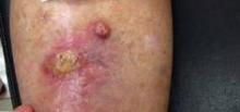
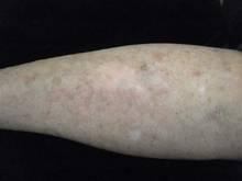
Dr. Roth gave an example of a patient with two well differentiated SCC lesions on her leg that he treated with radiation therapy. Twelve fractions of radiation were delivered with the SRT-100 (Sensus Healthcare, Boca Raton, Fla.). The patient ulcerated and had a fairly brisk response but did not report any pain. Follow-up was conducted at months 1,2 and 5. "From my standpoint, it was a remarkably good response."
Results with the SRT-100 are very reproducible, Dr. Roth said. Energy imparted across the port or opening in the metal shield is consistent. The device delivers a flat field of radiation with a fairly fast drop-off deeper into the skin. The depth of the nonmelanoma skin cancer lesion generally determines the setting, Dr. Roth said.
The control unit is outside the radiation room, thus avoiding the need to work behind a leaded glass window. The control unit counts down duration of treatment and features an emergency stop. "I use a baby monitor to speak with patients during treatment," Dr. Roth said. "I did have one patient who yelled that her eye shields fell off. I hit the emergency button, entered the room, and put the shields back on." The unit holds the reading (pauses therapy) and then allows physicians to continue when ready.
"Radiation therapy has been a part of dermatology basically forever. We have been treating skin cancers since before I was born," Dr. Roth said. "Now that we have some more choices, this should be a part of dermatology again. We should take this back."
Dr. Roth is a consultant and researcher for Sensus, but purchased his SRT-100 at full price.
EXPERT ANALYSIS FROM THE ANNUAL MEETING OF THE FLORIDA SOCIETY OF DERMATOLOGY AND DERMATOLOGIC SURGERY
Multiple Milia Signal Need for Genetic Testing
ORLANDO – Loeys-Dietz syndrome is a rare genetic disease that can be life threatening if not caught early, said Dr. Milan J. Anadkat.
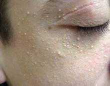
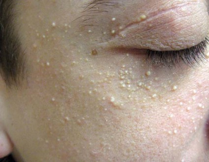
Children with the connective tissue disorder often present with up to 50 or more milia, as well as velvety and translucent skin, easy bruising, varicose veins, and atrophic scars. Craniosynostosis, cleft lip or palate, and ocular hypertelorism are among the potential craniofacial abnormalities.
"If you have a patient less than 10 years old who has these milia and craniofacial features, they need to be genetically tested," said Dr. Anadkat of the division of dermatology at Washington University in St. Louis.
The experience at Washington University includes at least 25 children with Loeys-Dietz syndrome. Exposure to multiple patients with such a relatively rare disorder is an advantage of practicing dermatology at a large academic center, Dr. Anadkat said at the annual meeting of the Florida Society of Dermatology and Dermatologic Surgery.
Loeys-Dietz syndrome, first described in 2005 by Dr. Bart Loeys and Dr. Hal Dietz, affects connective tissue throughout the body. There are different subtypes, receptor mutations, and phenotypes, but what is most remarkable is that 75% of cases present as de novo, spontaneous mutations.
"The importance of knowing about Loeys-Dietz syndrome has to do with the cardiovascular complications," he said. This autosomal dominant genetic disorder is part of the subset of aortic aneurysm syndromes that includes Marfan syndrome.
Aggressive cardiovascular disease, aortic dissection by the teenage years (or "decades earlier than what we see in Marfan syndrome"), and arterial tortuosity are among the serious, extracutaneous manifestations, said Dr. Anadkat. Because arterial tortuosity can occur anywhere in the body (not just around the heart), a total body vascular scan is warranted.
Presence of milia may facilitate an early distinction from Marfan syndrome. Other syndromes with milia to include in the differential diagnosis include Bazex-Dupré-Christol syndrome, Rombo syndrome, and Rasmussen syndrome.
Patients with Loeys-Dietz syndrome also can present with musculoskeletal anomalies such as joint laxity, club foot, pectus deformity, scoliosis, and cervical spine abnormalities. "This could explain how, for decades, we saw patients with Marfanlike abnormalities who did not have a genetic test positive for Marfan syndrome," Dr. Anadkat said.
He and his associates published a report on four unrelated patients with Loeys-Dietz syndrome who developed milia in early childhood and reported increases in their number over time (Arch. Dermatol. 2011:147:223-6). Although more needs to be studied regarding specific mutations and phenotypes, each of these patients had the same TGFBR2 mutation.
More information on the disease is available online at the Loeys-Dietz Foundation website.
Dr. Anadkat said that he had no relevant financial disclosures.
EXPERT ANALYSIS FROM THE ANNUAL MEETING OF THE FLORIDA SOCIETY OF DERMATOLOGY AND DERMATOLOGIC SURGERY
Don't Disregard High-Dose Brachytherapy for Skin Cancer
ORLANDO – Although Mohs surgery remains a mainstay of skin cancer treatment, some patients benefit from targeted brachytherapy of their basal or squamous cell carcinoma lesions, according to Dr. Michael E. Kasper.
Patients who are elderly, infirm, or on blood thinners are good candidates for noninvasive brachytherapy using high-dose, small surface applicators, Dr. Kasper said. This therapeutic strategy also works well for treating lesions in anatomic locations at risk for delayed surgical healing.
"We are treating various small lesions with these surface applicators," Dr. Kasper said. For example, superficial squamous cell carcinoma (SCC) lesions up to 2 cm can be targeted "where we feel comfortable about the visible margins," he said at the annual meeting of the Florida Society of Dermatology and Dermatologic Surgery.
"We also see patients who are tired of [invasive resection] or who are poor candidates for surgery, and that is really the bulk of our patients," Dr. Kasper said. He assured meeting attendees that his goal as a radiation oncologist is not to take skin cancer patients away from dermatologists. "We’re not interested in treating 40- or 50-year-olds and really competing," said Dr. Kasper of Lynn Cancer Institute at Boca Raton (Fla.) Regional Hospital. "We are interested in working with dermatologists and really helping you with those patients who might be neglected or who are at high risk of developing a serious recurrence."
Neglected patients may include nursing home residents who do not get medical attention for their skin cancer in its earlier stages, he said.
Available data point to good local control and cosmesis for a majority of patients, Dr. Kasper said. A typical patient might experience acute effects such as crusting and some mild erythema about 10 days to 2 weeks after brachytherapy of a well differentiated SCC lesion of the lower extremity. More brisk erythema also occurs in about 10%-15% of patients, Dr. Kasper said.
Late hypopigmentation also develops in about 10% of patients, he said. "We are also seeing a few telangiectasias, but it’s fairly mild."
Interpret postradiation therapy biopsies with caution, Dr. Kasper warned. Of the 240 patients treated to date at his institution over about 6.5 years, there were three documented recurrences, including a couple at 3 and 4 months. "While we are counting those as recurrences, they probably aren’t. That’s way too early to biopsy these lesions." Use discretion and ideally wait until you see clear progression prior to performing a postradiation biopsy, he said. "There certainly are false positives that occur due to delayed tumor regression. The cancer cells die when they reproduce and many of these are slow growing tumors. So we would not expect them to all be completely resolved at 3 or 4 months although clinically, on the surface, they can appear that way."
Historically brachytherapy was delivered as a low-dose treatment over a long period of time at many sessions. Low-dose brachytherapy is typically in the 0.4 to 2.0 Gy/hr range, medium dose is greater than 2 and up to 12 Gy/hr, and the high dose exceeds 12 Gy/hr.
Availability of high-dose rate brachytherapy was a "major breakthrough" because patients no longer had to lie in the hospital all weekend to receive treatment. "It was a bit controversial at first, but now there are really good data to show there are radiobiologic reasons why the high dose rate may actually be advantageous in killing cancer cells compared to this low trickle effect that we were using with low-dose regimens."
Dr. Kasper determined that up to 30 sessions of low-dose rate brachytherapy can be a real impediment to patient compliance and worked to design a safe and effective regimen delivered in fewer sessions. "We wanted to be at six treatments and worked backwards." Striking a balance between the dose-response rate and the potential late side effects was another consideration.
A meeting attendee asked about the relative cost of brachytherapy, compared with other treatment modalities. Brachytherapy is generally more expensive than Mohs surgery, Dr. Kasper replied. The avoidance of cancer recurrences will hopefully justify the higher initial costs, he said. He added that brachytherapy costs are coming down, more so in outpatient centers, compared with hospital settings.
Dr. Kasper said he receives consulting fees from Nucletron/Elekta.
EXPERT ANALYSIS FROM THE ANNUAL MEETING OF THE FLORIDA SOCIETY OF DERMATOLOGY AND DERMATOLOGIC SURGERY

