User login
Damian McNamara is a journalist for Medscape Medical News and MDedge. He worked full-time for MDedge as the Miami Bureau covering a dozen medical specialties during 2001-2012, then as a freelancer for Medscape and MDedge, before being hired on staff by Medscape in 2018. Now the two companies are one. He uses what he learned in school – Damian has a BS in chemistry and an MS in science, health and environmental reporting/journalism. He works out of a home office in Miami, with a 100-pound chocolate lab known to snore under his desk during work hours.
Systemic inflammation expands clinical challenge in IBD
ORLANDO – Prescribing optimal therapy for a patient with inflammatory bowel disease can be challenging under ordinary circumstances, but add an extra-intestinal manifestation and the complexity grows greater still. Until more evidence-based findings emerge to help guide and individualize systemic or combination therapy, one of the advantages of biologics – their ability to target the intestine – can be a drawback when patients present with other manifestations.
To highlight the challenges and propose management strategies, Corey A. Siegel, MD, presented the case of a 45-year-old man seeking care 8 years after a diagnosis of ileocolonic Crohn’s disease. Seven years earlier, after the patient failed 5-aminosalicylic acid medication and prednisone therapy, clinicians prescribed infliximab (Remicade, Janssen) monotherapy. He experienced a “great response,” Dr. Siegel said at the Advances in Inflammatory Bowel Diseases meeting.
“Unfortunately, he had a real-life delayed hypersensitivity reaction 2 years ago with no drug present at trough and good antibodies, equal to 12 on a drug tolerant assay,” added Dr. Siegel, director of the Inflammatory Bowel Disease Center at Dartmouth-Hitchcock Medical Center in Lebanon, N.H., and moderator of a case discussion panel session. So physicians initiated combination therapy with adalimumab (Humira, AbbVie) and methotrexate, but the patient “never felt as good as he did on infliximab, even with weekly dosing.”
A colonoscopy 9 months ago revealed mild to mildly active ileal and ascending colon disease. “So he still has residual disease present, even with weekly dosing of adalimumab,” Dr. Siegel explained. At the time, clinicians prescribed vedolizumab (Entyvio, Takeda) and the patient reported IBD symptom improvement. “He was doing better but not fantastic, and now his joints were bothering him, and they never bothered him before.” The man reported joint pain in his hands, knees, and hips. A more recent, follow-up colonoscopy revealed improvement, although mildly active disease was still present.
Do we need to dose-optimize vedolizumab or is it time to move on here? Dr. Siegel asked an expert panel: Bruce E. Sands, MD, David T. Rubin, MD, and Gary L. Lichtenstein, MD.
Instead of considering a third anti–tumor necrosis factor (anti-TNF) agent when the patient has already not responded fully to others in that class, Dr. Sands suggested ustekinumab (Stelara, Janssen). “It could be that ustekinumab would be a better choice versus dose escalation of vedolizumab, unless you add something to the dose-escalated vedolizumab to treat the arthralgias like celecoxib.”
Next, Dr. Siegel asked if the arthralgias are truly extra-intestinal manifestations of inflammatory bowel disease “or is it what my patients are telling me – what they see all over the Internet – that vedolizumab causes arthralgias or arthropathies?
“That is an important question,” said Dr. Rubin, section chief of gastroenterology, hepatology, and nutrition at the University of Chicago. “My question back to you is when did the joint pain start – right after vedolizumab was initiated? Right after steroids were tapered? Or after the patient was on stable dosing for some time?”
The arthralgias seemed to start right after the patient started vedolizumab, Dr. Siegel said. But when clinicians inquired a little further, the patient reported “it was when he came off the adalimumab that things really started.”
It could be that systemically active infliximab and adalimumab with methotrexate were essentially covering up an extra-intestinal manifestation that was later uncovered with the selective mechanism of vedolizumab, Dr. Rubin said. “If that’s true, I don’t think I would give him more vedolizumab to treat his joint pain – that certainly wouldn’t do it. I would make sure his disease is responding from mucosal view, then I would add methotrexate or even consider sulfasalazine.”
A meeting attendee said that the patient did very well with infliximab, and asked “can we ever go back?”
“Probably not easily,” Dr. Sands said. “For someone with a bona fide delayed hypersensitivity reaction – I wouldn’t go there, and I wouldn’t think that you could.”
Another attendee asked if there is a role for adding another biologic to vedolizumab. “We hope to initiate a study soon looking at combination of biologics, such as vedolizumab with adalimumab, with and without an immunomodulator, Dr. Sands replied. They cover somewhat different targets – so it’s sort of a ‘belts and suspenders approach’ … but there are no data whatsoever [yet].”
Dr. Siegel wrapped up the case discussion with the patient’s outcomes. “We did move him on to ustekinumab. He felt better and his arthralgias went away.”
The meeting was sponsored by the Crohn’s & Colitis Foundation of America.
Dr. Siegel and Dr. Rubin disclosed ties with AbbVie, Janssen, and Takeda. Dr. Sands disclosed ties with AbbVie, Janssen Biotech, and Takeda. Dr. Lichtenstein disclosed ties with AbbVie, Janssen Orthobiotech, and Takeda.
AT AIBD 2016
Key clinical point: Extra-intestinal manifestations of inflammatory bowel disease can leave gastroenterologists wondering about the best approach to treatment.
Major finding: Less gut-specific drug action may actually be better for these patients.
Data source: Panel discussion of challenging cases at AIBD 2016.
Disclosures: Dr. Siegel and Dr. Rubin disclosed ties with AbbVie, Janssen, and Takeda. Dr. Sands disclosed ties with AbbVie, Janssen Biotech, and Takeda. Dr. Lichtenstein disclosed ties with AbbVie, Janssen Orthobiotech, and Takeda.
Intensifying vedolizumab could counter loss of response in IBD
ORLANDO – In a study of 644 people with moderate to severe inflammatory bowel disease treated with vedolizumab (Entyvio, Takeda Pharmaceuticals), 346 achieved remission or a significant response. This 54% response rate includes 192 people with Crohn’s disease and 154 others with ulcerative colitis.
However, after a median of 143 days, some of the initial responders experienced a loss of response to vedolizumab therapy.
“In our real-world study of vedolizumab use in patients with IBD predominantly refractory to anti-TNF [tumor necrosis factor] therapy, loss of response was observed in about 40% of patients at 12 months,” said Eugenia Shmidt, MD, a gastroenterology fellow at the Icahn School of Medicine at Mount Sinai hospital in New York City.
To counter the loss of response, Dr. Shmidt and her colleagues shortened the dosing interval of vedolizumab to either 4 or 6 weeks in a subgroup of 36 patients who lost response. They also shortened the dosing interval to try to attempt an initial response in a subgroup of 47 people who did not respond to initial induction therapy in the first place. “Dose intensification led to a successful recapture of response in 32% of patients who had initial response and in 19% of patients who did not have an initial response,” Dr. Shmidt said at the Advances in Inflammatory Bowel Diseases meeting.
“We find it interesting that there was greater success in recapturing significant response in patients who responded to vedolizumab initially compared to those who did not initially respond,” she said when asked if she found any of her findings surprising. “This emphasizes a likely need for different management strategies for initial vedolizumab responders and nonresponders, as well as early recognition of the need for dose optimization,” she added.
The study included adults with mild to moderate inflammatory bowel disease, based on clinical factors or confirmed through endoscopy. There were no significant differences in severity of disease between patients who were able to recapture response versus those were not. The mean age among the 374 participants with Crohn’s disease was 39 years; similarly, the mean age in the group of 270 with ulcerative colitis was 41 years. Mean duration of disease was 15 years and 9 years in the two groups, respectively.
To identify risk factors associated with loss of response to vedolizumab, Dr. Shmidt and her colleagues performed univariable and multivariable Cox proportional hazard analyses. They found concomitant use of an immunomodulator for Crohn’s disease was protective against loss of response (hazard ratio, 0.44). In the ulcerative colitis group, a baseline serum albumin level below a normal value achieved lower vedolizumab response rates; a value below 3.2 g/dL was an independent predictor of cumulative loss of response over time (HR, 2.39).
Inflammatory bowel disease patients treated at higher volume centers, which were defined as enrolling at least 100 patients in the study, had higher rates of loss of response to vedolizumab (HR, 1.92 on multivariable analysis).
The multicenter VICTORY consortium coinvestigators in this study are affiliated with Mayo Clinic in Rochester, Minn.; Cleveland Clinic Foundation in Ohio; University of California, San Diego; New York University in New York City; Montefiore Medical Center/Albert Einstein College of Medicine in the Bronx, N.Y.; and Dartmouth-Hitchcock Medical Center in Lebanon, N.H.
Future studies are warranted to evaluate the pharmacodynamics and pharmacokinetics of vedolizumab, the authors noted, as well as to determine an optimal dosing strategy among people with high drug clearance.
The study was funded in part by Takeda Pharmaceuticals. Dr. Eugenia Shmidt did not have any relevant disclosures. The meeting was sponsored by the Crohn’s & Colitis Foundation of America.
Key clinical point: Some patients with moderate to severe inflammatory bowel disease experience loss of response to vedolizumab over time.
Major finding: At a median 143 days, 41% of patients with Crohn’s disease and 42% of those with ulcerative colitis who initially had a strong response or experienced remission with vedolizumab experienced subsequent loss of response.
Data source: Study of 644 patients with moderate to severe Crohn’s disease or ulcerative colitis followed for 12 months.
Disclosures: The study was funded in part by Takeda Pharmaceuticals. Dr. Eugenia Shmidt did not have any relevant disclosures.
Why ustekinumab dosing differs in Crohn’s disease
ORLANDO – Preclinical studies and years of clinical experience using the monoclonal antibody ustekinumab (Stelara, Janssen Biotech) in psoriasis and psoriatic arthritis offer important clues to any gastroenterologist perplexed by the official Food and Drug Administration indication, dosing frequency, and intensity for Crohn’s disease. Phase II and phase III findings also reveal where the monoclonal antibody may offer particular advantages, compared with other agents.
“Ustekinumab landed in your lap in September. You’re probably all trying to figure out how to get the ID formulation paid for with insurance,” William J. Sanborn, MD, professor and chief of the division of gastroenterology at the University of California, San Diego, said at the Advances in Inflammatory Bowel Diseases meeting. “But this is now the reality that you have this in your Crohn’s practice.”
The FDA approved ustekinumab to treat adults with moderately to severely active Crohn’s disease who 1) failed or were intolerant to immune modulators or corticosteroids but did not fail tumor necrosis factor (TNF) blockers or 2) failed or were intolerant to one or more TNF blockers. Dr. Sanborn and colleagues observed a significant induction of clinical response in a subgroup of patients who previously failed a TNF blocker in an early efficacy study (Gastroenterology. 2008;135:1130-41). “This is where the idea of initially focusing on TNF failures came from,” he added at the meeting sponsored by the Crohn’s & Colitis Foundation of America.
Induction dosing in Crohn’s disease is intravenous versus subcutaneous in psoriasis and psoriatic arthritis, in part because of the same study. “It looked like relatively better bioavailability and relatively better effect with intravenous dosing,” Dr. Sanborn said. “In Crohn’s disease, it’s a completely different animal.”
Official induction dosing is approximately 6 mg/kg in three fixed doses according to patient weight in Crohn’s disease. The 6-mg/kg dose yielded the most consistent response, compared with 1-mg/kg or 3-mg/kg doses in a subsequent phase IIb study (N Engl J Med. 2012;367:1519-28).
The most consistent induction results at weeks 6 and 8 were observed with 6 mg/kg ustekinumab versus 1 mg/kg or 3 mg/kg.
Dr. Sanborn and coinvestigators also saw “numeric differences in drug versus placebo for remission at 6 and 8 weeks “but it was not that clear from the phase II trial what the remission efficacy was, so that needed more exploration to really understand.”
Another distinction for ustekinumab in Crohn’s disease is the approved maintenance dosing of 90 mg subcutaneously every 8 weeks versus a 12-week interval recommended for psoriasis. “Why so much more in Crohn’s disease, and is that necessary?” Dr. Sanborn asked.
Based on changes in C-reactive protein levels and a “rapid drop” in Crohn’s Disease Activity Index scores by 4 weeks, “clearly efficacy was there for induction,” he said. Ustekinumab has a “quick onset – analogous to the TNF blockers.”
“These were quite encouraging data, and paved the way to move on to phase III [studies],” Dr. Sanborn said. The preclinical studies up to this point focused on patients with Crohn’s disease who previously failed TNF blockers. However, “in clinical practice, we would be interested to know if it would work in anti-TNF naive or nonfailures as well.”
So two subsequent studies assessed safety and efficacy in a TNF blocker–failure population (UNITI-1 trial. Inflamm. Bowel Dis. 2016 Mar;22 Suppl 1:S1) and a non-TNF failure population of patients who did fail previous conventional therapy such as steroids or immunomodulators (UNITI-2 trial).
Clinical response and remission steadily rose following induction up to a significant difference versus placebo at 8 weeks in the non–TNF failure population. “Remember, in the phase IIa study, the remission rates were not as clear-cut, so this really nails down this as a good drug in both patient populations,” Dr. Sanborn said.
To evaluate long-term maintenance, investigators rerandomized all participants in the UNITI-1 and UNITI-2 studies. They saw a 15% gain in clinical remission out to week 44, compared with placebo. Dr. Sanborn noted that ustekinumab has a relatively long half-life, so the difference in patients switched to placebo may not have been as striking. “In practice it’s important to know the on-time and off-time of this agent, and I think the clinical trials make that clear.”
The trials also show that 12-week dosing works, Dr. Sanborn said. “You see about 20% gain for every 8-week dosing. You get extra 5% or 10% extra on all outcome measures at 8 weeks, compared to 12 weeks dosing, with no difference in safety signals.” He added, “So more intensive dosing of 90 mg every 8 weeks is what ended up getting approved in the United States.”
Safety profile
So what does all the preclinical evidence suggest about safety of ustekinumab? The UNITI trials combined included more than 1,000 patients, and there were no deaths, Dr. Sanborn said. “Usually with TNF blockers in 1,000 patients you would see a few deaths.”
Patient withdrawals from the preclinical studies were also relatively low, Dr. Sanborn reported. “With ustekinumab monotherapy, drug withdrawal is only 3% or 4%, so it seems to be different from TNF blockers in that sense [too].”
In addition, the rates of adverse events were similar between placebo (83.5%) and ustekinumab’s combined every 8 week and every 12 week dosing groups through 44 weeks (81.0%), Dr. Sanborn said. The rates of serious adverse events were likewise similar, 15.0% and 11.0%, respectively. Reported malignancy included two cases of basal cell skin cancers, one in the placebo group and one in the every-8-week dosing group, he added.
“So all those black box warnings you’re used to worrying about with TNF blockers – serious infections, about opportunistic infections, malignancy – there is no black box warning with this agent around that.”
Dr. Sanborn noted that the FDA labeling reports infections. “We know Crohn’s disease patients are [also] getting azathioprine, steroids, methotrexate, so you will see some infections, but there wasn’t a consistent opportunistic infection signal.”
One case of reversible posterior leukoencephalopathy syndrome is included on the labeling. Dr. Sanborn also put this in perspective: “With all the experience in psoriasis and psoriatic arthritis, and the clinical trials [in IBD], there is just one case. So the relationship is not very clear.”
“The safety signals with ustekinumab are really very good. It seems to be an extremely safe agent – we really don’t see much in terms of infections,” Brian Feagan, MD, an internist and gastroenterologist at the University of Western Ontario in London, said in a separate presentation at the conference. “We don’t have a lot of long-term experience with ustekinumab in Crohn’s disease, but we have a lot of experience in psoriasis, and it’s a safe drug.”
“Ustekinumab may be our first really valid monotherapy, with less immunogenicity,” Dr. Feagan said.
EXPERT ANALYSIS FROM AIBD 2016
Initial data on biosimilars in IBD are encouraging, but more needed
ORLANDO – The rapidly evolving field of biologic and biosimilar agents can leave gastroenterologists reeling in terms of what role biosimilars will play in practice and how best to utilize them for a patient with ulcerative colitis or Crohn’s disease. Limited data so far – primarily case series and limited postmarketing findings – suggest that a biosimilar can be as safe and effective as the innovator biologic. However, more rigorous research and more research overall are needed to confirm this comparative efficacy and to address unanswered questions around switching and cost effectiveness.
The uncertainty stems in part from a lack of prospective, randomized comparison studies to guide evidence-based clinical practice, Miguel D. Regueiro, MD, gastroenterologist and IBD clinical medical director at the University of Pittsburgh Medical Center.
As clinicians await more definitive evidence, a number of limited studies have been published in the past year assessing biosimilars in inflammatory bowel disease (IBD). “The clinical response ranges were high, in most studies more than two-thirds [of patients] had a response, but again there are no head-to-head studies,” Dr. Regueiro said at the meeting sponsored by the Crohn’s & Colitis Foundation of America.
“We don’t have the data yet in IBD.”
A review of the literature on anti-tumor necrosis factor antibody biosimilar Inflectra (infliximab-dyyb, Celltrion) supports its bioequivalence to infliximab (Remicade, Janssen) in ankylosing spondylitis (Aliment Pharmacol Ther. 2015;42:1158-69). However, dosing regimens and use of concomitant medications differ in the IBD population, the investigators noted, so the case series and short-duration postmarketing results published in the literature leave unanswered questions for gastroenterologists.
Just because biosimilars appear bioequivalent so far in IBD, it does not mean this new class of agents is free of controversy. When the U.S. Food and Drug Administration approved Inflectra in April 2016 based on evidence in ankylosing spondylitis and rheumatoid arthritis, it extrapolated the approval to all conditions for which infliximab is approved, including ulcerative colitis and Crohn’s disease. However, this “highly controversial approach has been criticized by various rheumatology and gastroenterology professional societies around the world,” according to authors of a critical review on biosimilars in IBD (Inflamm Bowel Dis. 2016;22:2513-26).
Clinicians can safely switch patients from infliximab to Inflectra, according to the noninferiority phase IV NOR-SWITCH study conducted in Norway. Researchers assessed almost 500 patients on stable infliximab treatment for a minimum of 6 months prior to switching. However, this was a multi-indication study that included inflammatory bowel disease patients and many others, Dr. Regueiro said. “When you read the fine print in these studies, especially with Crohn’s disease, you start to see differences,” he added.
Although the evidence is encouraging so far, “we do not have prospective, comparative studies yet.”
During a Q&A panel session, a meeting attendee asked if clinicians will be able to use the same laboratory assay used to check a biologic’s drug and antibody levels for its biosimilar. “Our understanding is you will not be able to use the same assay as you do for infliximab. There are companies developing assays specifically for the biosimilars, and I think they will be available in addition to the ones for the innovator agents.”
AT AIBD 2016
Key clinical point: Initial research on biosimilars in ulcerative colitis and Crohn’s disease suggests similar efficacy and safety to originator biologic agents, which researchers hope to confirm in larger, prospective studies underway.
Major finding: The clinical response ranges were high – in most studies more than two-thirds of patients had a response to a biosimilar agent.
Data source: Literature review, expert opinion.
Disclosures: Dr. Regueiro reported he is a consultant for AbbVie, Janssen, Pfizer, Takeda, and UCB.
Sonovaginography bests negative ‘sliding sign’ in predicting deep infiltrating endometriosis
ORLANDO – Direct visualization with sonovaginography had greater success in predicting rectal/rectosigmoid deep infiltrating endometriosis than did negative transvaginal ultrasound uterine “sliding sign,” according to the findings of a prospective study of 189 women.
“Both performed quite well,” but sonovaginography was superior for predicting rectal deep infiltrating endometriosis on all measures, including accuracy – 92% vs. 88%, said Bassem Gerges, MBBS, an ob.gyn. at the University of Sydney, Kingswood.
Dr. Gerges and his colleagues evaluated 189 women of reproductive age who were scheduled for operative laparoscopy at a tertiary referral center for women. The patients had a history of chronic pelvic pain and/or endometriosis and presented between 2009 and 2013.
The women first had transvaginal ultrasound to determine if their uterine sliding sign was positive or negative, followed by sonovaginography to assess the posterior pelvic compartment for rectal or rectosigmoid deep infiltrating endometriosis. All patients then underwent laparoscopic surgery for endometriosis.
Laparoscopy revealed pouch of Douglas obliteration in 47 of the 189 women and rectal and/or rectosigmoid deep infiltrating endometriosis in 43 women.
The sensitivity of sonovaginography to predict deep infiltrating endometriosis was 88%, compared with 74% for the sliding-sign approach. Specificity was the same with the two methods at 93%. The positive predictive value was 79% vs. 74%, respectively, and the negative predictive value was 97% vs. 93%.
“These findings can help clinicians with preoperative planning,” Dr. Gerges said at the meeting, which was sponsored by AAGL.
Dr. Gerges and his colleagues also identified 11 false-negative cases in which the sliding sign was positive but laparoscopy confirmed rectal deep infiltrating endometriosis.
Previous research suggests that, in women with suspected endometriosis, a negative transvaginal ultrasound uterine sliding sign can predict rectal or rectosigmoid deep infiltrating endometriosis (Ultrasound Obstet Gynecol. 2013;41[6]:692-5, J Ultrasound Med. 2014;33:315-21). A negative sliding sign indicates the presence of uterorectal adhesions and whether the pouch of Douglas might be obliterated. The current study, however, suggested that sonovaginography might be the better method.
Dr. Gerges reported having no relevant financial disclosures.
AT THE AAGL GLOBAL CONGRESS
Plasma energy ablation yields pregnancy rates similar to cystectomy
ORLANDO – The first study to directly compare plasma energy treatment of ovarian endometriomas to cystectomy demonstrates similar postintervention pregnancy rates, suggesting that plasma energy ablation may be a comparable treatment option.
Researchers evaluated 104 women seeking pregnancy after 1 year or more of infertility. Women presented with unilateral or bilateral ovarian endometriomas larger than 3 cm between January 2009 and June 2014. Clinicians treated 64 patients with plasma energy ablation and another 40 with cystectomy and followed them to compare pregnancy rates.
After at least 1 year of follow-up, pregnancy rates were 68% following plasma energy ablation, compared with 80% after cystectomy. Of the 76 pregnancies, 24 were due to spontaneous conception, including 40% of pregnancies in the plasma energy group and 18% in the cystectomy group. Even after adjustment for multiple factors, the type of intervention had no statistically significant impact on achieving a subsequent pregnancy.
“Ablation using plasma energy may be considered a valuable tool that allows a high pregnancy rate,” Basma Darwish, MD, an ob.gyn. at Rouen (France) University Hospital, said at the meeting, which was sponsored by AAGL.
These similar outcomes were observed despite a higher prevalence of risk predictors for infertility in the plasma energy group at baseline. For instance, women in this cohort were significantly older and had significantly higher revised American Fertility Society (rAFS) classification scores, as well as higher rates of pouch of Douglas obliteration, deep endometriosis, and colorectal localizations.
“Endometrial ablation using plasma energy allows good postoperative [pregnancy] rates, that is well known,” Dr. Darwish said, but the technique has not been directly compared with cystectomy outcomes.
Pregnancy rates remained similar in both groups at 24 and 36 months. The probability of pregnancy was 61% in the plasma energy group versus 69% in the cystectomy group at 24 months. At 36 months, these rates changed to 84% and 78%, respectively.
A unique property of plasma energy ablation is “very limited thermal spread, both in depth and laterally,” Dr. Darwish said.
A lack of randomization is a potential limitation of the study. “Each surgeon chose the technique he or she was best at, which may have explained our good outcomes.” Dr. Darwish said. Strengths of the study include a prospective design and follow-up to 5 years. In addition, the six centers involved in the study included both private and public hospitals, “so it’s a good reflection of what happens in real life.”
The investigators plan to conduct a randomized controlled trial to confirm these findings.
AT THE AAGL GLOBAL CONGRESS
Narrow band imaging could expand endometriosis detection
ORLANDO – Narrow band imaging detects neovascularization associated with endometriosis and can be a useful adjunct to laparoscopic white light evaluation, a prospective cohort trial of 53 women with pelvic pain suggested.
The women in the study had no deep infiltrating endometriosis on preoperative ultrasound. Investigators then conducted a standard laparoscopic survey of the pelvis with white light to identify areas of suspected superficial endometriosis, followed by secondary analysis with narrow band imaging.
In the group of 32 patients, follow-up biopsy results confirmed endometriosis in 24 women, including 7 who also had lesions detected by narrow band imaging. Six of these seven were positive for endometriosis on histology. The women were enrolled in the study from September 2014 to October 2015.
“We found that the [narrow band imaging] is useful in detecting additional areas in patients who had histopathology-proven endometriosis,” said Tony J. Ma, MBBS, a fellow at Mercy Hospital for Women in Melbourne.
In the group of 21 women with no white light lesions, narrow band imaging detected four suspicious lesions. However, these four were not positive for endometriosis.
Narrow band imaging is a mixture of blue and green light, opposite of red color. “It causes blood vessels to be more visually prominent,” Dr. Ma said at the meeting sponsored by AAGL. White light laparoscopy for detection of endometriosis “is the gold standard … but depends on experience of [the] surgeon and severity of the disease.”
These findings support those of a 2008 study that reported a high detection rate of lesions with narrow band imaging, Dr. Ma said. In this earlier prospective cohort study of 20 women, 7 patients with endometriosis who had been ruled out by white light evaluation had a positive histologic finding with narrow band imaging (J Minim Invasive Gynecol. 2008 Sep-Oct;15[5]:636-9).
“Narrow band imaging is a simple, noninvasive adjunct that can assist in the identification of additional sites of endometriosis at laparoscopy,” Dr. Ma said. “The shortened depth of field with narrow band imaging requires the operator to inspect the surface quite closely.”
The study topic is an important one because endometriosis is such a challenge to diagnose and treat, said study discussant Sawsan As-Sanie, MD, a minimally invasive gynecologic surgeon at the University of Michigan, Ann Arbor. She estimated that with the current approach – visualization by white light followed by histopathology – “about 50%-70% of patients we think have endometriosis ultimately do.”
But several unanswered questions remain, Dr. As-Sanie said, such as whether the narrow band imaging findings are clinically relevant and whether they will lead to improved patient outcomes.
“We don’t know the answer yet,” she said. “The end game is really only relevant if we improve patient outcomes.”
Dr. Ma reported having no relevant financial disclosures.
AT THE AAGL GLOBAL CONGRESS
New-onset pain rare after Essure placement
ORLANDO – While some women report new-onset pelvic pain after placement of an Essure sterilization device, results of a retrospective study suggest this pain is actually associated with placement of the device in about 1% of cases.
Among 1,430 women who had an Essure micro-insert (Bayer) placed at a tertiary care hospital in Canada from June 2002 to June 2013, 62 secondary surgeries were performed, including some for removal of fallopian tubes and removal of the device.
In total, 27 patients reported new-onset pelvic pain after Essure placement and another 11 reported worsening of previous pain. Upon further workup, 15 of the 27 women in the new-onset pain group had another possible explanation for their pain, including surgical or pathology findings of endometriosis or adenomyosis. The investigators concluded there was a link between the pain and the device in just 12 (0.8%) of the women.
Among these dozen patients, the investigators linked the pain in eight women to perforation or migration of the Essure device. Investigators found no other obvious cause for the new-onset pain in the remaining four patients and attributed it to the Essure device.
Set realistic expectations, take a comprehensive pain history, and reassure women when they report post-Essure placement pain, James Robinson, MD, a minimally invasive gynecologic surgeon at Medstar Washington Hospital in Washington, D.C., advised at the meeting, which was sponsored by AAGL.
Dr. Robinson pointed out that there is no standardized approach to managing women with complaints of pain or guidelines on how best to remove the device. Imaging to confirm proper placement and to rule out other sources of pelvic pain, followed by medical or surgical management as warranted, can be effective strategies.
“There is less science here – it’s more the art of medicine, I think,” he said.
Dr. Robinson filled in as a presenter for one of the study coauthors, John A. Thiel, MD, of the University of Saskatchewan, Saskatoon, who was unable to attend the conference.
The study by Dr. Thiel and his colleagues suggests a thorough examination will typically reveal other reasons for pelvic pain and rules out the Essure device as the cause, Dr. Robinson said. The full findings of the study are published in the Journal of Minimally Invasive Gynecology (2016 Nov-Dec;23[7]:1158-1162).
Does removal help?
In a recently published case series of 29 women who had their Essure device removed laparoscopically because of pain, 23 reported relief following excision (Contraception. 2016 Aug;94[2]:190-2).
“Again, a subset had misplaced inserts or had another condition such as endometriosis,” Dr. Robinson said.
The majority of women whose pain resolved with removal of the devices reported their pain early on, so there is an important takeaway from this,” Dr. Robinson said. “We need to listen to our patients when they report pain shortly after device placement … and respond to that.”
Dr. Robinson advised physicians to be ready to surgically remove the device if that is warranted. “I think a lot of people doing these procedures are not comfortable taking their patient back to the operating room or don’t know who to send them to,” he said. “If you are going to place the device, you should be able to take it out or know someone who can.”
The bigger picture
Even though the Essure device is not frequently the cause of pelvic pain, physicians needs to be aware that some patients are likely to assume that it is.
Dr. Robinson pointed to a case in which a woman who had the Essure micro-insert placed 7 years earlier presented with a complaint of a bilateral tingling sensation over the course of 6 months. Online research led her to suspect Essure as the cause of her symptoms. However, on further investigation, it turned out she had relatively high levels of lead in her system from leaky pipes in her home. “It wasn’t an Essure issue,” he said. “But because it’s out there, people will jump to the conclusion that the foreign body is likely the cause of their problem.”
Ob.gyns. should become familiar with websites such as essureproblems.webs.com, which chronicle problems patients have reported with the device, he said.
When talking to patients, start with informed consent and listen to their concerns, Dr. Robinson advised. As part of the counseling about Essure permanent birth control, discuss the risks and benefits of alternatives, such as laparoscopic tubal ligation, long-acting reversible contraception, and vasectomy.
“I took the time to listen to the FDA hearing in Sept 2015 and … it moved me to listen to those patients, and I’ve been a huge advocate of Essure sterilization. I felt for a while I would never do another tubal ligation,” Dr. Robinson said. “But when you listen to patients who have real complaints, what sticks out in your mind is so many of these people are upset because no one took them seriously and listened to them.”
Key clinical point:
Major finding: Of 1,430 women who had the Essure inserts placed, just 12 had new-onset pain that was found to be related to the device.
Data source: Retrospective cohort study of 1,430 women treated at a tertiary care hospital in Canada.
Disclosures: Dr. Robinson reported having no financial disclosures.
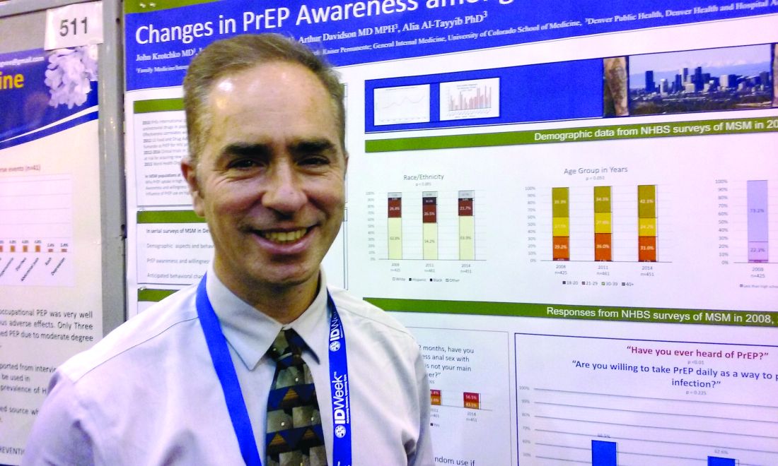
PrEP adoption lagging behind awareness in high-risk population
NEW ORLEANS – Awareness about preexposure prophylaxis (PrEP) is steadily increasing among men who have sex with men at high risk for HIV infection, but that increased knowledge did not translate into greater willingness to take the daily pill nor did it increase engagement in high-risk behaviors.
Using questions from the National HIV Behavioral Surveillance System (NHBS), investigators surveyed men who have sex with men in urban settings in 3-year cycles. Awareness about HIV PrEP, once-daily emtricitabine/tenofovir (Truvada) increased from 21% in 2008 to 28% in 2011 to 46% in 2014.
The increase from 2011 to 2014 was statistically significant (P less than .001). The Food and Drug Administration approved the preventive regimen in 2012.
Increased knowledge did not translate to greater willingness to take the daily pill – which has held steady at about 60% of over time.
The number of men who self-reported as HIV negative and sexually active in the previous 12 months included in the survey varied from 421 in 2008, to 461 in 2011, to 451 in 2014.
For people at elevated risk for HIV infection, PrEP also represents an opportunity to take greater control over behavior, according to a recent review (Curr Opin HIV AIDS. 2016;11:3-9). “When you get people in for counseling or condoms, you give them a sense of control,” said Dr. Krotchko of Denver Health Medical Center.
Most survey respondents said they anticipated they would use condoms just as frequently as before if taking PrEP (82%, 78%, and 78%, in 2008, 2011, and 2014, respectively). Similarly, the majority of respondents anticipated having the same number of sexual partners if taking PrEP (92%, 85%, and 89%). These differences were not statistically significant.
The findings indicate availability of HIV PrEP is not increasing unhealthy behaviors, as some may fear. “Riskier behavior while on PrEP has not been borne out by the literature,” Dr. Krotchko said.
Strengths of the study include directly targeting a high-risk population and identifying those with high-risk behaviors who could benefit from use of HIV PrEP. Self-reported anticipated changes may not reflect future behavior in all cases, a potential limitation, Dr. Krotchko pointed out.
The NHBS survey is funded by the Centers for Disease Control and Prevention. IDWeek 2016 comprises the combined meetings of the Infectious Diseases Society of America, the Society for Healthcare Epidemiology of America, the HIV Medicine Association, and the Pediatric Infectious Diseases Society.
AT IDWEEK 2016
Key clinical point: Adoption and use of HIV PrEP continues to lag behind studies showing its effectiveness in the high-risk population of men who have sex with men.
Major finding: Despite increasing awareness, willingness to use HIV PrEP among MSM remained steady at about 60% over time in a series of national behavioral health surveys.
Data source: The National HIV Behavioral Surveillance System.
Disclosures: Dr. Krotchko had no relevant disclosures.
Ultrasound effective in diagnosing occult hernia in women
ORLANDO – Ultrasound appears to be an effective tool in diagnosing occult hernia in women with unexplained chronic pelvic pain, according to a retrospective cohort study of 96 women.
“As gynecologists, we are likely to see those patients with chronic pelvic pain in our clinic before other specialists due to the location of their pain,” said Joelle Aoun, MD, an ob.gyn. in the division of minimally invasive gynecologic surgery at Henry Ford Health System in Detroit. “So it’s very important to recognize women with a high clinical suspicion for occult hernia and evaluate them in order to prevent a delayed diagnosis and prolonged suffering.”
Hernias can be more difficult to diagnose in women than in men, Dr. Aoun said at the meeting sponsored by AAGL, and the literature offers conflicting findings since most hernia studies are conducted in men or with mixed gender cohorts.
Dr. Aoun and her coinvestigators conducted a retrospective cohort study from January 2005 to July 2016, identifying 96 women with chronic pelvic pain and focal inguinal tenderness. Protruding fat or visceral tissue on physical exam or observed visually led clinicians to suspect a hernia. A single sonographer performed the musculoskeletal ultrasound.
Investigators diagnosed an occult hernia in more than half of the patients (51 women) based on the physical exam and ultrasound findings. Diagnoses included inguinal, femoral, Spigelian, and umbilical hernias.
All women with an ultrasound-diagnosed hernia were referred to general surgery. A majority – 69% of women – underwent surgical exploration. The remaining 31% of women who declined tended to have lower pain scores, Dr. Aoun said. Surgeons confirmed the hernia diagnosis in 97% of the women, or 34 out of the 35 women who had consented to surgery.
The group with a hernia was older and more likely to have arthritis, but otherwise did not differ significantly from the nonhernia cohort.
“We believe musculoskeletal ultrasound is valuable as an initial imaging modality due to its high predictive value, low cost, and noninvasiveness,” Dr. Aoun said.
Chronic pelvic pain is not uncommon, affecting approximately 15% of women during their reproductive years often with significant impacts on quality of life, workplace productivity, and health care utilization, Dr. Aoun said. This presentation also accounts for about 10% outpatient gynecology consultations and approximately 40% of laparoscopies in the United States, she added.
Dr. Aoun and her colleagues are planning a subsequent study of all the women who opted not to undergo surgery to determine their follow-up pain profiles based on chart review and phone interviews.
Dr. Aoun reported having no relevant financial disclosures.
AT THE AAGL GLOBAL CONGRESS
Key clinical point:
Major finding: About 53% of women with chronic pelvic pain were diagnosed with an occult hernia following physical exam and ultrasound. Surgeons confirmed the hernia diagnosis in 34 out of 35 patients who consented to surgery.
Data source: A retrospective cohort study of 96 women with chronic pelvic pain and focal inguinal tenderness.
Disclosures: Dr. Aoun reported having no relevant financial disclosures.


