User login
Study found two-way link between IBD and cervical cancer
Women with Crohn’s disease had about a 53% greater risk of developing cervical cancer compared with controls, and women with inflammatory bowel disease had a significantly greater risk of having had cervical neoplasia years earlier, according to a large population-based study reported in the April issue of Clinical Gastroenterology and Hepatology (http://dx.doi.org/10.1016/j.cgh.2014.07.036).
“We found a two-way association between inflammatory bowel disease, notably Crohn’s disease, and neoplastic lesions of the uterine cervix. This observation is not explained by differences in screening activity,” said Dr. Christine Rungoe at Statens Serum Institut in Copenhagen and her associates. “Patients with IBD should be encouraged to follow the screening program for cervical neoplasia, and clinicians should be aware of the slightly increased risk of HPV-related cervical lesions in IBD patients.”
Studies of IBD and cervical neoplasia have yielded mixed results as to a possible association. Some experts have postulated that underlying immunologic changes or the use of immunosuppressive drugs in IBD could thwart patients’ ability to clear HPV infections, thereby increasing their risk of developing cervical neoplasia. To explore that possibility, Dr. Rungoe and her associates compared rates of cervical dysplasia or cervical cancer among 27,408 women newly diagnosed with ulcerative colitis or Crohn’s disease and 1,508,334 controls without IBD. They identified cases and controls from a national patient registry of about 4 million women living in Denmark during 1979-2011. They also calculated the likelihood of a cervical neoplasia diagnosis preceding IBD.
Source: American Gastroenterological Association
Women with Crohn’s disease had a 26% higher rate of low-grade intraepithelial lesions of the cervix, a 28% greater incidence of high-grade lesions, and a 53% greater risk of cervical cancer compared with controls, the researchers reported (incidence rate ratios and 95% confidence intervals, respectively: 1.26, 1.07-1.48; 1.28, 1.13-1.45; and 1.53, 1.04-2.27). Women with ulcerative colitis also had about a 12%-15% increase in risk of developing cervical dysplasia, compared with controls (IRR for low-grade lesions, 1.15; 95% CI, 1.00-1.32; IRR for high-grade lesions, 1.12; 95% CI, 1.01-1.25), but no significant increase in cervical cancer risk.
Notably, women newly diagnosed with IBD had a “markedly elevated” odds of having been diagnosed with cervical neoplasia up to 10 years beforehand, the investigators reported. “This is a novel finding that may suggest a yet unexplored common susceptibility to IBD and cervical neoplasia, rather than an etiologic role of IBD or its treatment in development of cervical neoplasia,” they said.
Treatment with common IBD therapies such as azathioprine, mesalamine, and corticosteroids did not affect rates of cervical neoplasia, but women with Crohn’s disease who had used tumor necrosis factor–alpha antagonists had an 85% increase in high-grade intraepithelial cervical lesions. They also had a 2% increase in risk of these lesions for each filled prescription for hormonal contraceptives.
The frequency of cervical screening was slightly higher among women with ulcerative colitis, compared with controls, but was similar between controls and women with Crohn’s disease, the investigators noted.
The study was funded in part by the Danish Council of Independent Research. The investigators reported having no relevant financial disclosures.
The possibility that intraepithelial neoplasia or dysplasia of the uterine cervix might occur more frequently in women with inflammatory bowel disease (IBD) was raised almost 10 years ago. It stands to reason that some women with Crohn's disease or ulcerative colitis might be at increased risk of cervical dysplasia - after all, the primary driver of cervical neoplasia is infection with human papillomavirus, many patients with IBD are on drugs that suppress the immune system, and other immunosuppressive states (for example, HIV infection, post organ transplant) have been associated with higher rates of cervical dysplasia and cancer. However, the results of studies on this question have been conflicting.
These researchers from the Statens Serum Institut in Copenhagen have harnessed the power of the nationwide Danish medical informatics system to answer many epidemiologic questions about various aspects of IBD. The researchers identified a cohort of more than 18,000 women with ulcerative colitis, more than 8,000 women with Crohn's, and more than 1.5 million women with neither, and "followed" them through a pathology registry for cervical dysplasia and through a cancer registry for cervical cancer. Access to a prescription registry allowed stratification of risk based on medication use. Careful review of the methods section of the paper suggests that this study was well designed and executed.
Women with ulcerative colitis were about 15% more likely than controls to develop dysplasia, but the cancer risk was not increased. Women with Crohn's disease were about 25% more likely to develop dysplasia relative to controls and more than 50% more likely to develop cervical cancer. There were no significant differences in neoplasia risk when stratified by medication use, although there were trends toward increased risk of high-grade cervical dysplasia in women with Crohn's disease who were prescribed azathioprine or anti-tumor necrosis factor agents. Interestingly, the risk of cervical neoplasia was elevated in women well before the diagnosis of IBD.
The study confirms that there is an elevated risk of cervical dysplasia and cancer among women with IBD, and that the risk seems slightly higher in those with Crohn's disease. The finding of the increased risk of neoplasia well before the diagnosis of IBD suggests that perhaps a relative state of immunosuppression exists in patients who are ultimately diagnosed with IBD. In some respects, I found this to be the most intriguing aspect of the paper, and it needs to be explored further in both prospective and retrospective studies.
Dr. Edward V. Loftus Jr., AGAF is professor of medicine and director of the inflammatory bowel disease interest group, division of gastroenterology and hepatology, at the Mayo Clinic, Rochester, Minn. He has consulted for and received research support from UCB, AbbVie, and Janssen.
Women with Crohn’s disease had about a 53% greater risk of developing cervical cancer compared with controls, and women with inflammatory bowel disease had a significantly greater risk of having had cervical neoplasia years earlier, according to a large population-based study reported in the April issue of Clinical Gastroenterology and Hepatology (http://dx.doi.org/10.1016/j.cgh.2014.07.036).
“We found a two-way association between inflammatory bowel disease, notably Crohn’s disease, and neoplastic lesions of the uterine cervix. This observation is not explained by differences in screening activity,” said Dr. Christine Rungoe at Statens Serum Institut in Copenhagen and her associates. “Patients with IBD should be encouraged to follow the screening program for cervical neoplasia, and clinicians should be aware of the slightly increased risk of HPV-related cervical lesions in IBD patients.”
Studies of IBD and cervical neoplasia have yielded mixed results as to a possible association. Some experts have postulated that underlying immunologic changes or the use of immunosuppressive drugs in IBD could thwart patients’ ability to clear HPV infections, thereby increasing their risk of developing cervical neoplasia. To explore that possibility, Dr. Rungoe and her associates compared rates of cervical dysplasia or cervical cancer among 27,408 women newly diagnosed with ulcerative colitis or Crohn’s disease and 1,508,334 controls without IBD. They identified cases and controls from a national patient registry of about 4 million women living in Denmark during 1979-2011. They also calculated the likelihood of a cervical neoplasia diagnosis preceding IBD.
Source: American Gastroenterological Association
Women with Crohn’s disease had a 26% higher rate of low-grade intraepithelial lesions of the cervix, a 28% greater incidence of high-grade lesions, and a 53% greater risk of cervical cancer compared with controls, the researchers reported (incidence rate ratios and 95% confidence intervals, respectively: 1.26, 1.07-1.48; 1.28, 1.13-1.45; and 1.53, 1.04-2.27). Women with ulcerative colitis also had about a 12%-15% increase in risk of developing cervical dysplasia, compared with controls (IRR for low-grade lesions, 1.15; 95% CI, 1.00-1.32; IRR for high-grade lesions, 1.12; 95% CI, 1.01-1.25), but no significant increase in cervical cancer risk.
Notably, women newly diagnosed with IBD had a “markedly elevated” odds of having been diagnosed with cervical neoplasia up to 10 years beforehand, the investigators reported. “This is a novel finding that may suggest a yet unexplored common susceptibility to IBD and cervical neoplasia, rather than an etiologic role of IBD or its treatment in development of cervical neoplasia,” they said.
Treatment with common IBD therapies such as azathioprine, mesalamine, and corticosteroids did not affect rates of cervical neoplasia, but women with Crohn’s disease who had used tumor necrosis factor–alpha antagonists had an 85% increase in high-grade intraepithelial cervical lesions. They also had a 2% increase in risk of these lesions for each filled prescription for hormonal contraceptives.
The frequency of cervical screening was slightly higher among women with ulcerative colitis, compared with controls, but was similar between controls and women with Crohn’s disease, the investigators noted.
The study was funded in part by the Danish Council of Independent Research. The investigators reported having no relevant financial disclosures.
FROM CLINICAL GASTROENTEROLOGY AND HEPATOLOGY
Key clinical point: Inflammatory bowel disease – particularly Crohn’s disease – might increase risk of cervical cancer.
Major finding: Women with Crohn’s disease had an estimated 53% increase in risk of developing cervical cancer, compared with controls.
Data source: Population-based cohort study of 27,408 women with inflammatory bowel disease and 1,508,334 controls.
Disclosures: The study was funded in part by the Danish Council of Independent Research. The investigators reported having no relevant financial disclosures.

Sitting while watching television tied to poor sleep, obstructive sleep apnea risk
Every extra hour per day spent sitting while watching television increased the odds of sleep problems and obstructive sleep apnea (OSA) risk by about 12%-15%, even after accounting for body mass index and physical activity, researchers reported in the March issue of Chest.
“Overall, this investigation represents a first step in understanding the relationship between sitting and sleep,” said Matthew Burman, Ph.D., at the Arizona State University in Phoenix and his associates. “To our knowledge, no studies in adults have explored the relationship between sitting, sleep disturbance, and obstructive sleep apnea. Sitting time and television viewing appear to be novel and important risk factors for sleep quality and OSA risk.”

The cross-sectional study analyzed data on 1,000 adults aged 23-60 years who had responded to the National Sleep Foundation’s 2013 Sleep in America poll. The researchers examined associations between daily time sitting, time sitting while watching television, and quality and amount of sleep, daytime sleepiness, and OSA. To avoid overreporting television viewing, they looked at time spent sitting during activities such as using a computer, reading, socializing, and traveling.
After controlling for factors such as body mass index and physical activity, every extra hour per day of sitting was tied to a 6% rise in the odds of poor sleep quality (odds ratio, 1.06; 95% confidence interval, 1.01-1.11), But overall sitting time was not linked with sleep duration, OSA risk category, or daytime sleepiness, said the researchers.
However, when they look specifically at time spent sitting watching television, they found that every extra hour a day significantly increased the odds of taking at least 30 minutes to fall asleep (OR, 1.15; 95% CI, 1.04-1.27), waking up too early in the morning (OR, 1.12; 95% CI, 1.03-1.23), sleeping poorly (OR, 1.12; 95% CI, 1.02-1.24), and being at high risk for OSA (OR, 1.15; 95% CI, 1.04-1.28) based on the STOP (snoring, tiredness during the daytime, observed apnea, and reported high blood pressure) measure.
Notably, regular physical activity helped mitigate the risk of OSA associated with watching television (P = .04). “The relationship between television viewing and OSA risk was not as strong among physically active individuals as it was among physically inactive individuals,” said the investigators.
The study did not look at the timing of sitting relative to bedtime, the researchers said. “Sitting time increases throughout the day, and it is possible that excess sitting close to bedtime may have more harmful effects on sleep. Future studies should examine the association of timing of sitting with sleep. Being physically active rather than sitting near bedtime might be associated with reduction in some risks,” they added.
The National Sleep Foundation and the National Institutes of Health funded the study. The authors declared no relevant conflicts of interest.
Every extra hour per day spent sitting while watching television increased the odds of sleep problems and obstructive sleep apnea (OSA) risk by about 12%-15%, even after accounting for body mass index and physical activity, researchers reported in the March issue of Chest.
“Overall, this investigation represents a first step in understanding the relationship between sitting and sleep,” said Matthew Burman, Ph.D., at the Arizona State University in Phoenix and his associates. “To our knowledge, no studies in adults have explored the relationship between sitting, sleep disturbance, and obstructive sleep apnea. Sitting time and television viewing appear to be novel and important risk factors for sleep quality and OSA risk.”
The cross-sectional study analyzed data on 1,000 adults aged 23-60 years who had responded to the National Sleep Foundation’s 2013 Sleep in America poll. The researchers examined associations between daily time sitting, time sitting while watching television, and quality and amount of sleep, daytime sleepiness, and OSA. To avoid overreporting television viewing, they looked at time spent sitting during activities such as using a computer, reading, socializing, and traveling.
After controlling for factors such as body mass index and physical activity, every extra hour per day of sitting was tied to a 6% rise in the odds of poor sleep quality (odds ratio, 1.06; 95% confidence interval, 1.01-1.11), But overall sitting time was not linked with sleep duration, OSA risk category, or daytime sleepiness, said the researchers.
However, when they look specifically at time spent sitting watching television, they found that every extra hour a day significantly increased the odds of taking at least 30 minutes to fall asleep (OR, 1.15; 95% CI, 1.04-1.27), waking up too early in the morning (OR, 1.12; 95% CI, 1.03-1.23), sleeping poorly (OR, 1.12; 95% CI, 1.02-1.24), and being at high risk for OSA (OR, 1.15; 95% CI, 1.04-1.28) based on the STOP (snoring, tiredness during the daytime, observed apnea, and reported high blood pressure) measure.
Notably, regular physical activity helped mitigate the risk of OSA associated with watching television (P = .04). “The relationship between television viewing and OSA risk was not as strong among physically active individuals as it was among physically inactive individuals,” said the investigators.
The study did not look at the timing of sitting relative to bedtime, the researchers said. “Sitting time increases throughout the day, and it is possible that excess sitting close to bedtime may have more harmful effects on sleep. Future studies should examine the association of timing of sitting with sleep. Being physically active rather than sitting near bedtime might be associated with reduction in some risks,” they added.
The National Sleep Foundation and the National Institutes of Health funded the study. The authors declared no relevant conflicts of interest.
Key clinical point: Sitting while watching television appears to increase the risk of sleep disturbances and obstructive sleep apnea.
Major finding: Every extra hour per day spent sitting while watching television increased the odds of sleep problems and OSA risk by about 12%-15%.
Data source: Multiregional cross-sectional study of 1,000 adults.
Disclosures: The National Sleep Foundation and the National Institutes of Health funded the study. The authors declared no relevant conflicts of interest.

Probiotics showed slight promise in post-resection Crohn’s prevention
A mixture of eight probiotic bacterial strains only somewhat outperformed placebo for preventing endoscopic recurrence after ileal resection in Crohn’s disease patients, according to a multicenter, randomized trial.
After 90 days of treatment, 9.3% of patients who received the probiotic mixture (VSL#3) had developed severe endoscopic recurrence, compared with 15.7% of the placebo group (P = .19), reported Dr. Richard Fedorak of the University of Alberta, Edmonton, and his associates.
The recurrence rate for the placebo group was about two-thirds lower than what the researchers had expected based on the sample size calculation, they noted. But the probiotic blend was linked to significantly significant decreases in colonic mucosal levels of proinflammatory cytokines, they reported (Clin. Gastroenterol. Hepatol. 2014 Nov. 6 [doi:10.1016/j.cgh.2014.10.031]).
Investigators have tested probiotics as a preventive therapy for Crohn’s disease because patients with active disease have less diverse intestinal microbiota, compared with those with quiescent disease or healthy controls. Past studies of single-strain probiotics have shown them to be no better than placebo for preventing endoscopic recurrence.
But in one small study, rifampin followed by VSL#3 outperformed mesalamine at 1 year (Gastroenterology 2000;118:A781), the researchers noted. “This mixture could confer protective effects where single-strain or lactobacillus-only formulations had failed,” they hypothesized.
To test that theory, the investigators randomized 120 patients with Crohn’s disease who had undergone ileal resection and ileocolonic anastomosis to twice-daily VSL#3 or placebo. Treatment began within 30 days after surgery and continued for 90 days, after which all patients received open-label VSL#3 for another 9 months.
Among patients who had nonsevere endoscopic lesions at day 90, 1-year rates of severe endoscopic recurrence were 10% for the early VSL#3 group, compared with 26.7% for the late VSL#3 group (P = .09), said the researchers. Likewise, combined rates of severe recurrence on days 90 and 365 were not statistically different, they reported. However, the early VSL#3 group had lower mucosal levels of 13 pro-inflammatory cytokines, compared with patients who received placebo until day 90 (P < .05). Measures of Crohn’s disease activity and disease-related quality of life scores were similar for both groups.
“Early treatment with VSL#3 had a larger effect than late treatment,” concluded the investigators. “Future larger studies will be needed to confirm the effect of VSL#3 in prevention of postoperative recurrence.”
The study was funded by VSL Pharmaceuticals, the Canadian Institutes of Health Research, and Crohn’s and Colitis Foundation of Canada. Dr. Fedorak reported having served on a speakers bureau for VSL Pharmaceuticals. The other authors declared no relevant conflicts of interest.
FROM CLINICAL GASTROENTEROLOGY AND HEPATOLOGY
Key clinical point: A mixture of eight bacterial probiotic strains somewhat outperformed placebo for preventing endoscopic recurrence in patients with Crohn’s disease.
Major finding: At day 90, severe endoscopic recurrence affected 9.3% of the treatment group and 15.7% of the placebo group (P =. 19)
Data source: Multicenter, randomized, double-blind study of 119 patients who had undergone ileal resection for Crohn’s disease.
Disclosures: The study was funded by VSL Pharmaceuticals, the Canadian Institutes of Health Research, and Crohn’s and Colitis Foundation of Canada. Dr. Fedorak reported having served on a speaker bureau for VSL Pharmaceuticals. The other authors declared no relevant conflicts of interest.
Indocyanine green technique detects sentinel nodes in breast cancers
Indocyanine green dye caused no major side effects and was almost 99% concordant with radioisotope technetium for sentinel lymph node detection in early-stage breast cancer, according to researchers.
The results extend promising findings from an earlier meta-analysis of the tracer in a variety of tumor types, said Dr. Domenico Samorani of Santarcangelo di Romagna Hospital, Rimini, Italy.
“The method seems reproducible, safe, eliminates exposure to ionizing radiation, and is potentially cost-saving, despite requiring specialist training. [Also,] it could be an option for breast cancer centers with no nuclear medicine supply,” wrote Dr. Samorani and colleagues.
Sentinel lymph node biopsy has largely replaced axillary lymph node dissection for staging breast cancer, as it causes much less morbidity and is associated with similar rates of survival and locoregional recurrence. Vital blue dyes and radioisotope technetium (99mTc) are the most commonly used enhancers, but the blue dyes can cause allergic reactions and skin necrosis, and 99mTc is costly and requires special logistics and handling because of its radioactivity, the investigators noted. For these reasons, there has been renewed interest in indocyanine green dye (ICG) as an alternative, they said (Eur. J. Surg. Oncol. 2015;41:64-70).
For the study, the investigators evaluated 589 lymph nodes from 301 patients with clinically node-negative, invasive or microinvasive early breast cancer confirmed by core biopsy. All patients underwent 99mTc-guided sentinel node detection, which served as the gold standard for comparison. To perform ICG-guided detection, the researchers diluted 25 mg of ICG PULSION with 5 mL distilled water and then injected empirical doses of 0.4 to 1.2 mL of the solution subcutaneously above the tumor site for unicentric cancers, or around the areola for multicentric disease. Then the researchers used an infrared-emitting camera (Photodynamic Eye, Hamamatsu Photonics, Hamamatsu, Japan) to visualize the lymphatic drainage pathway and localize sentinel nodes for removal.
Overall, 98.7% of sentinel nodes that were 99mTc positive also were ICG positive (95% confidence interval, 97.1%-99.5%), a high degree of concordance that reflected past study results, the investigators said. Notably, the ICG-guided technique identified at least one sentinel node for 297 patients (98.7%), compared with 287 patients (95.4%) for the 99mTc method (P < .05). Thus, the use of ICG prevented removal of the entire axilla for 10 patients, the researchers wrote.
For six patients, the ICG method identified a metastatic node that 99mTc failed to identify. Therefore, ICG provided an advantage for 16 cases (5.3%). No patients experienced allergic reactions, 3.2% developed seromas, and 2.5% developed paresthesia, the researchers added.
Use of ICG instead of 99mTc for sentinel lymph node detection has several advantages: It does not require involving a nuclear medicine department, uses less radioactive material, minimizes issues around waste disposal, and can be performed in the operating room immediately after the induction of general anesthesia, said the researchers. And if combined with radio-guided occult lesion localization (ROLL), it avoids placing two radioactive tracings at the injection site, thereby facilitating tumor detection, the researchers noted.
Clinicians who are implementing ICG-guided sentinel node detection should consider combining it with 99mTc to avoid missing nodes during the learning process, the researchers emphasized.
They reported no funding sources and no conflicts of interest.
FROM THE EUROPEAN JOURNAL OF SURGICAL ONCOLOGY
Key clinical point: The fluorescent dye indocyanine green tracer showed promise in detecting sentinel lymph nodes in early-stage breast cancer.
Major finding: The dye caused no major side effects and was almost 99% concordant with radioisotope technetium.
Data source: Prospective validation trial evaluating 589 lymph nodes from 301 patients with clinically node-negative, invasive early breast cancer.
Disclosures: The investigators reported no funding sources and no conflicts of interest.
Four-port laparoscopic cholecystectomy linked to less fatigue
Single-port and four-port laparoscopic cholecystectomy were associated with similar self-reported pain scores and physiologic measures of recovery, according to a double-blinded, randomized trial published online in the Journal of the American College of Surgery.
But patients in the four-port arm reported significantly less fatigue (P = .009), and 40% needed postoperative narcotics, compared with 60% of patients who underwent single-port laparoscopy (P = .056), reported Dr. Juliane Bingener and her associates at Mayo Clinic, Rochester, N.Y.
“The data from this study may assist patients and surgeons in the discussion on how to choose a procedure,” said the researchers. “If fast postoperative recovery is the most important goal, four-port cholecystectomy may be more advantageous than single-port laparoscopic cholecystectomy. If cosmesis is the most important factor, single-port entry may be chosen” (J. Am. Coll. Surg. 2015 March 2 [doi:10.1016/j.jamcollsurg.2015.02.022]).
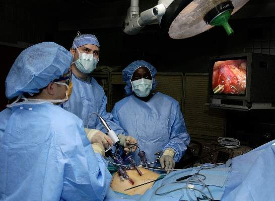
The study comprised 110 patients, of whom half underwent single-port and half underwent four-port laparoscopic cholecystectomy, all performed by the same surgeon. The primary outcome was patient-reported pain 7 days after the procedure, as measured by the visual analog scale (VAS). In all, 81% of patients were female, and median age was 47.5 years, the researchers reported.
Postoperative VAS pain scores rose significantly for both groups, compared with baseline, but did not significantly differ between the groups, said the investigators (single-port: 4.2 ± 2.4; four-port: 4.2 ± 2.2; P = .83). Cytokine levels and variations in heart rate also were similar between the groups.
However, measures of fatigue on the linear analog self-assessment were better with four-port entry, compared with the single-port approach (3.1 ± 2.1, versus 4.2 ± 2.2, respectively; P = .009), the investigators reported. And 49% of patients in the single-port group reported severe fatigue (a score greater than 5), compared with only 22% of the four-port group (P = .005).
“While this could be due to alpha-error, the fact that the group with the higher fatigue levels (single port) also seemed to be taking more oral narcotic pain medication before discharge … while maintaining similar pain scores in the postoperative period and otherwise having a similar distribution of factors affecting postoperative pain … suggests that the narcotic pain medication may have indeed had an influence on the fatigue scores,” the investigators said.
The National Institutes of Health and the National Center for Advancing Translational Sciences funded the research. The authors declared no conflicts of interest.
Single-port and four-port laparoscopic cholecystectomy were associated with similar self-reported pain scores and physiologic measures of recovery, according to a double-blinded, randomized trial published online in the Journal of the American College of Surgery.
But patients in the four-port arm reported significantly less fatigue (P = .009), and 40% needed postoperative narcotics, compared with 60% of patients who underwent single-port laparoscopy (P = .056), reported Dr. Juliane Bingener and her associates at Mayo Clinic, Rochester, N.Y.
“The data from this study may assist patients and surgeons in the discussion on how to choose a procedure,” said the researchers. “If fast postoperative recovery is the most important goal, four-port cholecystectomy may be more advantageous than single-port laparoscopic cholecystectomy. If cosmesis is the most important factor, single-port entry may be chosen” (J. Am. Coll. Surg. 2015 March 2 [doi:10.1016/j.jamcollsurg.2015.02.022]).
The study comprised 110 patients, of whom half underwent single-port and half underwent four-port laparoscopic cholecystectomy, all performed by the same surgeon. The primary outcome was patient-reported pain 7 days after the procedure, as measured by the visual analog scale (VAS). In all, 81% of patients were female, and median age was 47.5 years, the researchers reported.
Postoperative VAS pain scores rose significantly for both groups, compared with baseline, but did not significantly differ between the groups, said the investigators (single-port: 4.2 ± 2.4; four-port: 4.2 ± 2.2; P = .83). Cytokine levels and variations in heart rate also were similar between the groups.
However, measures of fatigue on the linear analog self-assessment were better with four-port entry, compared with the single-port approach (3.1 ± 2.1, versus 4.2 ± 2.2, respectively; P = .009), the investigators reported. And 49% of patients in the single-port group reported severe fatigue (a score greater than 5), compared with only 22% of the four-port group (P = .005).
“While this could be due to alpha-error, the fact that the group with the higher fatigue levels (single port) also seemed to be taking more oral narcotic pain medication before discharge … while maintaining similar pain scores in the postoperative period and otherwise having a similar distribution of factors affecting postoperative pain … suggests that the narcotic pain medication may have indeed had an influence on the fatigue scores,” the investigators said.
The National Institutes of Health and the National Center for Advancing Translational Sciences funded the research. The authors declared no conflicts of interest.
FROM JOURNAL OF THE AMERICAN COLLEGE OF SURGERY
Key clinical point: Four-port laparoscopic cholecystectomy was associated with less fatigue than was single-port laparoscopic cholecystectomy.
Major finding: Seven days after surgery, self-reported fatigue was lower for the four-port entry group compared with the single-port group (P = .009).
Data source: Double-blinded, randomized controlled trial of 110 patients who underwent single-port or four-port laparoscopic cholecystectomy.
Disclosures: The National Institutes of Health and the National Center for Advancing Translational Sciences funded the research. The authors declared no conflicts of interest.

Untreated OSA upped risk of need for repeat revascularization after PCI
Patients with untreated, moderate to severe obstructive sleep apnea who underwent percutaneous coronary intervention were more than twice as likely to undergo repeat revascularization during the next 4.8 years, compared with patients on continuous positive airway pressure (CPAP), researchers reported in the March issue of Chest.
The first-in-kind finding “provides new evidence that untreated moderate-severe OSA [obstructive sleep apnea] is an independent risk factor for repeat revascularization after PCI [percutaneous coronary intervention] and that CPAP can reduce this risk.,” said Dr. Xiaofan Wu at the Beijing Anzhen Hospital at Capital Medical University in Beijing and her associates. “Interestingly, the data show that untreated mild OSA was not associated with an increased risk of repeat revascularization, suggesting a dose-effect relationship between OSA severity and risk of complications after PCI.”
Untreated OSA has been linked with many cardiovascular problems. Patients have high levels of sympathetic excitation, oxidative stress, inflammatory mediators, endothelial dysfunction, and attenuated endothelial repair, which all can promote atherogenesis, hypertension, arrhythmogenesis, and cardiac death, the researchers noted. But the effect of untreated OSA on PCI outcomes was not well understood, they said (Chest 2015;147:708-18).
Their study retrospectively followed 390 patients with OSA who had undergone PCI. The cohorts included 128 patients with moderate to severe OSA that was successfully treated with CPAP, 167 patients with untreated, moderate to severe OSA, and 95 patients with untreated mild OSA. The investigators used subjective patient reports to assess adherence to CPAP. In all, 83.6% of treated patients had used CPAP for at least 6 months, and the rest had used CPAP for 3-6 months, they said.
Over a median follow-up of 4.8 years, 25.1% of patients with untreated, moderate to severe OSA underwent repeat revascularization, compared with 14.1% of patients on CPAP for similarly severe OSA (P = .019), the investigators reported. In the adjusted analysis, untreated patients had more than double the likelihood of repeat revascularization during the follow-up period (hazard ratio, 2.13; 95% confidence interval, 1.19-3.81; P = .011).
Mortality and rates of major adverse cardiac and cerebrovascular events were similar among the groups, said the researchers. “Although untreated moderate to severe OSA was not associated with an increased risk of death in this cohort, we believe that timely diagnosis and treatment in patients undergoing PCI can serve as a clinically relevant method of secondary prevention to decrease the risk of repeat revascularization,” they said.
The work was funded by the Program for New Century Excellent Talents in University from the Ministry of Education of China, National Natural Science Foundation of China, the Capital Health Research and Development Fund of China, and National Institutes of Health. One coauthor reported relationships with Koninklijke Philips NV and Philips/Respironics. The other authors declared no relevant conflicts of interest.
Key clinical point: Untreated, moderate to severe sleep apnea significantly increased the risk of repeat revascularization after percutaneous coronary intervention.
Major finding: Untreated patients had more than double the likelihood of repeat revascularization during the follow-up period (HR, 2.13; 95% CI, 1.19-3.81; P = .011).
Data source: Retrospective observational study of 390 adults with OSA who had undergone PCI.
Disclosures: The work was supported by the Program for New Century Excellent Talents in University from the Ministry of Education of China, National Natural Science Foundation of China, the Capital Health Research and Development Fund of China, and National Institutes of Health. One coauthor reported relationships with Koninklijke Philips NV and Philips/Respironics. The other authors declared no relevant conflicts of interest.
Automated scoring from home sleep test monitors misclassified OSA severity
Automated scoring data from two type-3 home sleep test monitors misclassified the severity of obstructive sleep apnea, especially in patients with mild to moderate disease, researchers reported in the March issue of Chest.
“Agreement between automated and manual scoring of home sleep tests varies as a function of the portable device and definition of disordered breathing event used,” said Dr. Nisha Aurora and her associates at the Johns Hopkins University in Baltimore. “Although modest agreement exists between automated and manual scoring, input by a sleep specialist or a certified polysomnologist in reviewing home sleep study results may help to improve diagnostic accuracy and classification of OSA [obstructive sleep apnea], particularly if there is mild disease.”
About 10%-15% of the general population has OSA, and home sleep testing has become an important part of ambulatory care for the condition. The American Academy of Sleep Medicine recommends that raw data from home sleep test devices be reviewed by either a board-certified sleep specialist or a clinician who fulfills eligibility criteria for the sleep medicine certification examination. Few studies, however, have actually compared automated and manual scoring data from these devices, the researchers said (Chest 2015;147:719-27).
For their analysis, Dr. Aurora and her associates evaluated automated and manual scoring data for 200 patients without a previous OSA diagnosis who used one of two home sleep test monitors: the ApneaLink Plus monitor from ResMed or the Embletta device from Embla Systems. To assess the apnea-hypopnea index (AHI), the researchers defined disordered-breathing events based on both the >3% and >4% thresholds for oxygen desaturation.
Automated scoring consistently underscored disordered-breathing events, regardless of which threshold was used to define AHI, the researchers found. For the ApneaLink Plus monitor, the average difference in AHI between manual and automated scoring was 6.1 events per hour (95% confidence interval, 4.9-7.3) for the 3% threshold and 4.6 events per hour (95% CI, 3.5-5.6) for the 4% threshold, they reported. For the Embletta monitor, manual and automated scoring varied by an average of 5.3 (95% CI, 3.2-7.3) and 8.4 (95% CI, 7.2-9.6) events per hour, respectively.
The difference between automated and manual scoring narrowed substantially when considering oxygen desaturation index instead of AHI, the researchers reported.
The investigators then used AHI to assess OSA severity. For the ApneaLink Plus monitor, relying on automated readings led to a <10% rate of clinically significant misclassification of OSA severity, regardless which oxygen desaturation threshold was used. For the Embletta device, rates of clinically significant misclassification were 29% and 41% for the 3% and 4% thresholds, respectively. “As expected, disease misclassification was most notable for subjects with mild disease irrespective of the device used,” they wrote.
The National Institutes of Health funded the work. One coauthor reported relationships with ResMed and Koninklijke Philips NV (Respironics). The other authors declared no relevant conflicts of interest.
FROM CHEST
Key clinical point: Automated scoring data from two type-3 home sleep test monitors misclassified the severity of obstructive sleep apnea.
Major finding: Automated scoring consistently underscored disordered-breathing events, compared with manual scoring, for both monitors, regardless of whether the apnea-hypopnea index was defined based on the >3% or >4% oxygen desaturation threshold.
Data source: Evaluation of the ApneaLink Plus (ResMed) and Embletta (Embla Systems) type-3 devices in 200 subjects who had not been previously diagnosed with OSA.
Disclosures: The National Institutes of Health funded the work. One coauthor reported relationships with ResMed and Koninklijke Philips NV (Respironics). The other authors declared no relevant conflicts of interest.
Study: No value in sending hernia sac specimens for routine pathology
Pathologic evaluations of hernia sac specimens from adult patients did not alter clinical management, and cost a medical center more than $75,000 over 4 years, according to a study published online in the American Journal of Surgery.
“The results from our study indicate that ‘routine’ evaluation of hernia sac specimens is likely neither indicated nor cost effective,” said Dr. Patrick Chesley of Madigan Army Medical Center, Fort Lewis, Wash., and his associates. “The rarity of changes in diagnosis and treatment from routine pathologic examination of a hernia sac does not justify this practice, and indicates that it may be omitted except in unique circumstances.”
The practice of sending hernia sac specimens for pathologic evaluation dates to 1926, when the American College of Surgeons Minimum Standard for Hospitals stated that all tissues removed during surgery should be examined and the results reported. The Joint Commission reiterated that recommendation in 1998, stating in its Comprehensive Accreditation Manual for Pathology and Clinical Laboratory Services that ‘‘specimens removed during surgery need to be evaluated for gross and microscopic abnormalities before a final diagnosis can be made.”
But the literature offers little support for the recommendation regarding hernia sac specimens, and institutions are starting to question the practice, Dr. Chesley and his associates wrote (Am. J. Surg. 2015 Feb. 12 [doi: 10.1016/j.amjsurg.2014.12.019]).
In one study, for example, pathologists reviewed 1,020 hernia sac specimens and found that only one had yielded an unexpected result – an atypical lipoma that did not affect patient management. Another study reviewed more than 2,000 hernia repairs and found that only 34% cases underwent pathologic review, with no resulting changes in treatment or management of any case.
For their study, Dr. Chesley and his coinvestigators retrospectively reviewed operative reports and medical records for 1,216 inguinal, incisional, umbilical, and ventral hernia repairs, all of which occurred at a single medical center between 2007 and 2011. More than half (55.4%) of cases were inguinal hernia repairs, 21.5% were umbilical, 11.4% were incisional, and 11.7% were ventral. In 20% of cases, surgeons sent hernia sac specimens for pathologic evaluation. Of these, 96% were selected for routine examination and 4% were selected because of concerns about possible gross abnormalities, the researchers said. Regardless of the reason for pathologic evaluation, none of the 246 examinations produced findings that reportedly altered clinical management, they said. Furthermore, pathologic evaluations cost patients about $300 to $350 each, for a total bill of more than $75,000 during the course of the study.
“These data reflect previous results from the pediatric surgical literature, and support the notion that routine pathologic evaluation of hernia sac specimens is not indicated,” the researchers concluded. But the recommendation should only apply to routine pathologic examinations, and surgeons should continue to treat abnormal intraoperative findings during hernia repair as indications for pathologic evaluation at their own discretion, they said.
Because the study was retrospective, the researchers could not ensure completeness of the data, they said. They also lacked a standardized method for reporting the reasons for specimen collection.
They reported no funding sources and declared no conflicts of interest.
FROM THE AMERICAN JOURNAL OF SURGERY
Key clinical point: Pathologic examination of hernia sac specimens can be omitted except in unique cases.
Major finding: Pathologic evaluation yielded no information that changed clinical management.
Data source: Four-year, single-center retrospective analysis of hernia sac specimens from 246 adults.
Disclosures: The investigators reported no funding sources and declared no conflicts of interest.
Noisy OR linked to communication gaps, surgical site infections
Noise in the OR has been on the rise for decades, prompting researchers to look into the links between sound levels, health impacts on surgeons, and patient outcomes.
A recent literature review found that noise levels in operating rooms frequently exceed OSHA guidelines for safe work environments. The OSHA recommendation for hospitals in general is 45 time-weighted average decibels (dbA) with a maximum peak of 140 dbA for “impulsive noise” events (Anesthesiology 2014;121:894-8). The sources of noise are many: moving of equipment, clashing metal instruments, pumps, suction apparatus, air warming units, monitors, alarms, nonclinical conversation, and background music. The review found evidence of the impact of noise on communication among staff, concentration of the surgeon, performance of complex tasks by surgeons and anesthesiologists and, potentially, patient outcomes. Noise was found to be greater at the beginning and closing of procedures, and routine peak levels reported were in excess of 100 dBA for various procedures and as high as 131 dBA in some instances.
Another study linked communication gaps in the OR to high noise levels (J. Am. Coll. Surg. 2013:216:933-8). The amount of noise from equipment, staff conversation, and background music was found to have a negative impact on surgeon performance, especially when complex tasks were in process.
Noise pollution during surgery is now being studied in terms of patient outcomes, postop complications, and surgical-site infections (SSIs) in particular.

A pilot study in the United Kingdom looked at noise in the OR as a possible surrogate marker for intraoperative behavior and a potential predictor of SSIs. With a small sample of 35 elective open abdominal procedures, a significant correlation was detected between noise levels and the 30-day postop SSI rate in these patients (Br. J. Surg. 2011;98:1021-5).
Another study has found that high noise levels in the operating room were linked to surgical site infections after outpatient hernia repair (Surgery 2015 Feb. 28 [doi: 10.1016/j.surg.2014.12.026]). “Noise levels were substantially greater in patients with surgical site infections from time point of 50 minutes onwards, which correlated to when wound closure was occurring,” wrote Dr. Shamik Dholakia and his associates at the Milton Keynes General Hospital in Buckinghamshire, England.
The researchers found that music and conversation were the two main contributors to increased noise levels in the OR. “Both of these factors may have distracting influences on the operator and contribute to lower levels of concentration, and so need to be considered as potential issues that could be improved to reduce error,” they said.
Past studies have linked surgical site infections (SSI) with lapses in aseptic technique, which can occur if the OR team is distracted. To assess the role of OR noise in SSI risk, the investigators prospectively studied 64 male, otherwise healthy outpatients who underwent left-side inguinal hernia repair at a single hospital. All patients received a prophylactic dose of 1.2 grams of co-amoxiclav antibiotic, and the same kind of mesh and suture were used for all cases, the investigators said.
The researchers used a decibel meter to measure sound levels in the OR during surgeries, and interviewed patients weekly for the next 30 days to detect SSI.
Five patients (7.8%) developed SSI, all of which were superficial. Swabs from these patients grew mixed skin flora that were sensitive to penicillin, said the researchers.
The mean level for background noise was 47.6 dB before the procedure started. On average, noise levels during procedures for which patients developed SSI were 11.337 dB higher than during other procedures. “Although the actual formula is complex, the accepted rule is that an increase of 10 decibels is perceived to be approximately twice as loud,” the investigators said. “Hence, we believe that this increase in loudness is significant enough to effect the operation. On the basis of our results, we hypothesize that poor concentration caused by high levels of noise may affect one’s ability to perform adequate aseptic closure and increase the probability of developing SSI.”
Surgical assistants usually closed the surgical wounds, which was notable because music’s effects on concentration vary with experience, the researchers noted. “Music may have a soothing, calming, and positive effect on senior experienced operators, whereas it may be distracting and reduce ability to concentrate on junior operators who are not experienced and still learning how to perform the procedure,” they said. “This finding is an important one that the lead surgeon needs to be aware of.”The investigators reported no funding sources or conflicts of interest.
Hospitals are noisy. Noise intensities in operating rooms frequently exceed those found in the hospital’s boiler room.
As pointed out in the accompanying news article, noise in the operating room can be harmful both to the patient and to the health care provider. Evidence is growing of the negative impact on performance and health of nurses, anesthesiologists, and surgeons caused by exposure to the levels of noise commonly experienced in modern operating rooms.
Much of the noise is staff generated. Music and conversations unrelated to clinical care can add close to 90 dBA to the substantial ambient noises produced by air-conditioning systems and monitors. Any attempt at clinically relevant discussion must exceed this background cacophony to be heard.
One big step toward a remedy is simple and inexpensive. All staff should be aware of the harmful effects of excessive noise. Sources of unnecessary noise should be identified and minimized. It has been suggested that a “sterile cockpit” environment (as routinely employed in commercial airplanes) be imposed during critical moments of a surgical procedure. Short of this, educational efforts, such as this brief news report, will go a long way toward cleaning up the noise pollution commonly encountered in our operating rooms.
Dr. Jonathan Katz is clinical professor of anesthesiology, Yale University Medical Center, New Haven, Conn., and attending anesthiologist at St. Vincent Medical Center, Bridgeport, Conn.
Noise in the OR has been on the rise for decades, prompting researchers to look into the links between sound levels, health impacts on surgeons, and patient outcomes.
A recent literature review found that noise levels in operating rooms frequently exceed OSHA guidelines for safe work environments. The OSHA recommendation for hospitals in general is 45 time-weighted average decibels (dbA) with a maximum peak of 140 dbA for “impulsive noise” events (Anesthesiology 2014;121:894-8). The sources of noise are many: moving of equipment, clashing metal instruments, pumps, suction apparatus, air warming units, monitors, alarms, nonclinical conversation, and background music. The review found evidence of the impact of noise on communication among staff, concentration of the surgeon, performance of complex tasks by surgeons and anesthesiologists and, potentially, patient outcomes. Noise was found to be greater at the beginning and closing of procedures, and routine peak levels reported were in excess of 100 dBA for various procedures and as high as 131 dBA in some instances.
Another study linked communication gaps in the OR to high noise levels (J. Am. Coll. Surg. 2013:216:933-8). The amount of noise from equipment, staff conversation, and background music was found to have a negative impact on surgeon performance, especially when complex tasks were in process.
Noise pollution during surgery is now being studied in terms of patient outcomes, postop complications, and surgical-site infections (SSIs) in particular.
A pilot study in the United Kingdom looked at noise in the OR as a possible surrogate marker for intraoperative behavior and a potential predictor of SSIs. With a small sample of 35 elective open abdominal procedures, a significant correlation was detected between noise levels and the 30-day postop SSI rate in these patients (Br. J. Surg. 2011;98:1021-5).
Another study has found that high noise levels in the operating room were linked to surgical site infections after outpatient hernia repair (Surgery 2015 Feb. 28 [doi: 10.1016/j.surg.2014.12.026]). “Noise levels were substantially greater in patients with surgical site infections from time point of 50 minutes onwards, which correlated to when wound closure was occurring,” wrote Dr. Shamik Dholakia and his associates at the Milton Keynes General Hospital in Buckinghamshire, England.
The researchers found that music and conversation were the two main contributors to increased noise levels in the OR. “Both of these factors may have distracting influences on the operator and contribute to lower levels of concentration, and so need to be considered as potential issues that could be improved to reduce error,” they said.
Past studies have linked surgical site infections (SSI) with lapses in aseptic technique, which can occur if the OR team is distracted. To assess the role of OR noise in SSI risk, the investigators prospectively studied 64 male, otherwise healthy outpatients who underwent left-side inguinal hernia repair at a single hospital. All patients received a prophylactic dose of 1.2 grams of co-amoxiclav antibiotic, and the same kind of mesh and suture were used for all cases, the investigators said.
The researchers used a decibel meter to measure sound levels in the OR during surgeries, and interviewed patients weekly for the next 30 days to detect SSI.
Five patients (7.8%) developed SSI, all of which were superficial. Swabs from these patients grew mixed skin flora that were sensitive to penicillin, said the researchers.
The mean level for background noise was 47.6 dB before the procedure started. On average, noise levels during procedures for which patients developed SSI were 11.337 dB higher than during other procedures. “Although the actual formula is complex, the accepted rule is that an increase of 10 decibels is perceived to be approximately twice as loud,” the investigators said. “Hence, we believe that this increase in loudness is significant enough to effect the operation. On the basis of our results, we hypothesize that poor concentration caused by high levels of noise may affect one’s ability to perform adequate aseptic closure and increase the probability of developing SSI.”
Surgical assistants usually closed the surgical wounds, which was notable because music’s effects on concentration vary with experience, the researchers noted. “Music may have a soothing, calming, and positive effect on senior experienced operators, whereas it may be distracting and reduce ability to concentrate on junior operators who are not experienced and still learning how to perform the procedure,” they said. “This finding is an important one that the lead surgeon needs to be aware of.”The investigators reported no funding sources or conflicts of interest.
FROM SURGERY
Key clinical point: Higher noise levels in the operating room correlated with surgical site infections (SSI) after outpatient hernia repair.
Major finding: On average, noise levels during procedures for which patients developed SSI were 11.337 decibels higher than during other procedures.
Data source: Prospective study of 64 young, healthy males who underwent outpatient left-sided hernia repair.
Disclosures: The investigators reported no funding sources or conflicts of interest.

VIDEO: Expert compares current atopic dermatitis guidelines
SAN FRANCISCO – Guidelines on atopic dermatitis from the American Academy of Dermatology and the Joint Task Force on Practice Parameters differ in a few important ways.
Dr. Peter Lio of Northwestern University, Chicago, summarized similarities and differences between the guidelines, discussed subtypes of atopic dermatitis, and shared tips on educating parents about food allergies and eczema in an interview at the annual meeting of the American Academy of Dermatology.
The video associated with this article is no longer available on this site. Please view all of our videos on the MDedge YouTube channel
AT THE AAD ANNUAL MEETING