User login
Progesterone, estrogen benefit postmenopausal cognition
Twelve weeks of estradiol or progesterone yielded distinct cognitive benefits in recently menopausal women, compared with placebo, according to a randomized, double-blind study reported in Psychoneuroendocrinology.
“Despite uncertainty about the potential cognitive benefits of postmenopausal hormone use, as women continue to live longer and healthier lives beyond the end of their reproductive years, hormones will continue to be prescribed for symptomatic indications,” said Dr. Alison Berent-Spillson of the University of Michigan, Ann Arbor, and her associates. “In contrast to previous studies that have found negative cognitive effects of treatment with synthetic progestins, our results point to potential cognitive benefits of both estrogen and progesterone.”
Few studies have examined unopposed progesterone treatment in postmenopausal women, and none have included functional neuroimaging (fMRI), even though fMRI can detect neurobiologic differences before patients exhibit measurable behavioral changes on task assessments, the investigators said. Past studies of postmenopausal hormone therapy and cognition have reported inconsistent results, they noted. Therefore, the investigators carried out a pilot study of 29 women who were within 38 months of their final menstrual period, and thus were inside the “critical window” when hormone therapy is thought to offer maximum benefit. Participants were randomized to 12 weeks of 1 mg oral estradiol or 200 mg oral progesterone. Each therapy was counterbalanced with placebo for the same amount of time, separated by a 12-week washout period. At the end of each 12-week treatment period, the women underwent verbal and visual cognitive function and working memory tests, as well as functional MRI of verbal and visual working memory processing (Psychoneuroendocrinology 2015; 59:25-36).
Compared with placebo, postmenopausal progesterone therapy was associated with a greater verbal working memory composite score and with greater fMRI activation of the right prefrontal cortex during the verbal working memory task (P = .014), the investigators reported. Women on progesterone also had significantly greater activation of the left prefrontal cortex (P = .001) and the right hippocampus (P = .003) during the visual working memory tasks, compared with the placebo group. Estradiol therapy and placebo did not differ significantly in terms of verbal or visual learning, recall, or working memory composite scores. However, estradiol was associated with increased activation of the left prefrontal cortex (P = .006) and the left hippocampus (P = .037) compared with placebo during the verbal working memory tasks, the researchers said.
Progesterone might affect the hippocampus by promoting neurogenesis and neuron survival, although its effects on the postmenopausal brain remain largely unstudied, the investigators said. Estrogen seems to offer cognitive effects during menopause through its actions on the hippocampus, but a longer treatment period might be needed for such benefits to emerge during testing, they said. “While we did not detect differences in verbal ability between the estrogen and placebo treatment arms, we saw increased activation after estrogen treatment in the left prefrontal cortex during the verbal processing task, a region associated with verbal processing,” they emphasized. “This may reflect more efficient encoding, as women remembered more words from the verbal task after estrogen than placebo, although this difference did not reach statistical significance.”
Although the study was small, its crossover design maximized the statistical power, said the researchers. They recommended larger studies with longer treatment durations in order to better detect emergent differences in cognitive ability between treatment groups and relationships between performance on tasks and regional brain activation patterns.
The National Institutes of Health and the Phil F. Jenkins Foundation funded the study. The investigators declared no conflicts of interest.
FROM PSYCHONEUROENDOCRINOLOGY
Key clinical point: Compared with placebo, progesterone was associated with a greater verbal working memory composite score and greater activation of the right prefrontal cortex during that task.
Major finding: Women on progesterone also had significantly greater activation of the left prefrontal cortex and the right hippocampus during the visual working memory tasks, compared with the placebo group.
Data source: Double-blind placebo-controlled randomized pilot study of 29 recently postmenopausal women.
Disclosures: The National Institutes of Health and the Phil F. Jenkins Foundation supported the research. The investigators declared no conflicts of interest.
Predictors identified for progression of moderate RA on methotrexate
Patients with moderately active rheumatoid arthritis who were positive for rheumatoid factor and had baseline C-reactive protein levels above 40 mg/L had about a 60% chance of progressing on methotrexate monotherapy, investigators reported in RMD Open.
Elevated C-reactive protein (CRP) levels are known to predict radiographic progression in active rheumatoid arthritis (RA), but this study is the first to extend the finding to moderately active disease, said Dr. Bruno Fautrel of Pierre and Marie Curie University, Paris, and his associates. Understanding baseline predictors of radiographic progression could help clinicians to decide which patients need early, aggressive treatment and who can be spared intensive therapies, the researchers added.

“Studies investigating the risk of disease progression in patients with established and stable moderate disease activity are very limited,” they noted. “While it is established that moderate disease activity is not an adequate target for patients with RA, in real life, those with moderate RA represent a substantial proportion of patients in clinical practice.”
The study was a subgroup analysis of 96 patients with moderate RA from the methotrexate arm of the multicenter, phase III, randomized, double-blind, TEMPO (Trial of Etanercept and Methotrexate with Radiographic Patient Outcomes) trial. Patients had sustained moderate RA, defined as mean disease activity score in 28 joints ranging from 3.2 to 5.1 during the last 6 months of the first year. The researchers scored radiographs of hands, wrists, and feet based on the modified Total Sharp Score (mTSS) method, and defined radiographic progression as an increase from baseline mTSS of at least 3.0 after at least 2 years of methotrexate treatment. Patients averaged 55 years of age, and 84% were women, the investigators said (RMD Open 2015;1:e000018 doi: 10.1136/rmdopen-2014-000018).
Overall, about 26% of patients had signs of significant radiographic progression after 2 years of methotrexate treatment, and 34% had significantly progressed after 3 years, the researchers reported. Stratifying patients by baseline rheumatoid factor (RF) and CRP levels showed that those who were RF-positive at baseline with CRP levels above 40 mg/L were significantly more likely to progress at year 2 (51%; 95% confidence interval, 32%-69%) and year 3 (61%; 95% CI, 42%-78%) than were other patients, they said. In fact, only 33% of patients who were RF-positive and had baseline CRP levels below 10 mg/L had significantly progressed at year 3, as had only 10% of patients who were RF-negative and had baseline CRP levels below 10 mg/L. The median change in mTSS at year 3 was 3.6 for the highest-risk group, compared with zero for the lowest-risk group. Baseline erosion scores did not predict radiographic progression, probably because TEMPO patients had erosive disease at baseline, the researchers said.
The study was a post hoc analysis with a small sample size, and the mean methotrexate dose at baseline was only 7.5 mg/week, although the weekly dose rose to an average of 15 mg by week 24 of the study. Future studies should examine progression on etanercept monotherapy and on methotrexate-etanercept dual therapy, the investigators said.
Pfizer supported the research. Dr. Fautrel reported financial relationships with Pfizer, AbbVie, Bristol-Myers Squibb, Merck Sharp & Dohme, Roche, Sobi, and UCB. All three coauthors reported current or former employment with Pfizer.
Patients with moderately active rheumatoid arthritis who were positive for rheumatoid factor and had baseline C-reactive protein levels above 40 mg/L had about a 60% chance of progressing on methotrexate monotherapy, investigators reported in RMD Open.
Elevated C-reactive protein (CRP) levels are known to predict radiographic progression in active rheumatoid arthritis (RA), but this study is the first to extend the finding to moderately active disease, said Dr. Bruno Fautrel of Pierre and Marie Curie University, Paris, and his associates. Understanding baseline predictors of radiographic progression could help clinicians to decide which patients need early, aggressive treatment and who can be spared intensive therapies, the researchers added.
“Studies investigating the risk of disease progression in patients with established and stable moderate disease activity are very limited,” they noted. “While it is established that moderate disease activity is not an adequate target for patients with RA, in real life, those with moderate RA represent a substantial proportion of patients in clinical practice.”
The study was a subgroup analysis of 96 patients with moderate RA from the methotrexate arm of the multicenter, phase III, randomized, double-blind, TEMPO (Trial of Etanercept and Methotrexate with Radiographic Patient Outcomes) trial. Patients had sustained moderate RA, defined as mean disease activity score in 28 joints ranging from 3.2 to 5.1 during the last 6 months of the first year. The researchers scored radiographs of hands, wrists, and feet based on the modified Total Sharp Score (mTSS) method, and defined radiographic progression as an increase from baseline mTSS of at least 3.0 after at least 2 years of methotrexate treatment. Patients averaged 55 years of age, and 84% were women, the investigators said (RMD Open 2015;1:e000018 doi: 10.1136/rmdopen-2014-000018).
Overall, about 26% of patients had signs of significant radiographic progression after 2 years of methotrexate treatment, and 34% had significantly progressed after 3 years, the researchers reported. Stratifying patients by baseline rheumatoid factor (RF) and CRP levels showed that those who were RF-positive at baseline with CRP levels above 40 mg/L were significantly more likely to progress at year 2 (51%; 95% confidence interval, 32%-69%) and year 3 (61%; 95% CI, 42%-78%) than were other patients, they said. In fact, only 33% of patients who were RF-positive and had baseline CRP levels below 10 mg/L had significantly progressed at year 3, as had only 10% of patients who were RF-negative and had baseline CRP levels below 10 mg/L. The median change in mTSS at year 3 was 3.6 for the highest-risk group, compared with zero for the lowest-risk group. Baseline erosion scores did not predict radiographic progression, probably because TEMPO patients had erosive disease at baseline, the researchers said.
The study was a post hoc analysis with a small sample size, and the mean methotrexate dose at baseline was only 7.5 mg/week, although the weekly dose rose to an average of 15 mg by week 24 of the study. Future studies should examine progression on etanercept monotherapy and on methotrexate-etanercept dual therapy, the investigators said.
Pfizer supported the research. Dr. Fautrel reported financial relationships with Pfizer, AbbVie, Bristol-Myers Squibb, Merck Sharp & Dohme, Roche, Sobi, and UCB. All three coauthors reported current or former employment with Pfizer.
FROM RMD OPEN
Key clinical point:Elevated C-reactive protein and rheumatoid factor levels predicted radiographic progression of moderate rheumatoid arthritis despite methotrexate treatment.
Major finding: RF positivity and baseline CRP levels above 40 mg/L predicted significant radiographic progression after 2 and 3 years (P less than .05 for all associations).
Data source: Subgroup analysis of 96 patients from the methotrexate arm of the multicenter, double-blind, randomized, 3-year TEMPO study.
Disclosures: Pfizer supported the research. Dr. Fautrel reported financial relationships with Pfizer, AbbVie, Bristol-Myers Squibb, Merck Sharp & Dohme, Roche, Sobi, and UCB. All three coauthors reported current or former employment with Pfizer.

Surgical bolt cutters quickly cut titanium ring
Clinicians can use a pair of large surgical bolt cutters to safely and quickly cut a titanium ring from a patient’s swollen finger, and then can pull apart the edges with two paper clips, according to a letter published online in Emergency Medicine Journal.
“Our method used simple equipment that is readily available in most hospitals at all times, took less than 30 seconds to perform, and could be performed by a sole operator without damage to the underlying finger,” wrote Dr. Andrej Salibi and Dr. Andrew Morritt at Sheffield (England) Teaching Hospital NHS Foundation (2015 Aug 13; doi: 10.1136/emermed-2015-204962).
Ring constriction is a fairly common problem that can cause necrosis and loss of the digit if the ring is not removed. Basic ring cutters can sever gold and silver, but not titanium, which has become popular for rings because it is hypoallergenic, durable, lightweight, and strong – so strong that diamond-tipped saws or drills can take up to 15 minutes to cut these rings, the surgeons noted. Many facilities also lack access to such equipment, and it generates enough heat that an assistant must irrigate the surrounding skin to prevent burns, they said.
Their case report described a patient who bathed in warm water at a spa and developed a painful, swollen finger that was constricted by a titanium wedding band. Elevation and lubrication at the emergency department failed to remove the ring, as did finger binding and use of a manual ring cutter.
“The fire service was called and attempted removal using specialized cutting equipment, which also failed,” the surgeons wrote. “The patient was then admitted under the plastic surgery service for hand elevation, and further attempts 8 hours later blunted two manual ring cutters.” At this point, they borrowed a large pair of bolt cutters from the operating room, which quickly severed the ring without harming the finger. Then they applied lateral traction with a pair of paper clips and removed it, they said.
The authors declared no funding sources or conflicts of interest.
FROM EMERGENCY MEDICINE JOURNAL
A 3-hour tour: Patients report long round-trips for rheumatologic care
A total of 7% of Medicare beneficiaries who visited a rheumatologist in 2009 traveled at least 3 hours round-trip to do so, according to a study published online in Seminars in Arthritis and Rheumatism.
“This study confirms that a small but significant proportion of patients in the [United States] have long travel times to visit a rheumatologist, and that the most important determinant of long travel times is supply of rheumatologists,” said Dr. Gabriela Schmajuk and her associates at the University of California, San Francisco. Access to care was especially limited in the Mountain region of the United States, the investigators said. “Assessing the best methods for attracting and retaining rheumatologists to low-supply areas and providing high-quality care to rural beneficiaries will be crucial challenges for policy makers and professional societies,” they added.

Despite recent rises in numbers of U.S. patients with arthritis and other rheumatic conditions, no prior study has reported national estimates for access to rheumatology care, the researchers noted.
They studied Medicare Part B insurance claims for 41,693 patients who visited a rheumatologist at least once in 2009. Data were from the Chronic Conditions Data Warehouse, a Medicare and Medicaid research database. The researchers used the amount of time patients traveled to see a rheumatologist as a proxy for access to care, and estimated travel times in Google Maps by calculating distances between the center of each beneficiary’s zip code and the center of the zip code of their rheumatologist’s office (Sem. Arthritis Rheum. 2015 Jul 28. doi: 10.1016/j.semarthrit.2015.07.007).
Medicare patients traveled a median of 22 minutes to see a rheumatologist in 2009 (interquartile range, 12-40 minutes), for a median one-way distance of 9.3 miles (interquartile range, 4-22 miles), and an average of 3.4 visits per patient (standard deviation, 2.8 visits), said the investigators. But nearly 3,000 (7%) patients traveled at least 90 minutes one way for their appointments, they reported. Rural areas of the country had substantially higher proportions of patients who faced long times for care, compared with urban areas, even after the investigators controlled for baseline differences among patients, they said. In particular, 17% of patients who lived in hospital service areas that lacked rheumatologists traveled at least 90 minutes one way, compared with 3%-4% of patients in areas with better access to care. “Although living in an area with a low supply of rheumatologists was a critical determinant of long travel times, other factors, including being male, white, and living in lower socioeconomic status areas were also important,” the investigators added.
Other studies that have examined driving distance to rheumatologists as a proxy for access to care have reported similar results for the impacts of gender and race (Arthritis Care Res. 2014;66[11]:1634-43) or socioeconomic status (Arthritis Care Res. 2015;67[2]:230-9), they noted.
The estimated number of rheumatologists came within 10% of the number listed in the 2010 membership directory for the American College of Rheumatology, but travel times might have been underestimated for lower-income patients because these individuals might have been more likely to travel by public transportation, the researchers noted. The study also did not account for patients who received rheumatology care from general providers, the authors said.
A total of 7% of Medicare beneficiaries who visited a rheumatologist in 2009 traveled at least 3 hours round-trip to do so, according to a study published online in Seminars in Arthritis and Rheumatism.
“This study confirms that a small but significant proportion of patients in the [United States] have long travel times to visit a rheumatologist, and that the most important determinant of long travel times is supply of rheumatologists,” said Dr. Gabriela Schmajuk and her associates at the University of California, San Francisco. Access to care was especially limited in the Mountain region of the United States, the investigators said. “Assessing the best methods for attracting and retaining rheumatologists to low-supply areas and providing high-quality care to rural beneficiaries will be crucial challenges for policy makers and professional societies,” they added.
Despite recent rises in numbers of U.S. patients with arthritis and other rheumatic conditions, no prior study has reported national estimates for access to rheumatology care, the researchers noted.
They studied Medicare Part B insurance claims for 41,693 patients who visited a rheumatologist at least once in 2009. Data were from the Chronic Conditions Data Warehouse, a Medicare and Medicaid research database. The researchers used the amount of time patients traveled to see a rheumatologist as a proxy for access to care, and estimated travel times in Google Maps by calculating distances between the center of each beneficiary’s zip code and the center of the zip code of their rheumatologist’s office (Sem. Arthritis Rheum. 2015 Jul 28. doi: 10.1016/j.semarthrit.2015.07.007).
Medicare patients traveled a median of 22 minutes to see a rheumatologist in 2009 (interquartile range, 12-40 minutes), for a median one-way distance of 9.3 miles (interquartile range, 4-22 miles), and an average of 3.4 visits per patient (standard deviation, 2.8 visits), said the investigators. But nearly 3,000 (7%) patients traveled at least 90 minutes one way for their appointments, they reported. Rural areas of the country had substantially higher proportions of patients who faced long times for care, compared with urban areas, even after the investigators controlled for baseline differences among patients, they said. In particular, 17% of patients who lived in hospital service areas that lacked rheumatologists traveled at least 90 minutes one way, compared with 3%-4% of patients in areas with better access to care. “Although living in an area with a low supply of rheumatologists was a critical determinant of long travel times, other factors, including being male, white, and living in lower socioeconomic status areas were also important,” the investigators added.
Other studies that have examined driving distance to rheumatologists as a proxy for access to care have reported similar results for the impacts of gender and race (Arthritis Care Res. 2014;66[11]:1634-43) or socioeconomic status (Arthritis Care Res. 2015;67[2]:230-9), they noted.
The estimated number of rheumatologists came within 10% of the number listed in the 2010 membership directory for the American College of Rheumatology, but travel times might have been underestimated for lower-income patients because these individuals might have been more likely to travel by public transportation, the researchers noted. The study also did not account for patients who received rheumatology care from general providers, the authors said.
FROM SEMINARS IN ARTHRITIS AND RHEUMATISM
Key clinical point: A small but significant percentage of Medicare patients travel for long periods for rheumatology care.
Major finding: A total of 7% percent of beneficiaries traveled at least 3 hours round-trip to see their rheumatologists.
Data source: Analysis of 41,693 Medicare Part B beneficiaries who saw a rheumatologist at least once in 2009.
Disclosures: The National Institutes of Health supported the work. The investigators declared no competing interests.

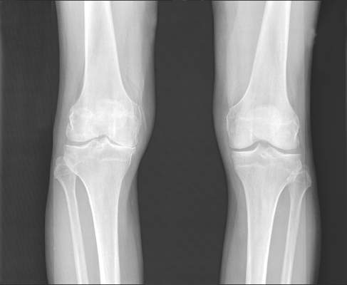
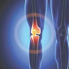
Optimized analgesia, exercise cut pain in severe knee OA
A 6-week protocol of optimized analgesia followed by a 12-week exercise program significantly improved pain and functional limitations for patients with knee osteoarthritis, researchers reported in Arthritis Care and Research.
The study was the first to explore how to achieve sufficient pain relief for patients with severely painful knee OA to participate in exercise therapy, and it achieved that goal for 78% of patients, said Joyce van Tunen of the Amsterdam Rehabilitation Center and associates. “The newly developed intervention protocol was feasible, which means that patients were able to participate in exercise therapy despite their severe pain at baseline,” the researchers said. “Although the results are promising, they need to be confirmed in a randomized controlled trial.”

Guidelines recommend combining pharmacologic and nonpharmacologic modalities to improve osteoarthritis outcomes, and past studies have suggested that acetaminophen, NSAIDs, and glucosamine therapy might augment the benefits of exercise in OA, they said.
The study included 49 patients with severe knee OA whose pain scored at least 7 on a 10-point scale. Patients received standardized analgesia with acetaminophen and then were stepped up to NSAIDs, weak opioids, and intra-articular steroid injections if their pain did not improve to a 5 or lower. Patients had 2 weeks to adapt to each new prescription without further changes, and their doses were cut if they maintained pain scores of 4 or less for a month or longer. The protocol was based on the World Health Organization analgesic ladder and the Beating osteoARThritis strategy for stepped care in hip and knee OA, the investigators noted (Arthritis Care Res. 2015 Aug 3 doi: 10.1002/acr.22682).
After 6 weeks, patients continued with analgesia and started a 12-week exercise program of two 60-minute sessions per week. The first 6 weeks of the program focused on muscle strength, while the last 6 weeks aimed to maximize strength while adding functional and aerobic exercises. Patients also were asked to do physical therapy exercises at home on the days they did not take part in supervised exercise, the researchers said.
At the end of the 18-week intervention, 72% of patients reported improvement on a combined global scale, the study showed. Average pain scores improved by 30% (P < .001) and activity limitations improved by 17% (P < .001). Fully 78% of patients were able to follow the exercise program, and these patients improved their physical limitations by an extra 10% (P = .004), compared with patients who could not complete the program. Patients who were not able to finish the exercise program also had significantly worse radiographic signs of OA, compared with the others (P = .03), and tended to be younger; had higher body mass indices; and reported more pain, anxiety, and depression, the researchers said. These patients might need surgical interventions or therapy to help them learn to better cope with pain, they added.
Most of the patients had used analgesics irregularly and at suboptimal doses at baseline, in part because they feared adverse effects. However, they experienced no serious side effects from the analgesia protocol or the exercise program, the investigators noted.
The Dutch Arthritis Association funded the work. The investigators reported having no conflicts of interest.
Therapy of osteoarthritis often remains a frustration to both the patient and physician. In clinical trials as well as the clinical setting, no single nonsurgical therapeutic approach has consistently been of significant benefit.

|
| Dr. Roy Altman |
Summaries of the medical literature are reflected by unimpressive P values, effect sizes, and other statistical methods. Yet the clinician has to use the tools available, so that several partially effective programs are often employed in the clinical setting, most often in combination – sometimes called multimodal therapy. Therapeutic guidelines are based on the literature but usually only allude to a multimodal approach.
In this study, the investigators examined a combined nonpharmacologic and pharmacologic program over an 18-week period. They easily demonstrated significant benefit of the multimodal approach. No study is perfect, and this one has multiple drawbacks, not the least of which is the lack of any control or comparison group to determine if the benefits seen are from one or both programs. Despite the drawbacks in their study design, they have contributed to filling that gap in the literature. The program of medications to reduce pain enough to increase the physical rehabilitation is logical and reflects a real-world setting.
Dr. Roy D. Altman is professor emeritus in the division of rheumatology and immunology at the University of California, Los Angeles. He reported having no relevant disclosures.
Therapy of osteoarthritis often remains a frustration to both the patient and physician. In clinical trials as well as the clinical setting, no single nonsurgical therapeutic approach has consistently been of significant benefit.
|
|
| Dr. Roy Altman |
Summaries of the medical literature are reflected by unimpressive P values, effect sizes, and other statistical methods. Yet the clinician has to use the tools available, so that several partially effective programs are often employed in the clinical setting, most often in combination – sometimes called multimodal therapy. Therapeutic guidelines are based on the literature but usually only allude to a multimodal approach.
In this study, the investigators examined a combined nonpharmacologic and pharmacologic program over an 18-week period. They easily demonstrated significant benefit of the multimodal approach. No study is perfect, and this one has multiple drawbacks, not the least of which is the lack of any control or comparison group to determine if the benefits seen are from one or both programs. Despite the drawbacks in their study design, they have contributed to filling that gap in the literature. The program of medications to reduce pain enough to increase the physical rehabilitation is logical and reflects a real-world setting.
Dr. Roy D. Altman is professor emeritus in the division of rheumatology and immunology at the University of California, Los Angeles. He reported having no relevant disclosures.
A 6-week protocol of optimized analgesia followed by a 12-week exercise program significantly improved pain and functional limitations for patients with knee osteoarthritis, researchers reported in Arthritis Care and Research.
The study was the first to explore how to achieve sufficient pain relief for patients with severely painful knee OA to participate in exercise therapy, and it achieved that goal for 78% of patients, said Joyce van Tunen of the Amsterdam Rehabilitation Center and associates. “The newly developed intervention protocol was feasible, which means that patients were able to participate in exercise therapy despite their severe pain at baseline,” the researchers said. “Although the results are promising, they need to be confirmed in a randomized controlled trial.”
Guidelines recommend combining pharmacologic and nonpharmacologic modalities to improve osteoarthritis outcomes, and past studies have suggested that acetaminophen, NSAIDs, and glucosamine therapy might augment the benefits of exercise in OA, they said.
The study included 49 patients with severe knee OA whose pain scored at least 7 on a 10-point scale. Patients received standardized analgesia with acetaminophen and then were stepped up to NSAIDs, weak opioids, and intra-articular steroid injections if their pain did not improve to a 5 or lower. Patients had 2 weeks to adapt to each new prescription without further changes, and their doses were cut if they maintained pain scores of 4 or less for a month or longer. The protocol was based on the World Health Organization analgesic ladder and the Beating osteoARThritis strategy for stepped care in hip and knee OA, the investigators noted (Arthritis Care Res. 2015 Aug 3 doi: 10.1002/acr.22682).
After 6 weeks, patients continued with analgesia and started a 12-week exercise program of two 60-minute sessions per week. The first 6 weeks of the program focused on muscle strength, while the last 6 weeks aimed to maximize strength while adding functional and aerobic exercises. Patients also were asked to do physical therapy exercises at home on the days they did not take part in supervised exercise, the researchers said.
At the end of the 18-week intervention, 72% of patients reported improvement on a combined global scale, the study showed. Average pain scores improved by 30% (P < .001) and activity limitations improved by 17% (P < .001). Fully 78% of patients were able to follow the exercise program, and these patients improved their physical limitations by an extra 10% (P = .004), compared with patients who could not complete the program. Patients who were not able to finish the exercise program also had significantly worse radiographic signs of OA, compared with the others (P = .03), and tended to be younger; had higher body mass indices; and reported more pain, anxiety, and depression, the researchers said. These patients might need surgical interventions or therapy to help them learn to better cope with pain, they added.
Most of the patients had used analgesics irregularly and at suboptimal doses at baseline, in part because they feared adverse effects. However, they experienced no serious side effects from the analgesia protocol or the exercise program, the investigators noted.
The Dutch Arthritis Association funded the work. The investigators reported having no conflicts of interest.
FROM ARTHRITIS CARE AND RESEARCH
Key clinical point: Patients with knee osteoarthritis and severe pain improved significantly with an optimized standard protocol of analgesia and exercise therapy.
Major finding: Average pain scores improved by 30% (P < .001) and activity limitations improved by 17% (P < .001).
Data source: Single-center prospective study of 49 patients with knee OA and severe pain.
Disclosures: The Dutch Arthritis Association funded the work. The investigators reported having no conflicts of interest.
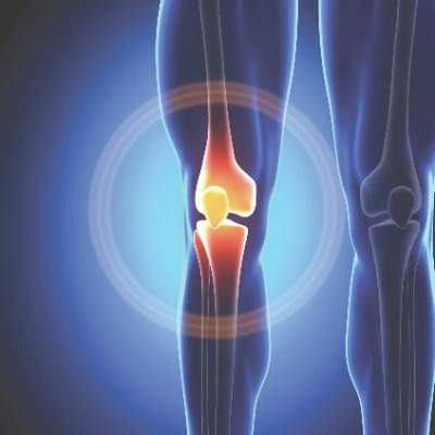
New analysis suggests functional decline for most people with knee OA
Accounting for missing data revealed functional declines over time in patients with newly diagnosed knee osteoarthritis, according to researchers.
The findings contradict more optimistic results from other longitudinal studies of knee OA, said Britt Elin Øiestad, Ph.D., of Oslo and Akershus University College of Applied Sciences and her associates. “The adjusted analyses showed either stable or worsening physical function, which is more in line with what is observed in the clinic,” the investigators said.
Their analysis included data from 802 participants in either the Multicenter Osteoarthritis Study (MOST) or the Osteoarthritis Initiative (OAI) who developed symptomatic knee OA during the course of the studies.
Approximately two-thirds of affected patients were women. Patients averaged about 63-66 years of age, had average body mass index of about 30-31 kg/m2, and had not undergone total knee replacement surgery at baseline or total hip replacement at any time. The researchers used a multiple imputation method to account for missing physician visits and a local regression smoothing curve to predict clinical status just prior to total knee replacement surgery (Arthritis Care Res. 2015 Aug 3. doi: 10.1002/acr.22674). Patients in MOST who developed knee OA showed no significant change over 4 years in scores on the Western Ontario and McMaster Universities Osteoarthritis Index (WOMAC) Physical Function (pf) subscale, while their Five Times Sit to Stand Test and 20-Meter Walk Test results rose by 1.5 seconds over 5-6 years (P less than .003), the investigators said. Adjusted results from OAI were similar, revealing significant worsening over time in WOMAC-pf and 20-Meter Walk Test results. “In crude results in which we did not impute missing values or pre–total knee replacement physical function status, the trajectory of physical function was more favorable,” the researchers added. “We found that imputing missing values and predicting pre–total knee replacement function reduced some of the bias seen in the unadjusted analyses, which incorrectly suggested improvement in physical function in people with knee osteoarthritis.”
The Research Council of Norway and the National Institutes of Health funded the study. The authors did not report funding sources or conflicts of interest.
FROM ARTHRITIS CARE & RESEARCH
Key clinical point:A new analysis shows either stable or worsening physical function after new-onset knee osteoarthritis, contradicting more optimistic results from other longitudinal studies of knee OA.
Major finding: In adjusted analyses, patients showed stable to significantly worse results over time in WOMAC-pf, Five Times Sit to Stand, and the 20-Meter Walk Test. Crude data suggested a more positive trajectory.
Data source: Longitudinal analysis of 802 patients in the Multicenter Osteoarthritis Study or the Osteoarthritis Initiative.
Disclosures: The Research Council of Norway and the National Institutes of Health funded the study. The authors did not report funding sources or conflicts of interest.
Study links statin use to lower mortality in RA patients
Among adults with rheumatoid arthritis, starting a statin prescription led to a 21% drop in risk of dying from any cause, compared with not using statins, according to a study published online Aug. 5 in Annals of the Rheumatic Diseases.
The size of the protective effect resembled results from trials of patients without RA and somewhat exceeded those from population-level analyses of statins as preventive therapy, reported Dr. Sara Schoenfeld of Harvard Medical School, Boston.
“Although the differences were small, this finding may not be surprising, as patients with RA are at a higher risk for cardiovascular disease than the general population, and might benefit from the dual anti-inflammatory and lipid-lowering effects of statins in a way that the general population might not,” wrote Dr. Schoenfeld and her colleagues.

Few studies have examined statin use in patients with RA, and a recent randomized trial, TRACE-RA (Ann Rheum Dis. 2015;74:688), was halted early because of a low event rate, the investigators noted.
To further explore the issue, they compared matched cohorts of RA patients who were at least 20 years old, were listed in a general practice medical records database from the United Kingdom, had used at least one disease-modifying antirheumatic drug between 2000 and 2012, and had either started statins or not during the year they were added to the study.
The investigators excluded current or former statin users to help prevent selection bias, and they excluded patients with missing data on relevant risk factors, such as body mass index or smoking status (Ann Rheum Dis. 2015 Aug. 5 doi: 10.1136/annrheumdis-2015-207714).
Over a median of 4.5 years of follow-up, 432 of 2,943 patients with RA who started statins died, for an incidence rate of 32.6 deaths per 1,000 person-years, which was substantially less than the rate of 40.6 per 1,000 person-years among those who did not use statins, the investigators reported. Thus, starting statins was linked to a 21% lower likelihood of all-cause mortality (hazard ratio, 0.79; 95% confidence interval, 0.68 to 0.91), they said. The hazard ratio was similar when they defined RA based on diagnostic code only, without requiring use of DMARDs for the case definition (HR, 0.81; 95% CI, 0.74 to 0.90).
“Our findings expand previous evidence for the beneficial effects of statins in RA, which have been indirectly drawn from studies evaluating intermediate markers of cardiovascular disease and premature mortality in RA, antirheumatic and lipid findings from studies evaluating RA disease outcomes, and studies evaluating statin effects in other patient populations, such as the JUPITER trial,” the researchers said.
The comparison groups were well balanced in terms of baseline demographic traits, comorbidities, total cholesterol levels, and use of cardiovascular medications, nonsteroidal anti-inflammatory drugs, glucocorticoids, and biological agents, but the medical records database usually lacked information on cause of death, the researchers noted. “We hypothesize that the lower mortality rate associated with statin use stems from the reduction of cardiovascular-specific mortality in patients with RA, and this speculation calls for future studies that examine cause-specific mortality outcomes,” they concluded.
The National Institutes of Health partly funded the work. The investigators declared having no competing interests.
Among adults with rheumatoid arthritis, starting a statin prescription led to a 21% drop in risk of dying from any cause, compared with not using statins, according to a study published online Aug. 5 in Annals of the Rheumatic Diseases.
The size of the protective effect resembled results from trials of patients without RA and somewhat exceeded those from population-level analyses of statins as preventive therapy, reported Dr. Sara Schoenfeld of Harvard Medical School, Boston.
“Although the differences were small, this finding may not be surprising, as patients with RA are at a higher risk for cardiovascular disease than the general population, and might benefit from the dual anti-inflammatory and lipid-lowering effects of statins in a way that the general population might not,” wrote Dr. Schoenfeld and her colleagues.
Few studies have examined statin use in patients with RA, and a recent randomized trial, TRACE-RA (Ann Rheum Dis. 2015;74:688), was halted early because of a low event rate, the investigators noted.
To further explore the issue, they compared matched cohorts of RA patients who were at least 20 years old, were listed in a general practice medical records database from the United Kingdom, had used at least one disease-modifying antirheumatic drug between 2000 and 2012, and had either started statins or not during the year they were added to the study.
The investigators excluded current or former statin users to help prevent selection bias, and they excluded patients with missing data on relevant risk factors, such as body mass index or smoking status (Ann Rheum Dis. 2015 Aug. 5 doi: 10.1136/annrheumdis-2015-207714).
Over a median of 4.5 years of follow-up, 432 of 2,943 patients with RA who started statins died, for an incidence rate of 32.6 deaths per 1,000 person-years, which was substantially less than the rate of 40.6 per 1,000 person-years among those who did not use statins, the investigators reported. Thus, starting statins was linked to a 21% lower likelihood of all-cause mortality (hazard ratio, 0.79; 95% confidence interval, 0.68 to 0.91), they said. The hazard ratio was similar when they defined RA based on diagnostic code only, without requiring use of DMARDs for the case definition (HR, 0.81; 95% CI, 0.74 to 0.90).
“Our findings expand previous evidence for the beneficial effects of statins in RA, which have been indirectly drawn from studies evaluating intermediate markers of cardiovascular disease and premature mortality in RA, antirheumatic and lipid findings from studies evaluating RA disease outcomes, and studies evaluating statin effects in other patient populations, such as the JUPITER trial,” the researchers said.
The comparison groups were well balanced in terms of baseline demographic traits, comorbidities, total cholesterol levels, and use of cardiovascular medications, nonsteroidal anti-inflammatory drugs, glucocorticoids, and biological agents, but the medical records database usually lacked information on cause of death, the researchers noted. “We hypothesize that the lower mortality rate associated with statin use stems from the reduction of cardiovascular-specific mortality in patients with RA, and this speculation calls for future studies that examine cause-specific mortality outcomes,” they concluded.
The National Institutes of Health partly funded the work. The investigators declared having no competing interests.
FROM ANNALS OF THE RHEUMATIC DISEASES
Key clinical point:Starting statins might substantially decrease mortality among adults with rheumatoid arthritis.
Major finding: Patients who started statins had a subsequent 21% lower risk of all-cause mortality than did those who did not.
Data source: Matched cohort study of 5,886 patients with rheumatoid arthritis who at baseline had never used statins.
Disclosures: The National Institutes of Health partly funded the work. The investigators declared no competing interests.

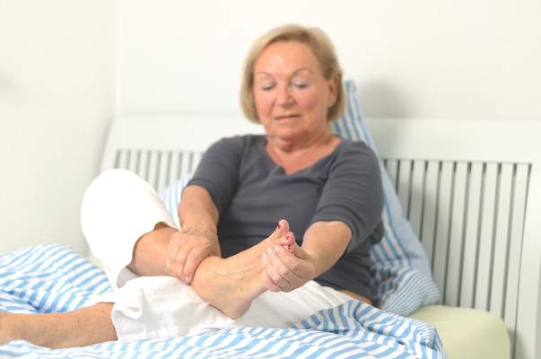
Study reveals distinct forms of foot osteoarthritis
Osteoarthritis of the foot takes one of two forms: isolated disease of the first metatarsophalangeal joint or more extensive disease of that and several other joints of the midfoot, researchers reported online in Arthritis Care & Research.
The latter, known as polyarticular foot osteoarthritis (OA), disproportionately affects women, is associated with worse pain and disability, compared with localized disease, and tends to co-occur with nodal hand OA, said Trishna Rathod of Keele University, Staffordshire, England, and her associates.
Studies of OA phenotypes have yielded targeted treatments (Ann Intern Med. 2009;150[10]:661-9andOsteoarthritis Cartilage. 2014;22[suppl S431]) for other joints, but had not yet characterized foot OA, the researchers said. Therefore, they surveyed 533 adults who reported foot pain in the prior year and scored radiographs of their first metatarsophalangeal, first and second cuneometatarsal, navicular first cuneiform, and talonavicular joints. The patients did not have psoriatic or rheumatoid arthritis and averaged 65 years of age (Arthritis Care Res. [Hoboken] 2015 Aug. 3. doi: 10.1002/acr.22677).
In all, 15% of patients had polyarticular foot OA, 22% had isolated OA of the first metatarsophalangeal joint, and 64% had no or minimal foot arthritis, the investigators reported. About 77% of patients with polyarticular disease were women, compared with approximately half of those in the other groups (P = .001). After the researchers controlled for age and sex, polyarticular disease was significantly linked to nodal hand OA (P = .04), higher body mass index (P = .002), worse scores on a 10-point foot pain scale (6 vs. 4.9 and 5.2 for the other classes; P = .02), and worse pain and disability scores on the Manchester Foot Pain and Disability Index (P = .002 and .007), they said.
“As is the case for OA at other small joint sites, particularly the hands, patterning of individual joint involvement in radiographic foot OA is polyarticular and strongly symmetrical,” the researchers concluded. “Patterns of joint involvement in radiographic foot OA have indicated a distinction between individuals with isolated [first] metatarsophalangeal joint OA and those with a more widespread form of OA ... which also includes one or both first metatarsophalangeal joints.”
The Arthritis Research UK Programme Grant and the West Midlands North CLRN supported the work. The investigators declared no competing interests.
FROM ARTHRITIS CARE & RESEARCH
Key clinical point:Osteoarthritis of the foot manifests as either isolated disease of the first metatarsophalangeal joint or as more debilitating disease of that joint and several others of the midfoot.
Major finding: Affected patients had either polyarticular foot OA or isolated OA of the first metatarsophalangeal joint, and the former was associated with significantly worse pain and functional limitations.
Data source: Prospective observational cohort study of 533 adults who reported foot pain in the previous year.
Disclosures: The Arthritis Research UK Programme Grant and the West Midlands North CLRN supported the work. The investigators declared no competing interests.

Recommended RA targets tied to better health outcomes
Patients with rheumatoid arthritis who achieved recommended targets on measures of disease activity functioned better, had better quality of life, and used fewer health care resources, compared with patients with low disease activity levels, according to researchers.
Relevant targets included scoring 3.3 or less on the Simplified Disease Activity Index (SDAI), 2.8 or less on the Clinical Disease Activity Index (CDAI), and less than 2.6 on the Disease Activity Score in 28 joints using C-reactive protein (DAS28-CRP), said Dr. Evo Alemao of Bristol-Myers Squibb, Princeton, N.J., and associates. If patient-clinician teams cannot meet these targets, they should aim for low levels of disease activity because higher levels were associated with even worse physical functioning and health-related quality of life, they said.

Clinical studies have increasingly focused on target measures of RA activity, but their short durations and highly adherent, severely affected patient populations have limited generalizability, the investigators noted. To bridge the gap, they analyzed outcomes data from the observational, longitudinal, prospective Brigham and Women’s Hospital Rheumatoid Arthritis Sequential Study, which has followed almost 1,300 RA patients seen at a single hospital center for up to 5 years.
The average age of the patients was 56 years. They had experienced RA symptoms for a median of 15 years (Arthritis Care Res. (Hoboken) 2015 Aug 3 doi: 10.1002/acr.22678). In the controlled analysis, patients who met recommended SDAI and CDAI targets functioned better physically than did those with low disease activity (average improvement on the Modified Health Activity Questionnaire, .047 and .073, respectively; P values, .01 and .0003), the researchers reported. The DAS28-CRP target did not show that effect, but all three targets were linked to better EQ-5D scores compared with low, moderate, or high disease activity (range of improvement, .022 to .096; P less than .003).
Furthermore, patients who met recommended targets were 36%-45% less likely to be hospitalized (P less than .05) and 23%-45% less likely to report using durable medical equipment (P less than .01) than were patients with residual disease activity, the researchers said.
Future work should look at relationships between target measures and pain, fatigue, and Total Sharp Score outcomes, and also should analyze cost data, the investigators said.
Bristol-Myers Squibb funded the study and write-up, and Crescendo Bioscience and UCB funded the BRASS Registry. Dr. Alemao reported being employed by BMS. Four coauthors reported financial ties with BMS, Amgen, AbbVie, Genentech, Crescendo Bioscience, Medimmune, and UCB. The rest reported having no conflicts of interest.
Patients with rheumatoid arthritis who achieved recommended targets on measures of disease activity functioned better, had better quality of life, and used fewer health care resources, compared with patients with low disease activity levels, according to researchers.
Relevant targets included scoring 3.3 or less on the Simplified Disease Activity Index (SDAI), 2.8 or less on the Clinical Disease Activity Index (CDAI), and less than 2.6 on the Disease Activity Score in 28 joints using C-reactive protein (DAS28-CRP), said Dr. Evo Alemao of Bristol-Myers Squibb, Princeton, N.J., and associates. If patient-clinician teams cannot meet these targets, they should aim for low levels of disease activity because higher levels were associated with even worse physical functioning and health-related quality of life, they said.
Clinical studies have increasingly focused on target measures of RA activity, but their short durations and highly adherent, severely affected patient populations have limited generalizability, the investigators noted. To bridge the gap, they analyzed outcomes data from the observational, longitudinal, prospective Brigham and Women’s Hospital Rheumatoid Arthritis Sequential Study, which has followed almost 1,300 RA patients seen at a single hospital center for up to 5 years.
The average age of the patients was 56 years. They had experienced RA symptoms for a median of 15 years (Arthritis Care Res. (Hoboken) 2015 Aug 3 doi: 10.1002/acr.22678). In the controlled analysis, patients who met recommended SDAI and CDAI targets functioned better physically than did those with low disease activity (average improvement on the Modified Health Activity Questionnaire, .047 and .073, respectively; P values, .01 and .0003), the researchers reported. The DAS28-CRP target did not show that effect, but all three targets were linked to better EQ-5D scores compared with low, moderate, or high disease activity (range of improvement, .022 to .096; P less than .003).
Furthermore, patients who met recommended targets were 36%-45% less likely to be hospitalized (P less than .05) and 23%-45% less likely to report using durable medical equipment (P less than .01) than were patients with residual disease activity, the researchers said.
Future work should look at relationships between target measures and pain, fatigue, and Total Sharp Score outcomes, and also should analyze cost data, the investigators said.
Bristol-Myers Squibb funded the study and write-up, and Crescendo Bioscience and UCB funded the BRASS Registry. Dr. Alemao reported being employed by BMS. Four coauthors reported financial ties with BMS, Amgen, AbbVie, Genentech, Crescendo Bioscience, Medimmune, and UCB. The rest reported having no conflicts of interest.
FROM ARTHRITIS CARE & RESEARCH
Key clinical point: Patients with rheumatoid arthritis who achieved recommended disease activity targets had significantly better physical functioning, health-related quality of life, and use of health care resources than did those with low disease activity.
Major finding: Modified Health Activity Questionnaire scores were significantly better for patients who scored 3.3 or less on the Simplified Disease Activity Index and 2.8 or less on the Clinical Disease Activity Index (P values, .01 and .0003), compared with patients with low disease activity.
Data source: Analysis of outcomes for 1,297 patients from the prospective, observational, longitudinal Brigham and Women’s Hospital Rheumatoid Arthritis Sequential Study.
Disclosures: Bristol-Myers Squibb funded the study and write-up, and Crescendo Bioscience and UCB funded the BRASS Registry. Dr. Alemao reported being employed by BMS. Four coauthors reported financial ties with BMS, Amgen, AbbVie, Genentech, Crescendo Bioscience, Medimmune, and UCB. The rest reported having no conflicts of interest.

HPV Vaccine Coverage Continues to Lag
Human papillomavirus vaccine coverage is up slightly, but 40% of girls and 58% of boys aged 13-17 years have not begun the series, according to survey data from the Centers for Disease Control and Prevention.
“Despite overall progress in vaccination coverage among adolescents, HPV vaccination coverage continues to lag behind Tdap and meningococcal conjugate vaccine coverage at state and national levels,” said Dr. Sarah Reagan-Steiner and her associates at the CDC, who analyzed data from the 2014 National Immunization Survey–Teen. “Differences in coverage estimates by vaccine indicate missed opportunities for administering HPV vaccine at visits when Tdap or meningococcal conjugate vaccines are given. Routinely recommending HPV vaccine at ages 11-12 years, during the same visit and with the same emphasis used for other vaccines, is critical.”
The 2014 NIS-Teen assessed vaccination coverage for 20,827 U.S. adolescents aged 13-17 years, the investigators said. The survey involved random-digit dialing the landlines and cell phones of parents to collect demographic data, and mailing follow-up surveys to their children’s clinicians to gather vaccination data (MMWR. 2015 Jul 31;64[29]:784-92.)
From 2013 to 2014, vaccination coverage rose for all routinely recommended vaccines for adolescents, the researchers reported. Coverage for at least one Tdap dose ranged from almost 95% in Connecticut to 71% in Idaho and Mississippi, and coverage for at least one dose of meningococcal conjugate vaccine ranged from 95% in Pennsylvania to 46% in Mississippi. First-dose HPV coverage among adolescent girls rose by 3.3% overall, but ranged from only 38% in Kansas to 76% in Rhode Island.
Rates of HPV vaccination were much larger in several jurisdictions, including two cities (Chicago and Washington) and two states (Georgia and Utah) that received funding to increase HPV vaccination coverage, the researchers said. Strategies that helped increase HPV vaccine coverage included adding HPV vaccination to cancer control plans, partnering with cancer prevention stakeholders, and holding clinician-to-clinician information sessions to stress the need to make strong vaccine recommendations when patients reach 11-12 years of age.
Study limitations included household response rates (60% for landline surveys and 31% for cell phone surveys, and 52%-57% of surveys gathering adequate data from clinicians), Dr. Reagan-Steiner and her associates noted. They reported no conflicts of interest.
FROM MORBIDITY AND MORTALITY WEEKLY REPORT