User login
Women recovered half their ovarian reserve 13 months after chemotherapy
SALT LAKE CITY – Women who resumed menstruating after undergoing chemotherapy for breast cancer usually recovered about half their ovarian reserve 13 months later, according to interim results from a prospective cohort study.
“These findings provide important references for counseling patients before chemotherapy about their expectations for ovarian recovery. Patients who were not able to freeze enough oocytes before chemotherapy can expect to be able to stimulate approximately half as many for cryopreservation post chemo,” Joseph Letourneau, MD, and his colleagues at the University of California, San Francisco wrote in a poster presented at the annual meeting of the American Society for Reproductive Medicine.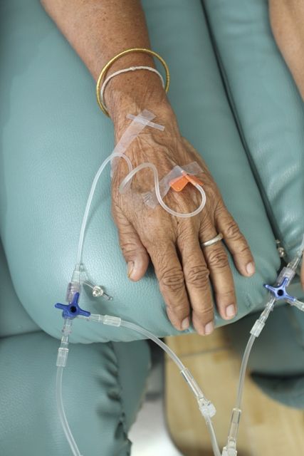
Chemotherapy increases the risk of infertility and early menopause. But in past studies, some 70% to 80% of women regained at least some level of ovarian function after completing treatment, the researchers noted. Over the course of 6-12 months, quiescent follicles mature to the antral stage, in which exposure to follicle-stimulating hormone triggers their rapid growth and maturation. Thus, antral follicle count is an accepted marker of ovarian reserve that reliably predicts how many oocytes will be retrieved during in vitro fertilization and fertility preservation, according to the investigators.
Based on these observations, they tracked antral follicle counts over time among 199 patients who were seen for fertility preservation consultations before starting cyclophosphamide-based chemotherapy for breast cancer, and who resumed menstruating afterward. Before chemotherapy, these women had an average antral follicle count of 15, with a standard deviation of 12. They averaged 35 years of age, with a standard deviation of 5 years.
A total of 66 women returned after chemotherapy for follow-up, and underwent an average of four antral follicle counts, with a standard deviation of two, the researchers said. These measurements showed that for up to 12 months after finishing chemotherapy, patients typically had only about 14% to 25% of their baseline ovarian reserve. By month 13, however, antral follicle counts rose to an average of 52% of baseline, and remained at this level through month 18 and beyond.
Treatment with leuprolide during chemotherapy appeared to increase ovarian recovery from about month 7 onward, the researchers reported. Between 7 and 9 months after chemotherapy, antral follicle counts averaged 32% of baseline among patients who had received leuprolide, but were about 8% of baseline among patients who had not received leuprolide. Between months 13 and 18, leuprolide recipients recovered about 74% of their ovarian reserve, while other patients recovered about 35% (P = .09). Beyond month 18, leuprolide recipients recovered an average of 69% of their baseline antral follicle count and other patients recovered an average of 4 (P = .07).
“Women who did not take [leuprolide] during chemotherapy represent 75% of our study population,” the researchers said. “[These women] appeared to have lower antral follicle count recovery, despite resumption of menses, than those whose menses resumed after chemotherapy with concurrent [leuprolide].”
Dr. Letourneau reported having no relevant financial disclosures.
Key clinical point:
Major finding: Average antral follicle counts rose to 52% of baseline, on average, by 13 months after patients completed chemotherapy.
Data source: A prospective cohort study of 66 patients who resumed menstruating and returned for at least one follow-up visit after chemotherapy.
Disclosures: Dr. Letourneau reported having no relevant financial disclosures.
Study links low diastolic blood pressure to myocardial damage, coronary heart disease
Low diastolic blood pressure (DBP) was significantly associated with myocardial injury and incident coronary heart disease, especially when the systolic blood pressure was 120 mm or higher, investigators reported.
Compared with a DBP of 80 to 89 mm Hg, DBP below 60 mm Hg more than doubled the odds of high-sensitivity cardiac troponin-T levels equaling or exceeding 14 ng per mL, and increased the risk of incident coronary heart disease (CHD) by about 50%, in a large observational study. Associations were strongest when baseline systolic blood pressure was at least 120 mm Hg, signifying elevated pulse pressure, reported Dr. John McEvoy of the Ciccarone Center for the Prevention of Heart Disease, Hopkins University, Baltimore, and associates (J Am Coll Cardiol 2016;68[16]:1713–22).
Their study included 11,565 individuals tracked for 21 years through the Atherosclerosis Risk in Communities Cohort, an observational population-based study of adults from in North Carolina, Mississippi, Minnesota, and Maryland. The researchers excluded participants with known baseline cardiovascular disease or heart failure. High-sensitivity cardiac troponin-T levels were measured at three time points between 1990 and 1992, 1996 and 1998, and 2011 and 2013. Participants averaged 57 years old at enrollment, 57% were female, and 25% were black (J Am Coll Cardiol. 2016 Oct 18. doi: 10.1016/j.jacc.2016.07.754).
Compared with baseline DBP of 80 to 89 mm Hg, DBP under 60 mm Hg was associated with a 2.2-fold greater odds (P = .01) of high-sensitivity cardiac troponin-T levels equal to or exceeding 14 ng per mL during the same visit – indicating prevalent myocardial damage – even after controlling for race, sex, body mass index, smoking and alcohol use, triglyceride and cholesterol levels, diabetes, glomerular filtration rate, and use of antihypertensives and lipid-lowering drugs, said the researchers. The odds of myocardial damage remained increased even when DBP was 60 to 69 mm Hg (odds ratio, 1.5; P = .05). Low DBP also was associated with myocardial damage at any given systolic blood pressure.
Furthermore, low DBP significantly increased the risk of progressively worsening myocardial damage, as indicated by a rising annual change in high-sensitivity cardiac troponin-T levels over 6 years. The association was significant as long as DBP was under 80 mm Hg, but was strongest when DBP was less than 60 mm Hg. Diastolic blood pressure under 60 mm Hg also significantly increased the chances of incident CHD and death, but not stroke.
Low DBP was most strongly linked to subclinical myocardial damage and incident CHD when systolic blood pressure was at least 120 mm Hg, indicating elevated pulse pressure, the researchers reported. Systolic pressure is “the main determinant of cardiac afterload and, thus, a primary driver of myocardial energy requirements,” while low DBP reduces myocardial energy supply, they noted. Therefore, high pulse pressure would lead to the greatest mismatch between myocardial energy demand and supply.
“Among patients being treated to SBP goals of 140 mm Hg or lower, attention may need to be paid not only to SBP, but also, importantly, to achieved DBP. Diastolic and systolic BP are inextricably linked, and our results highlighted the importance of not ignoring the former and focusing only on the latter, instead emphasizing the need to consider both in the optimal treatment of adults with hypertension.,”
The study was supported by the National Institutes of Health/National Institute of Diabetes and Digestive and Kidney Diseases and by the National Heart, Lung, and Blood Institute. Roche Diagnostics provided reagents for the cardiac troponin assays. Dr. McEvoy had no disclosures. One author disclosed ties to Roche; one author disclosed ties to Roche, Abbott Diagnostics, and several other relevant companies; and two authors are coinvestigators on a provisional patent filed by Roche for use of biomarkers in predicting heart failure. The other four authors had no disclosures.
The average age in the study by McEvoy et al. was 57 years. One might anticipate that in an older population, the side effects from lower BPs [blood pressures] due to drug therapy such as hypotension or syncope would be greater, and the potential for adverse cardiovascular events due to a J-curve would be substantially increased compared with what was seen in the present study. Similarly, an exacerbated potential for lower DBP to be harmful might be expected in patients with established coronary artery disease.
The well done study ... shows that lower may not always be better with respect to blood pressure control and, along with other accumulating evidence, strongly suggests careful thought before pushing blood pressure control below current guideline targets, especially if the diastolic blood pressure falls below 60 mm Hg while the pulse pressure is[greater than] 60 mm Hg.
Deepak L. Bhatt, MD, MPH, is at Brigham and Women’s Hospital Heart & Vascular Center, Boston. He disclosed ties to Amarin, Amgen, AstraZeneca, Bristol-Myers Squibb, Eisai, and a number of other pharmaceutical and medical education companies. His comments are from an accompanying editorial (J Am Coll Cardiol. 2016 Oct 18;68[16]:1723-1726).
Low diastolic blood pressure (DBP) was significantly associated with myocardial injury and incident coronary heart disease, especially when the systolic blood pressure was 120 mm or higher, investigators reported.
Compared with a DBP of 80 to 89 mm Hg, DBP below 60 mm Hg more than doubled the odds of high-sensitivity cardiac troponin-T levels equaling or exceeding 14 ng per mL, and increased the risk of incident coronary heart disease (CHD) by about 50%, in a large observational study. Associations were strongest when baseline systolic blood pressure was at least 120 mm Hg, signifying elevated pulse pressure, reported Dr. John McEvoy of the Ciccarone Center for the Prevention of Heart Disease, Hopkins University, Baltimore, and associates (J Am Coll Cardiol 2016;68[16]:1713–22).
Their study included 11,565 individuals tracked for 21 years through the Atherosclerosis Risk in Communities Cohort, an observational population-based study of adults from in North Carolina, Mississippi, Minnesota, and Maryland. The researchers excluded participants with known baseline cardiovascular disease or heart failure. High-sensitivity cardiac troponin-T levels were measured at three time points between 1990 and 1992, 1996 and 1998, and 2011 and 2013. Participants averaged 57 years old at enrollment, 57% were female, and 25% were black (J Am Coll Cardiol. 2016 Oct 18. doi: 10.1016/j.jacc.2016.07.754).
Compared with baseline DBP of 80 to 89 mm Hg, DBP under 60 mm Hg was associated with a 2.2-fold greater odds (P = .01) of high-sensitivity cardiac troponin-T levels equal to or exceeding 14 ng per mL during the same visit – indicating prevalent myocardial damage – even after controlling for race, sex, body mass index, smoking and alcohol use, triglyceride and cholesterol levels, diabetes, glomerular filtration rate, and use of antihypertensives and lipid-lowering drugs, said the researchers. The odds of myocardial damage remained increased even when DBP was 60 to 69 mm Hg (odds ratio, 1.5; P = .05). Low DBP also was associated with myocardial damage at any given systolic blood pressure.
Furthermore, low DBP significantly increased the risk of progressively worsening myocardial damage, as indicated by a rising annual change in high-sensitivity cardiac troponin-T levels over 6 years. The association was significant as long as DBP was under 80 mm Hg, but was strongest when DBP was less than 60 mm Hg. Diastolic blood pressure under 60 mm Hg also significantly increased the chances of incident CHD and death, but not stroke.
Low DBP was most strongly linked to subclinical myocardial damage and incident CHD when systolic blood pressure was at least 120 mm Hg, indicating elevated pulse pressure, the researchers reported. Systolic pressure is “the main determinant of cardiac afterload and, thus, a primary driver of myocardial energy requirements,” while low DBP reduces myocardial energy supply, they noted. Therefore, high pulse pressure would lead to the greatest mismatch between myocardial energy demand and supply.
“Among patients being treated to SBP goals of 140 mm Hg or lower, attention may need to be paid not only to SBP, but also, importantly, to achieved DBP. Diastolic and systolic BP are inextricably linked, and our results highlighted the importance of not ignoring the former and focusing only on the latter, instead emphasizing the need to consider both in the optimal treatment of adults with hypertension.,”
The study was supported by the National Institutes of Health/National Institute of Diabetes and Digestive and Kidney Diseases and by the National Heart, Lung, and Blood Institute. Roche Diagnostics provided reagents for the cardiac troponin assays. Dr. McEvoy had no disclosures. One author disclosed ties to Roche; one author disclosed ties to Roche, Abbott Diagnostics, and several other relevant companies; and two authors are coinvestigators on a provisional patent filed by Roche for use of biomarkers in predicting heart failure. The other four authors had no disclosures.
From the Journal of the American College of Cardiology
Key clinical point: Low diastolic blood pressure is associated with myocardial injury and incident coronary heart disease.
Major finding: Diastolic blood pressure below 60 mm Hg more than doubled the odds of high-sensitivity cardiac troponin-T levels equaling or exceeding 14 ng per mL and increased the risk of incident coronary heart disease by about 50%, compared to diastolic blood pressure of 80 to 89 mm Hg. Associations were strongest when pressure was elevated (above 60 mm Hg).
Data source: A prospective observational study of 11,565 adults followed for 21 years as part of the Atherosclerosis Risk in Communities cohort.
Disclosures: The study was supported by the National Institutes of Health/National Institute of Diabetes and Digestive and Kidney Diseases and by the National Heart, Lung, and Blood Institute. Roche Diagnostics provided reagents for the cardiac troponin assays. Dr. McEvoy had no disclosures. One author disclosed ties to Roche; one author disclosed ties to Roche, Abbott Diagnostics, and several other relevant companies; and two authors are coinvestigators on a provisional patent filed by Roche for use of biomarkers in predicting heart failure. The other four authors had no disclosures.

CDC study finds worrisome trends in hospital antibiotic use
U.S. hospitals have not cut overall antibiotic use and have significantly increased the use of several broad-spectrum agents, according to a first-in-kind analysis of national hospital administrative data.
“We identified significant changes in specific antibiotic classes and regional variation that may have important implications for reducing antibiotic-resistant infections,” James Baggs, PhD, and colleagues from the Centers for Disease Control and Prevention, Atlanta, reported in the study, published online on September 19 in JAMA Internal Medicine.
The retrospective study included approximately 300 acute care hospitals in the Truven Health MarketScan Hospital Drug Database, which covered 34 million pediatric and adult patient discharges equating to 166 million patient-daysIn all, 55% of patients received at least one antibiotic dose while in the hospital, and for every 1,000 patient-days, 755 days included antibiotic therapy, the investigators said. Overall antibiotic use rose during the study period by only 5.6 average days of therapy per 1,000 patient-days, which was not statistically significant.
However, the use of third and fourth-generation cephalosporins rose by a mean of 10.3 days of therapy per 1,000 patient-days (95% confidence interval, 3.1 to 17.5), and hospitals also used significantly more macrolides (mean rise, 4.8 days of therapy per 1,000 patient-days; 95% confidence interval, 2.0 to 7.6 days), glycopeptides, (22.4; 17.5 to 27.3); β-lactam/β-lactamase inhibitor combinations (18.0; 13.3 to 22.6), carbapenems (7.4; 4.6 to 10.2), and tetracyclines (3.3; 2.0 to 4.7)
Inpatient antibiotic use also varied significantly by region, the investigators said. Hospitals in rural areas used about 16 more days of antibiotic therapy per 1,000 patient-days compared with those in urban areas. Hospitals in Mid-Atlantic states (New Jersey, New York, Pennsylvania) and Pacific Coast states (Alaska, California, Hawaii, Oregon, and Washington) used the least antibiotics (649 and 665 days per 1,000 patient-days, respectively), while Southwest Central states (Arkansas, Louisiana, Oklahoma, and Texas) used the most (823 days).
The CDC provided funding for the study. The researchers had no disclosures.
The dramatic variation in antibiotic prescribing across individual clinicians, regions in the United States, and internationally indicates great potential for improvement. ... In the article by Baggs et al, inpatient antibiotic prescribing in some regions of the United States is roughly 20% lower than other regions. On a per capita basis, Swedes consume less than half the antibiotics per capita than Americans.
Growing patterns of antibiotic resistance have driven calls for more physician education and new diagnostics. While these efforts may help, it is important to recognize that many emotionally salient factors are driving physicians to inappropriately prescribe antibiotics. Future interventions need to counterbalance these factors using tools from behavioral science to reduce the use of inappropriate antibiotics.
Ateev Mehrotra, MD, MPH, and Jeffrey A. Linder, MD, MPH, are at Harvard University, Boston. They had no disclosures. These comments are from an editorial that accompanied the study ( JAMA Intern Med. 2016 Sept 19. doi: 10.1001/jamainternmed.2016.6254).
U.S. hospitals have not cut overall antibiotic use and have significantly increased the use of several broad-spectrum agents, according to a first-in-kind analysis of national hospital administrative data.
“We identified significant changes in specific antibiotic classes and regional variation that may have important implications for reducing antibiotic-resistant infections,” James Baggs, PhD, and colleagues from the Centers for Disease Control and Prevention, Atlanta, reported in the study, published online on September 19 in JAMA Internal Medicine.
The retrospective study included approximately 300 acute care hospitals in the Truven Health MarketScan Hospital Drug Database, which covered 34 million pediatric and adult patient discharges equating to 166 million patient-daysIn all, 55% of patients received at least one antibiotic dose while in the hospital, and for every 1,000 patient-days, 755 days included antibiotic therapy, the investigators said. Overall antibiotic use rose during the study period by only 5.6 average days of therapy per 1,000 patient-days, which was not statistically significant.
However, the use of third and fourth-generation cephalosporins rose by a mean of 10.3 days of therapy per 1,000 patient-days (95% confidence interval, 3.1 to 17.5), and hospitals also used significantly more macrolides (mean rise, 4.8 days of therapy per 1,000 patient-days; 95% confidence interval, 2.0 to 7.6 days), glycopeptides, (22.4; 17.5 to 27.3); β-lactam/β-lactamase inhibitor combinations (18.0; 13.3 to 22.6), carbapenems (7.4; 4.6 to 10.2), and tetracyclines (3.3; 2.0 to 4.7)
Inpatient antibiotic use also varied significantly by region, the investigators said. Hospitals in rural areas used about 16 more days of antibiotic therapy per 1,000 patient-days compared with those in urban areas. Hospitals in Mid-Atlantic states (New Jersey, New York, Pennsylvania) and Pacific Coast states (Alaska, California, Hawaii, Oregon, and Washington) used the least antibiotics (649 and 665 days per 1,000 patient-days, respectively), while Southwest Central states (Arkansas, Louisiana, Oklahoma, and Texas) used the most (823 days).
The CDC provided funding for the study. The researchers had no disclosures.
FROM JAMA INTERNAL MEDICINE
Key clinical point: Inpatient antibiotic use did not decrease between 2006 and 2012, and the use of several broad-spectrum agents rose significantly.
Major finding: Hospitals significantly decreased their use of fluoroquinolones and first- and second-generation cephalosporins, but these trends were offset by significant rises in the use of vancomycin, carbapenem, third- and fourth-generation cephalosporins, and β-lactam/β- lactamase inhibitor combinations.
Data source: A retrospective study of administrative hospital discharge data for about 300 US hospitals from 2006 through 2012.
Disclosures: The Centers for Disease Control and Prevention provided funding. The researchers had no disclosures.
Resveratrol cut androgen levels in small PCOS trial
SALT LAKE CITY – Resveratrol significantly decreased androgen levels and insulin resistance among women with polycystic ovary syndrome (PCOS), according to a first-in-kind randomized, double-blind placebo-controlled trial of 30 patients.
After 3 months of daily oral treatment with 1.5 g of the antioxidant polyphenol, testosterone levels fell an average of 23%, dehydroepiandrosterone sulfate (DHEAS) levels dropped 22%, fasting insulin levels decreased by 32%, and insulin sensitivity improved by 66%, Antoni Duleba, MD, said at the annual meeting of the American Society for Reproductive Medicine. No such improvements occurred in the placebo group, he and his coinvestigators reported simultaneously in the Journal of Clinical Endocrinology & Metabolism (doi: 10.1210/jc.2016-1858).

Past work has found that resveratrol inhibits mRNA expression of Cyp17a1 and reduces androgen production by ovarian theca-interstitial cells. To build on those findings, Dr. Duleba worked with researchers at Poznan (Poland) University of Medical Sciences to enroll 30 women with PCOS based on the Rotterdam criteria. Treatment and placebo groups resembled each other at baseline in terms of age, body mass index, androgen levels, lipid profiles, and levels of follicle-stimulating hormone, luteinizing hormone, prolactin, sex hormone–binding globulin, fasting glucose, and insulin, as well as scores on an insulin sensitivity index.
For the primary outcome measure – total testosterone level – the resveratrol group averaged 0.53 ng/mL at baseline and 0.41 ng/mL at 3-month follow-up, a statistically significant decrease (P = .01). In contrast, testosterone levels in the placebo group remained essentially unchanged, averaging 0.48 ng/mL at baseline and 0.49 ng/mL at follow-up. The difference in effect between the resveratrol and placebo groups was statistically significant (P = .04).
Similarly, average DHEAS levels dropped from 8.1 to 6.3 micromol/L in the resveratrol group (a 22% decline), but increased by 10% in the placebo group.
Resveratrol did not significantly affect progesterone levels, which is consistent with prior findings, Dr. Duleba said. Nor was resveratrol associated with significant changes in body mass index, lipid profile, markers of inflammation or endothelial function, ovarian volume, or gonadotropins.
“We were disappointed that we didn’t see gross changes in ovarian morphology on ultrasound,” he added. Whether those changes might occur with longer treatment is unknown, but for now, “we can only be sure of declining androgen and insulin levels, and improvements in insulin sensitivity.”
The study won a Scientific Congress Prize from ASRM.
RevGenetics provided the resveratrol for the study. Dr. Duleba reported having no relevant financial disclosures.
SALT LAKE CITY – Resveratrol significantly decreased androgen levels and insulin resistance among women with polycystic ovary syndrome (PCOS), according to a first-in-kind randomized, double-blind placebo-controlled trial of 30 patients.
After 3 months of daily oral treatment with 1.5 g of the antioxidant polyphenol, testosterone levels fell an average of 23%, dehydroepiandrosterone sulfate (DHEAS) levels dropped 22%, fasting insulin levels decreased by 32%, and insulin sensitivity improved by 66%, Antoni Duleba, MD, said at the annual meeting of the American Society for Reproductive Medicine. No such improvements occurred in the placebo group, he and his coinvestigators reported simultaneously in the Journal of Clinical Endocrinology & Metabolism (doi: 10.1210/jc.2016-1858).
Past work has found that resveratrol inhibits mRNA expression of Cyp17a1 and reduces androgen production by ovarian theca-interstitial cells. To build on those findings, Dr. Duleba worked with researchers at Poznan (Poland) University of Medical Sciences to enroll 30 women with PCOS based on the Rotterdam criteria. Treatment and placebo groups resembled each other at baseline in terms of age, body mass index, androgen levels, lipid profiles, and levels of follicle-stimulating hormone, luteinizing hormone, prolactin, sex hormone–binding globulin, fasting glucose, and insulin, as well as scores on an insulin sensitivity index.
For the primary outcome measure – total testosterone level – the resveratrol group averaged 0.53 ng/mL at baseline and 0.41 ng/mL at 3-month follow-up, a statistically significant decrease (P = .01). In contrast, testosterone levels in the placebo group remained essentially unchanged, averaging 0.48 ng/mL at baseline and 0.49 ng/mL at follow-up. The difference in effect between the resveratrol and placebo groups was statistically significant (P = .04).
Similarly, average DHEAS levels dropped from 8.1 to 6.3 micromol/L in the resveratrol group (a 22% decline), but increased by 10% in the placebo group.
Resveratrol did not significantly affect progesterone levels, which is consistent with prior findings, Dr. Duleba said. Nor was resveratrol associated with significant changes in body mass index, lipid profile, markers of inflammation or endothelial function, ovarian volume, or gonadotropins.
“We were disappointed that we didn’t see gross changes in ovarian morphology on ultrasound,” he added. Whether those changes might occur with longer treatment is unknown, but for now, “we can only be sure of declining androgen and insulin levels, and improvements in insulin sensitivity.”
The study won a Scientific Congress Prize from ASRM.
RevGenetics provided the resveratrol for the study. Dr. Duleba reported having no relevant financial disclosures.
AT 2016 ASRM
Key clinical point:
Major finding: Average total testosterone levels dropped 23% in the treatment group and remained unchanged in the placebo group (P = .04).
Data source: A single-center, double-blind, randomized controlled trial of 30 women with polycystic ovary syndrome.
Disclosures: RevGenetics provided the resveratrol for the study. Dr. Duleba reported having no relevant financial disclosures.
Psychological stress did not harm IVF outcomes
SALT LAKE CITY – Self-reported psychological distress and biological markers of stress did not affect the chances of pregnancy after in vitro fertilization, according to a single-center prospective cohort study of 186 women.
Rates of ultrasound-confirmed pregnancy were 36% among first-time IVF recipients and 30% among women undergoing their third round of IVF, and did not differ based on self-reported depressive symptoms, stress, anxiety, or serum levels of adrenocorticotropic hormone (ACTH), cortisol, or interleukin-6, Maria Costantini-Ferrando, MD, PhD, said at the annual meeting of the American Society for Reproductive Medicine.
Nonetheless, IVF recipients reported more depressive symptoms than oocyte donors did, especially after a negative pregnancy test, reported Dr. Costantini-Ferrando, a reproductive endocrinologist in Englewood, N.J.
“Patients undergoing IVF do experience real psychological distress. The efficiency of IVF in controlling the hypothalamic-pituitary-adrenal and the hypothalamic-pituitary-gonadal axes may overshadow any deleterious effect of heightened stress, but it will not eliminate patients’ experience of it,” she said.
Patients need support and follow-up to help lessen the psychological burden of treatment and decrease drop-out, she said.
Women in the study were aged 22-44 years, and the IVF recipients had a mean age of 37 years. In all, 75 women (40%) were first-time IVF patients, 29% were undergoing their third cycle of IVF, and 31% were oocyte donors. Among the IVF recipients, 22% were in psychotherapy, and 4% were on psychotropic medications. None of the women had other medical comorbidities. Patients were assessed at baseline, time of oocyte retrieval, time of embryo transfer, and on the day of the pregnancy test.
Patients with infertility reported significantly worse depressive symptoms at baseline compared with oocyte donors, with average Beck Depression Inventory scores of 11, compared with 2.3 (P less than .01). This difference persisted at the time of vaginal oocyte retrieval. First-time and third-time IVF recipients tended to have similar Beck Depression Inventory scores until pregnancy testing, when the average scores of third-time recipients increased by about five additional points.
Scores on the daily stress questionnaire echoed these findings. Patients who were receiving IVF reported significantly more stress than did oocyte donors, with the highest increase at the time of the pregnancy test among patients with previous IVF failures. However, both recipients and donors reported mild to moderate anxiety based on the State-Trait Anxiety Inventory, Dr. Costantini-Ferrando said.
All groups had low interleukin-6 levels when starting IVF, but donors had the highest levels of both ACTH and cortisol. Levels of these hormones dropped over time, and then rose at the time of pregnancy testing. That trend suggests that IVF was an acute “procedural” stressor for both recipients and donors, but that the magnitude of the stress response lessened somewhat over time with repeated exposure to the stressor, Dr. Costantini-Ferrando noted.
“The complexity of this interaction may explain why it is difficult to elucidate the true impact of stress on IVF outcome,” she said.
Ultimately, these findings support a shift in focus, she added. Instead of asking if stress decreases reproductive potential, clinicians and researchers can instead explore how to address psychological distress to maximize reproductive potential. “This is important to keep patients engaged in infertility treatment and facilitate the difficult process of decision making during treatment,” she added.
The paper won a prize from the ASRM Mental Health Professional Group.
The National Institutes of Health provided funding for the study. Dr. Costantini-Ferrando reported having no relevant financial disclosures.
Key clinical point:
Major finding: Rates of pregnancy were 36% among first-time IVF recipients and 30% among third-time IVF recipients, and did not correlate with self-reported depressive symptoms, stress, anxiety, or serum levels of adrenocorticotropic hormone, cortisol, or interleukin-6.
Data source: A single-center prospective study of 186 IVF patients.
Disclosures: The National Institutes of Health provided funding for the study. Dr. Costantini-Ferrando reported having no financial disclosures.
Low serum AMH predicted miscarriage after IVF
SALT LAKE CITY – Low serum levels of anti-Müllerian hormone predicted miscarriage after in vitro fertilization and embryo transfer, according to a single-center prospective study of more than 2,000 women.
This relationship remained statistically significant after the study controlled for age and response to controlled ovarian hyperstimulation, which suggests that AMH is a marker of the reproductive potential of oocytes, not just of oocyte quantity, Bruno Tarasconi, MD, said at the annual meeting of the American Society for Reproductive Medicine.
“Low” serum AMH was defined as less than 1.61 ng per mL, but specific levels vary by test type, so “this is not a definite cutoff,” Dr. Tarasconi emphasized. “What we need to know is that patients with low AMH are probably experiencing some problems in the ovary that affect oocyte quality.”
The study comprised 2,365 women undergoing 2,688 IVF cycles with fresh oocytes. Levels of AMH were measured by ELISA (AMH Gen II, Beckman Coulter) within 12 months before IVF on cycle days 2 and 4, said Dr. Tarasconi of University of Paris Ouest in France.
Women younger than 34 years old, women 34-36 years old, and women 37 years and older had the highest rates of miscarriage when they had low serum AMH levels. For the two older age groups, low AMH was associated with about a 33% miscarriage rate – about twice the rate of miscarriage among women whose AMH measured at least 5.6 ng per mL. This difference reached statistical significance for both age groups (P less than .05).
Among women younger than 34 years, low serum AMH was associated with a 22% rate of miscarriage, compared with about 13% among women with higher AMH levels. This difference did not reach significance because of a lack of power, Dr. Tarasconi said. “However, binary logistic regression of the whole population confirmed the association between low serum AMH and miscarriage, independent of patient’s age and oocyte yield, with the P value less than .001.”
Low circulating AMH can reflect an above-average prevalence of oocyte abnormalities, which are linked to miscarriage, Dr. Tarasconi noted. “The fact that oocyte yield failed to alter the relationship between AMH and miscarriage suggests that altered per-follicle AMH production, not oocyte or embryo availability, fosters pregnancy loss,” he added. However, the researchers did not test either preimplantation embryos or the expelled gestational sacs to directly link miscarriages to abnormalities such as aneuploidy, he noted.
Low AMH levels also were associated with lower rates of pregnancy after IVF, regardless of age, as has been previously reported. Patients in the study were of normal karyotype, with no history of recurrent pregnancy loss, uterine abnormalities, or family history of genetic or congenital disorders, Dr. Tarasconi noted.
He reported having no financial disclosures.
Key clinical point:
Major finding: The miscarriage rate was about 33% in women with low serum AMH levels after IVF, about twice that of women with high AMH levels.
Data source: A single-center prospective study of 2,365 women undergoing 2,688 IVF cycles with fresh oocytes.
Disclosures: Dr. Tarasconi reported having no financial disclosures.
Novel fractional laser eased genitourinary syndrome of menopause
DENVER – Novel fractional laser therapy achieved significant 12-month reductions in vaginal dryness, burning, pain, and itching associated with menopause, Eric Sokol, MD, reported at Pelvic Floor Disorders Week, sponsored by the American Urogynecologic Society.
Most patients – 92% – said they were satisfied or very satisfied with the treatment, said Dr. Sokol of Stanford (Calif.) University. “Treatments take 45 seconds and are painless,” he added. “We are part of a group that is planning a larger multicenter randomized trial of this laser, as well as some histologic studies.”
The two-center pilot study included 30 women with vulvovaginal atrophy treated with a novel fractional carbon dioxide laser system called SmartXide2 V2LR (MonaLisa Touch). The system has a maximum power of 60 W and emits laser energy at a 10,600-nm wavelength, Dr. Sokol noted. Patients underwent three treatments spaced by 6 weeks, and used 10-point visual analogue scales to score baseline and subsequent levels of vaginal pain, burning, itching, dryness, dyspareunia, and dysuria.

Average scores on the Female Sexual Function Index rose from 11.3 at baseline to 21.25 at 12 months, a statistically significant improvement.
Laser therapy caused no major adverse events, but about 10% of patients developed slight vaginal discharge or minor spotting after treatment, Dr. Sokol reported. Patients were not allowed to use lubricants or estrogens during the study.
The study was supported by DEKA M.E.L.A. Srl, using an investigator-initiated protocol. Patients did not pay for treatment. Dr. Sokol reported that he has no financial disclosures related to this study. His coinvestigator is a paid consultant for Cynosure.
DENVER – Novel fractional laser therapy achieved significant 12-month reductions in vaginal dryness, burning, pain, and itching associated with menopause, Eric Sokol, MD, reported at Pelvic Floor Disorders Week, sponsored by the American Urogynecologic Society.
Most patients – 92% – said they were satisfied or very satisfied with the treatment, said Dr. Sokol of Stanford (Calif.) University. “Treatments take 45 seconds and are painless,” he added. “We are part of a group that is planning a larger multicenter randomized trial of this laser, as well as some histologic studies.”
The two-center pilot study included 30 women with vulvovaginal atrophy treated with a novel fractional carbon dioxide laser system called SmartXide2 V2LR (MonaLisa Touch). The system has a maximum power of 60 W and emits laser energy at a 10,600-nm wavelength, Dr. Sokol noted. Patients underwent three treatments spaced by 6 weeks, and used 10-point visual analogue scales to score baseline and subsequent levels of vaginal pain, burning, itching, dryness, dyspareunia, and dysuria.
Average scores on the Female Sexual Function Index rose from 11.3 at baseline to 21.25 at 12 months, a statistically significant improvement.
Laser therapy caused no major adverse events, but about 10% of patients developed slight vaginal discharge or minor spotting after treatment, Dr. Sokol reported. Patients were not allowed to use lubricants or estrogens during the study.
The study was supported by DEKA M.E.L.A. Srl, using an investigator-initiated protocol. Patients did not pay for treatment. Dr. Sokol reported that he has no financial disclosures related to this study. His coinvestigator is a paid consultant for Cynosure.
Key clinical point:
Major finding: Reductions in dryness and dyspareunia were especially marked, dropping by an average of about 75% and 66%, respectively.
Data source: A two-center pilot study of 30 women with genitourinary syndrome of atrophy (previously known as vaginal atrophy).
Disclosures: The study was supported by DEKA M.E.L.A. Srl, using an investigator-initiated protocol. Patients did not pay for treatment. Dr. Sokol reported that he has no financial disclosures related to this study. His coinvestigator is a paid consultant for Cynosure.
PD-L1 positivity correlated with pembrolizumab response in advanced melanoma
Programmed death ligand–1 expression correlated positively and significantly with pembrolizumab response in advanced melanoma, based on an analysis of 405 patients from the international, multicohort, open-label phase I KEYNOTE-001 trial.
Among patients for whom 33%-65% of tumor cells expressed PD-L1, the objective rate of response was 57%, but the rate was only 8% among patients whose specimens did not express PD-L1, Adil Daud, MD, of the University of California, San Francisco, and associates reported. Taken together, the findings suggest that melanoma is most likely to respond to pembrolizumab when specimens show at least 10% positivity, the investigators reported (J Clin Oncol. 2016 Oct 10. doi: 10.1200/JCO.2016.67.2477).

Among the 405 patients who also were evaluable for tumor response, the overall objective response rate was 33% (95% confidence interval, 28%-37%). Grouping patients based on the melanoma scoring system for PD-L1 expression showed that PD-L1 positivity correlated significantly with the objective response rate (P less than .001). Furthermore, a higher PD-L1 melanoma score correlated significantly with both progression-free survival (hazard ratio, 0.76; 95% CI, 0.7-0.82) and overall survival (HR, 0.76; 95% CI, 0.69-0.83), with P values less than .001 for each association.
Median progression-free survival was 5.6 months in PD-L1–positive patients and 2.8 months in PD-L1–negative patients, while median overall survival was 30 months in PD-L1–positive patients and 12.6 months in PD-L1–negative patients, the researchers reported. “The high prevalence of PD-L1 positivity observed in this study, along with the durable responses observed in PD-L1–negative tumors, suggest that pembrolizumab treatment should not be limited to patients with PD-L1–positive tumors,” they concluded. “Ongoing clinical trials with correlative studies will further delineate the role of PD-L1 expression in melanoma.”
Merck sponsored the trial. Dr. Daud disclosed ties to Merck, OncoSec, Novartis, Genentech, Bristol-Myers Squibb, and Array BioPharma.
Programmed death ligand–1 expression correlated positively and significantly with pembrolizumab response in advanced melanoma, based on an analysis of 405 patients from the international, multicohort, open-label phase I KEYNOTE-001 trial.
Among patients for whom 33%-65% of tumor cells expressed PD-L1, the objective rate of response was 57%, but the rate was only 8% among patients whose specimens did not express PD-L1, Adil Daud, MD, of the University of California, San Francisco, and associates reported. Taken together, the findings suggest that melanoma is most likely to respond to pembrolizumab when specimens show at least 10% positivity, the investigators reported (J Clin Oncol. 2016 Oct 10. doi: 10.1200/JCO.2016.67.2477).
Among the 405 patients who also were evaluable for tumor response, the overall objective response rate was 33% (95% confidence interval, 28%-37%). Grouping patients based on the melanoma scoring system for PD-L1 expression showed that PD-L1 positivity correlated significantly with the objective response rate (P less than .001). Furthermore, a higher PD-L1 melanoma score correlated significantly with both progression-free survival (hazard ratio, 0.76; 95% CI, 0.7-0.82) and overall survival (HR, 0.76; 95% CI, 0.69-0.83), with P values less than .001 for each association.
Median progression-free survival was 5.6 months in PD-L1–positive patients and 2.8 months in PD-L1–negative patients, while median overall survival was 30 months in PD-L1–positive patients and 12.6 months in PD-L1–negative patients, the researchers reported. “The high prevalence of PD-L1 positivity observed in this study, along with the durable responses observed in PD-L1–negative tumors, suggest that pembrolizumab treatment should not be limited to patients with PD-L1–positive tumors,” they concluded. “Ongoing clinical trials with correlative studies will further delineate the role of PD-L1 expression in melanoma.”
Merck sponsored the trial. Dr. Daud disclosed ties to Merck, OncoSec, Novartis, Genentech, Bristol-Myers Squibb, and Array BioPharma.
Key clinical point: PD-L1 expression correlates with pembrolizumab response in advanced melanoma.
Major finding: Higher levels of PD-L1 staining correlated positively with tumor response (P less than .001).
Data source: Analyses of 405 patients from KEYNOTE-001, an international, multicohort, open-label phase I trial of pembrolizumab in advanced melanoma.
Disclosures: Merck sponsored the trial. Dr. Daud disclosed ties to Merck, OncoSec, Novartis, Genentech, Bristol-Myers Squibb, and Array BioPharma.
Registry helps track pelvic organ prolapse outcomes in the U.S.
DENVER – Catherine Bradley, MD, said during a presentation at Pelvic Floor Disorders Week, sponsored by the American Urogynecologic Society.
“This is a unique collaborative registry created in response to U.S. industry requirements. There are many benefits to using this approach, but also many challenges. It’s a work in progress,” said Dr. Bradley, of the University of Iowa, Iowa City. She chairs the American Urogynecologic Society Research Council, which helps oversee the registry.

The purpose of the PFDR is to fulfill requirements from the Food and Drug Administration for postmarketing studies of the vaginal mesh procedure, to give surgeons a pelvic organ prolapse treatment database to track individual and aggregate outcomes for research and quality improvement purposes, and to create a framework for future clinical studies, according to Dr. Bradley.
As such, the PFDR comprises two interrelated registries, Dr. Bradley noted. The PFDR-I includes industry-sponsored studies, while the PFDR-R is the independent research registry of the American Urogynecologic Society. The PFDR previously included a third quality improvement registry, which in January 2016 was moved to a separate platform called AQUIRE.
The PFDR-I is tracking 1,620 patients who underwent 1,386 procedures for pelvic organ prolapse as part of four studies sponsored by three manufacturers, according to Dr. Bradley. The PFDR-R, for its part, has eight active sites and has enrolled 179 patients with pelvic organ prolapse, 154 of whom underwent mesh surgery and 25 of whom received vaginal pessaries, she said.
The PFDR-R is voluntary and has faced some barriers in the 10 months since its launch, Dr. Bradley said. Participation increases workload for physicians as well as data entry personnel, and patients must provide informed consent to be entered into the registry. But the benefits of participating are also substantial, she emphasized. The PFDR-R will enable surgeons to track their own outcomes in terms of caseload, anatomic outcomes, symptoms and quality of life, and adverse events. They can download their own data, compare their outcomes to others as part of highly granular benchmarking initiatives, and propose and conduct studies of the entire registry.
The PFDR is supported by ACell, ASTORA Women’s Health, Boston Scientific, and Coloplast. Dr. Bradley reported having no conflicts of interest.
DENVER – Catherine Bradley, MD, said during a presentation at Pelvic Floor Disorders Week, sponsored by the American Urogynecologic Society.
“This is a unique collaborative registry created in response to U.S. industry requirements. There are many benefits to using this approach, but also many challenges. It’s a work in progress,” said Dr. Bradley, of the University of Iowa, Iowa City. She chairs the American Urogynecologic Society Research Council, which helps oversee the registry.
The purpose of the PFDR is to fulfill requirements from the Food and Drug Administration for postmarketing studies of the vaginal mesh procedure, to give surgeons a pelvic organ prolapse treatment database to track individual and aggregate outcomes for research and quality improvement purposes, and to create a framework for future clinical studies, according to Dr. Bradley.
As such, the PFDR comprises two interrelated registries, Dr. Bradley noted. The PFDR-I includes industry-sponsored studies, while the PFDR-R is the independent research registry of the American Urogynecologic Society. The PFDR previously included a third quality improvement registry, which in January 2016 was moved to a separate platform called AQUIRE.
The PFDR-I is tracking 1,620 patients who underwent 1,386 procedures for pelvic organ prolapse as part of four studies sponsored by three manufacturers, according to Dr. Bradley. The PFDR-R, for its part, has eight active sites and has enrolled 179 patients with pelvic organ prolapse, 154 of whom underwent mesh surgery and 25 of whom received vaginal pessaries, she said.
The PFDR-R is voluntary and has faced some barriers in the 10 months since its launch, Dr. Bradley said. Participation increases workload for physicians as well as data entry personnel, and patients must provide informed consent to be entered into the registry. But the benefits of participating are also substantial, she emphasized. The PFDR-R will enable surgeons to track their own outcomes in terms of caseload, anatomic outcomes, symptoms and quality of life, and adverse events. They can download their own data, compare their outcomes to others as part of highly granular benchmarking initiatives, and propose and conduct studies of the entire registry.
The PFDR is supported by ACell, ASTORA Women’s Health, Boston Scientific, and Coloplast. Dr. Bradley reported having no conflicts of interest.
AT PFD WEEK 2016
No rise in complications with concomitant gynecologic cancer, PFD surgery
DENVER – Concomitantly treating pelvic floor disorders during surgery for gynecologic cancer does not increase the risk of postoperative complications, according to an analysis of 4 years of data from the American College of Surgeons’ National Surgical Quality Improvement Program.
Among 23,501 surgical gynecologic cancer patients, 556 (2.4%) underwent concomitant surgery for symptomatic pelvic organ prolapse or urinary incontinence, Katarzyna Bochenska, MD, reported in a poster at Pelvic Floor Disorders Week, sponsored by the American Urogynecologic Society. This subgroup had similar 30-day rates of reoperation, venous thromboembolism, and infectious, pulmonary, and cardiac complications as patients who had surgery only for gynecologic cancer, reported Dr. Bochenska and her associates at Northwestern University in Chicago.
Urinary incontinence and symptomatic pelvic organ prolapse often accompany gynecologic cancer, the researchers noted. In one study, more than half of women with gynecologic cancer reported urinary incontinence, while 11% described feeling a bulge of tissue from their vaginas (Obstet Gynecol. 2013 Nov;122[5]:976-80). But few studies have examined outcomes after concomitant surgery for pelvic floor disorders and gynecologic cancer, according to the researchers.
In the current study, the Northwestern University researchers used postoperative ICD-9 codes to identify patients in the American College of Surgeons’ National Surgical Quality Improvement Program (ACS NSQIP) who underwent surgery for uterine, cervical, ovarian, or vulvar or vaginal cancer between 2010 and 2014. Most patients had uterine or ovarian cancer, while the most common pelvic floor disorder procedures included anterior and/or posterior colporrhaphy, laparoscopic colpopexy, and midurethral slings.
None of the complications studied differed significantly between the groups. Rates of 30-day reoperation were 1.1% among concomitant surgery patients and 2.3% among patients who underwent only cancer surgery (P = .09). Rates of procedure-related infections such as sepsis, deep wound infections, and abscesses also were similar between groups (3.1% and 3.9%, respectively), as were rates of postoperative urinary tract infections (1.8% and 3.2%), pulmonary complications (0.7% and 1.3%), and cardiac complications (0.2% and 0.4%), with all P-values exceeding .05.
Patients who underwent concomitant surgery for pelvic floor disorders were an average of about 3.5 years older than other patients, but otherwise resembled them in term of body mass index and prevalence of comorbidities, such as diabetes, chronic obstructive pulmonary disease, and hypertension.
“Our data suggest that serious postoperative complication rates are not increased in this population,” the researchers concluded. “Therefore, gynecologic surgeons should consider offering concomitant treatment for pelvic floor symptoms at the time of gynecologic cancer surgery.”
Dr. Bochenska and her associates did not report information on funding sources or financial disclosures.
AT PFD WEEK 2016
Key clinical point:
Major finding: Women who underwent concomitant surgeries had similar rates of infectious, pulmonary, and cardiac complications as those who underwent surgery only for gynecologic cancer, with all P-values exceeding .05.
Data source: A study of 23,501 gynecologic cancer patients in the ACS National Surgical Quality Improvement Program dataset.
Disclosures: Dr. Bochenska and her associates did not report information on funding sources or financial disclosures.