User login
Psychosis: First-episode variety in adolescence ‘insidious’
Serious mental illness can present slowly and in ways that might not look serious, which is why primary care physicians would do well to educate themselves about what psychosis looks like.
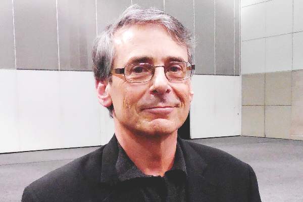
The problem, according to Dr. David Pickar, psychiatrist and former (retired) intramural research director for the National Institute of Mental Health, is the lack of information about recognizing the signs and symptoms, and about proper interventions.
“Knowing about it is enormously important for all docs,” Dr. Pickar says. “What’s fascinating is many of the first breaks occur, not necessarily quietly, but can be a little insidious. They can be brought to primary care. It is not uncommon. With serious mental illness, particularly schizophrenia, 1% of the population has it. That makes it a very common disorder.”
Serious mental illness can present slowly and in ways that might not look serious, which is why primary care physicians would do well to educate themselves about what psychosis looks like.
The problem, according to Dr. David Pickar, psychiatrist and former (retired) intramural research director for the National Institute of Mental Health, is the lack of information about recognizing the signs and symptoms, and about proper interventions.
“Knowing about it is enormously important for all docs,” Dr. Pickar says. “What’s fascinating is many of the first breaks occur, not necessarily quietly, but can be a little insidious. They can be brought to primary care. It is not uncommon. With serious mental illness, particularly schizophrenia, 1% of the population has it. That makes it a very common disorder.”

VIDEO: How to navigate value-based care payer contracts
AUSTIN, TEX. – The shift from volume- to value-based care has become a regular hot topic among the medical community. But one rarely discussed question is how quality-based care will impact physician contracts with health plans, according to Bloomfield Hills, Mich., health law attorney Mark S. Kopson.
In a video interview at an American Health Lawyers Association meeting, Mr. Kopson discusses how to navigate payer contracts when operating within value-based care models. He addresses ideal terms to include in quality-based care contracts and how to mitigate legal risks with health plans.
“The contract language that works in volume-based contracts doesn’t work in value-based contracts,” Mr. Kopson explains. “We have to make a distinction between the two and recognize that there are risks and issues that have to be addressed in value-based arrangements.”
The video associated with this article is no longer available on this site. Please view all of our videos on the MDedge YouTube channel
On Twitter @legal_med
EXPERT ANALYSIS FROM THE PHYSICIANS AND HOSPITALS LAW INSTITUTE

VIDEO: Are you an effective care team player?
SAN DIEGO – Hospitalists spend one-quarter of their practice time on team-related activities, yet some could stand to improve their performance as a patient-centered care team member, Dr. Kevin J. O’Leary said in a video interview at the annual meeting of the Society of Hospital Medicine.
Research shows that other team professionals, especially nurses, are not pleased with how hospitalists engage and collaborate with them, said Dr. O’Leary, chief of the division of hospital medicine at Northwestern University, Chicago.
To improve communication and coordination of care, Dr. O’Leary, who is also associate chair for quality in the department of medicine at the medical school, offered some teamwork interventions, including unit-based co-leadership, that hospitalists can try at their facilities. He reported having no financial disclosures.
The video associated with this article is no longer available on this site. Please view all of our videos on the MDedge YouTube channel
AT HOSPITAL MEDICINE 16

VIDEO: How proposed patient substance use privacy rule impacts physicians
Health providers have until April 11 to offer perspective on a proposed rule that would loosen restrictions over the redisclosure of patient records involving substance abuse.
The U.S. Department of Health and Human Services in early February announced suggested revisions to the Confidentiality of Alcohol and Drug Abuse Patient Records regulations, known as 42 CFR Part 2. The changes, published Feb. 9 in the Federal Register, aim to facilitate the transfer of health information within new care models while protecting patient privacy.
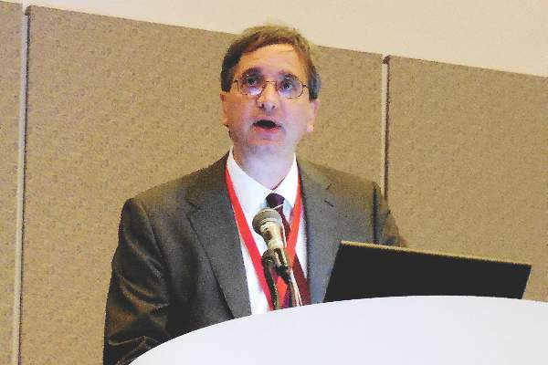
The proposed rule seeks to broaden the consent process for re-releasing substance use records to make it easier for health information exchanges to obtain health data, Chicago health law attorney Gerald “Jud” DeLoss said at an American Health Lawyers Association (AHLA) meeting.
In this video interview at the AHLA conference, Mr. DeLoss breaks down the proposed modifications and discusses how the rule, if made final, would affect physician practices. He also addresses how the rule would impact the intersection of behavioral health and primary care treatment.
“I think the proposed rule goes a long way in terms of advancing the goals of integrated care between behavioral health and primary care,” Mr. DeLoss said.
The video associated with this article is no longer available on this site. Please view all of our videos on the MDedge YouTube channel
On Twitter @legal_med
Health providers have until April 11 to offer perspective on a proposed rule that would loosen restrictions over the redisclosure of patient records involving substance abuse.
The U.S. Department of Health and Human Services in early February announced suggested revisions to the Confidentiality of Alcohol and Drug Abuse Patient Records regulations, known as 42 CFR Part 2. The changes, published Feb. 9 in the Federal Register, aim to facilitate the transfer of health information within new care models while protecting patient privacy.
The proposed rule seeks to broaden the consent process for re-releasing substance use records to make it easier for health information exchanges to obtain health data, Chicago health law attorney Gerald “Jud” DeLoss said at an American Health Lawyers Association (AHLA) meeting.
In this video interview at the AHLA conference, Mr. DeLoss breaks down the proposed modifications and discusses how the rule, if made final, would affect physician practices. He also addresses how the rule would impact the intersection of behavioral health and primary care treatment.
“I think the proposed rule goes a long way in terms of advancing the goals of integrated care between behavioral health and primary care,” Mr. DeLoss said.
The video associated with this article is no longer available on this site. Please view all of our videos on the MDedge YouTube channel
On Twitter @legal_med

VIDEO: Focused breast cancer radiation maintains efficacy, cuts AEs
AMSTERDAM – Modifying the standard radiation dosage to women with early-stage breast cancer so that the tumor-bed dosage remained the same but eliminated the dosage delivered to surrounding breast tissue led to similar 5-year efficacy and reduced adverse effects in a multicenter U.K. trial that involved more than 2,000 patients.
“Partial-breast radiotherapy was noninferior to whole-breast radiotherpy for reducing local recurrences and reduced the severity of overall breast appearance and breast hardness,” Dr. Charlotte Coles said at the European Breast Cancer Congress. “I consider this practice changing,” concluded Dr. Coles of Cambridge (England) University Hospitals.

While some experts at the meeting agreed with Dr. Coles’ assessment of the findings, others took a more skeptical view, focusing on the median follow-up of just 5 years.
“There is still some question about the long-term results for local recurrence rates,” said Dr. Robert Mansel, professor of surgical oncology at Cardiff (Wales) University. In addition, “the rationale for doing this is to reduce side effects to the whole breast, but the results showed little difference in side effects” between women who received the standard radiation dosage and those who received a reduced dosage, “so the rationale for doing this is not quite there,” said Dr. Mansel, the session’s designated discussant.
The Intensity Modulated Partial Breast Radiotherapy for Women with Early Breast Cancer (IMPORT LOW) trial enrolled 2,018 patients with early breast cancer at 30 U.K. centers during 2007-2010. Patients underwent breast conservation surgery and were at least 50 years old, had a tumor of 3 cm or less, had an invasive and unifocal adenocarcinoma that was grade I, II, or III, and had lymph nodes that were either negative or had micrometastases (pN0 or pN1). The trial randomized these women to either standard, whole-breast radiation with 40 gray, a reduced-dosage regimen that delivered 40 gray to the tumor bed but 36 gray to the rest of the breast, or a partial regimen that delivered 40 gray to the tumor bed and no radiation at all to the rest of the ipsilateral breast. All three regimens were administered as 15 fractions.

After a median follow-up of 71 months, the 674 women randomized to the reduced regimen had a 26% reduction in their absolute local recurrence rate compared with the 674 whole-breast controls. The 670 women randomized to the partial regimen had an absolute reduction of 39%. Both of these relative reductions were statistically significant. These efficacy results also met the study’s prespecified primary endpoint for noninferiority for controlling local recurrences in the ipsilateral breast, Dr. Coles reported.
For the study’s secondary endpoints of various measures of adverse effects to the normal breast tissue, the partial-dosage regimen led to a statistically significant reduction in patient assessment of changed breast appearance (P = .005) and a reduced rate of patient perception of increased breast firmness (P = .001). The adverse-effects endpoints also showed a significant reduction with the partial regimen in physician assessment of breast edema (P = .0053). The overall rate of patients free of any adverse effect in their normal breast tissue after 5 years was 72% in the control patients, 79% in those who received the reduced-dose regimen (P = .042), and 81% in those who received the partial dose (P = .004).
An expert panel is scheduled to review new evidence on breast radiotherapy later in March, and these new data may produce a change in the recommended irradiation protocol, Dr. Coles said.
On Twitter @mitchelzoler
I believe that these 5-year results are sufficient to make the partial-dose radiation regimen our new standard treatment. The advantage is to better spare the normal breast tissue from radiation damage while maintaining similar local control compared with standard radiotherapy that irradiates the entire breast.
In the results reported by Dr. Coles, partial breast irradiation produced excellent local control with more limited toxicity. It should be used for women with an early, small, low-risk breast cancer detected by screening with no or very limited lymph node metastases. I estimate that about a third of women with breast cancer fall into this category. There seems to be no reason to not apply these findings to any woman who meets the enrollment criteria for the study.
One limitation of this study is that it involved a median 5-year follow-up, whereas we usually focus on 10-year follow-up data. However, we know that local-recurrence rates are usually low during years 6-10 following breast irradiation for these types of patients, so I think the 5-year data are sufficient.
The video associated with this article is no longer available on this site. Please view all of our videos on the MDedge YouTube channel
Dr. Emiel J.T. Rutgers is a surgical oncologist and clinical director of the Netherlands Cancer Institute in Amsterdam. He had no disclosures. He made these comments during an interview.
AMSTERDAM – Modifying the standard radiation dosage to women with early-stage breast cancer so that the tumor-bed dosage remained the same but eliminated the dosage delivered to surrounding breast tissue led to similar 5-year efficacy and reduced adverse effects in a multicenter U.K. trial that involved more than 2,000 patients.
“Partial-breast radiotherapy was noninferior to whole-breast radiotherpy for reducing local recurrences and reduced the severity of overall breast appearance and breast hardness,” Dr. Charlotte Coles said at the European Breast Cancer Congress. “I consider this practice changing,” concluded Dr. Coles of Cambridge (England) University Hospitals.
While some experts at the meeting agreed with Dr. Coles’ assessment of the findings, others took a more skeptical view, focusing on the median follow-up of just 5 years.
“There is still some question about the long-term results for local recurrence rates,” said Dr. Robert Mansel, professor of surgical oncology at Cardiff (Wales) University. In addition, “the rationale for doing this is to reduce side effects to the whole breast, but the results showed little difference in side effects” between women who received the standard radiation dosage and those who received a reduced dosage, “so the rationale for doing this is not quite there,” said Dr. Mansel, the session’s designated discussant.
The Intensity Modulated Partial Breast Radiotherapy for Women with Early Breast Cancer (IMPORT LOW) trial enrolled 2,018 patients with early breast cancer at 30 U.K. centers during 2007-2010. Patients underwent breast conservation surgery and were at least 50 years old, had a tumor of 3 cm or less, had an invasive and unifocal adenocarcinoma that was grade I, II, or III, and had lymph nodes that were either negative or had micrometastases (pN0 or pN1). The trial randomized these women to either standard, whole-breast radiation with 40 gray, a reduced-dosage regimen that delivered 40 gray to the tumor bed but 36 gray to the rest of the breast, or a partial regimen that delivered 40 gray to the tumor bed and no radiation at all to the rest of the ipsilateral breast. All three regimens were administered as 15 fractions.
After a median follow-up of 71 months, the 674 women randomized to the reduced regimen had a 26% reduction in their absolute local recurrence rate compared with the 674 whole-breast controls. The 670 women randomized to the partial regimen had an absolute reduction of 39%. Both of these relative reductions were statistically significant. These efficacy results also met the study’s prespecified primary endpoint for noninferiority for controlling local recurrences in the ipsilateral breast, Dr. Coles reported.
For the study’s secondary endpoints of various measures of adverse effects to the normal breast tissue, the partial-dosage regimen led to a statistically significant reduction in patient assessment of changed breast appearance (P = .005) and a reduced rate of patient perception of increased breast firmness (P = .001). The adverse-effects endpoints also showed a significant reduction with the partial regimen in physician assessment of breast edema (P = .0053). The overall rate of patients free of any adverse effect in their normal breast tissue after 5 years was 72% in the control patients, 79% in those who received the reduced-dose regimen (P = .042), and 81% in those who received the partial dose (P = .004).
An expert panel is scheduled to review new evidence on breast radiotherapy later in March, and these new data may produce a change in the recommended irradiation protocol, Dr. Coles said.
On Twitter @mitchelzoler
AT EBCC 10
Key clinical point: Maintaining the standard breast cancer radiation dosage to the tumor bed while eliminating the dosage to the rest of the breast maintained efficacy and improved posttreatment breast appearance.
Major finding: Women not receiving radiation to their normal breast tissue had a 39% reduction in local recurrences, compared with controls.
Data source: The IMPORT LOW study, which randomized 2,018 women with early breast cancer at 30 U.K. centers for a median of 5 years.
Disclosures: IMPORT LOW received no commercial support. Dr. Coles and Dr. Mansel has no disclosures.

WATCH: It's All in Your Hospitalist Contract
Steve Harris, Esq., legal columnist for The Hospitalist, explains the ins and outs of a hospitalist contract.
The video associated with this article is no longer available on this site. Please view all of our videos on the MDedge YouTube channel
Steve Harris, Esq., legal columnist for The Hospitalist, explains the ins and outs of a hospitalist contract.
The video associated with this article is no longer available on this site. Please view all of our videos on the MDedge YouTube channel
Steve Harris, Esq., legal columnist for The Hospitalist, explains the ins and outs of a hospitalist contract.
The video associated with this article is no longer available on this site. Please view all of our videos on the MDedge YouTube channel
Fibromyalgia: Finding a new narrative

VIDEO: Ischemic-stroke thrombectomy use widens and refines
LOS ANGELES – The use of endovascular thrombectomy in the United States to treat appropriate patients with acute ischemic stroke mushroomed during the past year, following several early-2015 reports that collectively documented the dramatic clinical benefit of the treatment.
As endovascular thrombectomy use grows, stroke centers are also refining and reshaping delivery of the treatment in concert with administration of intravenous tissue plasminogen activator (TPA; alteplase; Activase), which remains a key partner in producing best outcomes for acute ischemic-stroke patients with a proximal occlusion of a large cerebral artery. Collapsing delivery of the two treatments into a more seamless and streamlined process shaves critical minutes to treatment delivery, an approach called parallel processing. Recent findings have also emboldened stroke specialists to seriously consider simplifying the brain imaging that stroke patients receive prior to these treatments, a step that could further cut time to intervention while also making thrombectomy even more widely available.
Use of thrombectomy surges
The biggest endovascular thrombectomy news of the past year is how it has taken off for treating selected patients with acute ischemic stroke. “The rollout over the past year has been explosive. Everything pretty much shut down after the negative trial results in 2013, but now more hospitals are offering thrombectomy,” said Dr. Thomas A. Kent, professor of neurology and director of stroke research and education at Baylor College of Medicine in Houston, in an interview at the International Stroke Conference sponsored by the American Heart Association.
The best documentation of this surge came in a poster presented at the conference by researchers at the University of California, San Francisco. They analyzed data on treatment of 357,973 patients with acute ischemic stroke who were hospitalized at any one of 161 U.S. academic medical centers during October 2009-July 2015 and included in the University Healthsystem Consortium database. They tracked the percentage of patients treated endovascularly during each calendar quarter of the study period.
During 2009-2013, use of endovascular treatment rose steadily but gradually, from 1.5% of stroke patients in 2009 to 3.1% during the fourth quarter of 2012. Then, following three reports of no benefit from endovascular treatment presented at the International Stroke Conference in February 2013 – the IMS III, MR RESCUE, and SYNTHESIS trials – the endovascular rate dropped immediately and quickly bottomed out at a level of 2.6% that remained steady through the third quarter of 2014. But when the positive endovascular results from the MR CLEAN study became public in the final week of 2014, endovascular use began to quickly rise again, and then began to skyrocket during the first quarter of 2015 with three additional positive trial results reported during the Stroke Conference in February 2015. By the end of the second quarter of 2015, usage stood at 4.7%, representing a projected year-over-year increase of about 150% for all of 2015, compared with 2014, reported Dr. Anthony S. Kim, a vascular neurologist and medical director of the Stroke Center at the University of California, San Francisco, and his associates.
To put these percentages in perspective, experts estimate that roughly 10%-15% of all stroke patients qualify for thrombectomy intervention.
Their data also showed that the percentage of hospitals included in the database that performed endovascular therapies for stroke rose steadily from about 40% of centers in 2009 to nearly 60% by mid-2015.

“Endovascular therapy with newer-generation devices is increasingly part of standard treatment for acute ischemic stroke,” they said in their poster. In addition, they cited a “new urgency to evaluate regional access to embolectomy [another name for thrombectomy] nationally and to identify system-based solutions to improve access in underserved areas.”
Several stroke experts interviewed at the conference added their own anecdotal view of thrombectomy’s rapidly expanding use for appropriate acute ischemic stroke patients during 2015, and the need for continued effort to broaden its U.S. availability.
“The number of thrombectomies fell off after the negative 2013 trials and stayed flat until a year ago, but then jumped up. It has been very dramatic,” said Dr. Wade S. Smith, professor of neurology and director of the neurovascular service at the University of California, San Francisco.
“Thrombectomy use tremendously increased since February 2015,” said Dr. Mark J. Alberts, professor of neurology and medical director of the neurology service at the University of Texas Southwestern Medical Center in Dallas, in a video interview during the conference. But despite this growth, “the major challenge [today] is geography;” that is, reaching patients in suburban and rural areas who are not as close to the primarily urban medical centers that currently offer the procedure.
“We now have about 100 certified comprehensive stroke centers in the U.S.,” and by definition comprehensive stroke centers have the capability of treating patients with endovascular thrombectomy, noted Dr. Jeffrey Saver, professor of neurology and director of the stroke unit at the University of California, Los Angeles.
“Certification of these centers did not begin until about 2-3 years ago. But we probably need 300-400 of these centers” to provide thrombectomy to most U.S. stroke patients, he said. “A lot of additional hospitals are close to certification. I anticipate that over the next 1-2 years we will be in the neighborhood of having the number of centers we need,” Dr. Saver said in an interview.
Making thrombectomy better
In addition to expanding availability, the specifics of how endovascular thrombectomy gets delivered is evolving. A major trend is movement toward a “parallel processing” model, in which patients with an acute clinical presentation of a stroke amenable for endovascular treatment simultaneously undergo CT angiography to confirm and localize the large-artery clot causing their stroke, receive intravenous TPA, and undergo preparation for the endovascular access needed to remove the clot.
A pooled analysis of the recent, positive endovascular thrombectomy trials that was presented at the conference showed how quick you need to be to obtain a benefit from the procedures. “This gives us a starting point to further improve the target metrics for imaging and puncture times,” Dr. Saver said. “We want to shorten door-to-needle times for TPA and door-to-puncture times for thrombectomy, and the processes that need to be addressed for rapid delivery of both of these are very similar. We need for patients to only make a pit stop in the ED; we need to have the catheterization team ready to go in the thrombectomy suite within 30 minutes; and we need to emphasize speed in access to the target clot rather than time-consuming diagnostic angiography.”
“We now face the issue of how to best integrate TPA treatment and clot removal.” Dr. Kent said. “People are still trying to work that out. With parallel processing there is some overuse of resources: Some patients recover with TPA alone and don’t need thrombectomy. We are getting closer to the cardiology model of MI treatment. It’s now clear that there needs to be a simple, safe, effective way to do both TPA treatment and thrombectomy. We need to model ourselves on the cardiology experience.”
“If you can deal with the TPA decision in the same room without moving patients from room to room, from a scanner to a catheterization suite, you can really shorten the time to treatment,” Dr. Smith explained. “This is identical to the model that cardiologists have developed. We should now consider taking stroke patients directly to the angiography room in addition to administering TPA. We still need cross-sectional imaging, but the quality of the image from an angiography suite is probably sufficient to make a TPA decision. So you can start TPA while you are getting arterial access. The idea is simultaneous approaches to the patient instead of serial.”
“The whole system moves at the same time to eliminate wasted time,” Dr. Alberts summed up.
One of the big questions that has come up in this effort to speed up treatment and carve the quickest route to endovascular thrombectomy is whether TPA remains necessary. The skeptics’ position is, why waste time administering TPA if you’re also going to take out the offending clot?
The answer, at least for now, is that all signs indicate that giving TPA helps and is worth delivering.
“The 2015 thrombectomy trials had big differences among them in the dosage of TPA administered, and in the percentage of patients who received TPA. When 100% of patients received TPA they had the best outcomes,” Dr. Kent said. “There was a clear synergistic relationship between thrombectomy and TPA. There has been a trend to think about sending patients straight to thrombectomy and skipping TPA, but my colleagues and I think that we need to hold off on doing that. For now, if a patient is eligible to receive TPA they should get it and then quickly move to endovascular therapy. We are not yet ready to know it’s okay to go straight to endovascular treatment. In SWIFT-PRIME, it was pretty clear that the good outcomes were attributable to both [thrombectomy plus TPA]. Treating patients with TPA helps soften the clot to make it easier to remove, and improves flow through collateral arteries.”
“Our data in Memphis show that patients do better with thrombectomy plus intravenous TPA than on TPA alone,” agreed Dr. Lucas Elijovich, a neurologist at the University of Tennessee Health Science Center in Memphis, in an interview.
Simpler imaging also saves time
Although it’s not yet proven, another new wrinkle in working up acute ischemic-stroke patients for TPA and thrombectomy treatment is the idea that simpler and more widely available CT imaging, especially CT angiography of cerebral arteries, may suffice for confirming and localizing the culprit clot.
This concept received a significant boost at the International Stroke Conference in data reported from the Pragmatic Ischaemic Stroke Thrombectomy Evaluation (PISTE) trial, yet another study that compared treatment with TPA alone with TPA plus endovascular thrombectomy, this time in 65 randomized patients treated at any of 11 U.K. centers. PISTE had a low enrollment level because the trial stopped prematurely, in July 2015, following the news that several fully completed trials had collectively established the superiority of endovascular thrombectomy plus TPA, thereby making it unethical to continue yet another randomized study.
This premature stoppage prevented PISTE from itself producing a statistically significant difference for its primary efficacy endpoint in favor of the combined treatment, although the results did show a nominal advantage to using thrombectomy plus TPA over TPA alone that was fully consistent with the other studies, Dr. Keith W. Muir reported at the conference.
But what made the PISTE results especially notable was that the trial achieved this consistent outcome with a “simpler” imaging protocol for patients during their workup that used only CT angiography, avoiding the cerebral CT perfusion imaging or MRI used in several of the other TPA-plus-thrombectomy versus TPA-only trials, noted Dr. Muir, professor of neuroscience and head of the stroke imaging group at the University of Glasgow.
“PISTE raises the question of how much imaging is necessary,” Dr. Kent commented.
“The PISTE results are exciting. A lot of us believe that all we need to know is that there is a blockage in a target vessel,” Dr. Smith said. “If we have that information, then we can identify a population of patients who will benefit from [thrombectomy]. CT angiography is simple and can easily fit into work flows.”
“PISTE used a very simple imaging system that makes thrombectomy even more applicable and generalizable to less resourced health systems,” Dr. Saver said. “Although the results from PISTE were not internally statistically significant because the trial ended early, the results were consistent with the external studies of thrombectomy, so it provides further evidence for benefit from thrombectomy.” And because the consistent results were achieved with simpler imaging it suggests simpler imaging may be all that’s needed.
“That’s a major question to wrestle with,” Dr. Saver suggested. “We need addition trials with a head-to-head comparison of simpler and more sophisticated imaging so we can tailor treatment to patients who would benefit from simpler and faster imaging.”
Dr. Kent had no disclosures. Dr. Kim has received research funding from SanBio and Biogen. Dr. Smith served on the data safety and monitoring board for a trial funded by Stryker. Dr. Alberts has been a consultant to Genentech. Dr. Saver has been a consultant to Stryker, Neuravi, Cognition Medical, Boehringer Ingelheim, and Medtronic. Dr. Elijovich has been a consultant to Stryker and Codman and received research support from Siemens. Dr. Muir has received research support from ReNeuron and unrestricted grants from Codman and Covidien.
The video associated with this article is no longer available on this site. Please view all of our videos on the MDedge YouTube channel
On Twitter @mitchelzoler
LOS ANGELES – The use of endovascular thrombectomy in the United States to treat appropriate patients with acute ischemic stroke mushroomed during the past year, following several early-2015 reports that collectively documented the dramatic clinical benefit of the treatment.
As endovascular thrombectomy use grows, stroke centers are also refining and reshaping delivery of the treatment in concert with administration of intravenous tissue plasminogen activator (TPA; alteplase; Activase), which remains a key partner in producing best outcomes for acute ischemic-stroke patients with a proximal occlusion of a large cerebral artery. Collapsing delivery of the two treatments into a more seamless and streamlined process shaves critical minutes to treatment delivery, an approach called parallel processing. Recent findings have also emboldened stroke specialists to seriously consider simplifying the brain imaging that stroke patients receive prior to these treatments, a step that could further cut time to intervention while also making thrombectomy even more widely available.
Use of thrombectomy surges
The biggest endovascular thrombectomy news of the past year is how it has taken off for treating selected patients with acute ischemic stroke. “The rollout over the past year has been explosive. Everything pretty much shut down after the negative trial results in 2013, but now more hospitals are offering thrombectomy,” said Dr. Thomas A. Kent, professor of neurology and director of stroke research and education at Baylor College of Medicine in Houston, in an interview at the International Stroke Conference sponsored by the American Heart Association.
The best documentation of this surge came in a poster presented at the conference by researchers at the University of California, San Francisco. They analyzed data on treatment of 357,973 patients with acute ischemic stroke who were hospitalized at any one of 161 U.S. academic medical centers during October 2009-July 2015 and included in the University Healthsystem Consortium database. They tracked the percentage of patients treated endovascularly during each calendar quarter of the study period.
During 2009-2013, use of endovascular treatment rose steadily but gradually, from 1.5% of stroke patients in 2009 to 3.1% during the fourth quarter of 2012. Then, following three reports of no benefit from endovascular treatment presented at the International Stroke Conference in February 2013 – the IMS III, MR RESCUE, and SYNTHESIS trials – the endovascular rate dropped immediately and quickly bottomed out at a level of 2.6% that remained steady through the third quarter of 2014. But when the positive endovascular results from the MR CLEAN study became public in the final week of 2014, endovascular use began to quickly rise again, and then began to skyrocket during the first quarter of 2015 with three additional positive trial results reported during the Stroke Conference in February 2015. By the end of the second quarter of 2015, usage stood at 4.7%, representing a projected year-over-year increase of about 150% for all of 2015, compared with 2014, reported Dr. Anthony S. Kim, a vascular neurologist and medical director of the Stroke Center at the University of California, San Francisco, and his associates.
To put these percentages in perspective, experts estimate that roughly 10%-15% of all stroke patients qualify for thrombectomy intervention.
Their data also showed that the percentage of hospitals included in the database that performed endovascular therapies for stroke rose steadily from about 40% of centers in 2009 to nearly 60% by mid-2015.
“Endovascular therapy with newer-generation devices is increasingly part of standard treatment for acute ischemic stroke,” they said in their poster. In addition, they cited a “new urgency to evaluate regional access to embolectomy [another name for thrombectomy] nationally and to identify system-based solutions to improve access in underserved areas.”
Several stroke experts interviewed at the conference added their own anecdotal view of thrombectomy’s rapidly expanding use for appropriate acute ischemic stroke patients during 2015, and the need for continued effort to broaden its U.S. availability.
“The number of thrombectomies fell off after the negative 2013 trials and stayed flat until a year ago, but then jumped up. It has been very dramatic,” said Dr. Wade S. Smith, professor of neurology and director of the neurovascular service at the University of California, San Francisco.
“Thrombectomy use tremendously increased since February 2015,” said Dr. Mark J. Alberts, professor of neurology and medical director of the neurology service at the University of Texas Southwestern Medical Center in Dallas, in a video interview during the conference. But despite this growth, “the major challenge [today] is geography;” that is, reaching patients in suburban and rural areas who are not as close to the primarily urban medical centers that currently offer the procedure.
“We now have about 100 certified comprehensive stroke centers in the U.S.,” and by definition comprehensive stroke centers have the capability of treating patients with endovascular thrombectomy, noted Dr. Jeffrey Saver, professor of neurology and director of the stroke unit at the University of California, Los Angeles.
“Certification of these centers did not begin until about 2-3 years ago. But we probably need 300-400 of these centers” to provide thrombectomy to most U.S. stroke patients, he said. “A lot of additional hospitals are close to certification. I anticipate that over the next 1-2 years we will be in the neighborhood of having the number of centers we need,” Dr. Saver said in an interview.
Making thrombectomy better
In addition to expanding availability, the specifics of how endovascular thrombectomy gets delivered is evolving. A major trend is movement toward a “parallel processing” model, in which patients with an acute clinical presentation of a stroke amenable for endovascular treatment simultaneously undergo CT angiography to confirm and localize the large-artery clot causing their stroke, receive intravenous TPA, and undergo preparation for the endovascular access needed to remove the clot.
A pooled analysis of the recent, positive endovascular thrombectomy trials that was presented at the conference showed how quick you need to be to obtain a benefit from the procedures. “This gives us a starting point to further improve the target metrics for imaging and puncture times,” Dr. Saver said. “We want to shorten door-to-needle times for TPA and door-to-puncture times for thrombectomy, and the processes that need to be addressed for rapid delivery of both of these are very similar. We need for patients to only make a pit stop in the ED; we need to have the catheterization team ready to go in the thrombectomy suite within 30 minutes; and we need to emphasize speed in access to the target clot rather than time-consuming diagnostic angiography.”
“We now face the issue of how to best integrate TPA treatment and clot removal.” Dr. Kent said. “People are still trying to work that out. With parallel processing there is some overuse of resources: Some patients recover with TPA alone and don’t need thrombectomy. We are getting closer to the cardiology model of MI treatment. It’s now clear that there needs to be a simple, safe, effective way to do both TPA treatment and thrombectomy. We need to model ourselves on the cardiology experience.”
“If you can deal with the TPA decision in the same room without moving patients from room to room, from a scanner to a catheterization suite, you can really shorten the time to treatment,” Dr. Smith explained. “This is identical to the model that cardiologists have developed. We should now consider taking stroke patients directly to the angiography room in addition to administering TPA. We still need cross-sectional imaging, but the quality of the image from an angiography suite is probably sufficient to make a TPA decision. So you can start TPA while you are getting arterial access. The idea is simultaneous approaches to the patient instead of serial.”
“The whole system moves at the same time to eliminate wasted time,” Dr. Alberts summed up.
One of the big questions that has come up in this effort to speed up treatment and carve the quickest route to endovascular thrombectomy is whether TPA remains necessary. The skeptics’ position is, why waste time administering TPA if you’re also going to take out the offending clot?
The answer, at least for now, is that all signs indicate that giving TPA helps and is worth delivering.
“The 2015 thrombectomy trials had big differences among them in the dosage of TPA administered, and in the percentage of patients who received TPA. When 100% of patients received TPA they had the best outcomes,” Dr. Kent said. “There was a clear synergistic relationship between thrombectomy and TPA. There has been a trend to think about sending patients straight to thrombectomy and skipping TPA, but my colleagues and I think that we need to hold off on doing that. For now, if a patient is eligible to receive TPA they should get it and then quickly move to endovascular therapy. We are not yet ready to know it’s okay to go straight to endovascular treatment. In SWIFT-PRIME, it was pretty clear that the good outcomes were attributable to both [thrombectomy plus TPA]. Treating patients with TPA helps soften the clot to make it easier to remove, and improves flow through collateral arteries.”
“Our data in Memphis show that patients do better with thrombectomy plus intravenous TPA than on TPA alone,” agreed Dr. Lucas Elijovich, a neurologist at the University of Tennessee Health Science Center in Memphis, in an interview.
Simpler imaging also saves time
Although it’s not yet proven, another new wrinkle in working up acute ischemic-stroke patients for TPA and thrombectomy treatment is the idea that simpler and more widely available CT imaging, especially CT angiography of cerebral arteries, may suffice for confirming and localizing the culprit clot.
This concept received a significant boost at the International Stroke Conference in data reported from the Pragmatic Ischaemic Stroke Thrombectomy Evaluation (PISTE) trial, yet another study that compared treatment with TPA alone with TPA plus endovascular thrombectomy, this time in 65 randomized patients treated at any of 11 U.K. centers. PISTE had a low enrollment level because the trial stopped prematurely, in July 2015, following the news that several fully completed trials had collectively established the superiority of endovascular thrombectomy plus TPA, thereby making it unethical to continue yet another randomized study.
This premature stoppage prevented PISTE from itself producing a statistically significant difference for its primary efficacy endpoint in favor of the combined treatment, although the results did show a nominal advantage to using thrombectomy plus TPA over TPA alone that was fully consistent with the other studies, Dr. Keith W. Muir reported at the conference.
But what made the PISTE results especially notable was that the trial achieved this consistent outcome with a “simpler” imaging protocol for patients during their workup that used only CT angiography, avoiding the cerebral CT perfusion imaging or MRI used in several of the other TPA-plus-thrombectomy versus TPA-only trials, noted Dr. Muir, professor of neuroscience and head of the stroke imaging group at the University of Glasgow.
“PISTE raises the question of how much imaging is necessary,” Dr. Kent commented.
“The PISTE results are exciting. A lot of us believe that all we need to know is that there is a blockage in a target vessel,” Dr. Smith said. “If we have that information, then we can identify a population of patients who will benefit from [thrombectomy]. CT angiography is simple and can easily fit into work flows.”
“PISTE used a very simple imaging system that makes thrombectomy even more applicable and generalizable to less resourced health systems,” Dr. Saver said. “Although the results from PISTE were not internally statistically significant because the trial ended early, the results were consistent with the external studies of thrombectomy, so it provides further evidence for benefit from thrombectomy.” And because the consistent results were achieved with simpler imaging it suggests simpler imaging may be all that’s needed.
“That’s a major question to wrestle with,” Dr. Saver suggested. “We need addition trials with a head-to-head comparison of simpler and more sophisticated imaging so we can tailor treatment to patients who would benefit from simpler and faster imaging.”
Dr. Kent had no disclosures. Dr. Kim has received research funding from SanBio and Biogen. Dr. Smith served on the data safety and monitoring board for a trial funded by Stryker. Dr. Alberts has been a consultant to Genentech. Dr. Saver has been a consultant to Stryker, Neuravi, Cognition Medical, Boehringer Ingelheim, and Medtronic. Dr. Elijovich has been a consultant to Stryker and Codman and received research support from Siemens. Dr. Muir has received research support from ReNeuron and unrestricted grants from Codman and Covidien.
The video associated with this article is no longer available on this site. Please view all of our videos on the MDedge YouTube channel
On Twitter @mitchelzoler
EXPERT ANALYSIS FROM THE INTERNATIONAL STROKE CONFERENCE

VIDEO: Study links hair loss in black women with genetics
WASHINGTON – Almost 41% of black women surveyed described hair loss that was consistent with central centrifugal cicatricial alopecia (CCCA), but only about 9% said they had been diagnosed with the condition, Dr. Yolanda Lenzy reported at the annual meeting of the American Academy of Dermatology.
In a video interview at the meeting, Dr. Lenzy of the University of Connecticut, Farmington, discussed the results of a hair survey she conducted with the Black Women’s Health Study at Boston University’s Slone Epidemiology Center. Nearly 6,000 women have completed the survey to date.
“For many years, it was thought to be due to hair styling practices,” but there are new data showing that genetics can be an important cause, she said, referring to research from South Africa indicating that CCCA can be inherited in an autosomal dominant fashion.
Dr. Lenzy, who practices dermatology in Chicopee, Mass., used a central hair loss photographic scale in the study, which also can be helpful in the office to monitor hair loss and “to quantify how much hair loss a person has … in terms of: Are they getting worse? Do they go from stage 3 to stage 5 or stage 1 to stage 3?”
The video associated with this article is no longer available on this site. Please view all of our videos on the MDedge YouTube channel
AT AAD 16

Minimally invasive system smooths cellulite for up to 3 years
WASHINGTON – The key to treating cellulite may be as simple as cutting a tiny tether.
Vertical bundles of fibrous septae that run through subdermal fat and anchor the skin seem to cause the dimples and bulges that annoy up to 95% of women. Releasing those fibers with a minimally invasive instrument causes an immediate smoothing of the skin – an improvement that appears to last for at least 3 years, according to data presented at the annual meeting of the American Academy of Dermatology.
“At 3 years, the data are remarkably consistent,” said Dr. Michael Kaminer of Yale University, New Haven, Conn. “No one was unsatisfied with the result, and in fact, 93% of the patients were still satisfied or very satisfied. So I would say this meets the criteria of something that is reasonable to offer to our patients.
The Cellfina system can treat up to 30 dimples in a single hour-long session, according to Dr. Kaminer, who discussed the procedure in a video interview. The procedure costs anywhere from $2,500-$6,000 per session, depending on how many dimples are treated.
The device consists of a circular area that provides enough suction to elevate the skin over each dimple. One disposable click-in module contains a 22-gauge multibore needle that delivers local anesthetic. After the anesthetic is delivered, a second module clicks into place. This contains an 18-gauge probe tipped with a very small triangular blade. Inserted 5-6 mm below the skin, the probe can be pivoted back and forth, cutting the septae under the dimple. This releases their constricting effect on the skin, which immediately rebounds and appears smooth. The system does not remove any fat.
Cellfina’s earlier data earned it a 2-year Food and Drug Administration clearance for cellulite treatment. The data presented at the meeting confirm its effect at 3 years, Dr. Kaminer said, and form the basis of an application for a 3-year clearance.
The follow-up study comprised 45 women who had undergone the procedure and were reevaluated by several means: a blinded physician rating of before and after pictures, and a 1- 5 point patient self-evaluation scale, with a change of at least 1 point rated as “satisfactory.”
These women represented all six Fitzpatrick skin types. They were a mean of 40 years old at baseline and had a mean body mass index of 25 kg/m2.
Independent, blinded physicians were given before-and-after photos of all patients and asked to identify which photo showed change. All of the physicians correctly identified all of the “after” photos.
“This was not a subtle change they were seeing in the pictures,” Dr. Kaminer said. “It’s a very, very noticeable improvement that a physician who doesn’t treat cellulite would recognize as something having been done.”
At the 1-year follow-up, patients reported an average improvement of 2 points on the severity scale. This was largely maintained at 3 years; 42 patients (93%) remained satisfied. Of the entire group, 19 patients said they were very satisfied, 23 were “satisfied,” and the rest were neutral. No one was unsatisfied, he noted.
“What this means is that about half of patients had an amazing result and the rest had a very good result,” Dr. Kaminer said. He added that none of the women had requested any retreatment. “Most felt like the improvement they experienced was good enough not to need more.”
In fact, he said, the 3-year follow-up photos seem to show that the contours of the treated areas continued to improve. “It certainly looks that way. We have no idea why this would be,” he said but suggested that once women feel better about the appearance of their legs and rear, they may be more motivated to make such beneficial lifestyle changes as improved diet and regular exercise.
The system has mostly been used for buttock cellulite, but that’s expanding. Dr. Kaminer said. “We are treating a lot of outer thighs now, and we’re starting to move into anterior thighs and seeing excellent results.”
He added that the procedure is very safe and quite easy for patients to tolerate. Postprocedural pain is minimal and managed with nonprescription acetaminophen. Aftercare consists only of wearing a light compression garment – like a body shaper – for a week after the procedure. Adverse events included slight ecchymosis that cleared rapidly; one patient experienced scratching for pruritus.
Some women, however, will experience a residual firmness in treated areas. It’s not clear what this is, Dr. Kaminer said, but it recedes spontaneously over 6-12 months. “If it’s really bothering them, you can inject a little steroid into it. It can sometimes go away with massage.”
Informing patients about the possibility is key, he added. “If you tell them they might experience it, it’s really not a big deal to them.”
Dr. Kaminer received financial benefits during the initial development and deployment of Cellfina, but said he has no financial ties to the company now.
The video associated with this article is no longer available on this site. Please view all of our videos on the MDedge YouTube channel
AT AAD 2016
Key clinical point: The Cellfina cellulite treatment system appears to confer a lasting benefit.
Major finding: Three years after the procedure, 93% of women were either “satisfied” or “very satisfied” with their results.
Data source: The follow-up study comprised 45 women.
Disclosures: Dr. Kaminer had financial ties with the company during the product’s development and early deployment. He is no longer associated with it.