User login
Summer is over, more health care changes are afoot
CMS has released its proposed rule (see related articles and a commentary) and included changes as substantial as I have seen in the last two decades. Additionally, the Affordable Care Act has been under continued attack despite its majority support from our citizenry. Loss of the individual mandate, allowance of “skinny” health plans, a rewrite of association plan rules, elimination of cost-sharing reductions and premium support – all have contributed to a shifting away from socialized medical costs and toward a system of individual responsibility for health. Depending on one’s political philosophy (and income), that may be bad or good.

Our article list this month will be interesting to many. The AGA produced a Clinical Practice Update about tumor seeding with endoscopic procedures. This should give us pause and make us reconsider our endoscopic practices. My wife (an endoscopy nurse in Minneapolis) has been asking for years whether pulling a PEG tube past an esophageal cancer might cause tumor seeding, and physicians have reassured her that there is no cause for worry. Turns out she was right (as usual). Deaths from liver disease in the U.S. have seen a dramatic increase since 1999, driven substantially by increasing alcohol use. Fecal transplants in irritable bowel syndrome? Possibly helpful, as reported in an article from Digestive Disease Week.®
As summer comes to an end, we head into a tumultuous fall that will be dominated by November elections.
John I. Allen, MD, MBA, AGAF
Editor in Chief
CMS has released its proposed rule (see related articles and a commentary) and included changes as substantial as I have seen in the last two decades. Additionally, the Affordable Care Act has been under continued attack despite its majority support from our citizenry. Loss of the individual mandate, allowance of “skinny” health plans, a rewrite of association plan rules, elimination of cost-sharing reductions and premium support – all have contributed to a shifting away from socialized medical costs and toward a system of individual responsibility for health. Depending on one’s political philosophy (and income), that may be bad or good.
Our article list this month will be interesting to many. The AGA produced a Clinical Practice Update about tumor seeding with endoscopic procedures. This should give us pause and make us reconsider our endoscopic practices. My wife (an endoscopy nurse in Minneapolis) has been asking for years whether pulling a PEG tube past an esophageal cancer might cause tumor seeding, and physicians have reassured her that there is no cause for worry. Turns out she was right (as usual). Deaths from liver disease in the U.S. have seen a dramatic increase since 1999, driven substantially by increasing alcohol use. Fecal transplants in irritable bowel syndrome? Possibly helpful, as reported in an article from Digestive Disease Week.®
As summer comes to an end, we head into a tumultuous fall that will be dominated by November elections.
John I. Allen, MD, MBA, AGAF
Editor in Chief

Sometimes talk is useless
“Alex, I understand that you are upset that you left your little bulldozer at home. Let’s try to think of something else you can play with here at the restaurant that is kind of like a bulldozer.”

Sounds like a reasonable strategy to calm an unruly preschooler, and it might have been had it not been the fifth attempt in a 45-minute dialogue between a mother and her overtired, misbehaving 3-year-old. There had been a lot of “I understand how you feel” and “use your words” woven into a gag-worthy and futile effort to forge a collaborative parent-child solution to the problem of an exhausted preschooler who is up past his bedtime in a public place.
My wife and I enjoy a night out with friends and prefer a quiet dining atmosphere. However, some evenings we eat earlier and choose a restaurant we know appeals to families with young children. At those meals, we anticipate being serenaded by a loud background buzz punctuated by the occasional shriek or short bout of crying. We expect a degree of childish behavior to come with the territory, and watching the dramas unfold brings back fond “been there, done that” memories. But, listening to those behaviors being horribly mismanaged can ruin even the most tolerant adult’s appetite in less time than it takes a parent to say, “I can see you’re unhappy, and we need to talk about why.”
In an op-ed piece, a psychotherapist asks the legitimate question, and provides the correct quick answer (“Which Is Better, Rewards or Punishments? Neither,” New York Times, Aug. 21, 2018). I couldn’t agree more. In my experience, rewards have a very short half-life and can become disastrously inflationary in the blink of an eye. On the other hand, punishments can be either too heavy-handed or so irrational that the child fails to make a logical connection between his misbehavior and his sentence.
Unfortunately, many child behavior advisers, including the op-ed author, offer alternatives to rewards and punishment that are unworkable in real-world circumstances, such as the restaurant scenario my wife and I endured.

While it sounds very democratic to ask a 3-year-old why he is misbehaving, more often than not it should be the parent who is asking what he or she could have done differently to avoid the situation. It is likely the child has been allowed to become overtired and/or the parent may be in denial about his/her child’s intolerance for stimulating environments.
Too often the parent takes too long to realize that the water is spilling over the dam and it is time to head for shore. Children who are overtired and having a tantrum can’t participate in a rational discussion about their feelings. If that dialogue needs to happen, and that is seldom, it should be the next day after the parent has time to consider his or her own mistakes.
When it comes to managing misbehaving children, I prefer well-tailored consequences and my favorite is a humanely crafted time-out. When presented and executed properly, a time-out can break the cycle of misbehavior and give both parent and child a chance to reconsider their positions.
But at 7 p.m. on a Friday evening in a busy restaurant, neither a time-out nor philosophizing with a 3-year-old is going to work. It’s time to ask for the check and head home to bed.
Dr. Wilkoff practiced primary care pediatrics in Brunswick, Maine, for nearly 40 years. He has authored several books on behavioral pediatrics, including “How to Say No to Your Toddler.” Email him at [email protected].
“Alex, I understand that you are upset that you left your little bulldozer at home. Let’s try to think of something else you can play with here at the restaurant that is kind of like a bulldozer.”
Sounds like a reasonable strategy to calm an unruly preschooler, and it might have been had it not been the fifth attempt in a 45-minute dialogue between a mother and her overtired, misbehaving 3-year-old. There had been a lot of “I understand how you feel” and “use your words” woven into a gag-worthy and futile effort to forge a collaborative parent-child solution to the problem of an exhausted preschooler who is up past his bedtime in a public place.
My wife and I enjoy a night out with friends and prefer a quiet dining atmosphere. However, some evenings we eat earlier and choose a restaurant we know appeals to families with young children. At those meals, we anticipate being serenaded by a loud background buzz punctuated by the occasional shriek or short bout of crying. We expect a degree of childish behavior to come with the territory, and watching the dramas unfold brings back fond “been there, done that” memories. But, listening to those behaviors being horribly mismanaged can ruin even the most tolerant adult’s appetite in less time than it takes a parent to say, “I can see you’re unhappy, and we need to talk about why.”
In an op-ed piece, a psychotherapist asks the legitimate question, and provides the correct quick answer (“Which Is Better, Rewards or Punishments? Neither,” New York Times, Aug. 21, 2018). I couldn’t agree more. In my experience, rewards have a very short half-life and can become disastrously inflationary in the blink of an eye. On the other hand, punishments can be either too heavy-handed or so irrational that the child fails to make a logical connection between his misbehavior and his sentence.
Unfortunately, many child behavior advisers, including the op-ed author, offer alternatives to rewards and punishment that are unworkable in real-world circumstances, such as the restaurant scenario my wife and I endured.
While it sounds very democratic to ask a 3-year-old why he is misbehaving, more often than not it should be the parent who is asking what he or she could have done differently to avoid the situation. It is likely the child has been allowed to become overtired and/or the parent may be in denial about his/her child’s intolerance for stimulating environments.
Too often the parent takes too long to realize that the water is spilling over the dam and it is time to head for shore. Children who are overtired and having a tantrum can’t participate in a rational discussion about their feelings. If that dialogue needs to happen, and that is seldom, it should be the next day after the parent has time to consider his or her own mistakes.
When it comes to managing misbehaving children, I prefer well-tailored consequences and my favorite is a humanely crafted time-out. When presented and executed properly, a time-out can break the cycle of misbehavior and give both parent and child a chance to reconsider their positions.
But at 7 p.m. on a Friday evening in a busy restaurant, neither a time-out nor philosophizing with a 3-year-old is going to work. It’s time to ask for the check and head home to bed.
Dr. Wilkoff practiced primary care pediatrics in Brunswick, Maine, for nearly 40 years. He has authored several books on behavioral pediatrics, including “How to Say No to Your Toddler.” Email him at [email protected].
Speaking the unspeakable: Talking to children about parental mental illness
You probably think you know how to talk with a child about death. But somehow talking about a parent’s mental illness may seem more difficult. Even medical professionals, as most people, can find themselves feeling more judgmental or uneasy talking about mental illness than about physical problems. But with a prevalence of about one in four people having mental disorders, we need to be prepared for this discussion.

Sometimes family members, or even parents themselves, have asked me to tell a child about a parent’s mental illness or substance use. They know the child is confused and scared but don’t know what to say about this still-hushed issue. Other times, children’s behaviors show that they are struggling – by their aggression, depression, decline in school performance, anger, anxiety, or running away – and I find out only by asking that they are experiencing life with a mentally ill parent.
Both are times to get more information about the nature of the parent’s symptoms, what is being done about it; what the child has seen, heard, or experienced; the child’s safety; and what intrinsic or cultural attitudes the family has about the problem. It is best if the affected parents themselves are able to talk realistically about the illness with the child. Sometimes we, as pediatric professionals, can help get the conversation started or may need to step in.
Teens can understand the nature of mental illness as a brain disorder, but often have acquired misinformation from peers, the other parent who is hurt and angry, or the Internet that needs to be respectfully corrected. They may be less willing to have such a discussion than would younger children, as they may have experienced verbal or physical aggression from the parent, embarrassment in front of peers, or teasing by classmates. They may have developed coping strategies of writing off the parent, defiance, aggressive responses, or denial.
It is important to find a relaxed moment and begin by saying, “May I ask what you have noticed about your parent’s behavior?” and “What did you make of it?” If they do not answer, you could add, “You know, like acting different or strange.” It is very valuable to be able to name it, if you know the diagnosis, to make the illness more objective. Teens are typically egocentric and wonder if they are prone to develop a similar condition, as well as anxious about who will be there for them if the parent does not get better; both are good questions to address.
Both adolescents and school-aged children are very attuned to when things in life are “unfair,” and having an ill parent certainly qualifies. It is important for whoever has this discussion to empathize, and to acknowledge that it is unfair and that feeling angry, sad, or confused is natural – without giving them permission for misbehavior. It may be easier for some children to have a journal in which to write questions to have an adult answer later. Any child can be given hope by knowing that the parent is getting help (or that you will work on this), especially if the parents themselves say they are trying to get better. The children need to know that although mental illness tends to get better and worse at unexpected times, mental health can be improved. It is essential that children of school age and older have a clear plan for what to do if the parent loses control or is in danger. This might include getting out of the house and calling the parent’s partner, a trusted neighbor, or 911.
While teens may feel guilty about their anger or things they might have said or done in reaction to the ill parent, school-aged and preschool children are more likely to feel guilty that they somehow contributed to the parent’s condition through misbehavior or some imagined influence. Eliciting these thoughts may simply require asking, “What thoughts have you had about why this happened?” or having another family member prompting them by saying, “I have been wondering if there is something I should have done. Have you wondered about that, too?”
What is harder to explain is the impression children may get that their depressed or psychotic or drug-abusing parents no longer love them; and the parents may have said hurtful things to them. School-aged children can be told and can understand an illness of “the feelings and thinking parts of the brain” as being similar to other physical illnesses, such as the flu, that “make people act tired or grouchy or say things they do not mean.” Children of all ages need to be reassured that, inside, their parents still love them, and “it is the illness talking” if they act or speak otherwise. In the case of substance abuse disorders, which might seem more of a choice by the parent than would other mental illnesses, it can be helpful to point out that “drugs and alcohol can be stronger than people and can take over their brains.” The National Alliance on Mental Illness (www.nami.org) offers support programs that may be helpful for older children.

Sometimes families may resist telling a child about parental mental illness because of personal beliefs, cultural stigma, or privacy concerns. I try to emphasize that by being honest in giving children an understanding of their parents’ mental illness, one is helping them trust the adults in their lives. They need the truth and also to practice formulating things to say to counter those who have incorrect ideas, or who insult or make fun of them. They need the truth about mental illness to make sense of their experiences so they can feel lovable and hopeful about their own futures and can recognize illness symptoms in themselves. Giving them words, ensuring their safety and support, and strengthening their coping can help buffer the impact of this common adverse childhood experience to help prevent long-term negative effects.
Dr. Howard is assistant professor of pediatrics at Johns Hopkins University, Baltimore, and creator of CHADIS (www.CHADIS.com). She had no other relevant disclosures. Dr. Howard’s contribution to this publication was as a paid expert to MDedge News. E-mail her at [email protected].
You probably think you know how to talk with a child about death. But somehow talking about a parent’s mental illness may seem more difficult. Even medical professionals, as most people, can find themselves feeling more judgmental or uneasy talking about mental illness than about physical problems. But with a prevalence of about one in four people having mental disorders, we need to be prepared for this discussion.
Sometimes family members, or even parents themselves, have asked me to tell a child about a parent’s mental illness or substance use. They know the child is confused and scared but don’t know what to say about this still-hushed issue. Other times, children’s behaviors show that they are struggling – by their aggression, depression, decline in school performance, anger, anxiety, or running away – and I find out only by asking that they are experiencing life with a mentally ill parent.
Both are times to get more information about the nature of the parent’s symptoms, what is being done about it; what the child has seen, heard, or experienced; the child’s safety; and what intrinsic or cultural attitudes the family has about the problem. It is best if the affected parents themselves are able to talk realistically about the illness with the child. Sometimes we, as pediatric professionals, can help get the conversation started or may need to step in.
Teens can understand the nature of mental illness as a brain disorder, but often have acquired misinformation from peers, the other parent who is hurt and angry, or the Internet that needs to be respectfully corrected. They may be less willing to have such a discussion than would younger children, as they may have experienced verbal or physical aggression from the parent, embarrassment in front of peers, or teasing by classmates. They may have developed coping strategies of writing off the parent, defiance, aggressive responses, or denial.
It is important to find a relaxed moment and begin by saying, “May I ask what you have noticed about your parent’s behavior?” and “What did you make of it?” If they do not answer, you could add, “You know, like acting different or strange.” It is very valuable to be able to name it, if you know the diagnosis, to make the illness more objective. Teens are typically egocentric and wonder if they are prone to develop a similar condition, as well as anxious about who will be there for them if the parent does not get better; both are good questions to address.
Both adolescents and school-aged children are very attuned to when things in life are “unfair,” and having an ill parent certainly qualifies. It is important for whoever has this discussion to empathize, and to acknowledge that it is unfair and that feeling angry, sad, or confused is natural – without giving them permission for misbehavior. It may be easier for some children to have a journal in which to write questions to have an adult answer later. Any child can be given hope by knowing that the parent is getting help (or that you will work on this), especially if the parents themselves say they are trying to get better. The children need to know that although mental illness tends to get better and worse at unexpected times, mental health can be improved. It is essential that children of school age and older have a clear plan for what to do if the parent loses control or is in danger. This might include getting out of the house and calling the parent’s partner, a trusted neighbor, or 911.
While teens may feel guilty about their anger or things they might have said or done in reaction to the ill parent, school-aged and preschool children are more likely to feel guilty that they somehow contributed to the parent’s condition through misbehavior or some imagined influence. Eliciting these thoughts may simply require asking, “What thoughts have you had about why this happened?” or having another family member prompting them by saying, “I have been wondering if there is something I should have done. Have you wondered about that, too?”
What is harder to explain is the impression children may get that their depressed or psychotic or drug-abusing parents no longer love them; and the parents may have said hurtful things to them. School-aged children can be told and can understand an illness of “the feelings and thinking parts of the brain” as being similar to other physical illnesses, such as the flu, that “make people act tired or grouchy or say things they do not mean.” Children of all ages need to be reassured that, inside, their parents still love them, and “it is the illness talking” if they act or speak otherwise. In the case of substance abuse disorders, which might seem more of a choice by the parent than would other mental illnesses, it can be helpful to point out that “drugs and alcohol can be stronger than people and can take over their brains.” The National Alliance on Mental Illness (www.nami.org) offers support programs that may be helpful for older children.
Sometimes families may resist telling a child about parental mental illness because of personal beliefs, cultural stigma, or privacy concerns. I try to emphasize that by being honest in giving children an understanding of their parents’ mental illness, one is helping them trust the adults in their lives. They need the truth and also to practice formulating things to say to counter those who have incorrect ideas, or who insult or make fun of them. They need the truth about mental illness to make sense of their experiences so they can feel lovable and hopeful about their own futures and can recognize illness symptoms in themselves. Giving them words, ensuring their safety and support, and strengthening their coping can help buffer the impact of this common adverse childhood experience to help prevent long-term negative effects.
Dr. Howard is assistant professor of pediatrics at Johns Hopkins University, Baltimore, and creator of CHADIS (www.CHADIS.com). She had no other relevant disclosures. Dr. Howard’s contribution to this publication was as a paid expert to MDedge News. E-mail her at [email protected].
Hand, foot, and mouth disease: From self-limited to fatal
Hand, foot, and mouth disease (HFMD) has become a global challenge since its first description in 1957 in New Zealand and Canada. Clinicians readily recognize the characteristic syndrome in young children: fever associated with a papulovesicular rash affecting the palms or soles, or both, usually in spring, summer, or fall. In most cases the disease is self-limited, and brief. However, aseptic meningitis, brainstem encephalitis, acute flaccid paralysis or autonomic nervous system deregulation or cardiorespiratory failure may complicate the clinical course.

HFMD is caused by enterovirus A (formerly called human enterovirus A) which consists of 25 serotypes including multiple coxsackie A viruses, multiple enteroviruses, simian enteroviruses, and baboon enterovirus A13. The clinical spectrum spans from herpangina, characterized by fever and painful mouth ulcers most prominent in the posterior oral cavity (uvula, tonsils, soft plates, and anterior pharyngeal folds), to HFMD with papulovesicular rash on palms and soles with or without mouth ulcers/vesicles.1 In atypical cases, the rash may be maculopapular and may include the buttocks, knees or elbows.
In the United States, the predominant cause is coxsackie A16. However, coxsackie A6 appears to be emerging; often more than one HFMD causing virus is circulating concurrently and clinically indistinguishable. Globally, especially, in Asia, enterovirus 1 is a major cause of HFMD and more often associated with prominent central nervous system involvement. Disease can be sporadic or epidemic. An outbreak is usually defined as two or more cases within a defined geographic area; global epidemics as large as 1.5 million cases (Taiwan, 1998) have been reported, and outbreaks in China involving tens of thousands with multiple deaths have been reported.
In 2015, an outbreak of HFMD occurred during basic military training at Lackland Air Force Base, Bexar County, Texas, due to coxsackie A6.2 The illness was characterized by prodromal symptoms of fever and malaise followed by erosive stomatitis and a rash that began on the palms and soles. The rate of infection among trainees was 4.7% (50 of 1,054 persons).
The differential diagnosis includes aphthous ulcers and herpetic gingivostomatitis.3 Aphthous ulcers are seen more commonly in older children and adolescents, are often recurrent, are not seasonal, and are not associated with rash. Herpes simplex virus gingivostomatitis usually has a febrile prodrome, perioral lesions are frequent in addition to gum and tongue involvement, and gingival bleeding is common. HFMD usually has an incubation period of 3-5 days and fever, malaise, and myalgia prodrome followed by onset of oral and dermatologic manifestations in sequence. The skin rash has features that may overlap with varicella, erythema multiforme (EM) or drug eruption. Varicella usually involves the face before spreading to the extremities, and the lesions are characterized by umbilication and subsequent crusting. EM is characterized by target lesions and drug eruptions are morbilliform or maculopapular. The majority of cases of HFMD are diagnosed clinically; polymerase chain reaction testing is available and best performed on throat or vesicle specimens. Serologic testing for A16 and enterovirus 71 (IgM) is available. Infected patients shed virus for 2-4 weeks and virus is stable in the environment resulting in fecal-oral or oral-oral transmission.
Atypical features of HFMD include occurrence in the winter (outbreak in Alabama in 2011/2012) or an atypical distribution of rash involving the antecubital and popliteal fossae distribution of rash, or “eczema coxsackium” – the accentuation of rash in areas previously affected by atopic dermatitis. Additional features may include nail dystrophies that manifest as Beau lines (deep grooved lines that run from side to side on the fingernail or the toenail) and nail shedding.
A spectrum of neurologic complications has been observed, more frequently with EV71 and more frequently in Asia. The spectrum includes aseptic meningitis and brainstem encephalitis. Progressive cardiopulmonary failure also can be observed in severe cases. The hallmark of severe disease is often presentation with high fever, sweating, mottled skin, and tachycardia. Early signs of CNS involvement include myoclonic jerks, ataxia, and “wandering eyes.”3 Elevated white blood count and/or hyperglycemia may distinguish children with severe disease from benign disease. Anecdotal reports of response to treatment with high-dose methylprednisolone and intravenous immune globulin suggest that the neurologic disease may be an autoimmune phenomenon.

The clinician’s primary role is to accurately diagnose HFMD, provide supportive care for fever and dehydration, and identify those with early signs or laboratory features heralding a more severe course of disease.3 The Centers for Disease Control and Prevention recommends frequent hand washing after toileting and changing diapers, disinfecting surfaces such as toys, avoiding close contact with infected individuals or sharing of personal items for all affected patients. No antiviral treatment is available although improvement following early treatment with acyclovir has been reported anecdotally. Intravenous immunoglobulin has been used in severe cases in Asia with retrospective data analysis suggesting a potential for improvement when administered prior to cardiopulmonary arrest.1
Dr. Pelton is professor of pediatrics and epidemiology at Boston University. Dr. Pelton said he had no relevant financial disclosures. Email him at [email protected].
References
1. Cleveland Clinic Journal of Medicine 2014;81(9):537-43.
2. Morbidity and Mortality Weekly Report MMWR. 2016 Jul 8;65(26);678-80.
3. A Guide to clinical management and public health response for hand, foot and mouth disease (HFMD).
Hand, foot, and mouth disease (HFMD) has become a global challenge since its first description in 1957 in New Zealand and Canada. Clinicians readily recognize the characteristic syndrome in young children: fever associated with a papulovesicular rash affecting the palms or soles, or both, usually in spring, summer, or fall. In most cases the disease is self-limited, and brief. However, aseptic meningitis, brainstem encephalitis, acute flaccid paralysis or autonomic nervous system deregulation or cardiorespiratory failure may complicate the clinical course.
HFMD is caused by enterovirus A (formerly called human enterovirus A) which consists of 25 serotypes including multiple coxsackie A viruses, multiple enteroviruses, simian enteroviruses, and baboon enterovirus A13. The clinical spectrum spans from herpangina, characterized by fever and painful mouth ulcers most prominent in the posterior oral cavity (uvula, tonsils, soft plates, and anterior pharyngeal folds), to HFMD with papulovesicular rash on palms and soles with or without mouth ulcers/vesicles.1 In atypical cases, the rash may be maculopapular and may include the buttocks, knees or elbows.
In the United States, the predominant cause is coxsackie A16. However, coxsackie A6 appears to be emerging; often more than one HFMD causing virus is circulating concurrently and clinically indistinguishable. Globally, especially, in Asia, enterovirus 1 is a major cause of HFMD and more often associated with prominent central nervous system involvement. Disease can be sporadic or epidemic. An outbreak is usually defined as two or more cases within a defined geographic area; global epidemics as large as 1.5 million cases (Taiwan, 1998) have been reported, and outbreaks in China involving tens of thousands with multiple deaths have been reported.
In 2015, an outbreak of HFMD occurred during basic military training at Lackland Air Force Base, Bexar County, Texas, due to coxsackie A6.2 The illness was characterized by prodromal symptoms of fever and malaise followed by erosive stomatitis and a rash that began on the palms and soles. The rate of infection among trainees was 4.7% (50 of 1,054 persons).
The differential diagnosis includes aphthous ulcers and herpetic gingivostomatitis.3 Aphthous ulcers are seen more commonly in older children and adolescents, are often recurrent, are not seasonal, and are not associated with rash. Herpes simplex virus gingivostomatitis usually has a febrile prodrome, perioral lesions are frequent in addition to gum and tongue involvement, and gingival bleeding is common. HFMD usually has an incubation period of 3-5 days and fever, malaise, and myalgia prodrome followed by onset of oral and dermatologic manifestations in sequence. The skin rash has features that may overlap with varicella, erythema multiforme (EM) or drug eruption. Varicella usually involves the face before spreading to the extremities, and the lesions are characterized by umbilication and subsequent crusting. EM is characterized by target lesions and drug eruptions are morbilliform or maculopapular. The majority of cases of HFMD are diagnosed clinically; polymerase chain reaction testing is available and best performed on throat or vesicle specimens. Serologic testing for A16 and enterovirus 71 (IgM) is available. Infected patients shed virus for 2-4 weeks and virus is stable in the environment resulting in fecal-oral or oral-oral transmission.
Atypical features of HFMD include occurrence in the winter (outbreak in Alabama in 2011/2012) or an atypical distribution of rash involving the antecubital and popliteal fossae distribution of rash, or “eczema coxsackium” – the accentuation of rash in areas previously affected by atopic dermatitis. Additional features may include nail dystrophies that manifest as Beau lines (deep grooved lines that run from side to side on the fingernail or the toenail) and nail shedding.
A spectrum of neurologic complications has been observed, more frequently with EV71 and more frequently in Asia. The spectrum includes aseptic meningitis and brainstem encephalitis. Progressive cardiopulmonary failure also can be observed in severe cases. The hallmark of severe disease is often presentation with high fever, sweating, mottled skin, and tachycardia. Early signs of CNS involvement include myoclonic jerks, ataxia, and “wandering eyes.”3 Elevated white blood count and/or hyperglycemia may distinguish children with severe disease from benign disease. Anecdotal reports of response to treatment with high-dose methylprednisolone and intravenous immune globulin suggest that the neurologic disease may be an autoimmune phenomenon.
The clinician’s primary role is to accurately diagnose HFMD, provide supportive care for fever and dehydration, and identify those with early signs or laboratory features heralding a more severe course of disease.3 The Centers for Disease Control and Prevention recommends frequent hand washing after toileting and changing diapers, disinfecting surfaces such as toys, avoiding close contact with infected individuals or sharing of personal items for all affected patients. No antiviral treatment is available although improvement following early treatment with acyclovir has been reported anecdotally. Intravenous immunoglobulin has been used in severe cases in Asia with retrospective data analysis suggesting a potential for improvement when administered prior to cardiopulmonary arrest.1
Dr. Pelton is professor of pediatrics and epidemiology at Boston University. Dr. Pelton said he had no relevant financial disclosures. Email him at [email protected].
References
1. Cleveland Clinic Journal of Medicine 2014;81(9):537-43.
2. Morbidity and Mortality Weekly Report MMWR. 2016 Jul 8;65(26);678-80.
3. A Guide to clinical management and public health response for hand, foot and mouth disease (HFMD).
The impact of tuition-free medical education
On Aug.16, the New York University School of Medicine announced it was offering full-tuition scholarships to all current and future students in its MD Program, regardless of need or merit – and that this policy also would apply to all matriculating students for the remainder of their medical school education at NYU.

This bold initiative, they stated, was being instituted to simultaneously address the rising costs of medical education while still attracting the best and brightest to careers in medicine. In doing so, NYU School of Medicine (at which I hold a faculty appointment) became the first Top 10–ranked medical school in the nation to do so.
The symbolism of this announcement was noticeable: It was made at the medical school’s annual white coat ceremony, when each new student is presented with a white lab coat to mark the beginning of their medical education and training.
I count myself among many medical professionals and, indeed, others outside of medicine who have long advocated for free tuition for medical education, at the very least for those who have little or no means to pay. This particularly painful burden of debt often serves as a deterrent to many individuals who are considering a career in medicine or medical research.
According to the Association of American Medical Colleges,
What might develop as a result of NYU’s decision and that of several other schools who have adopted debt-reduction policies?
First, that these programs might have a ripple effect at other medical schools, and create a movement for more students to earn a medical degree without incurring a crushing financial burden. Some other schools, like Columbia University, already have taken steps, such as replacing all student loans with scholarships creating a “debt-free” medical school. It would, indeed, be a powerful message if other schools developed similar creative solutions to this problem.
Second, there is hope that debt relief will encourage more medical school graduates to pursue careers in such specialties as family medicine, psychiatry, pediatrics, and geriatrics – because they will not have the additional financial pressure to pursue careers in more lucrative specialties in order to pay off debt. While many medical school graduates point to other issues like complex reimbursement as a greater deterrent to a specific specialty choice, I certainly hope that debt relief will have a positive effect in shifting the subspecialty paradigm.
Third, these actions might incentivize the federal government to establish an AmeriCorps-type program, in which the cost of a medical education is covered in return for a commitment to practice medicine for a period of time in underserved areas of the country. Such an approach also might motivate more medical school graduates to pursue careers in primary care specialties and help address some of the ongoing concerns related to the uneven distribution of physicians in the United States.
Another issue that often comes up is the impact of debt burden on “burnout” among medical students. This is a complex subject – and one that actually affects physicians beyond medical school and into residency training and medical practice.
There is no doubt that debt weighs heavily on the minds of medical students – and many enter medical school having sustained significant debt already from previous education in colleges and universities. However, the causes and influences on burnout in training are multifactorial. Earlier in medical school, the impact of debt obligation may be less apparent because of the other challenges students face when beginning medical school.
However, once trainees begin residency and fellowship training – and especially during early career years when many are beginning to have families – concerns about increasing financial strain become even more prominent. For many young physicians, already stressed by other extenuating factors, it would be a tremendous relief not to have that debt pursuing them.
As someone who teaches and mentors medical students and residents, I firmly believe that most students pursue a career in medicine for altruistic reasons: to help cure illness and take care of patients, to make new scientific discoveries, and to train the next generation of physicians who will follow them into the medical profession. Unfortunately, outside economic influences – such as increased competition among health care systems, shrinking reimbursements, loss of joy and meaning in medicine, increasing isolation of the caregiver from the patient and the significant cost of a medical education – lead many physicians to burn out prematurely and, for some, to leave the profession altogether.
By eliminating medical school debt, we can remove one of these constraints and make the practice of medicine as rewarding and gratifying as it has been in the past, and more accessible to those who truly wish to care for others.
Dr. Bernstein is a professor in the departments of psychiatry and neurology at New York University and a past president of the American Psychiatric Association.
On Aug.16, the New York University School of Medicine announced it was offering full-tuition scholarships to all current and future students in its MD Program, regardless of need or merit – and that this policy also would apply to all matriculating students for the remainder of their medical school education at NYU.
This bold initiative, they stated, was being instituted to simultaneously address the rising costs of medical education while still attracting the best and brightest to careers in medicine. In doing so, NYU School of Medicine (at which I hold a faculty appointment) became the first Top 10–ranked medical school in the nation to do so.
The symbolism of this announcement was noticeable: It was made at the medical school’s annual white coat ceremony, when each new student is presented with a white lab coat to mark the beginning of their medical education and training.
I count myself among many medical professionals and, indeed, others outside of medicine who have long advocated for free tuition for medical education, at the very least for those who have little or no means to pay. This particularly painful burden of debt often serves as a deterrent to many individuals who are considering a career in medicine or medical research.
According to the Association of American Medical Colleges,
What might develop as a result of NYU’s decision and that of several other schools who have adopted debt-reduction policies?
First, that these programs might have a ripple effect at other medical schools, and create a movement for more students to earn a medical degree without incurring a crushing financial burden. Some other schools, like Columbia University, already have taken steps, such as replacing all student loans with scholarships creating a “debt-free” medical school. It would, indeed, be a powerful message if other schools developed similar creative solutions to this problem.
Second, there is hope that debt relief will encourage more medical school graduates to pursue careers in such specialties as family medicine, psychiatry, pediatrics, and geriatrics – because they will not have the additional financial pressure to pursue careers in more lucrative specialties in order to pay off debt. While many medical school graduates point to other issues like complex reimbursement as a greater deterrent to a specific specialty choice, I certainly hope that debt relief will have a positive effect in shifting the subspecialty paradigm.
Third, these actions might incentivize the federal government to establish an AmeriCorps-type program, in which the cost of a medical education is covered in return for a commitment to practice medicine for a period of time in underserved areas of the country. Such an approach also might motivate more medical school graduates to pursue careers in primary care specialties and help address some of the ongoing concerns related to the uneven distribution of physicians in the United States.
Another issue that often comes up is the impact of debt burden on “burnout” among medical students. This is a complex subject – and one that actually affects physicians beyond medical school and into residency training and medical practice.
There is no doubt that debt weighs heavily on the minds of medical students – and many enter medical school having sustained significant debt already from previous education in colleges and universities. However, the causes and influences on burnout in training are multifactorial. Earlier in medical school, the impact of debt obligation may be less apparent because of the other challenges students face when beginning medical school.
However, once trainees begin residency and fellowship training – and especially during early career years when many are beginning to have families – concerns about increasing financial strain become even more prominent. For many young physicians, already stressed by other extenuating factors, it would be a tremendous relief not to have that debt pursuing them.
As someone who teaches and mentors medical students and residents, I firmly believe that most students pursue a career in medicine for altruistic reasons: to help cure illness and take care of patients, to make new scientific discoveries, and to train the next generation of physicians who will follow them into the medical profession. Unfortunately, outside economic influences – such as increased competition among health care systems, shrinking reimbursements, loss of joy and meaning in medicine, increasing isolation of the caregiver from the patient and the significant cost of a medical education – lead many physicians to burn out prematurely and, for some, to leave the profession altogether.
By eliminating medical school debt, we can remove one of these constraints and make the practice of medicine as rewarding and gratifying as it has been in the past, and more accessible to those who truly wish to care for others.
Dr. Bernstein is a professor in the departments of psychiatry and neurology at New York University and a past president of the American Psychiatric Association.
Options for treatment of bipolar disorder during pregnancy
The management of bipolar disorder during pregnancy is a critical clinical situation demanding great attention to issues such as reproductive safety of psychiatric medications used by women with bipolar disorder to maintain emotional well-being, compared with the established risk of relapse if patients stopped those medications.

Treatment of bipolar disorder frequently includes mainstay treatment with mood stabilizers such as sodium valproate, lithium, lamotrigine, and second-generation atypical antipsychotics. While we have robust information regarding the reproductive safety of sodium valproate, it is a teratogen with a very high risk for neural tube defects. In contrast, data over the 15 years have been very supportive of the reproductive safety of lamotrigine. The last decade has seen growing use of second-generation antipsychotics, so-called atypical antipsychotics. There has been growing interest in the reproductive safety of these medicines given their use both for acute mania and for prophylaxis of bipolar disorder; they also are used as an adjunct to treat patients with major depression. Atypical antipsychotics are widely used off-label to treat obsessive compulsive disorder, other anxiety disorders, and a spectrum of psychiatric illness.
Until relatively recently, data on the reproductive safety of second-generation atypical antipsychotics has been relatively sparse, with the small number of prospective studies yielding a small total number of patients. Over the same period of time, the National Pregnancy Registry for Atypical Antipsychotics (NPRAA) at Massachusetts General Hospital (MGH) was established, modeled after the North American Antiepileptic Drug Registry as a prospective registry of women with histories of first trimester exposure to atypical antipsychotics.
Over the last several years, the MGH NPRAA has accumulated very rigorous, prospectively ascertained data on outcomes following first trimester exposure to the atypical antipsychotics. Given the high prevalence of the use of this class of medications in reproductive-age women, data on the reproductive safety of atypical antipsychotics has been anxiously awaited and also has been relatively reassuring based on sources such as the NPRAA and also analyses of data from large administrative databases. For example, a recent paper published in JAMA Psychiatry by KF Huybrechts and her colleagues of 1,360,101 pregnant women who were enrolled in the Medicaid Analytic Extract database found an adjusted relative risk of 1.05 for congenital malformations in births for patients exposed to atypical antipsychotics (2016;73[9]:938-46).
Patients most often present with questions not about the reproductive safety of a class of medications, but about the safety of a particular medicine. A recent paper from our own group published in the American Journal of Psychiatry using data from the MGH NPRAA–described outcome of fetal exposure to quetiapine with a total of 152 women exposed to quetiapine and 205 unexposed patients. These patients were prospectively followed and compared with controls not exposed to the atypical antipsychotic but having a history of psychiatric morbidity. There was a 1.29% risk of major malformations in women exposed to quetiapine vs. 1.43% in the unexposed population (2018 Aug 16. doi: 10.1176/appi.ajp.2018.18010098).
The positive features of the MGH NPRAA include the careful rigorous assessments of patients over time as well as review of their obstetric, neonatal, and pediatric records up to 6 months, with blinded adjudication of outcome. The limitation of the small sample size remains with findings including relatively wide confidence intervals. With that being said, included in the paper in the discussion section is a pooled analysis of prospective data regarding quetiapine from the world’s literature that supports the findings of even this small prospective study in our registry, namely flat risk or absence of data suggesting that quetiapine is a major teratogen (pooled risk ratio, 1.03; 95% confidence interval, 0.89-1.19).
The delineation of risk for atypical antipsychotics is an extremely important area of research from a clinical point of view because it may help inform choices made by women with bipolar disorder who are well and maintained on these medicines as they wrestle with risk of relapse when agents are discontinued on one hand and reproductive safety concerns on the other.
Although not as widely used as perhaps a decade ago, data on the reproductive safety of lithium only continue to grow and become more refined. Use of lithium, a known teratogen with studies dating back to the 1970s, has an increased risk for cardiovascular malformations with the classic reference being to the small heightened risk of Ebstein’s anomaly (0.05%-0.1%). More recent studies from large administrative databases have been published with new data regarding risk of fetal exposure to lithium.
Two recent studies on lithium help to clarify some lingering questions about lithium use during pregnancy and risk for cardiovascular malformations. In one study published in the New England Journal of Medicine, researchers have demonstrated a small increased risk for cardiac malformations associated with using lithium during the first trimester (2017;376:2245-54). After researchers controlled for potential confounding factors, the adjusted risk ratio for cardiac malformations among infants exposed to lithium was 1.65 (95% CI, 1.02-2.68), compared with nonexposed infants. In a second study published in Lancet Psychiatry (2018 Aug;5[8]:644-52), a primary data meta-analysis of pregnant women and their children from six international cohorts in Denmark, Sweden, and Canada, there was no significant difference in major cardiac malformations between the lithium-exposed group, 2.1% (0.5%-3.7%), and the reference group, 1.6% (1.0%-2.1%).
Women with particularly brittle bipolar disorder or with histories of response to lithium may, in consultation with their doctors, consider use of lithium during pregnancy given the almost 50-year history of data accumulation on its reproductive safety, compared with some of the other mood stabilizers for which there is either confirmed teratogenicity (sodium valproate) or still incomplete data. Moreover, given the high risk for postpartum relapse of mood disorder in women who suffer from bipolar disorder, it is important to remember that the most robust data on prophylactic benefit of mood stabilizer during the peripartum period are with lithium.

Reproductive age women with bipolar disorder have for decades been caught between a teratologic rock and a clinical hard place. More recent data that have emerged from rigorously conducted registries and carefully analyzed administrative databases allow for more effective collaboration between patient and doctor as together they make personal decisions that match individual clinical situations with personal wishes.
Dr. Cohen is the director of the Ammon-Pinizzotto Center for Women’s Mental Health at Massachusetts General Hospital in Boston, which provides information resources and conducts clinical care and research in reproductive mental health. He has been a consultant to manufacturers of psychiatric medications. Email him at [email protected].
The management of bipolar disorder during pregnancy is a critical clinical situation demanding great attention to issues such as reproductive safety of psychiatric medications used by women with bipolar disorder to maintain emotional well-being, compared with the established risk of relapse if patients stopped those medications.
Treatment of bipolar disorder frequently includes mainstay treatment with mood stabilizers such as sodium valproate, lithium, lamotrigine, and second-generation atypical antipsychotics. While we have robust information regarding the reproductive safety of sodium valproate, it is a teratogen with a very high risk for neural tube defects. In contrast, data over the 15 years have been very supportive of the reproductive safety of lamotrigine. The last decade has seen growing use of second-generation antipsychotics, so-called atypical antipsychotics. There has been growing interest in the reproductive safety of these medicines given their use both for acute mania and for prophylaxis of bipolar disorder; they also are used as an adjunct to treat patients with major depression. Atypical antipsychotics are widely used off-label to treat obsessive compulsive disorder, other anxiety disorders, and a spectrum of psychiatric illness.
Until relatively recently, data on the reproductive safety of second-generation atypical antipsychotics has been relatively sparse, with the small number of prospective studies yielding a small total number of patients. Over the same period of time, the National Pregnancy Registry for Atypical Antipsychotics (NPRAA) at Massachusetts General Hospital (MGH) was established, modeled after the North American Antiepileptic Drug Registry as a prospective registry of women with histories of first trimester exposure to atypical antipsychotics.
Over the last several years, the MGH NPRAA has accumulated very rigorous, prospectively ascertained data on outcomes following first trimester exposure to the atypical antipsychotics. Given the high prevalence of the use of this class of medications in reproductive-age women, data on the reproductive safety of atypical antipsychotics has been anxiously awaited and also has been relatively reassuring based on sources such as the NPRAA and also analyses of data from large administrative databases. For example, a recent paper published in JAMA Psychiatry by KF Huybrechts and her colleagues of 1,360,101 pregnant women who were enrolled in the Medicaid Analytic Extract database found an adjusted relative risk of 1.05 for congenital malformations in births for patients exposed to atypical antipsychotics (2016;73[9]:938-46).
Patients most often present with questions not about the reproductive safety of a class of medications, but about the safety of a particular medicine. A recent paper from our own group published in the American Journal of Psychiatry using data from the MGH NPRAA–described outcome of fetal exposure to quetiapine with a total of 152 women exposed to quetiapine and 205 unexposed patients. These patients were prospectively followed and compared with controls not exposed to the atypical antipsychotic but having a history of psychiatric morbidity. There was a 1.29% risk of major malformations in women exposed to quetiapine vs. 1.43% in the unexposed population (2018 Aug 16. doi: 10.1176/appi.ajp.2018.18010098).
The positive features of the MGH NPRAA include the careful rigorous assessments of patients over time as well as review of their obstetric, neonatal, and pediatric records up to 6 months, with blinded adjudication of outcome. The limitation of the small sample size remains with findings including relatively wide confidence intervals. With that being said, included in the paper in the discussion section is a pooled analysis of prospective data regarding quetiapine from the world’s literature that supports the findings of even this small prospective study in our registry, namely flat risk or absence of data suggesting that quetiapine is a major teratogen (pooled risk ratio, 1.03; 95% confidence interval, 0.89-1.19).
The delineation of risk for atypical antipsychotics is an extremely important area of research from a clinical point of view because it may help inform choices made by women with bipolar disorder who are well and maintained on these medicines as they wrestle with risk of relapse when agents are discontinued on one hand and reproductive safety concerns on the other.
Although not as widely used as perhaps a decade ago, data on the reproductive safety of lithium only continue to grow and become more refined. Use of lithium, a known teratogen with studies dating back to the 1970s, has an increased risk for cardiovascular malformations with the classic reference being to the small heightened risk of Ebstein’s anomaly (0.05%-0.1%). More recent studies from large administrative databases have been published with new data regarding risk of fetal exposure to lithium.
Two recent studies on lithium help to clarify some lingering questions about lithium use during pregnancy and risk for cardiovascular malformations. In one study published in the New England Journal of Medicine, researchers have demonstrated a small increased risk for cardiac malformations associated with using lithium during the first trimester (2017;376:2245-54). After researchers controlled for potential confounding factors, the adjusted risk ratio for cardiac malformations among infants exposed to lithium was 1.65 (95% CI, 1.02-2.68), compared with nonexposed infants. In a second study published in Lancet Psychiatry (2018 Aug;5[8]:644-52), a primary data meta-analysis of pregnant women and their children from six international cohorts in Denmark, Sweden, and Canada, there was no significant difference in major cardiac malformations between the lithium-exposed group, 2.1% (0.5%-3.7%), and the reference group, 1.6% (1.0%-2.1%).
Women with particularly brittle bipolar disorder or with histories of response to lithium may, in consultation with their doctors, consider use of lithium during pregnancy given the almost 50-year history of data accumulation on its reproductive safety, compared with some of the other mood stabilizers for which there is either confirmed teratogenicity (sodium valproate) or still incomplete data. Moreover, given the high risk for postpartum relapse of mood disorder in women who suffer from bipolar disorder, it is important to remember that the most robust data on prophylactic benefit of mood stabilizer during the peripartum period are with lithium.
Reproductive age women with bipolar disorder have for decades been caught between a teratologic rock and a clinical hard place. More recent data that have emerged from rigorously conducted registries and carefully analyzed administrative databases allow for more effective collaboration between patient and doctor as together they make personal decisions that match individual clinical situations with personal wishes.
Dr. Cohen is the director of the Ammon-Pinizzotto Center for Women’s Mental Health at Massachusetts General Hospital in Boston, which provides information resources and conducts clinical care and research in reproductive mental health. He has been a consultant to manufacturers of psychiatric medications. Email him at [email protected].
The power of connection
Over the past several months, I’ve found myself increasingly reflective on the intersection of culture, relationships, and my professional work with those who have an autism spectrum disorder. Last winter, an adolescent boy treated by myself and other providers died by suicide. Adam (name changed) had been diagnosed with autism as a toddler and had struggled with anxiety and depression for several years; in the office, as he grew into an athletic teenager, Adam spoke more frequently about “not fitting in” with his peers and therapeutic focus was placed on building Adam’s sense of himself and fostering his self-confidence and perceived self-competence. His unexpected death was a tremendous shock, and his loved ones – including the clinical team – desperately searched for answers that could help add some understanding to the heartbreaking event.

Around the time of Adam’s death, I was teaching an undergraduate course about the neuroscience of relationships. The class was learning about the brain in love and the importance of social connectedness in overall health. We discussed the reward pathways, libido, notions of synchrony, the meaning of intimacy, prairie voles, dating trends amongst millennials, attachment principles, Harry Harlow’s work with primates, and the dangers associated with loneliness and isolation. Needless to say, my clinical work and my teaching were marked by similarities in theme, themes involving the importance of connection that were worth attending to – particularly as they have heavily influenced my interest in child and adolescent psychiatry since medical school.
In the spring of 2018, these themes were again revisited in the setting of several events. I attended the annual meeting of the Association of Directors of Medical Student Education in Psychiatry (ADMSEP), where Dr. Robert Englander provided an inspiring keynote address on the subject of love as a domain of competence in medical education. He referenced the practice of Metta meditation and how compassion and loving kindness meditation is being studied as a tool for healing and treatment. Certainly, preliminary results from examining the effectiveness of these interventions are promising.1 In June, many of us also were shaken by the deaths of both Kate Spade and Anthony Bourdain, occurring at a time when Centers for Disease Control and Prevention published data indicating rising suicide rates across most of the United States.2
Notably, relationship problems were reported as a major contributor to deaths by suicide. Concurrently, here in Vermont, the 2017 Youth Risk Behavior Survey results were released.Particularly striking were the numbers surrounding the rates of high school LGBT-identified students who thought about suicide (33%) and how they compared to cisgender heterosexual students’ reporting (8%).3 Making sense of these numbers is complicated and many factors appear to be informing the statistics. One can’t help but wonder about the impact of feeling marginalized and isolated on rates of suicide in certain populations. It’s also known that rural Americans have higher suicide rates compared with those living in metropolitan areas, and the lack of social integration and access to mental health care has been examined as a risk factor for these statistics.4,5
As I attempted to search for answers and reconcile national news with my clinical and teaching experiences, I was struck by something that Andrew Solomon eloquently captured in his June 2018 New Yorker article that again touches upon the theme of connection.6 Mr. Solomon writes “modernity is alienating” and about how he receives correspondence from those who struggle with depression. “What is most striking to me is how alone many of them are ... these people are so alone that they are effectively invisible to the rest of us ... many of them describe suicidal feelings,” he noted.
The power of connection in our day-to-day work is undeniable. The influence of human interaction and appreciating one’s unique narrative is a bedrock of clinical care and can unquestionably allow us to better understand individual suffering, deliver optimal care, and combat shame as Hannah Gadsby boldly shared in her recent Netflix comedy special “Nanette.” This shame can drive one to experience earth-shattering depressive episodes and influence thoughts of suicide. “We simply cannot make it on our own,” Ms. Gadsby explains, “we’re humans. We’re to be connected.” Humans are indeed hardwired for connection; isolation and disconnection can lead to significant health problems and are linked with mental health concerns. The former U.S. Surgeon General Vivek H. Murthy, MD, has referred to loneliness as an epidemic, and those with autism may be at increased risk for feeling lonely and isolated.7,8
Synthesizing thoughts about relationships, suicide, loneliness, love, well-being, and autism produces a complicated web of, well, connections. Suicide in the autism population hasn’t been well researched, but one 2016 study revealed sobering numbers about suicide being a leading cause of premature death in people with autism.9 How do these numbers associate with feelings of isolation, wanting to fit in, and troubles talking about emotions – all of which can characterize those with ASD? Data, not surprisingly, support the role of loneliness as a risk factor for the development of depression and suicidal ideation in those with ASD.10 In addition, social-communication challenges, even in the absence of an autism diagnosis, are related to depression and suicidality.11 Another recent study showed a relationship between autistic traits and depression symptoms, an association seemingly linked to being bullied.12 We cannot continue to hold onto the myth that individuals with autism don’t desire relationships and love because it’s these desires and not being able to fulfill them, limited opportunities to engage in meaningful experiences, and feeling different that can lead to negative outcomes.

Talking about suicide is critical in helping to prevent suicide, and there is a need for prevention and screening strategies tailored to specific communities and groups of individuals. How do we go about identifying specific risk factors and address them accordingly, especially in the setting of autism, where risk factors such as behavior change and changes in social interactions can be common features innate to the developmental disorder?
Changes in policy and practice are needed, and all people in crisis deserve supports that appreciate their unique challenges and individual strengths.13 Clinically, being mindful of the consequences of victimization and the power of connectedness and its role in health and well-being is critically important. The Centers for Disease Control and Prevention states that “connectedness is a common thread that weaves together many of the influences of suicidal behavior and has direct relevance for prevention,” and providers should strive to enhance positive connectedness between persons, between individuals, families and community organizations, and among organizations and social institutions.14
Spend time with patients asking about their relationships, desires, and satisfaction with social connections. Ask about loneliness and feelings of rejection while communicating to marginalized individuals that they matter. Finally, appreciate the benefits of safety planning.15 Developing a tailored safety plan with someone who is endorsing suicidal thinking, making follow-up connections (i.e., a telephone call), communicating concern, and instilling hope can save lives.
Dr. Dickerson, a child and adolescent psychiatrist, is assistant professor of psychiatry at the University of Vermont, Burlington, where he is director of the autism diagnostic clinic. Email him at [email protected].
References
1. Harv Rev Psychiatry. 2018 Jul/Aug;26(4):201-15.
2. MMWR Morb Mortal Wkly Rep. 2018 Jun 8;67(22):617-24.
3. www.healthvermont.gov/YRBS
4. MMWR Surveill Summ. 2017 Oct;66(18):1-16.
5. JAMA Pediatr. 2015 May;169(5):466-73.
6. “Anthony Bourdain, Kate Spade, and the Preventable Tragedies of Suicide,” By Anthony Solomon, The New Yorker. Jun 8, 2018.
7. “Work and the Loneliness Epidemic,” By Vivek H. Murthy, Harvard Business Review. Sep 28, 2017.
8. Child Dev. 2000 Mar-Apr;71(2):447-56.
9. Br J Psychiatry. 2016 Mar;208(3):232-8.
10. Depress Anxiety. 2018 Jul;35(7):648-57.
11. J Am Acad Child Adolesc Psychiatry. 2018 May;57(5):313-20.
12. JAMA Psychiatry. 2018 Aug 1;75(8):835-43.
13. The Lancet Psychiatry. 2017 Jun;4(6):e11.
14. “Promoting Individual, Family, and Community Connectedness to Prevent Suicidal Behavior,” Centers for Disease Control and Prevention, National Center for Injury Prevention and Control, www.cdc.gov/injury.
15. JAMA Psychiatry. 2018 Jul 11. doi: 10.1001/jamapsychiatry.2018.1776.
Over the past several months, I’ve found myself increasingly reflective on the intersection of culture, relationships, and my professional work with those who have an autism spectrum disorder. Last winter, an adolescent boy treated by myself and other providers died by suicide. Adam (name changed) had been diagnosed with autism as a toddler and had struggled with anxiety and depression for several years; in the office, as he grew into an athletic teenager, Adam spoke more frequently about “not fitting in” with his peers and therapeutic focus was placed on building Adam’s sense of himself and fostering his self-confidence and perceived self-competence. His unexpected death was a tremendous shock, and his loved ones – including the clinical team – desperately searched for answers that could help add some understanding to the heartbreaking event.
Around the time of Adam’s death, I was teaching an undergraduate course about the neuroscience of relationships. The class was learning about the brain in love and the importance of social connectedness in overall health. We discussed the reward pathways, libido, notions of synchrony, the meaning of intimacy, prairie voles, dating trends amongst millennials, attachment principles, Harry Harlow’s work with primates, and the dangers associated with loneliness and isolation. Needless to say, my clinical work and my teaching were marked by similarities in theme, themes involving the importance of connection that were worth attending to – particularly as they have heavily influenced my interest in child and adolescent psychiatry since medical school.
In the spring of 2018, these themes were again revisited in the setting of several events. I attended the annual meeting of the Association of Directors of Medical Student Education in Psychiatry (ADMSEP), where Dr. Robert Englander provided an inspiring keynote address on the subject of love as a domain of competence in medical education. He referenced the practice of Metta meditation and how compassion and loving kindness meditation is being studied as a tool for healing and treatment. Certainly, preliminary results from examining the effectiveness of these interventions are promising.1 In June, many of us also were shaken by the deaths of both Kate Spade and Anthony Bourdain, occurring at a time when Centers for Disease Control and Prevention published data indicating rising suicide rates across most of the United States.2
Notably, relationship problems were reported as a major contributor to deaths by suicide. Concurrently, here in Vermont, the 2017 Youth Risk Behavior Survey results were released.Particularly striking were the numbers surrounding the rates of high school LGBT-identified students who thought about suicide (33%) and how they compared to cisgender heterosexual students’ reporting (8%).3 Making sense of these numbers is complicated and many factors appear to be informing the statistics. One can’t help but wonder about the impact of feeling marginalized and isolated on rates of suicide in certain populations. It’s also known that rural Americans have higher suicide rates compared with those living in metropolitan areas, and the lack of social integration and access to mental health care has been examined as a risk factor for these statistics.4,5
As I attempted to search for answers and reconcile national news with my clinical and teaching experiences, I was struck by something that Andrew Solomon eloquently captured in his June 2018 New Yorker article that again touches upon the theme of connection.6 Mr. Solomon writes “modernity is alienating” and about how he receives correspondence from those who struggle with depression. “What is most striking to me is how alone many of them are ... these people are so alone that they are effectively invisible to the rest of us ... many of them describe suicidal feelings,” he noted.
The power of connection in our day-to-day work is undeniable. The influence of human interaction and appreciating one’s unique narrative is a bedrock of clinical care and can unquestionably allow us to better understand individual suffering, deliver optimal care, and combat shame as Hannah Gadsby boldly shared in her recent Netflix comedy special “Nanette.” This shame can drive one to experience earth-shattering depressive episodes and influence thoughts of suicide. “We simply cannot make it on our own,” Ms. Gadsby explains, “we’re humans. We’re to be connected.” Humans are indeed hardwired for connection; isolation and disconnection can lead to significant health problems and are linked with mental health concerns. The former U.S. Surgeon General Vivek H. Murthy, MD, has referred to loneliness as an epidemic, and those with autism may be at increased risk for feeling lonely and isolated.7,8
Synthesizing thoughts about relationships, suicide, loneliness, love, well-being, and autism produces a complicated web of, well, connections. Suicide in the autism population hasn’t been well researched, but one 2016 study revealed sobering numbers about suicide being a leading cause of premature death in people with autism.9 How do these numbers associate with feelings of isolation, wanting to fit in, and troubles talking about emotions – all of which can characterize those with ASD? Data, not surprisingly, support the role of loneliness as a risk factor for the development of depression and suicidal ideation in those with ASD.10 In addition, social-communication challenges, even in the absence of an autism diagnosis, are related to depression and suicidality.11 Another recent study showed a relationship between autistic traits and depression symptoms, an association seemingly linked to being bullied.12 We cannot continue to hold onto the myth that individuals with autism don’t desire relationships and love because it’s these desires and not being able to fulfill them, limited opportunities to engage in meaningful experiences, and feeling different that can lead to negative outcomes.
Talking about suicide is critical in helping to prevent suicide, and there is a need for prevention and screening strategies tailored to specific communities and groups of individuals. How do we go about identifying specific risk factors and address them accordingly, especially in the setting of autism, where risk factors such as behavior change and changes in social interactions can be common features innate to the developmental disorder?
Changes in policy and practice are needed, and all people in crisis deserve supports that appreciate their unique challenges and individual strengths.13 Clinically, being mindful of the consequences of victimization and the power of connectedness and its role in health and well-being is critically important. The Centers for Disease Control and Prevention states that “connectedness is a common thread that weaves together many of the influences of suicidal behavior and has direct relevance for prevention,” and providers should strive to enhance positive connectedness between persons, between individuals, families and community organizations, and among organizations and social institutions.14
Spend time with patients asking about their relationships, desires, and satisfaction with social connections. Ask about loneliness and feelings of rejection while communicating to marginalized individuals that they matter. Finally, appreciate the benefits of safety planning.15 Developing a tailored safety plan with someone who is endorsing suicidal thinking, making follow-up connections (i.e., a telephone call), communicating concern, and instilling hope can save lives.
Dr. Dickerson, a child and adolescent psychiatrist, is assistant professor of psychiatry at the University of Vermont, Burlington, where he is director of the autism diagnostic clinic. Email him at [email protected].
References
1. Harv Rev Psychiatry. 2018 Jul/Aug;26(4):201-15.
2. MMWR Morb Mortal Wkly Rep. 2018 Jun 8;67(22):617-24.
3. www.healthvermont.gov/YRBS
4. MMWR Surveill Summ. 2017 Oct;66(18):1-16.
5. JAMA Pediatr. 2015 May;169(5):466-73.
6. “Anthony Bourdain, Kate Spade, and the Preventable Tragedies of Suicide,” By Anthony Solomon, The New Yorker. Jun 8, 2018.
7. “Work and the Loneliness Epidemic,” By Vivek H. Murthy, Harvard Business Review. Sep 28, 2017.
8. Child Dev. 2000 Mar-Apr;71(2):447-56.
9. Br J Psychiatry. 2016 Mar;208(3):232-8.
10. Depress Anxiety. 2018 Jul;35(7):648-57.
11. J Am Acad Child Adolesc Psychiatry. 2018 May;57(5):313-20.
12. JAMA Psychiatry. 2018 Aug 1;75(8):835-43.
13. The Lancet Psychiatry. 2017 Jun;4(6):e11.
14. “Promoting Individual, Family, and Community Connectedness to Prevent Suicidal Behavior,” Centers for Disease Control and Prevention, National Center for Injury Prevention and Control, www.cdc.gov/injury.
15. JAMA Psychiatry. 2018 Jul 11. doi: 10.1001/jamapsychiatry.2018.1776.

Treating the effects of bruxism with botulinum toxin
Bruxism is grinding and clenching of the teeth with unconscious contractions of the temporal and masseter muscles while awake or during sleep. Bruxism occurs in 8%-16% of the population and is often an underdiagnosed condition that not only leads to dental problems but also to pain in the teeth, jaw, temporomandibular joint, and neck; headaches; and potentially, to tooth loss.

Although the pathogenesis of bruxism remains unclear, multiple factors, such as physical or psychological stress, malocclusion, sleep disorders and medication side effects, can cause bruxism. Treatment can be difficult given psychogenic and neurogenic components, and bruxism can be resistant to medical and behavioral therapy. There are various treatment options for bruxism, including oral splints; medications, such as muscle relaxants; antidepressants; and botulinum toxin. Multiple studies have shown that botulinum toxin injections into the masseter and temporalis muscles result in relaxation of the muscles and improvement of bruxism and the pain associated with chronic clenching and grinding.

In a recent study by Al-Wayli, 50 subjects who reported nocturnal bruxism were randomized to receive botulinum toxin versus conventional treatment (pharmacotherapy or oral splints). After 3 weeks, 2 months, 6 months, and 1 year, patients who received botulinum toxin had significantly less pain after only one treatment than did the traditional treatment group. Similarly, in a study by Lee et al., subjects randomized to receive botulinum toxin versus a placebo saline injections showed not only decreased pain but also decreased bruxism seen with nocturnal electromyography.
In our clinic, Botulinum toxin when injected into the temporalis and masseter muscles also helps with tension headaches and migraines related to clenching of the jaw. Albeit effective, the dose of botulinum toxin used in the aforementioned studies ranged between 25 U and 40 U of botulinum toxin and were lower than what we have found to be effective. Our patients receive 50 U botulinum toxin in each masseter muscle (100 U total). In a small minority of our patients, the temporalis muscle also needed 15-20 U per side as well. Clinical improvement starts within 3-5 days, and patients can expect to have relaxation of the muscle and decreased pain for 6 months. Side effects include mild swelling and bruising. Rarely, if the injection is not performed properly, the risorius muscle may be paralyzed, leading to an asymmetric smile. In addition, if the botulinum toxin is underdosed, the pain may not completely subside and the patient may report some symptoms returning within a couple of weeks of the initial treatment. Most patients also report thinning of the face and jaw, which is a much anticipated and appreciated result. Masseter hypertrophy with and without bruxism is treated similarly with botulinum toxin to sculpt the lower face.

Bruxism is a growing problem leading to facial pain, headaches, migraines, and significant dental pathology. Traditional treatments have been ineffective at treating the pain and masseter hypertrophy associated with chronic grinding and clenching. Botulinum toxin is a safe, effective, treatment with little downtime or side effects for treating both the neurogenic and muscular components of bruxism.
Dr. Lily Talakoub and Dr. Naissan Wesley and are co-contributors to this column. Dr. Talakoub is in private practice in McLean, Va. This month’s column is by Dr. Talakoub. Dr. Wesley practices dermatology in Beverly Hills, Calif. Write to them at [email protected]. They had no relevant disclosures.
References
Al-Wayli H. J Clin Exp Dent. 2017 Jan 1;9(1):e112-e117.
Asutay F et al. Pain Res Manag. 2017;2017:6264146. doi: 10.1155/2017/6264146.
Lee SJ et al. Am J Phys Med Rehabil. 2010 Jan;89(1):16-23.
Santamato A et al. J Chiropr Med. 2010 Sep;9(3):132-7.
Shetty S et al. J Indian Prosthodont Soc. 2010 Sep;10(3):141-8.
Tan EK et al. J Am Dent Assoc. 2000 Feb;131(2):211-6.
Bruxism is grinding and clenching of the teeth with unconscious contractions of the temporal and masseter muscles while awake or during sleep. Bruxism occurs in 8%-16% of the population and is often an underdiagnosed condition that not only leads to dental problems but also to pain in the teeth, jaw, temporomandibular joint, and neck; headaches; and potentially, to tooth loss.
Although the pathogenesis of bruxism remains unclear, multiple factors, such as physical or psychological stress, malocclusion, sleep disorders and medication side effects, can cause bruxism. Treatment can be difficult given psychogenic and neurogenic components, and bruxism can be resistant to medical and behavioral therapy. There are various treatment options for bruxism, including oral splints; medications, such as muscle relaxants; antidepressants; and botulinum toxin. Multiple studies have shown that botulinum toxin injections into the masseter and temporalis muscles result in relaxation of the muscles and improvement of bruxism and the pain associated with chronic clenching and grinding.
In a recent study by Al-Wayli, 50 subjects who reported nocturnal bruxism were randomized to receive botulinum toxin versus conventional treatment (pharmacotherapy or oral splints). After 3 weeks, 2 months, 6 months, and 1 year, patients who received botulinum toxin had significantly less pain after only one treatment than did the traditional treatment group. Similarly, in a study by Lee et al., subjects randomized to receive botulinum toxin versus a placebo saline injections showed not only decreased pain but also decreased bruxism seen with nocturnal electromyography.
In our clinic, Botulinum toxin when injected into the temporalis and masseter muscles also helps with tension headaches and migraines related to clenching of the jaw. Albeit effective, the dose of botulinum toxin used in the aforementioned studies ranged between 25 U and 40 U of botulinum toxin and were lower than what we have found to be effective. Our patients receive 50 U botulinum toxin in each masseter muscle (100 U total). In a small minority of our patients, the temporalis muscle also needed 15-20 U per side as well. Clinical improvement starts within 3-5 days, and patients can expect to have relaxation of the muscle and decreased pain for 6 months. Side effects include mild swelling and bruising. Rarely, if the injection is not performed properly, the risorius muscle may be paralyzed, leading to an asymmetric smile. In addition, if the botulinum toxin is underdosed, the pain may not completely subside and the patient may report some symptoms returning within a couple of weeks of the initial treatment. Most patients also report thinning of the face and jaw, which is a much anticipated and appreciated result. Masseter hypertrophy with and without bruxism is treated similarly with botulinum toxin to sculpt the lower face.
Bruxism is a growing problem leading to facial pain, headaches, migraines, and significant dental pathology. Traditional treatments have been ineffective at treating the pain and masseter hypertrophy associated with chronic grinding and clenching. Botulinum toxin is a safe, effective, treatment with little downtime or side effects for treating both the neurogenic and muscular components of bruxism.
Dr. Lily Talakoub and Dr. Naissan Wesley and are co-contributors to this column. Dr. Talakoub is in private practice in McLean, Va. This month’s column is by Dr. Talakoub. Dr. Wesley practices dermatology in Beverly Hills, Calif. Write to them at [email protected]. They had no relevant disclosures.
References
Al-Wayli H. J Clin Exp Dent. 2017 Jan 1;9(1):e112-e117.
Asutay F et al. Pain Res Manag. 2017;2017:6264146. doi: 10.1155/2017/6264146.
Lee SJ et al. Am J Phys Med Rehabil. 2010 Jan;89(1):16-23.
Santamato A et al. J Chiropr Med. 2010 Sep;9(3):132-7.
Shetty S et al. J Indian Prosthodont Soc. 2010 Sep;10(3):141-8.
Tan EK et al. J Am Dent Assoc. 2000 Feb;131(2):211-6.
What is your treatment plan?
The treatment choice is oral terbinafine for tinea capitis with kerion, a scalp dermatophyte infection with a concurrent inflammatory process. Tinea capitis is a very common infection with the peak occurrence at age 3-7 years. Tinea capitis is caused by a variety of dermatophyte species, most commonly by Trichophyton tonsurans or Microsporum canis. T. tonsurans is an endothrix infection, invading the hair shaft and superficial hair while M. canis is an ectothrix infection. T. tonsurans has a person to person transmission; in contrast, M. canis is a zoonotic infection most commonly acquired from infected pets.1 The epidemiology of tinea capitis is affected by multiple factors, including immigration patterns. For example, in Montreal, a study showed a sixfold increase of African dermatophyte species, M. audouinii and T. soudanense.2 Similarly, global variation has increased prevalence of T. violaceum, more commonly found in Europe and Africa, which has been seen in immigrant populations in the United States.1 Kerion is thought to be a hypersensitivity reaction to dermatophytes. Often, misdiagnosis of kerion can result in unnecessary antibiotic prescription and delays in initiation of antifungal therapy.3,4
Tinea capitis can present with focal, “patchy,” well-demarcated hair loss and overlying scale, broken-off hairs at the scalp, and often with pustules. It may be associated with occipital or posterior cervical lymphadenopathy. which presents as a painful boggy scalp mass with or without purulent drainage. Kerions can have associated fever. It is important to differentiate tinea capitis with kerion from processes requiring a different treatment course.3,5 Bacterial folliculitis may have erythema and pustules but only rarely causes hair loss. Similarly, alopecia areata can present with focal alopecia but lacks the scalp inflammation and pustules. Scalp psoriasis is quite scaly, but is more thickened, and usually does not have pustules, nor would purulent drainage be evident. There is a broader differential for other inflammatory focal alopecias including discoid lupus, lichen planopilaris, and dissecting cellulitis of the scalp, which can have similar pus-filled lumps, and cause permanent hair loss, but may be differentiated by associated conditions such as acne conglobata, hidradenitis suppurativa, and pilonidal sinus.

While some clinicians advocate clinical diagnosis of tinea capitis, we advocate confirmation of infection by fungal culture, which can identify the causative organism and influence therapy selection.1 The presence of a fungal infection can be confirmed on potassium hydroxide wet mount prep if hyphae and small spores are seen.6,7 Wood lamp examination will fluoresce if there are ectothrix species, however a negative fluorescence does not differentiate between an endothrix species or lack of infection.
It is important that tinea capitis with kerion is treated with systemic antifungal treatments to allow resolution of the infection, recovery of hair growth, and to prevent or minimize scarring. Systemic antifungal options include terbinafine, griseofulvin, and azoles. Terbinafine is becoming the treatment of choice given shorter duration of treatment with similar efficacy.1T. tonsurans also is thought to respond better to terbinafine than to griseofulvin.8 Griseofulvin is the historical treatment of choice because of its history of clinical safety and no need for laboratory testing. It is important to note that higher doses of griseofulvin (20-25 mg/kg) are recommended, as older lower dose regimens have high rates of failure. Griseofulvin may be more effective for treatment of Microsporum spp than is terbinafine.8 Fluconazole is the only oral antifungal agent that is approved for patients younger than 2 years of age, however it has lower cure rates, compared with terbinafine and griseofulvin.1 Tinea capitis with kerion does not generally require antibiotics unless there is superimposed bacterial infection. Kerion also do not require incision or drainage, which may increase scarring and complicate the clinical course.
Management of tinea capitis should include evaluation of any other household members for coinfection. Depending on the dermatophyte involved there can be risk of person-to-person transmission.5 Families should be educated about fomite transmission via shared combs or hats. Additionally, if a zoophilic dermatophyte is suspected, pets also should be appropriately examined and treated. Topical antifungals are insufficient to eradicate tinea capitis but can be used as adjunctive therapy. Kerion can be treated with oral prednisone in addition to oral antifungals if the lesions are very painful, however there is limited data on this treatment option.9

Ms. Kaushik is a research fellow and Dr. Eichenfield is professor of dermatology and pediatrics in the division of pediatric and adolescent dermatology at Rady Children’s Hospital and the University of California, both in San Diego. They have no conflicts of interest or relevant financial disclosures.
References
1. “Red Book: Report of the Committee on Infectious Diseases,” 31st Edition, (Elk Grove Village, Ill.: American Academy of Pediatrics, 2018, p. 1264).
2. Pediatr Dermatol. 2018 May;35(3):323-8
3. Arch Dis Child. 2016 May;101(5):503.
4. IDCases. 2018 Jun 28;14:e00418.
5. Int J Dermatol. 2018 Jan;57(1):3-9.
6. Pediatr Rev. 2007 May;28(5):164-74.
7. Clin Cosmet Investig Dermatol. 2010;3:89-98.
8. Cochrane Database Syst Rev. 2016 May 12;(5):CD004685.
9. Med Mycol. 1999 Apr;37(2):97-9.
The treatment choice is oral terbinafine for tinea capitis with kerion, a scalp dermatophyte infection with a concurrent inflammatory process. Tinea capitis is a very common infection with the peak occurrence at age 3-7 years. Tinea capitis is caused by a variety of dermatophyte species, most commonly by Trichophyton tonsurans or Microsporum canis. T. tonsurans is an endothrix infection, invading the hair shaft and superficial hair while M. canis is an ectothrix infection. T. tonsurans has a person to person transmission; in contrast, M. canis is a zoonotic infection most commonly acquired from infected pets.1 The epidemiology of tinea capitis is affected by multiple factors, including immigration patterns. For example, in Montreal, a study showed a sixfold increase of African dermatophyte species, M. audouinii and T. soudanense.2 Similarly, global variation has increased prevalence of T. violaceum, more commonly found in Europe and Africa, which has been seen in immigrant populations in the United States.1 Kerion is thought to be a hypersensitivity reaction to dermatophytes. Often, misdiagnosis of kerion can result in unnecessary antibiotic prescription and delays in initiation of antifungal therapy.3,4
Tinea capitis can present with focal, “patchy,” well-demarcated hair loss and overlying scale, broken-off hairs at the scalp, and often with pustules. It may be associated with occipital or posterior cervical lymphadenopathy. which presents as a painful boggy scalp mass with or without purulent drainage. Kerions can have associated fever. It is important to differentiate tinea capitis with kerion from processes requiring a different treatment course.3,5 Bacterial folliculitis may have erythema and pustules but only rarely causes hair loss. Similarly, alopecia areata can present with focal alopecia but lacks the scalp inflammation and pustules. Scalp psoriasis is quite scaly, but is more thickened, and usually does not have pustules, nor would purulent drainage be evident. There is a broader differential for other inflammatory focal alopecias including discoid lupus, lichen planopilaris, and dissecting cellulitis of the scalp, which can have similar pus-filled lumps, and cause permanent hair loss, but may be differentiated by associated conditions such as acne conglobata, hidradenitis suppurativa, and pilonidal sinus.
While some clinicians advocate clinical diagnosis of tinea capitis, we advocate confirmation of infection by fungal culture, which can identify the causative organism and influence therapy selection.1 The presence of a fungal infection can be confirmed on potassium hydroxide wet mount prep if hyphae and small spores are seen.6,7 Wood lamp examination will fluoresce if there are ectothrix species, however a negative fluorescence does not differentiate between an endothrix species or lack of infection.
It is important that tinea capitis with kerion is treated with systemic antifungal treatments to allow resolution of the infection, recovery of hair growth, and to prevent or minimize scarring. Systemic antifungal options include terbinafine, griseofulvin, and azoles. Terbinafine is becoming the treatment of choice given shorter duration of treatment with similar efficacy.1T. tonsurans also is thought to respond better to terbinafine than to griseofulvin.8 Griseofulvin is the historical treatment of choice because of its history of clinical safety and no need for laboratory testing. It is important to note that higher doses of griseofulvin (20-25 mg/kg) are recommended, as older lower dose regimens have high rates of failure. Griseofulvin may be more effective for treatment of Microsporum spp than is terbinafine.8 Fluconazole is the only oral antifungal agent that is approved for patients younger than 2 years of age, however it has lower cure rates, compared with terbinafine and griseofulvin.1 Tinea capitis with kerion does not generally require antibiotics unless there is superimposed bacterial infection. Kerion also do not require incision or drainage, which may increase scarring and complicate the clinical course.
Management of tinea capitis should include evaluation of any other household members for coinfection. Depending on the dermatophyte involved there can be risk of person-to-person transmission.5 Families should be educated about fomite transmission via shared combs or hats. Additionally, if a zoophilic dermatophyte is suspected, pets also should be appropriately examined and treated. Topical antifungals are insufficient to eradicate tinea capitis but can be used as adjunctive therapy. Kerion can be treated with oral prednisone in addition to oral antifungals if the lesions are very painful, however there is limited data on this treatment option.9
Ms. Kaushik is a research fellow and Dr. Eichenfield is professor of dermatology and pediatrics in the division of pediatric and adolescent dermatology at Rady Children’s Hospital and the University of California, both in San Diego. They have no conflicts of interest or relevant financial disclosures.
References
1. “Red Book: Report of the Committee on Infectious Diseases,” 31st Edition, (Elk Grove Village, Ill.: American Academy of Pediatrics, 2018, p. 1264).
2. Pediatr Dermatol. 2018 May;35(3):323-8
3. Arch Dis Child. 2016 May;101(5):503.
4. IDCases. 2018 Jun 28;14:e00418.
5. Int J Dermatol. 2018 Jan;57(1):3-9.
6. Pediatr Rev. 2007 May;28(5):164-74.
7. Clin Cosmet Investig Dermatol. 2010;3:89-98.
8. Cochrane Database Syst Rev. 2016 May 12;(5):CD004685.
9. Med Mycol. 1999 Apr;37(2):97-9.
A 10-year-old otherwise-healthy male presents for a progressing lesion on his scalp. One month prior to coming in, he developed some peeling and itch followed by loss of hair. This had worsened, becoming a painful and boggy mass on the back of his head with focal alopecia. He went to the local ED, where he had plain films of his skull, which were normal and was prescribed cephalexin. He has not shown any improvement after starting the antibiotics. He has had no fevers in this time, but the pain persists.
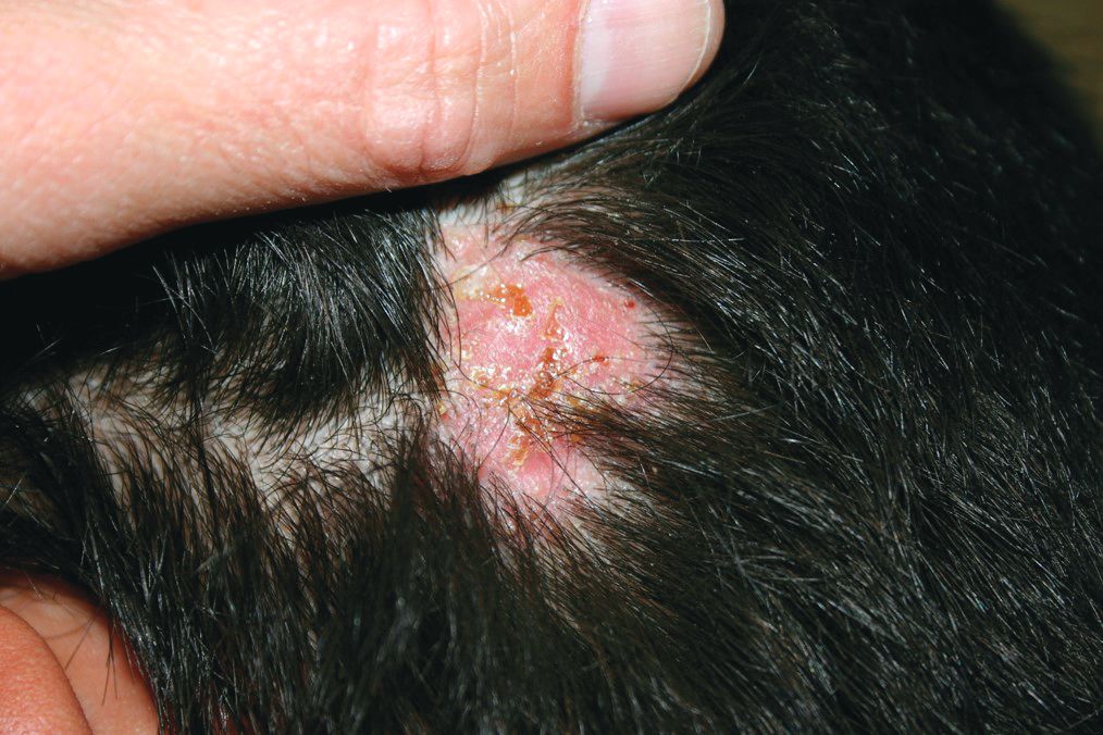
On physical exam, he is noted to have a hairless patch on a boggy left occipital mass, which is tender to palpation. There is a small amount of overlying honey-colored crusting. He has associated posterior occipital nontender lymphadenopathy.
The patient's older sister has a small area of scalp hair loss.
When your patients are your neighbors
“We’ll have one strawberry sugar cone, two chocolate swirls, and a mocha almond.”

“Dr. Wilkoff, I haven’t seen you in ... must be 5 years. You look great! How’s retirement going?”
“You look great too, Kim. The neighborhood has needed an ice cream parlor like yours for a long time. How’s the family?”
Alerted by the commotion of our catching up, Kim’s husband came outside to see what was going on. Looking at my wife, he said, “You know he saved our daughter’s life?”
Well, not exactly. A timely referral to a psychologist I knew was good with eating disorders had started the slow process of returning their anorectic daughter to health. I thanked him and tried to put a more historically correct spin on his story.
Most of my encounters with former patients and their parents aren’t as dramatic as this one at Kim’s Ice Cream Shack, but they always leave me with a warm, positive feeling that stays with me all day. Every now and then they include a compliment or a thank you, but most of the time the conversations are dominated by questions about how the other are doing and what our families are up to.
One of the perks of living in the town where you practice is that your patients also are your neighbors. Not every physician views this proximity as a positive, but for me, it was a gift that has kept on giving after I retired.
Our house phone number was always listed in the phone book, and I can count on the fingers of two hands how many times in 40 years that I received what I would consider an inappropriate or invasive call. Our office offered evening and weekend hours and a generous schedule of phone-in call times. But I’m convinced that it was the neighbor-to-neighbor relationship that kept the work/home balance intact. Even though I may have helped a plumber and his wife with their sick children, that professional arrangement didn’t include a free pass to call him after hours if I knew my leaking faucet could wait until the weekend was over.
By the same token, when a patient or a customer is also your neighbor there is an unspoken ethic that the service you provide must be your best effort. That’s not an admission that I was in the habit of offering substandard care to “folks from away,” but there is special motivation when your work is being scrutinized by people you’re likely to see next week at the grocery store checkout.
Office visits with neighbors often tended to take longer because there was a tendency to drift off topic and ask about a sibling’s baseball game I had read about in the paper or how the lobster catch was running that season. On the other hand, I must admit that I did my share of reporting (really it was bragging) on my own children’s accomplishments.
But now I’m reaping the benefits of those extra minutes invested in the office, because I suspect former patients and their families are more likely to want to reminisce when we meet in a restaurant or at the farmers’ market.
If you are a young physician and worrying about finding a good work/life balance, I urge you to consider living and working and then staying on in a place in which your patients also will be your neighbors. It will enrich your work experience and repay you many times over when it’s time to retire.
If you can’t find that place, at least treat your patients as though they were your neighbors.
Dr. Wilkoff practiced primary care pediatrics in Brunswick, Maine, for nearly 40 years. He has authored several books on behavioral pediatrics, including “How to Say No to Your Toddler.” Email him at [email protected].
“We’ll have one strawberry sugar cone, two chocolate swirls, and a mocha almond.”
“Dr. Wilkoff, I haven’t seen you in ... must be 5 years. You look great! How’s retirement going?”
“You look great too, Kim. The neighborhood has needed an ice cream parlor like yours for a long time. How’s the family?”
Alerted by the commotion of our catching up, Kim’s husband came outside to see what was going on. Looking at my wife, he said, “You know he saved our daughter’s life?”
Well, not exactly. A timely referral to a psychologist I knew was good with eating disorders had started the slow process of returning their anorectic daughter to health. I thanked him and tried to put a more historically correct spin on his story.
Most of my encounters with former patients and their parents aren’t as dramatic as this one at Kim’s Ice Cream Shack, but they always leave me with a warm, positive feeling that stays with me all day. Every now and then they include a compliment or a thank you, but most of the time the conversations are dominated by questions about how the other are doing and what our families are up to.
One of the perks of living in the town where you practice is that your patients also are your neighbors. Not every physician views this proximity as a positive, but for me, it was a gift that has kept on giving after I retired.
Our house phone number was always listed in the phone book, and I can count on the fingers of two hands how many times in 40 years that I received what I would consider an inappropriate or invasive call. Our office offered evening and weekend hours and a generous schedule of phone-in call times. But I’m convinced that it was the neighbor-to-neighbor relationship that kept the work/home balance intact. Even though I may have helped a plumber and his wife with their sick children, that professional arrangement didn’t include a free pass to call him after hours if I knew my leaking faucet could wait until the weekend was over.
By the same token, when a patient or a customer is also your neighbor there is an unspoken ethic that the service you provide must be your best effort. That’s not an admission that I was in the habit of offering substandard care to “folks from away,” but there is special motivation when your work is being scrutinized by people you’re likely to see next week at the grocery store checkout.
Office visits with neighbors often tended to take longer because there was a tendency to drift off topic and ask about a sibling’s baseball game I had read about in the paper or how the lobster catch was running that season. On the other hand, I must admit that I did my share of reporting (really it was bragging) on my own children’s accomplishments.
But now I’m reaping the benefits of those extra minutes invested in the office, because I suspect former patients and their families are more likely to want to reminisce when we meet in a restaurant or at the farmers’ market.
If you are a young physician and worrying about finding a good work/life balance, I urge you to consider living and working and then staying on in a place in which your patients also will be your neighbors. It will enrich your work experience and repay you many times over when it’s time to retire.
If you can’t find that place, at least treat your patients as though they were your neighbors.
Dr. Wilkoff practiced primary care pediatrics in Brunswick, Maine, for nearly 40 years. He has authored several books on behavioral pediatrics, including “How to Say No to Your Toddler.” Email him at [email protected].