User login
Official Newspaper of the American College of Surgeons
On-label stent use looks safe in intracranial atherosclerotic disease
LOS ANGELES – A postmarketing study of the Wingspan stent shows that the safety of the device in the treatment of intracranial atherosclerotic disease (ICAD) is good enough to be a reasonable alternative to medical management in these patients, but only if the device is used on label.
The results may reassure some interventionalists who were alarmed by results from the Stenting versus Aggressive Medical Management for Preventing Recurrent Stroke in Intracranial Stenosis (SAMMPRIS) trial (N Engl J Med. 2011;365:993-1003), which showed a 30-day rate of stroke or death of 14.7%. The new study showed a frequency of 2.6%, so long as the device was used on label. Off-label use yielded a frequency of 23.9%.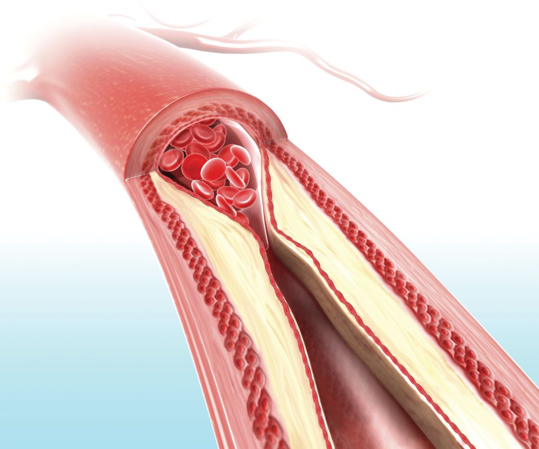
Up to 10% of strokes in the United States result from ICAD, and in China the rate is an estimated 20%-46%. The condition can also be treated medically. Early trials of the Wingspan device showed initial success with complication rates of 4.5%-6.2%, but the SAMMPRIS trial, which directly compared stenting to aggressive medical management, showed superior outcomes with medical treatment. The 30-day rate of stroke or death of 14.7% was too high to compete with medical therapy, which included aspirin 325 mg per day, clopidogrel 75 mg per day for 90 days after enrollment, and management of primary and secondary risk factors.
Dr. Alexander believes that the SAMMPRIS trial did not employ favorable patient selection. “ICAD is variable. Some patients present with hemodynamic compromise, where stenting is probably beneficial. Some present with embolic stroke, and some present with small-vessel perforator strokes that are unlikely to be responsive to stenting and better treated with medical therapy. All these patients were grouped together” in SAMMPRIS, said Dr. Alexander, who is director of the Neurovascular Center and endovascular surgery at Cedars-Sinai in Los Angeles.
The SAMMPRIS findings put a damper on stenting, and use of the procedure has dropped at many U.S. hospitals. But studies conducted prior to SAMMPRIS had shown much lower periprocedural morbidity, and those studies looked at on-label use of stenting. SAMMPRIS was off label.
Now the WEAVE study, which was an Food and Drug Administration–mandated, postmarketing surveillance study of Stryker’s Wingspan stent, suggests that the off-label use of the system in the SAMMPRIS trial may explain the poor results. SAMMPRIS had attempted to extend the approved boundaries of stenting by treating patients who presented with transient ischemic attacks only, patients who had failed medical therapy, and patients who had experienced a stroke in the past 7 days. In fact, about half of the patients were treated within 7 days of the previous event, sometimes within 24 hours.
Previous studies had shown that risk factors for poor outcomes included stenting within 10 days of the last event, stenting a posterior circulation target lesion, stenting presentations other than stroke, and sites with a low patient volume for stenting. Patient selection is vital to success, according to Dr. Alexander. Patients with hemodynamic compromise are good candidates for stenting, while those with perforator stroke alone are better off with medical therapy. Embolic stroke patients are candidates for either approach.
WEAVE looked at 152 consecutive patients treated on label at 24 institutions. The primary analysis group consisted of patients with a 70%-99% stenotic intracranial atherosclerotic lesion who were refractory to medical treatment, 22-80 years of age, and had a modified Rankin Scale (mRS) score of 3 or less at baseline. The treatment was performed at least 7 days after the last stroke. Finally, patients had to have experienced two or more strokes. This last requirement presented a problem for recruitment, according to Dr. Alexander. “That was never a criterion for any of the [previous] trials, so it’s not clear why FDA added that. That made it very difficult to enroll for this trial – to have patients who had two or more strokes and still had a functional mRS score,” he said.
The study protocol aimed for a frequency of 6.6% for periprocedural morbidity, defined as a stroke or death within 72 hours.
An interim analysis at 100 patients showed that the periprocedural morbidity frequency was below 4%, which met the agency’s requirement and allowed the trial to be halted once the trial enrolled 150 on-label patients. The total number of on-label patients reached 152, and the researchers analyzed the results from another 46 patients who were treated with stenting despite not meeting the study’s inclusion criteria, and these patients were considered to be off-label use. The final analysis showed that the on-label group had a periprocedural morbidity of 2.6%, compared with 23.9% in the off-label group (P = .0001).
The most glaring difference in the patient populations was that half of the off-label group received the stent within 7 days of experiencing a stroke. What might be the reason for worse outcomes when stenting is performed within 7 days? “There’s speculation that the plaques might be hot, and those patients might have a higher thrombotic risk with putting a foreign body in the vessel, or there’s capillary instability, so reperfusing a vessel that has a 99% stenosis has a higher risk for reperfusion hemorrhage,” Dr. Alexander said.
Experience may also be a factor. Interventionalists participating in the WEAVE trial had performed a stent using Wingspan an average of 37 times before the study began, compared with a mean of 10 cases for physicians in the SAMMPRIS trial. Those who had performed over 50 procedures had no periprocedural morbidity outcomes at all.
The study was funded by Stryker Neurovascular. Dr. Alexander has consulted for Stryker.
SOURCE: Alexander M et al. ISC 2018 Abstract LB13.
REPORTING FROM ISC 2018
Key clinical point:
Major finding: On-label 72-hour death and stroke rate was 2.6%, compared with 23.9% off label.
Data source: Postmarketing analysis of 198 consecutive patients.
Disclosures: The study was funded by Stryker Neurovascular. Dr. Alexander has consulted for Stryker.
Source: Alexander M et al. ISC 2018 Abstract LB13.
Hernia repair patients had less pain with lightweight mesh
and less discomfort at 1 year in a group of patients who had lightweight mesh, compared with patients who had heavyweight mesh, in a multicenter, randomized clinical trial.
Martin Rutegård, MD, of Umeå (Sweden) University, and his research associates reported that 3%-10% of all hernia surgeries “result in severe or moderately severe pain for more than a year after hernia surgery, which may have a significant impact on social activities, sex life, and quality of life. ... Interest in the use of lightweight meshes in groin hernia repair has increased in recent years, as it is assumed that this type of mesh may cause less discomfort and chronic pain.”

Patients were followed for 1-3 years and given questionnaires to report their outcomes. Patients were all male and were close in weight (mean body mass index, 25.2 kg/m2 in the lightweight-mesh group and 25.3 in the heavyweight-mesh group), age (59 and 58, respectively), and American Society of Anaesthesiologists classification of their hernia defect.
Of a total of 363 patients, 185 patients were randomized to the lightweight-mesh group and 178 patients to the heavyweight group. Investigators found that there were significant differences concerning awareness of a groin lump and groin discomfort, favoring the lightweight group 1 year after surgery. A total of 6% of the lightweight group reported the groin lump awareness at 1 year, vs. 18% of the heavyweight group. Groin discomfort was reported by 18% of the lightweight group and 28% of the heavyweight group.
After 1 year, that difference subsided. In terms of discomfort, the investigators found no statistically significant or clinically relevant differences between types of mesh, with 263/288 patients (91.3%) reporting improvement after 12 months, 19/288 patients (6.6%) experiencing no change, and 6/288 patients (2.1%) having worsened.
Additionally, there was no statistically significant difference in quality of life as measured by the EuroQol five dimensions (EQ-5D) between the different mesh groups. It was noted that all the patients had a statistically significantly better quality of life postoperatively from day 11 and onward, compared with before surgery. In addition, the investigators did not detect a significant difference between the mesh groups in their reported sexual life after surgery at 4 and 12 months subsequent to the operation.
The recurrence rate at the follow-up visit and clinical examination was 2.4% and equal between both groups.
The study was limited by possible bias of the expertise-based design and also some missing data, especially with regard to sexual life after surgery.
The study was funded by the Västerbotten County Council, VISARE NORR Fund, and Northern Country Councils Regional Federation. The investigators reported no conflicts of interest.
SOURCE: M. Rutegård et al. Hernia 2018 Jan 20. doi: 10.1007/s10029-018-1734-z.
and less discomfort at 1 year in a group of patients who had lightweight mesh, compared with patients who had heavyweight mesh, in a multicenter, randomized clinical trial.
Martin Rutegård, MD, of Umeå (Sweden) University, and his research associates reported that 3%-10% of all hernia surgeries “result in severe or moderately severe pain for more than a year after hernia surgery, which may have a significant impact on social activities, sex life, and quality of life. ... Interest in the use of lightweight meshes in groin hernia repair has increased in recent years, as it is assumed that this type of mesh may cause less discomfort and chronic pain.”
Patients were followed for 1-3 years and given questionnaires to report their outcomes. Patients were all male and were close in weight (mean body mass index, 25.2 kg/m2 in the lightweight-mesh group and 25.3 in the heavyweight-mesh group), age (59 and 58, respectively), and American Society of Anaesthesiologists classification of their hernia defect.
Of a total of 363 patients, 185 patients were randomized to the lightweight-mesh group and 178 patients to the heavyweight group. Investigators found that there were significant differences concerning awareness of a groin lump and groin discomfort, favoring the lightweight group 1 year after surgery. A total of 6% of the lightweight group reported the groin lump awareness at 1 year, vs. 18% of the heavyweight group. Groin discomfort was reported by 18% of the lightweight group and 28% of the heavyweight group.
After 1 year, that difference subsided. In terms of discomfort, the investigators found no statistically significant or clinically relevant differences between types of mesh, with 263/288 patients (91.3%) reporting improvement after 12 months, 19/288 patients (6.6%) experiencing no change, and 6/288 patients (2.1%) having worsened.
Additionally, there was no statistically significant difference in quality of life as measured by the EuroQol five dimensions (EQ-5D) between the different mesh groups. It was noted that all the patients had a statistically significantly better quality of life postoperatively from day 11 and onward, compared with before surgery. In addition, the investigators did not detect a significant difference between the mesh groups in their reported sexual life after surgery at 4 and 12 months subsequent to the operation.
The recurrence rate at the follow-up visit and clinical examination was 2.4% and equal between both groups.
The study was limited by possible bias of the expertise-based design and also some missing data, especially with regard to sexual life after surgery.
The study was funded by the Västerbotten County Council, VISARE NORR Fund, and Northern Country Councils Regional Federation. The investigators reported no conflicts of interest.
SOURCE: M. Rutegård et al. Hernia 2018 Jan 20. doi: 10.1007/s10029-018-1734-z.
FROM HERNIA
Key clinical point: The weight of mesh used for inguinal hernia repair did not impact outcomes.
Major finding: A total of 6% of the lightweight group reported groin lump awareness at 1 year, vs. 18% of the heavyweight group.
Study details: A randomized, multicenter study of 363 patients.
Disclosures: The study was funded by the Västerbotten County Council, VISARE NORR Fund, and Northern Country Councils Regional Federation. The investigators reported no conflicts of interest.
Source: Rutegård M et al. Hernia. 2018 Jan 20. doi: 10.1007/s10029-018-1734-z.
ERAS pathway can cut postdischarge opioid use
JACKSONVILLE, FLA. – An but also to reduce postdischarge opioid prescriptions.
The results of the enhanced recovery after surgery (ERAS) study were reported at the Association for Academic Surgical/Society of Academic Surgeons Academic Congress by Kathryn Hudak, a fourth-year medical student at the University of Alabama at Birmingham (UAB).

The researchers compared outcomes of 197 patients in the ERAS database at the institution who had colorectal surgery in 2015 with 198 patients who had surgery in 2013 and 2014 before the ERAS protocol was put in place.
Overall, the ERAS program had successes. “Using ERAS, we have shown a reduction in hospital length of stay and reduction in postoperative complications, [and] a reduction in hospital costs without any increase in readmissions or mortality,” Ms. Hudak said. Average length of stay decreased by 2 days and postoperative complications by 30%, study results showed.
“One purpose of ERAS is to control pain with as little need for opioids as possible,” she said. Pain management in the ERAS protocol used at UAB involved celecoxib, gabapentin, and acetaminophen before surgery; ketorolac and lidocaine during the operation; and alternating acetaminophen with other nonsteroidal anti-inflammatory drugs and oral oxycodone as needed after surgery. “If ERAS uses multimodal analgesia to avoid opioid use in the hospital, we wanted to know if we could see any effect in the use of opioids outside of the hospital,” Ms. Hudak said.
ERAS patients had more minimally invasive surgery (43.4% vs. 32.5%), more ostomies (38.9% vs. 25.9%), and lower rates of baseline opioid use (15.2% vs. 29.4%). So these patients would be expected to have a lower need for postdischarge pain medications.
For the study overall, 89.6% of patients in both groups were discharged with an opioid prescription but, Ms. Hudak said, “more of our ERAS patients were discharged without a prescription for an opioid – 14.1% vs. 7% in the pre-ERAS patients. “In our ERAS patients, we found a significantly different makeup in those discharge medications,” she said. “Many more patients were discharged on tramadol or a combination of tramadol and oxycodone or hydrocodone – again, using more of those low-potency opioids.”
The study revealed one unexpected finding, Ms. Hudak said. “We found that ERAS patients had a higher number of pills prescribed and OMEs [oral morphine equivalents], and we were surprised by this because we were expecting the opposite,” she said. Among those discharged with opioids, ERAS patients had an average oral morphine equivalent of 403 and 60.6 pills vs. 343 OMEs and 46.9 pills pre-ERAS (P less than .03). However, per-pill OME ratios were lower for the ERAS group: 6.9 vs. 7.6, Ms. Hudak said.
The study also followed up with patients a year after discharge, and found that 34% of ERAS patients needed an additional prescription while 44% of pre-ERAS patients required additional high-potency opioids, Hudak said.
“ERAS does seem to modify postdischarge opioid utilization, but we definitely need to work toward better standardization of opioid prescribing,” Ms. Hudak said. The UAB has since implemented a standardized protocol for residents to prescribe opioids after surgery based on a patient’s risk for postoperative pain, she said.
Ms. Hudak and her coauthors had no financial relationships to disclose.
JACKSONVILLE, FLA. – An but also to reduce postdischarge opioid prescriptions.
The results of the enhanced recovery after surgery (ERAS) study were reported at the Association for Academic Surgical/Society of Academic Surgeons Academic Congress by Kathryn Hudak, a fourth-year medical student at the University of Alabama at Birmingham (UAB).
The researchers compared outcomes of 197 patients in the ERAS database at the institution who had colorectal surgery in 2015 with 198 patients who had surgery in 2013 and 2014 before the ERAS protocol was put in place.
Overall, the ERAS program had successes. “Using ERAS, we have shown a reduction in hospital length of stay and reduction in postoperative complications, [and] a reduction in hospital costs without any increase in readmissions or mortality,” Ms. Hudak said. Average length of stay decreased by 2 days and postoperative complications by 30%, study results showed.
“One purpose of ERAS is to control pain with as little need for opioids as possible,” she said. Pain management in the ERAS protocol used at UAB involved celecoxib, gabapentin, and acetaminophen before surgery; ketorolac and lidocaine during the operation; and alternating acetaminophen with other nonsteroidal anti-inflammatory drugs and oral oxycodone as needed after surgery. “If ERAS uses multimodal analgesia to avoid opioid use in the hospital, we wanted to know if we could see any effect in the use of opioids outside of the hospital,” Ms. Hudak said.
ERAS patients had more minimally invasive surgery (43.4% vs. 32.5%), more ostomies (38.9% vs. 25.9%), and lower rates of baseline opioid use (15.2% vs. 29.4%). So these patients would be expected to have a lower need for postdischarge pain medications.
For the study overall, 89.6% of patients in both groups were discharged with an opioid prescription but, Ms. Hudak said, “more of our ERAS patients were discharged without a prescription for an opioid – 14.1% vs. 7% in the pre-ERAS patients. “In our ERAS patients, we found a significantly different makeup in those discharge medications,” she said. “Many more patients were discharged on tramadol or a combination of tramadol and oxycodone or hydrocodone – again, using more of those low-potency opioids.”
The study revealed one unexpected finding, Ms. Hudak said. “We found that ERAS patients had a higher number of pills prescribed and OMEs [oral morphine equivalents], and we were surprised by this because we were expecting the opposite,” she said. Among those discharged with opioids, ERAS patients had an average oral morphine equivalent of 403 and 60.6 pills vs. 343 OMEs and 46.9 pills pre-ERAS (P less than .03). However, per-pill OME ratios were lower for the ERAS group: 6.9 vs. 7.6, Ms. Hudak said.
The study also followed up with patients a year after discharge, and found that 34% of ERAS patients needed an additional prescription while 44% of pre-ERAS patients required additional high-potency opioids, Hudak said.
“ERAS does seem to modify postdischarge opioid utilization, but we definitely need to work toward better standardization of opioid prescribing,” Ms. Hudak said. The UAB has since implemented a standardized protocol for residents to prescribe opioids after surgery based on a patient’s risk for postoperative pain, she said.
Ms. Hudak and her coauthors had no financial relationships to disclose.
REPORTING FROM THE ACADEMIC SURGICAL CONGRESS
Key clinical point: Use of the enhanced recovery after surgery (ERAS) pathway reduces discharge prescriptions for opioids after colorectal surgery.
Major finding: 14.2% of ERAS patients were discharged without an opioid prescription vs. 7% for pre-ERAS patients.
Data source: An analysis of a single-institution ERAS database of 197 ERAS patients, compared with 198 patients who did not follow the ERAS pathway.
Disclosures: Ms. Hudak reported having no relevant financial disclosures.
Barbed sutures shorten cesarean closure time, reduce blood loss
DALLAS – Using knotless barbed sutures to close the uterine incision after cesarean delivery reduced operating time and blood loss, according to a recent randomized controlled study.
“On average, knotless barbed sutures were 1 minute and 43 seconds faster,” said David Peleg, MD, discussing the study results at the meeting sponsored by the Society for Maternal-Fetal Medicine. Average uterine closure time with knotless barbed sutures was 3 minutes and 37 seconds; with smooth sutures, average closure time was 5 minutes and 20 seconds (103-second difference; 95% confidence interval, 67.69-138.47 seconds; P less than .001).

Barbed sutures, in contrast, are usually made of monofilament with barbs created by the addition of tiny diagonal cuts made just partway through the suture material. When the suture is pulled through with the angle of the barbs, it pulls smoothly, but pulling back against the barb angle causes the barbs to protrude and catch against tissue, preventing slippage and eliminating the need for knots.
The fact that the monofilament has been partially cut to create the barbs can also reduce tensile strength, but some of the other disadvantages of smooth sutures are avoided, said Dr. Peleg.
The suture material used for the study was bidirectional, with the barbs running in opposite directions from the midline and a needle swaged onto each end; other barbed suture systems have an integral loop at one end of a unidirectionally barbed length of suture material that is used to anchor the first suture. The brand used was Stratafix.
The prospective study was necessarily unblinded, and compared the knotless barbed sutures with smooth sutures using polyglactin 910 braided material (Vicryl) for use during closure of the uterine incision during cesarean section procedures.
Patients were eligible if they were having an elective cesarean section after at least 38 weeks’ gestation, or if they were having a cesarean for the usual obstetric indications after laboring. Women with previous cesareans who failed a trial of labor were eligible.
The primary outcome was the length of time to close the uterine incision, measured from the start of suturing until hemostasis was achieved; the time included hemostatic suturing.
After a small pilot study that established a baseline suturing time with polyglactin 910 sutures of 6 minutes (standard deviation, 2 minutes and 10 seconds), Dr. Peleg and his collaborators determined that they would need to enroll at least 34 women per study arm to detect a decrease of 25% in suture time – to 4.5 minutes – with barbed sutures.
“The decrease in closure time is not linear,” said Dr. Peleg, so they increased their sample size by 50%, to 51 patients in each group, to ensure statistical significance of the results.
One of the challenges of a surgical randomized controlled trial is ensuring uniform technique; for this study, all patients had epidural or spinal anesthesia and antibiotics before opening. Surgeon clinical judgment was used to determine whether a Pfannenstiel or low transverse incision was made. In either case, there was no closure of the parietal peritoneum or the rectus muscles, and subcutaneous closure was used if tissues were greater than 2 cm in depth. Subcuticular stitches were used if possible.
Looking at secondary outcomes, there was a trend toward shorter total operative time with barbed sutures that didn’t reach statistical significance (20.1 min for barbed sutures vs. 23.1 for conventional, P = .062). However, fewer hemostatic sutures were required when barbed sutures were used: Extra sutures were used in 16 of the barbed group vs. 41 who had conventional sutures (P less than .001). Those receiving barbed sutures also had significantly less blood loss and estimated total blood loss during uterine closure than the conventional suture group (P = .005 and P = .002, respectively).
No study patients experienced serious postoperative complications; there were no infections, hematomas, or other wound complications, said Dr. Peleg of Bar-Ilan University, Zefat, Israel.
On the pro side for wider implementation of the use of barbed sutures for uterine closure stand the quicker closure and better hemostasis, along with the theoretical benefits of having no knots. Additionally, said Dr. Peleg, “there’s a gentle learning curve – it’s relatively easy to get used to the technique” of using barbed sutures. And, he said, “surgeons find them satisfying to use.”
However, he acknowledged the extra expense of barbed suture material – depending on the location and supplier, he estimated the cost could run from 7- to 20-fold for the barbed sutures, which he said cost $23.50 apiece. Also, he said, though the results were statistically significant, “Are they clinically significant? Does a difference in closure time of one minute 43 seconds, and a decrease in blood loss of 47 milliliters matter?”
Other considerations, he said, will require longer-term study. Polydioxanone, used for the barbed sutures, has a longer absorption time – a factor with unknown clinical implications in this application. Other longer-term outcomes, such as vaginal birth after cesarean success rates, rates of uterine rupture, the thickness of the uterine scar, and rates of adhesions and placenta accreta, will need to be tracked for years.
The authors reported no conflicts of interest and specifically reported that they had no relevant consulting or research agreements with suture manufacturers or marketers.
SOURCE: Peleg D et al. The Pregnancy Meeting Abstract 32.
DALLAS – Using knotless barbed sutures to close the uterine incision after cesarean delivery reduced operating time and blood loss, according to a recent randomized controlled study.
“On average, knotless barbed sutures were 1 minute and 43 seconds faster,” said David Peleg, MD, discussing the study results at the meeting sponsored by the Society for Maternal-Fetal Medicine. Average uterine closure time with knotless barbed sutures was 3 minutes and 37 seconds; with smooth sutures, average closure time was 5 minutes and 20 seconds (103-second difference; 95% confidence interval, 67.69-138.47 seconds; P less than .001).
Barbed sutures, in contrast, are usually made of monofilament with barbs created by the addition of tiny diagonal cuts made just partway through the suture material. When the suture is pulled through with the angle of the barbs, it pulls smoothly, but pulling back against the barb angle causes the barbs to protrude and catch against tissue, preventing slippage and eliminating the need for knots.
The fact that the monofilament has been partially cut to create the barbs can also reduce tensile strength, but some of the other disadvantages of smooth sutures are avoided, said Dr. Peleg.
The suture material used for the study was bidirectional, with the barbs running in opposite directions from the midline and a needle swaged onto each end; other barbed suture systems have an integral loop at one end of a unidirectionally barbed length of suture material that is used to anchor the first suture. The brand used was Stratafix.
The prospective study was necessarily unblinded, and compared the knotless barbed sutures with smooth sutures using polyglactin 910 braided material (Vicryl) for use during closure of the uterine incision during cesarean section procedures.
Patients were eligible if they were having an elective cesarean section after at least 38 weeks’ gestation, or if they were having a cesarean for the usual obstetric indications after laboring. Women with previous cesareans who failed a trial of labor were eligible.
The primary outcome was the length of time to close the uterine incision, measured from the start of suturing until hemostasis was achieved; the time included hemostatic suturing.
After a small pilot study that established a baseline suturing time with polyglactin 910 sutures of 6 minutes (standard deviation, 2 minutes and 10 seconds), Dr. Peleg and his collaborators determined that they would need to enroll at least 34 women per study arm to detect a decrease of 25% in suture time – to 4.5 minutes – with barbed sutures.
“The decrease in closure time is not linear,” said Dr. Peleg, so they increased their sample size by 50%, to 51 patients in each group, to ensure statistical significance of the results.
One of the challenges of a surgical randomized controlled trial is ensuring uniform technique; for this study, all patients had epidural or spinal anesthesia and antibiotics before opening. Surgeon clinical judgment was used to determine whether a Pfannenstiel or low transverse incision was made. In either case, there was no closure of the parietal peritoneum or the rectus muscles, and subcutaneous closure was used if tissues were greater than 2 cm in depth. Subcuticular stitches were used if possible.
Looking at secondary outcomes, there was a trend toward shorter total operative time with barbed sutures that didn’t reach statistical significance (20.1 min for barbed sutures vs. 23.1 for conventional, P = .062). However, fewer hemostatic sutures were required when barbed sutures were used: Extra sutures were used in 16 of the barbed group vs. 41 who had conventional sutures (P less than .001). Those receiving barbed sutures also had significantly less blood loss and estimated total blood loss during uterine closure than the conventional suture group (P = .005 and P = .002, respectively).
No study patients experienced serious postoperative complications; there were no infections, hematomas, or other wound complications, said Dr. Peleg of Bar-Ilan University, Zefat, Israel.
On the pro side for wider implementation of the use of barbed sutures for uterine closure stand the quicker closure and better hemostasis, along with the theoretical benefits of having no knots. Additionally, said Dr. Peleg, “there’s a gentle learning curve – it’s relatively easy to get used to the technique” of using barbed sutures. And, he said, “surgeons find them satisfying to use.”
However, he acknowledged the extra expense of barbed suture material – depending on the location and supplier, he estimated the cost could run from 7- to 20-fold for the barbed sutures, which he said cost $23.50 apiece. Also, he said, though the results were statistically significant, “Are they clinically significant? Does a difference in closure time of one minute 43 seconds, and a decrease in blood loss of 47 milliliters matter?”
Other considerations, he said, will require longer-term study. Polydioxanone, used for the barbed sutures, has a longer absorption time – a factor with unknown clinical implications in this application. Other longer-term outcomes, such as vaginal birth after cesarean success rates, rates of uterine rupture, the thickness of the uterine scar, and rates of adhesions and placenta accreta, will need to be tracked for years.
The authors reported no conflicts of interest and specifically reported that they had no relevant consulting or research agreements with suture manufacturers or marketers.
SOURCE: Peleg D et al. The Pregnancy Meeting Abstract 32.
REPORTING FROM THE PREGNANCY MEETING
Key clinical point: Closure time and blood loss were less with barbed sutures closing the uterine incision.
Major finding: Average uterine incision closure time was 3:37 minutes with knotless barbed sutures and 5:20 minutes with conventional smooth sutures.
Study details: Randomized controlled trial of 102 women undergoing cesarean section.
Disclosures: The study had no external sources of funding, and the study authors reported no relevant outside support.
Source: Peleg D et al. The Pregnancy Meeting Abstract 32.
These amniotic biomarkers predicted pPROM in women undergoing fetal surgery for spinal cord defect
DALLAS – Two molecular biomarkers in amniotic fluid seem to predict preterm premature rupture of membranes (pPROM) and subsequent premature delivery in women whose fetuses undergo surgical repair of spinal cord defects.
Matrix metalloproteinase–8 (MMP-8) and lactic acid levels were significantly higher in these women than in a comparator group of women who did not deliver a premature infant after the repair, Aikaterini Athanasiou, MD, said at the meeting sponsored by the Society for Maternal Fetal Medicine.
“Based on this pilot study, it appears that elevated amniotic levels of lactic acid and MMP-8 at time of surgery might identify a subset of women with increased susceptibility for pPROM and shorter time to delivery,” said Dr. Athanasiou, a fellow in obstetrics and gynecology at Cornell University, New York. She presented the work on behalf of primary author Antonio Moron, MD, PhD, of the Federal University of São Paulo.
Dr. Athanasiou was part of the Cornell team that conducted molecular assays on amniotic fluid samples drawn from 26 women carrying fetuses about to have corrective surgery for spinal defects in the fetuses. The women were all patients at the Federal Hospital of São Paulo. Samples were drawn immediately before surgery commenced, frozen, and shipped to Cornell for assay.
After surgical repair, 7 of the women (27%) later experienced pPROM and 19 did not.
At baseline, there were no significant differences between the groups. Women were a mean age of 32 years, with a mean of two prior pregnancies. There were no differences in prior cesarean and vaginal births, smoking status, and fetal gender. The defect was most commonly a myelomeningocele (about 70% of each group). Rachischisis was next most common, occurring in 27% of the pPROM group and 21% of the non-pPROM group. There were two cases of encephalocele, both in the non-pPROM group.
The length of surgery was not significantly different between those who experienced pPROM and those who did not (121 vs. 130 minutes). Wound healing time was 7 days for each group, as was the mean length of hospital stay.
Both groups went a mean of 57 days from surgery to delivery, although the fetal gestational age was almost a week younger in the pPROM group (33.7 vs. 34.4 weeks). These infants were also smaller at birth (2,247 g vs. 2,410 g).
The Cornell team examined five potential biomarkers in each amniotic fluid sample: MMP-8, MMP-9, MMP-2, lactic acid, and interleukin-6 (IL-6). The levels of IL-6, MMP-2, and MMP-9 were similar between groups.
However, lactic acid was significantly higher in the pPROM group (7.1 vs. 5.9 mU/mL). MMP-8 was also significantly elevated in the pPROM group (1.7 vs. 0.6 mcg/mL).
Dr. Athanasiou and her colleagues also observed an inverse relationship between MMP-8 levels and gestational age at delivery, which was statistically significant. There was also an inverse relationship between lactic acid levels and gestational age, but this did not reach statistical significance.
“While further investigations are needed to verify our findings, our data suggest that these differences are present before fetal surgery and that an increase in intra-amniotic anaerobic glycolysis as evidenced by elevated lactic acid may enhance MMP-8 production, which will weaken the maternal-fetal membranes,” Dr. Athanasiou said. “The mechanism may not be related to inflammation, as evidenced by the lack of association between pPROM and IL-6.”
She had no financial disclosures.
SOURCE: Moron A et al. Am J Obstet Gynecol. 2018 Jan:218(1);S64.
REPORTING FROM THE PREGNANCY MEETING
Key clinical point: Lactic acid and MMP-8 in amniotic fluid may have utility as biomarkers predictive of pPROM in women who undergo fetal surgery for spinal cord defects.
Major finding: Women who experienced pPROM had higher amniotic lactic acid (7.1 vs. 5.9 mU/mL) and MMP-8 (1.7 vs. 0.6 mcg/mL) at time of fetal surgery.
Study details: The prospective study comprised 26 women.
Disclosures: Dr. Athanasiou had no financial disclosures.
Source: Moron A et al. Am J Obstet Gynecol. 2018 Jan:218(1);S64.
Imaging methods for stroke thrombectomy eligibility yield similar results
LOS ANGELES – The benefits of mechanical thrombectomy observed in the DAWN trial for patients with acute ischemic stroke and a mismatch between core imaging and clinical presentation out to 24 hours appear to apply regardless of whether their eligibility is determined by CT perfusion or diffusion-weighted magnetic resonance imaging, according to a subanalysis of the trial data.
Diffusion-weighted magnetic resonance imaging (DW-MRI) is considered the gold standard, but it is not as widely available as CT perfusion (CTP) and previous studies have shown that MR is associated with longer times between stroke onset and treatment randomization. “Though MR was originally preferred in DAWN, it was pretty clear that CT perfusion was going to need to be employed in the trial as well,” Cathy Sila, MD, said during her presentation of the results of the subanalysis at the International Stroke Conference 2018, sponsored by the American Heart Association.
The research sought to determine if the two imaging methods perform similarly. CTP is more readily available, but it has some issues. In patients with severe heart failure, a severe proximal stenosis, or a contralateral severe stenosis, the technique may struggle to accurately image the core infarct, which has led some to wonder if the outcomes would be as good using CTP as selection criteria. “In our institution, we’ve had this conversation very frequently,” said Dr. Sila, who is a vascular neurologist and the director of the University Hospitals Systems stroke program in Cleveland.
To be eligible for DAWN, the core infarct had to correspond to at least a 30% decrease in regional blood flow in the CTP map, or an apparent diffusion coefficient of less than 620 on DW-MRI.
The researchers included all 206 patients in the DAWN study (N Engl J Med. 2018;378:11-21), separating them into DW-MRI or CTP groups based on which imaging method was used to randomize them during the trial. There were no statistically significant differences in any of the baseline characteristics between the two imaging groups.
The 26 sites participating in DAWN had clear differences in their preferences for imaging techniques; 19 exclusively used CTP, 4 used only DW-MRI, and 3 sites used a combination of both imaging methods.
There were no statistically significant differences between the two groups in any of the measured clinical outcomes, including neurologic deterioration in hospital (22.8% with CTP vs. 15.7% with DW-MRII, P = .286), symptomatic intracranial hemorrhage (4.1% with CTP vs. 4.8% with DW-MRI, P = 1.000), or death related to stroke (19.5% with CTP vs. 13.3% with DW-MRI, P = .263). Outcomes at 90 days proved to be similar between CTP and DW-MRI for achieving functional independence (29.3% vs. 34.9%, respectively; P = .445) and utility-weighted modified Rankin Scale scores (4.2 vs. 4.9, respectively; P = .172).
Multivariate analyses showed that 90-day functional independence was predicted by thrombectomy treatment, age, blood glucose level, baseline National Institutes of Health Stroke Scale score, and core lab ASPECTS (Alberta Stroke Program Early CT Score), but not the method of imaging.
“The efficacy and safety of mechanical thrombectomy for patients meeting those clinical mismatch criteria at 6-24 hours were comparable whether the small core infarcts were measured by diffusion imaging or cerebral blood flow imaging. I believe that future clinical trials aiming to extend the eligibility outside of this prespecified population should include both imaging modalities to determine whether these results are generalizable,” Dr. Sila said.
The DAWN study was funded by Stryker Neurovascular. Dr. Sila has reported receiving honoraria from Medtronic.
SOURCE: Sila C et al. ISC 2018, abstract LB11.
REPORTING FROM ISC 2018
Key clinical point: DW-MRI is the gold standard for imaging, but CTP is more widely available.
Major finding: Rates of neurologic deterioration in hospital, symptomatic intracranial hemorrhage, and death related to stroke were similar regardless of whether CT or MR imaging was used to assess patients’ infarcts.
Data source: A subanalysis of the DAWN randomized, controlled trial (n = 206).
Disclosures: The DAWN study was funded by Stryker Neurovascular. Dr. Sila reported receiving honoraria from Medtronic.
Source: Sila C et al. ISC 2018, abstract LB11.
ROBOT trial compares surgical approaches to esophagectomy
SAN FRANCISCO – Patients undergoing had less morbidity and pain and similarly good oncologic outcomes, when the surgery was performed by robot-assisted laparoscopy instead of by the open technique, a phase 3 clinical trial has found.
Investigators of the ROBOT (Robot-assisted Thoracolaparoscopic Esophagectomy vs. Open Transthoracic Esophagectomy) trial, led by Pieter C. van der Sluis, MD, a surgeon at the University Medical Center Utrecht, the Netherlands, randomized 112 patients with resectable esophageal cancer to open transthoracic esophagectomy – considered to be the gold standard – or robot-assisted minimally invasive thoracolaparoscopic esophagectomy.

“Robot-assisted minimally invasive thoracolaparoscopic esophagectomy versus open transthoracic esophagectomy improves postoperative outcome. There were no differences in oncologic outcomes, and our oncologic outcomes were in concordance with the highest standards nowadays,” Dr. van der Sluis summarized. “This trial provides evidence for the minimally invasive approach over the open approach, and especially the robot-assisted minimally invasive esophagectomy.”
The investigators will report a full cost comparison separately. “We see that costs are lower, though not significantly lower, with the robot,” he said, giving a preview. “We are going to show that the real costs of the operation are in the complications. When you have complications that involve the ICU and reoperations, some patients are in the hospital for months after the surgery. So by investing a little extra money in the surgical procedure, you might actually get it back by reducing the complications.”
When asked by an attendee why the trial did not compare robotic esophagectomy with thoracoscopic esophagectomy, Dr. van der Sluis noted that such comparison is complicated by many factors; for example, the challenge of finding surgeons skilled in both techniques, and the likelihood of small differences in outcomes, potentially requiring enrollment of thousands of patients to have adequate study power. “We concluded that such a trial might not be feasible,” he said.
Parsing the findings
“The complication rates [in this trial] are very high in the robotic and open groups, much higher than reported in some well-controlled prospective and retrospective studies,” commented session attendee Kenneth Meredith, MD, FACS, professor at Florida State University, Sarasota, and director of gastrointestinal oncology, Sarasota Memorial Institute for Cancer Care.
He wondered how extensive the investigators’ experience with robotics was and how many cases they had done on their learning curve. Data from his group suggest that surgeons must perform 29 cases of robotic esophagectomy before the complication rate drops (Dis Esophagus. 2017;30:1-7).
“That’s more then half of the patients in the robotic arm of their study,” he noted in an interview. “I find this needs to be explained. If the authors are past their learning curve, why were the complication rates so high?” Additionally, the 80% rate in the open group “is among the highest I’ve seen in many years.”
The lack of significant differences in complete resection rate and in lymph node harvest was also surprising, as he and other robotics users have found that this technique can improve these outcomes, Dr. Meredith added. This could likewise be a learning curve phenomenon.
Although ROBOT’s comparison of robotic with open esophagectomy is relevant, “it would have been more relevant to compare robotic to minimally invasive esophagectomy [MIE],” he maintained, as MIE has been shown to improve outcomes relative to open surgery (Lancet. 2012;379:1887-92).
“There are many high-volume centers in MIE but not necessarily robotics. The two are often mutually exclusive, and a multicenter trial in which each center performs high volumes of their respective technique, rather then mandating each center perform an operation they may not be facile in,” would be practical, Dr. Meredith concluded.
Study details
“The main objective in our trial was to reduce surgical trauma and reduce the percentage of complications,” Dr. van der Sluis told attendees of the symposium, sponsored by the American Gastroenterological Association, the American Society for Clinical Oncology, the American Society for Radiation Oncology, and the Society of Surgical Oncology.
Results showed that compared with peers in the open surgery group, patients in the robotic-assisted surgery group specifically had a lower rate of pulmonary complications (32% vs. 58%, P = .005), largely due to a reduction in rate of pneumonia (28% vs. 55%, P = .005), and a lower rate of cardiac complications (22% vs. 47%, P = .006), almost entirely due to a reduction in rate of atrial fibrillation (22% vs. 46%, P = .01).
There was a trend toward fewer wound infections with robotics (4% vs. 14%, P = .09), with a large difference in thoracic wound infections (0% vs. 9%, P = .06).
The two groups were statistically indistinguishable on rates of anastomotic leakage (24% and 20%) and recurrent laryngeal nerve injury (9% and 11%). The fairly high rate of anastomotic leakage was likely due to the center’s use of cervical anastomosis at the time of the trial, according to Dr. van der Sluis; they have since started using thoracic anastomosis, and will report results with that technique soon.
There was also no significant difference between groups in the rate of in-hospital mortality (4% with robotic surgery and 2% with open surgery), median hospital length of stay (14 and 16 days), and ICU length of stay (1 day in each group).
Patients in the robotics group more commonly had functional recovery within 2 weeks (70% vs. 51%, P = .04). And on the Quality of Life Questionnaire Core 30, they had better scores for health-related quality of life at discharge (57.9 vs 44.6, P = .02) and at 6 weeks (68.7 vs. 57.6, P = .03), and for physical functioning at discharge (54.5 vs. 41.0, P = .03) and 6 weeks (69.3 vs. 58.6, P = .049).
The two groups were similar on rates of R0 resection (93% and 96%) and median number of lymph nodes retrieved (27 and 25), reported Dr. van der Sluis. Pain during the first 14 days after surgery was lower for the robotics group (P = .003).
With a median follow-up of 40 months, the robotics and open groups did not differ significantly on disease-free survival (median, 26 and 28 months) and overall survival (not reached in either group).
Dr. van der Sluis disclosed no relevant conflicts of interest.
SOURCE: van der Sluis PC et al. 2018 GI Cancer Symposium, Abstract 156148
SAN FRANCISCO – Patients undergoing had less morbidity and pain and similarly good oncologic outcomes, when the surgery was performed by robot-assisted laparoscopy instead of by the open technique, a phase 3 clinical trial has found.
Investigators of the ROBOT (Robot-assisted Thoracolaparoscopic Esophagectomy vs. Open Transthoracic Esophagectomy) trial, led by Pieter C. van der Sluis, MD, a surgeon at the University Medical Center Utrecht, the Netherlands, randomized 112 patients with resectable esophageal cancer to open transthoracic esophagectomy – considered to be the gold standard – or robot-assisted minimally invasive thoracolaparoscopic esophagectomy.
“Robot-assisted minimally invasive thoracolaparoscopic esophagectomy versus open transthoracic esophagectomy improves postoperative outcome. There were no differences in oncologic outcomes, and our oncologic outcomes were in concordance with the highest standards nowadays,” Dr. van der Sluis summarized. “This trial provides evidence for the minimally invasive approach over the open approach, and especially the robot-assisted minimally invasive esophagectomy.”
The investigators will report a full cost comparison separately. “We see that costs are lower, though not significantly lower, with the robot,” he said, giving a preview. “We are going to show that the real costs of the operation are in the complications. When you have complications that involve the ICU and reoperations, some patients are in the hospital for months after the surgery. So by investing a little extra money in the surgical procedure, you might actually get it back by reducing the complications.”
When asked by an attendee why the trial did not compare robotic esophagectomy with thoracoscopic esophagectomy, Dr. van der Sluis noted that such comparison is complicated by many factors; for example, the challenge of finding surgeons skilled in both techniques, and the likelihood of small differences in outcomes, potentially requiring enrollment of thousands of patients to have adequate study power. “We concluded that such a trial might not be feasible,” he said.
Parsing the findings
“The complication rates [in this trial] are very high in the robotic and open groups, much higher than reported in some well-controlled prospective and retrospective studies,” commented session attendee Kenneth Meredith, MD, FACS, professor at Florida State University, Sarasota, and director of gastrointestinal oncology, Sarasota Memorial Institute for Cancer Care.
He wondered how extensive the investigators’ experience with robotics was and how many cases they had done on their learning curve. Data from his group suggest that surgeons must perform 29 cases of robotic esophagectomy before the complication rate drops (Dis Esophagus. 2017;30:1-7).
“That’s more then half of the patients in the robotic arm of their study,” he noted in an interview. “I find this needs to be explained. If the authors are past their learning curve, why were the complication rates so high?” Additionally, the 80% rate in the open group “is among the highest I’ve seen in many years.”
The lack of significant differences in complete resection rate and in lymph node harvest was also surprising, as he and other robotics users have found that this technique can improve these outcomes, Dr. Meredith added. This could likewise be a learning curve phenomenon.
Although ROBOT’s comparison of robotic with open esophagectomy is relevant, “it would have been more relevant to compare robotic to minimally invasive esophagectomy [MIE],” he maintained, as MIE has been shown to improve outcomes relative to open surgery (Lancet. 2012;379:1887-92).
“There are many high-volume centers in MIE but not necessarily robotics. The two are often mutually exclusive, and a multicenter trial in which each center performs high volumes of their respective technique, rather then mandating each center perform an operation they may not be facile in,” would be practical, Dr. Meredith concluded.
Study details
“The main objective in our trial was to reduce surgical trauma and reduce the percentage of complications,” Dr. van der Sluis told attendees of the symposium, sponsored by the American Gastroenterological Association, the American Society for Clinical Oncology, the American Society for Radiation Oncology, and the Society of Surgical Oncology.
Results showed that compared with peers in the open surgery group, patients in the robotic-assisted surgery group specifically had a lower rate of pulmonary complications (32% vs. 58%, P = .005), largely due to a reduction in rate of pneumonia (28% vs. 55%, P = .005), and a lower rate of cardiac complications (22% vs. 47%, P = .006), almost entirely due to a reduction in rate of atrial fibrillation (22% vs. 46%, P = .01).
There was a trend toward fewer wound infections with robotics (4% vs. 14%, P = .09), with a large difference in thoracic wound infections (0% vs. 9%, P = .06).
The two groups were statistically indistinguishable on rates of anastomotic leakage (24% and 20%) and recurrent laryngeal nerve injury (9% and 11%). The fairly high rate of anastomotic leakage was likely due to the center’s use of cervical anastomosis at the time of the trial, according to Dr. van der Sluis; they have since started using thoracic anastomosis, and will report results with that technique soon.
There was also no significant difference between groups in the rate of in-hospital mortality (4% with robotic surgery and 2% with open surgery), median hospital length of stay (14 and 16 days), and ICU length of stay (1 day in each group).
Patients in the robotics group more commonly had functional recovery within 2 weeks (70% vs. 51%, P = .04). And on the Quality of Life Questionnaire Core 30, they had better scores for health-related quality of life at discharge (57.9 vs 44.6, P = .02) and at 6 weeks (68.7 vs. 57.6, P = .03), and for physical functioning at discharge (54.5 vs. 41.0, P = .03) and 6 weeks (69.3 vs. 58.6, P = .049).
The two groups were similar on rates of R0 resection (93% and 96%) and median number of lymph nodes retrieved (27 and 25), reported Dr. van der Sluis. Pain during the first 14 days after surgery was lower for the robotics group (P = .003).
With a median follow-up of 40 months, the robotics and open groups did not differ significantly on disease-free survival (median, 26 and 28 months) and overall survival (not reached in either group).
Dr. van der Sluis disclosed no relevant conflicts of interest.
SOURCE: van der Sluis PC et al. 2018 GI Cancer Symposium, Abstract 156148
REPORTING FROM THE 2018 GI CANCERS SYMPOSIUM
Key clinical point: Patients with esophageal cancer undergoing esophagectomy are less likely to experience complications when the surgery is performed robotically.
Major finding: Compared with open transthoracic esophagectomy, robot-assisted minimally invasive thoracolaparoscopic esophagectomy had a lower rate of MCDC grade 2 or higher surgery-related postoperative complications (59% vs. 80%).
Data source: A single-center phase 3 randomized controlled trial among 112 patients with resectable esophageal cancer.
Disclosures: Dr. van der Sluis disclosed no relevant conflicts of interest.
Source: van der Sluis PC et al. 2018 GI Cancer Symposium, Abstract 156148
ACS Clinical Congress Report 2017
The American College of Surgeons (ACS) Clinical Congress is designed to offer a broad range of substantive opportunities for surgeons to interact with colleagues, participate in discussions, and learn about the latest innovations in our profession. The 2017 ACS Clinical Congress met all of these expectations.

The ACS Clinical Congress Report is a collection of news articles and videos from the meeting. This sampling of reportage by ACS Surgery News is meant to convey the essence of the meeting: new ideas, intense debate, and a profound commitment to professional development. We are especially proud to highlight the many substantive presentations by surgical trainees.
We hope this collection of articles will serve as a reminder of what the Clinical Congress is all about. The 2018 ACS Clinical Congress will be held in Boston, Oct. 21-25. Surgeons will be offered another great opportunity hear important presentations and updates, meet with colleagues, and witness firsthand the breaking news and unveiling of discoveries in our field. We hope you are inspired to attend.
Click on the PDF Download link above to read the report!
Karen E. Deveney, MD, FACS
Tyler G. Hughes, MD, FACS
Co-Editors, ACS Surgery News
Therese Borden
Managing Editor, ACS Surgery News
The American College of Surgeons (ACS) Clinical Congress is designed to offer a broad range of substantive opportunities for surgeons to interact with colleagues, participate in discussions, and learn about the latest innovations in our profession. The 2017 ACS Clinical Congress met all of these expectations.
The ACS Clinical Congress Report is a collection of news articles and videos from the meeting. This sampling of reportage by ACS Surgery News is meant to convey the essence of the meeting: new ideas, intense debate, and a profound commitment to professional development. We are especially proud to highlight the many substantive presentations by surgical trainees.
We hope this collection of articles will serve as a reminder of what the Clinical Congress is all about. The 2018 ACS Clinical Congress will be held in Boston, Oct. 21-25. Surgeons will be offered another great opportunity hear important presentations and updates, meet with colleagues, and witness firsthand the breaking news and unveiling of discoveries in our field. We hope you are inspired to attend.
Click on the PDF Download link above to read the report!
Karen E. Deveney, MD, FACS
Tyler G. Hughes, MD, FACS
Co-Editors, ACS Surgery News
Therese Borden
Managing Editor, ACS Surgery News
'This has to change': Surgeon pain, disability on the rise
The evidence has been accumulating for decades: Practicing surgery can exact a toll on surgeons’ physical well-being in ways that can shorten careers and contribute to professional dissatisfaction.
“Ergonomists have described the surgeon’s work environment and working conditions as equal to, if not at times harsher than, those of certain industrial workers,” wrote Sherise Epstein, MPH, and the coauthors of the systematic review and meta-analysis of studies addressing the issue. Ms. Epstein, currently a 4th-year medical student at the University of California, San Diego, and her team found a high prevalence of work-related musculoskeletal disorders and pain among surgeons and interventionalists, with pooled prevalence estimates for pain among these physicians ranging from 35% to 60% of all respondents. Cervical spine disease, rotator cuff pathology, and degenerative lumbar spine disease were each reported by nearly one in five respondents in the pooled analyses, with increasing prevalence over the duration of the study period (JAMA Surg. 2017 Dec 27. doi:10.1001/jamasurg.2017.49).
These findings, called “worrisome” by the study’s authors, come at a time when the demand for surgeons and interventionalists is expected soon to outstrip supply, and point to a major stressor for a profession already plagued by high rates of burnout and depression.
The results of the study are in line with previous work that finds a high – and increasing – rate of pain and disability among at-risk physicians, who endure long work hours, awkward positioning, and few opportunities for breaks.
The study also suggested a possible linkage between work-related pain and other factors, such as burnout and depression, that lead to attrition in the surgical profession.

Adrian Park, MD, FACS, a general surgeon who has long studied the issue of work-related musculoskeletal disorders (MSDs) among surgeons and interventionalists, was not surprised by the study’s findings. “This just goes to further the narrative, which is a consistent one: These aren’t just sporadic instances of individual surgeons having a tough time; this is an increasingly broadly recognized problem. ... These are the consequences of surgeons simply getting up and going to work each week,” Dr. Park said in an interview.
To take a long view of the published data on work-related injury and pain for at-risk physicians, Ms. Epstein and her collaborators identified 126 articles reporting physician work-related MSDs that met quality standards; the studies spanned 1974 to 2016, and 23 countries were represented.
A total of 21 articles were eventually included in the meta-analysis, which included responses from 5,828 physicians. Most of the respondents were male (78.5%), with a mean age of 46 years and a mean 12.8 years in practice. The mean number of hours spent in procedures per week was 14.4.
Sixteen cross-sectional studies included in the meta-analysis examined injury prevalence, with 14 studies eventually meeting eligibility criteria for quantitative synthesis. A total of 4,245 physicians were included in this portion of the analysis; 1,232 were orthopedic surgeons, 1,118 were interventional cardiologists, and 582 were general surgeons (a variety of other subspecialties made up the remainder).
The career prevalence of degenerative lumbar spine disease was estimated at 19%, with prevalence increasing over time in a series of surveys of interventional cardiologists, said Ms. Epstein and her colleagues. Among this group, the prevalence climbed from 6.5% in 1997 to 24.7% in 2015, an increase in prevalence of over 18%, they said. The investigators’ estimate of lumbar spine disease prevalence from their own data also showed an increase, to 21%, for studies published within the past decade.
Rotator cuff pathology during the study period was estimated at 18% overall, and carpal tunnel syndrome at 9% overall; carpal tunnel syndrome also appears to be increasing in prevalence, estimated at 12% within the last decade.
Musculoskeletal pain that was judged to be work related was common, as reported in 18 cross-sectional studies. Taken together, the studies used a total of five validated tools that measured both pain prevalence and related disability. Twelve of the studies met criteria for inclusion in the quantitative synthesis.
Neck, back, and shoulder pain were all common in this group of surgeons and interventionalists. Neck pain had a 60% estimated 12-month prevalence, and shoulder pain was almost as common, with a 52% prevalence; back pain was reported for about half (49%) of respondents.
Just 10 of the included studies addressed disability burden, said Ms. Epstein and her colleagues. Overall, 12% of physicians responding in this subset of studies reported that they required work modification, leave of absence, or early retirement because of work-related MSDs.
In a separate arm of the analysis, Ms. Epstein and her collaborators looked at studies that discussed ergonomic assessments and interventions, using a qualitative approach. A total of 101 studies, 50 of them in the United States, were included. None of the studies, they said, “included long-term surveillance for work-related MSDs”; rather, they focused on short-term outcomes and, in some cases, on real-time electromyographic data.
Of the 101 studies looking at ergonomics, about a third (n = 38) looked primarily at ergonomically designed devices or technology, while another third (n = 37) focused on educational or behavioral interventions and modifications. These were the studies included in the investigators’ analysis.
A lack of awareness of ergonomic recommendations on the part of the surgeons themselves was identified in several studies. Several specialties, publishing in a total of eight countries, have put forward “at least one article describing a need for ergonomics education during medical training.” And, said the investigators, “A few studies have found that ergonomics education during medical training appears feasible, accepted, and effective at changing behaviors and reducing symptoms.”
In addition to the usual limitations inherent to the meta-analysis research design (selection bias, self-reported data, small samples of individual studies), the study was limited by the elusive nature of the topic. Work-related pain and disability among surgeons remains underreported and underestimated, according to both Dr. Park and the study investigators. Further research is needed, and the investigators wrote: “Ultimately, this work should be integrated with research on preventing surgeon burnout and attrition given shared risk factors.”
Micro breaks can help
What’s the solution? Dr. Park says that some fixes are low tech, and can be put in place immediately. He and his team implemented a series of brief breaks, taken every half hour during a procedure, that they dubbed “targeted stretching micro breaks.” These are 45- to 50-second breaks during which a series of exercise physiologist–designed brief stretches are undertaken that do not require breaking scrub.
In a study of 66 surgeons and operating room staff, Dr. Park, chair of the department of surgery at Anne Arundel Medical Center, Annapolis, Md., and his collaborators found that surgeons reported less pain and fatigue, and improved mental focus, when they took micro breaks during a procedure. When surgeons took micro breaks, operative time was no longer than when they didn’t take the breaks (Ann Surg. 2017 Feb;265[2]:340-6).
“We do this all the time in our operating room now,” said Dr. Park. “And it makes a difference in terms of pain, in terms of alertness, in terms of stamina. And it costs nothing.”
Major redesign of tools and workspace
Other targets for technological improvement include offering better-designed handles for laparoscopic instruments. “We’ve had one size fits all forever. Why has it taken us so long to recognize that there’s not one size of surgical hand?” asked Dr. Park. He made the point that the influx of women – whose hands are smaller on average than men’s – into the surgical professions makes this a particularly pressing issue.
“The high-tech piece is going to be a better understanding of the operative work flow,” said Dr. Park, calling for “a meaningful revisiting of the surgeon-device interface and the surgeon-patient interface.”
A fundamental redesign of the operating room will eventually be in order, he said, contrasting the surgeon’s environments with that of an airline pilot. In the cockpit, the pilot sees a “heads-up” display of a limited amount of data, with an eye to controlling the cognitive workload for the operator. By contrast, said Dr. Park, in the OR, there are “lights flashing, and things pinging,” and pieces of equipment don’t “talk” to each other, presenting sometimes overwhelming sensory input that just potentiates the stress of the environment. Operating room design, he said, should move toward an ergonomically improved and stimulus-controlled environment.
As the leader of the surgical team, and as a physician whose foremost concern is the patient on the table, the surgeon may inadvertently have fallen into a role where his or her well-being is neglected.
Dr. Park suggested that, in the surgical profession, “it has never felt safe or appropriate to talk about ourselves.” But for the good of the profession, and ultimately for patient care, “this has to change.”
Ms. Epstein and her coauthors reported no conflicts of interest. .
We rarely think of surgeons as athletes and certainly we sometimes don’t look like athletes, but surgery is a performance sport that includes strength and grace. Many surgeons are also very active outside the OR. So, not only OR activity but all the other activities are stacked upon our bodies. Posture, strength training, and ergonomics need to be part of our training just as it would for a pitcher for the Kansas City Royals. You wouldn’t expect the pitcher’s arm to last long without coaching and training physically. That player’s career would be shortened. Why would we expect surgeons to be different? Our game is 90% mental, but the other half is physical (with respect to Yogi Berra).

We rarely think of surgeons as athletes and certainly we sometimes don’t look like athletes, but surgery is a performance sport that includes strength and grace. Many surgeons are also very active outside the OR. So, not only OR activity but all the other activities are stacked upon our bodies. Posture, strength training, and ergonomics need to be part of our training just as it would for a pitcher for the Kansas City Royals. You wouldn’t expect the pitcher’s arm to last long without coaching and training physically. That player’s career would be shortened. Why would we expect surgeons to be different? Our game is 90% mental, but the other half is physical (with respect to Yogi Berra).
We rarely think of surgeons as athletes and certainly we sometimes don’t look like athletes, but surgery is a performance sport that includes strength and grace. Many surgeons are also very active outside the OR. So, not only OR activity but all the other activities are stacked upon our bodies. Posture, strength training, and ergonomics need to be part of our training just as it would for a pitcher for the Kansas City Royals. You wouldn’t expect the pitcher’s arm to last long without coaching and training physically. That player’s career would be shortened. Why would we expect surgeons to be different? Our game is 90% mental, but the other half is physical (with respect to Yogi Berra).
The evidence has been accumulating for decades: Practicing surgery can exact a toll on surgeons’ physical well-being in ways that can shorten careers and contribute to professional dissatisfaction.
“Ergonomists have described the surgeon’s work environment and working conditions as equal to, if not at times harsher than, those of certain industrial workers,” wrote Sherise Epstein, MPH, and the coauthors of the systematic review and meta-analysis of studies addressing the issue. Ms. Epstein, currently a 4th-year medical student at the University of California, San Diego, and her team found a high prevalence of work-related musculoskeletal disorders and pain among surgeons and interventionalists, with pooled prevalence estimates for pain among these physicians ranging from 35% to 60% of all respondents. Cervical spine disease, rotator cuff pathology, and degenerative lumbar spine disease were each reported by nearly one in five respondents in the pooled analyses, with increasing prevalence over the duration of the study period (JAMA Surg. 2017 Dec 27. doi:10.1001/jamasurg.2017.49).
These findings, called “worrisome” by the study’s authors, come at a time when the demand for surgeons and interventionalists is expected soon to outstrip supply, and point to a major stressor for a profession already plagued by high rates of burnout and depression.
The results of the study are in line with previous work that finds a high – and increasing – rate of pain and disability among at-risk physicians, who endure long work hours, awkward positioning, and few opportunities for breaks.
The study also suggested a possible linkage between work-related pain and other factors, such as burnout and depression, that lead to attrition in the surgical profession.
Adrian Park, MD, FACS, a general surgeon who has long studied the issue of work-related musculoskeletal disorders (MSDs) among surgeons and interventionalists, was not surprised by the study’s findings. “This just goes to further the narrative, which is a consistent one: These aren’t just sporadic instances of individual surgeons having a tough time; this is an increasingly broadly recognized problem. ... These are the consequences of surgeons simply getting up and going to work each week,” Dr. Park said in an interview.
To take a long view of the published data on work-related injury and pain for at-risk physicians, Ms. Epstein and her collaborators identified 126 articles reporting physician work-related MSDs that met quality standards; the studies spanned 1974 to 2016, and 23 countries were represented.
A total of 21 articles were eventually included in the meta-analysis, which included responses from 5,828 physicians. Most of the respondents were male (78.5%), with a mean age of 46 years and a mean 12.8 years in practice. The mean number of hours spent in procedures per week was 14.4.
Sixteen cross-sectional studies included in the meta-analysis examined injury prevalence, with 14 studies eventually meeting eligibility criteria for quantitative synthesis. A total of 4,245 physicians were included in this portion of the analysis; 1,232 were orthopedic surgeons, 1,118 were interventional cardiologists, and 582 were general surgeons (a variety of other subspecialties made up the remainder).
The career prevalence of degenerative lumbar spine disease was estimated at 19%, with prevalence increasing over time in a series of surveys of interventional cardiologists, said Ms. Epstein and her colleagues. Among this group, the prevalence climbed from 6.5% in 1997 to 24.7% in 2015, an increase in prevalence of over 18%, they said. The investigators’ estimate of lumbar spine disease prevalence from their own data also showed an increase, to 21%, for studies published within the past decade.
Rotator cuff pathology during the study period was estimated at 18% overall, and carpal tunnel syndrome at 9% overall; carpal tunnel syndrome also appears to be increasing in prevalence, estimated at 12% within the last decade.
Musculoskeletal pain that was judged to be work related was common, as reported in 18 cross-sectional studies. Taken together, the studies used a total of five validated tools that measured both pain prevalence and related disability. Twelve of the studies met criteria for inclusion in the quantitative synthesis.
Neck, back, and shoulder pain were all common in this group of surgeons and interventionalists. Neck pain had a 60% estimated 12-month prevalence, and shoulder pain was almost as common, with a 52% prevalence; back pain was reported for about half (49%) of respondents.
Just 10 of the included studies addressed disability burden, said Ms. Epstein and her colleagues. Overall, 12% of physicians responding in this subset of studies reported that they required work modification, leave of absence, or early retirement because of work-related MSDs.
In a separate arm of the analysis, Ms. Epstein and her collaborators looked at studies that discussed ergonomic assessments and interventions, using a qualitative approach. A total of 101 studies, 50 of them in the United States, were included. None of the studies, they said, “included long-term surveillance for work-related MSDs”; rather, they focused on short-term outcomes and, in some cases, on real-time electromyographic data.
Of the 101 studies looking at ergonomics, about a third (n = 38) looked primarily at ergonomically designed devices or technology, while another third (n = 37) focused on educational or behavioral interventions and modifications. These were the studies included in the investigators’ analysis.
A lack of awareness of ergonomic recommendations on the part of the surgeons themselves was identified in several studies. Several specialties, publishing in a total of eight countries, have put forward “at least one article describing a need for ergonomics education during medical training.” And, said the investigators, “A few studies have found that ergonomics education during medical training appears feasible, accepted, and effective at changing behaviors and reducing symptoms.”
In addition to the usual limitations inherent to the meta-analysis research design (selection bias, self-reported data, small samples of individual studies), the study was limited by the elusive nature of the topic. Work-related pain and disability among surgeons remains underreported and underestimated, according to both Dr. Park and the study investigators. Further research is needed, and the investigators wrote: “Ultimately, this work should be integrated with research on preventing surgeon burnout and attrition given shared risk factors.”
Micro breaks can help
What’s the solution? Dr. Park says that some fixes are low tech, and can be put in place immediately. He and his team implemented a series of brief breaks, taken every half hour during a procedure, that they dubbed “targeted stretching micro breaks.” These are 45- to 50-second breaks during which a series of exercise physiologist–designed brief stretches are undertaken that do not require breaking scrub.
In a study of 66 surgeons and operating room staff, Dr. Park, chair of the department of surgery at Anne Arundel Medical Center, Annapolis, Md., and his collaborators found that surgeons reported less pain and fatigue, and improved mental focus, when they took micro breaks during a procedure. When surgeons took micro breaks, operative time was no longer than when they didn’t take the breaks (Ann Surg. 2017 Feb;265[2]:340-6).
“We do this all the time in our operating room now,” said Dr. Park. “And it makes a difference in terms of pain, in terms of alertness, in terms of stamina. And it costs nothing.”
Major redesign of tools and workspace
Other targets for technological improvement include offering better-designed handles for laparoscopic instruments. “We’ve had one size fits all forever. Why has it taken us so long to recognize that there’s not one size of surgical hand?” asked Dr. Park. He made the point that the influx of women – whose hands are smaller on average than men’s – into the surgical professions makes this a particularly pressing issue.
“The high-tech piece is going to be a better understanding of the operative work flow,” said Dr. Park, calling for “a meaningful revisiting of the surgeon-device interface and the surgeon-patient interface.”
A fundamental redesign of the operating room will eventually be in order, he said, contrasting the surgeon’s environments with that of an airline pilot. In the cockpit, the pilot sees a “heads-up” display of a limited amount of data, with an eye to controlling the cognitive workload for the operator. By contrast, said Dr. Park, in the OR, there are “lights flashing, and things pinging,” and pieces of equipment don’t “talk” to each other, presenting sometimes overwhelming sensory input that just potentiates the stress of the environment. Operating room design, he said, should move toward an ergonomically improved and stimulus-controlled environment.
As the leader of the surgical team, and as a physician whose foremost concern is the patient on the table, the surgeon may inadvertently have fallen into a role where his or her well-being is neglected.
Dr. Park suggested that, in the surgical profession, “it has never felt safe or appropriate to talk about ourselves.” But for the good of the profession, and ultimately for patient care, “this has to change.”
Ms. Epstein and her coauthors reported no conflicts of interest. .
Preoperative exercise lowers postoperative lung resection complications
with a systematic review suggesting it reduces postoperative complications and duration of hospital stay.
The review and meta-analysis, published in the February British Journal of Sports Medicine, looked at the impact of preoperative exercise in patients undergoing surgery for a range of cancers.
Their review of 13 interventional trials, involving 806 patients and six tumor types, found the postoperative benefits of exercise were evident only in patients undergoing lung resection.
Data from five randomized controlled trials and one quasirandomized trial in lung cancer patients showed a significant 48% reduction in postoperative complications, and a significant mean reduction of 2.86 days in hospital stay among patients undergoing lung resection, compared with controls.
“Postoperative complication is a major concern for patients undergoing oncological surgery,” wrote Dr. Daniel Steffens, from the Surgical Outcomes Research Centre at the Royal Prince Alfred Hospital, Sydney, and his coauthors. They suggested the benefits for patients undergoing lung resection were significant enough that exercise before surgery should be considered as standard preoperative care.
“Such findings may also [have impacts] on health care costs and on patients’ quality of life, and consequently, have important implications for patients, health care professionals and policy makers.”
The exercise regimens in the lung cancer studies mostly involved aerobic exercise, such as walking, and breathing exercises to train respiratory muscles, as well as use of an exercise bicycle. The exercises were undertaken in the 1-2 weeks before surgery, with a frequency ranging from three times a week to three times a day.
The authors noted that trials involving a higher frequency of exercise showed a larger effect size, which suggested there was a dose-response relationship.
There was little evidence of benefit in other tumor types. Two studies examined the benefits of preoperative pelvic floor muscle exercises in men undergoing radical prostatectomy and found significant benefits in quality of life, assessed using the International Continence Society Male Short form. However, the authors pointed out that the quality of evidence was very low.
One study investigated the effects of preoperative mouth-opening exercise training in patients undergoing surgery for oral cancer and found enhanced postoperative quality of life in these patients, but the researchers did not report estimates.
For patients undergoing surgery for colon cancer, colorectal liver metastases, and esophageal cancer, there was no benefit of exercise either in postoperative complications or duration of hospital stay. In all these studies, the authors rated the quality of evidence as “very low.”
“Despite the evidence suggesting that exercise improves physical and mental health in patients with cancer, there are only a limited number of trials investigating the effect of preoperative exercise on patients’ quality of life,” the authors wrote. “Therefore, the effect of preoperative exercise on quality of life at short-term and long-term postoperation should be explored in future trials.”
No conflicts of interest were declared.
SOURCE: Steffens D et al. Br J Sports Med. 2018 Feb 1. doi: 10.1136/bjsports-2017-098032
FROM THE BRITISH JOURNAL OF SPORTS MEDICINE
Key clinical point: Exercising before oncologic surgery appears to lower the risk of postoperative complications and reduce hospital stay for lung cancer patients.
Major finding: Patients who participated in preoperative exercise before lung cancer surgery had a 48% reduction in postoperative complications, compared with controls.
Data source: Systematic review and meta-analysis of 13 interventional trials involving 806 patients.
Disclosures: No conflicts of interest were declared.
Source: Steffens D et al. Br J Sports Med. 2018, Feb 1. doi: 10.1136/bjsports-2017-098032