User login
Official Newspaper of the American College of Surgeons
Spending on physician services shrunk again in 2013
WASHINGTON– Health spending in 2013 grew at the slowest rate since the federal government began keeping track in 1960, in part because of lower spending by both Medicare and private health insurance plans, especially on physician services.
The lingering effects of the 2009 recession played a role, and the Affordable Care Act had a negligible impact, adding to spending in some categories, and reducing it in others, according to economists with the Centers for Medicare & Medicaid Services. Their findings were published Dec. 3 in the journal Health Affairs (doi:10.1377/hlthaff.2014.1107).

Overall spending grew 3.6% in 2013, slower than the 4.1% it grew in 2012, Micah Hartman of the Office of the Actuary at CMS, said in a briefing with reporters. The nation’s health bill totaled $2.9 trillion in 2013, or about 17% of the gross domestic product.
Medicare spent $586 billion in 2013, accounting for 20% of the nation’s health tab. Expenditures by that program rose only 3.4%, compared with 4% in 2012, in part because of clampdowns on spending for fee for service. Medicare, along with most payers, spent less on both hospital and physician services in 2013.
The program’s physician spending was restrained by a 2% across-the-board cut mandated by sequestration, and a 0% increase in the Sustainable Growth Rate formula. Prices for physician services also had little impact, rising by just 0.1%, Mr. Hartman said.
Use and intensity of physician and hospital services across the health care system had rebounded a bit in 2012 from their recession-related lows, but shrunk again in 2013, the CMS economists reported.
But Medicaid spending on physician services grew by10% in 2013, compared with just under 3% growth in 2012. That was because of the temporary increase in Medicaid reimbursement to primary care physicians that was established by the ACA. That pay bump expires on Dec. 31.
Medicaid spending, which makes up 15% of the nation’s health bill, hit $450 billion in 2013, a 6% increase from the previous year. That continued an upward trend, as did enrollment, which grew by almost 3%, compared with just under 2% the year before. Enrollment is expected to sharply spike in 2014, when the ACA is more fully in effect, said David Lassman of the CMS Office of the Actuary.
Private insurers still account for the largest portion of America’s health budget, paying a third of the tab. They, too, spent less on physician services in 2013. Spending was also reined in by lower premiums and ACA provisions that kept a lid on rate increases.
The CMS economists noted that private insurers have been successful in shifting more consumers into high-deductible health plans, which may in turn be having a dampening effect on their use of health care. “Consumers enrolled in high-deductible plans tend to use services at a lower rate than those enrolled in plans with lower or no cost sharing,” they wrote.
As to the effects of the ACA, the economists said that the law helped reduce spending through productivity adjustments to Medicare fee-for-service payments, reduced Medicare Advantage payments, and increased prescription drug rebates for Medicaid. But it also raised costs through Medicaid expansion, increased Medicaid payments for primary care, and subsidized prescription drugs under Medicare Part D.
“The key question is whether health spending growth will accelerate once economic conditions improve significantly,” Mr. Hartman said in a statement. “Historical evidence suggests it will.”
The report updates the economists’ spending projections for 2013, which were issued in September.
On Twitter @aliciaault
WASHINGTON– Health spending in 2013 grew at the slowest rate since the federal government began keeping track in 1960, in part because of lower spending by both Medicare and private health insurance plans, especially on physician services.
The lingering effects of the 2009 recession played a role, and the Affordable Care Act had a negligible impact, adding to spending in some categories, and reducing it in others, according to economists with the Centers for Medicare & Medicaid Services. Their findings were published Dec. 3 in the journal Health Affairs (doi:10.1377/hlthaff.2014.1107).
Overall spending grew 3.6% in 2013, slower than the 4.1% it grew in 2012, Micah Hartman of the Office of the Actuary at CMS, said in a briefing with reporters. The nation’s health bill totaled $2.9 trillion in 2013, or about 17% of the gross domestic product.
Medicare spent $586 billion in 2013, accounting for 20% of the nation’s health tab. Expenditures by that program rose only 3.4%, compared with 4% in 2012, in part because of clampdowns on spending for fee for service. Medicare, along with most payers, spent less on both hospital and physician services in 2013.
The program’s physician spending was restrained by a 2% across-the-board cut mandated by sequestration, and a 0% increase in the Sustainable Growth Rate formula. Prices for physician services also had little impact, rising by just 0.1%, Mr. Hartman said.
Use and intensity of physician and hospital services across the health care system had rebounded a bit in 2012 from their recession-related lows, but shrunk again in 2013, the CMS economists reported.
But Medicaid spending on physician services grew by10% in 2013, compared with just under 3% growth in 2012. That was because of the temporary increase in Medicaid reimbursement to primary care physicians that was established by the ACA. That pay bump expires on Dec. 31.
Medicaid spending, which makes up 15% of the nation’s health bill, hit $450 billion in 2013, a 6% increase from the previous year. That continued an upward trend, as did enrollment, which grew by almost 3%, compared with just under 2% the year before. Enrollment is expected to sharply spike in 2014, when the ACA is more fully in effect, said David Lassman of the CMS Office of the Actuary.
Private insurers still account for the largest portion of America’s health budget, paying a third of the tab. They, too, spent less on physician services in 2013. Spending was also reined in by lower premiums and ACA provisions that kept a lid on rate increases.
The CMS economists noted that private insurers have been successful in shifting more consumers into high-deductible health plans, which may in turn be having a dampening effect on their use of health care. “Consumers enrolled in high-deductible plans tend to use services at a lower rate than those enrolled in plans with lower or no cost sharing,” they wrote.
As to the effects of the ACA, the economists said that the law helped reduce spending through productivity adjustments to Medicare fee-for-service payments, reduced Medicare Advantage payments, and increased prescription drug rebates for Medicaid. But it also raised costs through Medicaid expansion, increased Medicaid payments for primary care, and subsidized prescription drugs under Medicare Part D.
“The key question is whether health spending growth will accelerate once economic conditions improve significantly,” Mr. Hartman said in a statement. “Historical evidence suggests it will.”
The report updates the economists’ spending projections for 2013, which were issued in September.
On Twitter @aliciaault
FROM HEALTH AFFAIRS

Human experimentation: The good, the bad, and the ugly
Ever since the earliest medical practitioners treated the first patients, a tension has existed between potentially beneficial innovation and unintentional harm. For many centuries doctors relied on their own experience or intuition to determine what was best for those whom they treated. It was not until the 17th century that Francis Bacon introduced the scientific method that consisted of systematic observation and testing of hypotheses. In the case of clinical science, this provided an objective means of determining which treatments would be in the best interest of patients. Since then, society has greatly benefited from remarkable medical advancements based on what is essentially human experimentation, much of it noble, but unfortunately some episodes quite tragic, misguided, and even demonic.
The most notorious human research abuses were those perpetrated by the Nazi regime during the Holocaust. There were only 200 survivors from the 1,500 sets of twins forced to participate in Josef Mengele’s infamous twin experiments at the Auschwitz concentration camp. Many of these investigations were genetic experiments intended to prove the superiority of the Aryan race. Little useful scientific information was gained from these inhumane and evil studies.
However, totalitarianism is not a prerequisite for mistreatment of human subjects. The American research community has its own checkered past. Possibly the most well-known abuse is the Tuskegee syphilis experiments that were conducted between 1932 and 1972 by the U.S. Public Health Service. Four hundred impoverished African American males infected with syphilis, who were not fully informed about their disease, were closely followed in order to record the natural history of this deadly and debilitating illness. These patients were not treated with penicillin although the drug became available in 1947. As a result, over one-third of the subjects died of their disease, many of their wives contracted syphilis, and numerous children were unnecessarily born with congenital syphilis.
On the other end of the ethical scale are a number of noble researchers scattered throughout history who insisted on experimenting on themselves before submitting others to their treatments or procedures. A prime example is a courageous and creative German surgical intern, Werner Forssmann, who paved the path to heart surgery through self-experimentation. Even into the 20th century, it was taboo for a physician to touch the living heart. Thus, much of its physiology and pathophysiology remained shrouded in mystery. In 1929, Dr. Forssmann did a cut-down on his antecubital vein, inserted a ureteral catheter into the right side of his heart, and then descended a flight of stairs to confirm its position by x-ray. Later experiments, also performed on him, resulted in the first cardiac angiograms. Although heavily criticized by his superiors and the German medical establishment, Dr. Forssmann, an obscure urologist and general surgeon at the time, was eventually rewarded by sharing the Nobel Prize in 1956.
From the very beginning of surgery as a clinical science, surgeons have sat on the precipice of beneficial innovation versus unintentional harm to their patients. Because of the very nature of what they do, it has not usually been possible for them to self-experiment before testing their ideas on others. Every operation ever devised, occasionally with, but often without, animal experimentation, has had its initial human guinea pigs. In fact, surgeons have generally been given freer rein to try new and untested procedures or to modify older accepted ones. They have had greater license than have their counterparts who innovate with drugs and medical devices and are thus more tightly regulated by agencies such as the Food and Drug Administration.
In the best of circumstances, surgical patients are fully informed as to the potential consequences of a novel operation, both good and bad, and the results are carefully recorded to determine the benefit/harm ratio of the procedure. Ideally, though it is often not possible, the new approach is compared to a proven alternative therapy in a carefully designed trial. Unfortunately, such careful analysis has not always been done.
A glaring example of surgical human experimentation gone wrong is the frontal lobotomy story. In the early part of the 20th century, mental institutions in this country and throughout the world were filled with desperate patients for whom there were few therapeutic alternatives available. Many of these patients were incapable of giving meaningful informed consent. In 1935, frontal lobotomy was introduced by Antonio Moniz, a Portuguese neurologist, who later shared in a highly controversial Nobel Prize for his discovery. In 1946, an American neuropsychiatrist, Walter Freeman, modified the procedure so it could be done by psychiatrists with an ice pick–like instrument via a transorbital approach. A neurosurgeon performing a craniotomy, general anesthesia, and an operating room were no longer necessary, resulting in the rapid proliferation of this simpler operation despite its increasingly well-known and devastating side effects of loss of personality, decreased cognition, and even death. Only after more than 40,000 procedures were done in the United States did mounting criticism eventually lead to a ban on most lobotomies..
On the more noble side of surgical innovation, if Dr. Thomas Starzl and Dr. C. Walton Lillehei had not persisted despite failure after failure and death after death, liver transplantation and cardiac surgery would not have evolved to the lifesaving therapies they are today. These surgical pioneers and many others like them, who have persisted in the face of failure to develop new and useful approaches to surgical disease, can hardly be condemned for their human experiments that were disasters in the short term but enduring medical advancements in the long-term. Their initial patients were courageous, desperate, and hopefully well informed.
What separates these successful forerunners from those who promoted the lobotomy debacle? One factor may be history itself. Passed by Congress in response to the atrocities that had occurred earlier in the century, the National Research Act of 1974 mandated Institutional Review Boards (IRBs) in institutions conducting human research. Although the initial attempts at operating on the heart and transplanting the liver predated IRBs, much of the development of these specialties as we know them today took place under the watchful eye of these committees.
Whereas Freeman’s modifications made lobotomy a procedure that could be performed by almost anyone, cardiac surgery and liver transplantation required resources that could be provided only by major academic institutions.
While lobotomy almost became a traveling sideshow with poor documentation of results, the earliest attempts at heart surgery and liver transplantation were carefully recorded in the surgical literature for the entire academic community to analyze and ponder.
We owe much to those surgeons who persisted against great odds to develop our craft and to those patients with the courage to be a part of the great enterprise of surgical innovation. Without their daring, perseverance, and creativity, surgery would not have evolved to the diverse and noble specialty it is today. It is now incumbent upon us to make certain that future surgical innovation transpires only under an umbrella of safe, well-informed, and satisfactorily documented and controlled human experimentation.
Dr. Rikkers is the Editor in Chief of ACS Surgery News.

Survey: Independent physicians feel compelled to sell practices
Almost half of independent physicians expect to sell their practices within the next 10 years, though almost three-quarters would rather not do so, according to a new survey. Many think the trend of independent doctors moving into big health systems will reverse at some point, but financial realities and a changing policy landscape are at least momentarily endangering physicians outside of bigger systems and facilities.
In the survey, by health care consultant ProCare Systems, 44% of the 82 respondents expected to sell their practices within 10 years, but 73% said they don’t want to sell.
This trend is “a manifestation of the heavy hand of government intervention in every aspect of health care,” said Dr. Bill McClelland, the founder of a large otolaryngology group in Charlotte, N.C. He cited the logistical and administrative burden required of all physicians, from patient visit forms to lab work write-ups, and from HIPAA requirements to tax forms. “Is it any wonder that the independent physician feels overwhelmed and reaches out to anyone who can shelter him or her from this burden?”
Two-thirds of survey respondents said they felt that rising costs and downward payment pressure was the most or second-most pressing challenge to their independence. Maintaining referral streams – essentially, competing with larger health care systems for patients – was second on that list of challenges.
Independent physicians need to get creative to battle those pressures, said Dr. Wissam E. Nadra, of Lakeshore Pediatric Center in Denver, N.C. “We need to offer more conveniences to [our] patients, such as telemedicine services, concierge medicine, house calls, and integrative and holistic medicine,” he said.

Aside from making improvements in their own practices, independent physicians are getting on board with new practice and business models that could help relieve some of those pressures. Independent practice associations (IPAs) are becoming more and more common, and 49% of the survey respondents said they would be interested in such models, with particular focus on increased negotiating power with payers. Smaller numbers suggested they might be interested in practice management or shared equity models (28%), and simple mergers with other practices to take advantage of economies of scale (23%).
A 2012 report from consulting firm Accenture shed light on the ongoing trend away from independent practice. In 2000, 57% of U.S. physicians were independent; by 2012, that had dropped to 39%, and was projected to drop to 36% only a year later.
In spite of the current environment, many physicians believe the trend will eventually reverse itself, perhaps once some of those newer business models begin to take hold. In the new survey, 72% said they envision a reverse trend where doctors in bigger systems leave to become independent.
“I believe that independent practices will actually begin to proliferate again once they become part of an organized effort to collaborate information and resources,” said Dr. Larry F. Berman, an internist in Charlotte, N.C. “I learned long ago that [my practice] must be run as a business in order for it to proliferate and survive.”
At the root of all of this, of course, is how these trends might affect patient care and outcomes. Most survey respondents (88%) agreed that payment will eventually become dependent on clinical outcomes measures that demonstrate quality and value, which many independent physicians believe will favor them over bigger systems.
“Maintaining independence is important to me,” Dr. Berman said, “as I believe that it will provide better patient care for a fraction of the cost.”
Almost half of independent physicians expect to sell their practices within the next 10 years, though almost three-quarters would rather not do so, according to a new survey. Many think the trend of independent doctors moving into big health systems will reverse at some point, but financial realities and a changing policy landscape are at least momentarily endangering physicians outside of bigger systems and facilities.
In the survey, by health care consultant ProCare Systems, 44% of the 82 respondents expected to sell their practices within 10 years, but 73% said they don’t want to sell.
This trend is “a manifestation of the heavy hand of government intervention in every aspect of health care,” said Dr. Bill McClelland, the founder of a large otolaryngology group in Charlotte, N.C. He cited the logistical and administrative burden required of all physicians, from patient visit forms to lab work write-ups, and from HIPAA requirements to tax forms. “Is it any wonder that the independent physician feels overwhelmed and reaches out to anyone who can shelter him or her from this burden?”
Two-thirds of survey respondents said they felt that rising costs and downward payment pressure was the most or second-most pressing challenge to their independence. Maintaining referral streams – essentially, competing with larger health care systems for patients – was second on that list of challenges.
Independent physicians need to get creative to battle those pressures, said Dr. Wissam E. Nadra, of Lakeshore Pediatric Center in Denver, N.C. “We need to offer more conveniences to [our] patients, such as telemedicine services, concierge medicine, house calls, and integrative and holistic medicine,” he said.
Aside from making improvements in their own practices, independent physicians are getting on board with new practice and business models that could help relieve some of those pressures. Independent practice associations (IPAs) are becoming more and more common, and 49% of the survey respondents said they would be interested in such models, with particular focus on increased negotiating power with payers. Smaller numbers suggested they might be interested in practice management or shared equity models (28%), and simple mergers with other practices to take advantage of economies of scale (23%).
A 2012 report from consulting firm Accenture shed light on the ongoing trend away from independent practice. In 2000, 57% of U.S. physicians were independent; by 2012, that had dropped to 39%, and was projected to drop to 36% only a year later.
In spite of the current environment, many physicians believe the trend will eventually reverse itself, perhaps once some of those newer business models begin to take hold. In the new survey, 72% said they envision a reverse trend where doctors in bigger systems leave to become independent.
“I believe that independent practices will actually begin to proliferate again once they become part of an organized effort to collaborate information and resources,” said Dr. Larry F. Berman, an internist in Charlotte, N.C. “I learned long ago that [my practice] must be run as a business in order for it to proliferate and survive.”
At the root of all of this, of course, is how these trends might affect patient care and outcomes. Most survey respondents (88%) agreed that payment will eventually become dependent on clinical outcomes measures that demonstrate quality and value, which many independent physicians believe will favor them over bigger systems.
“Maintaining independence is important to me,” Dr. Berman said, “as I believe that it will provide better patient care for a fraction of the cost.”

An overlooked laboratory report
Question: Your office assistant misfiled a critical laboratory report showing dangerous hyperkalemia of 6.5 mEq/L. Unaware of the abnormality, you failed to notify the end-stage renal patient to return for treatment. In the meantime, the patient collapsed and died, and an autopsy revealed a fresh transmural myocardial infarct.
Which of the following statements is best?
A. You are negligent, because the standard of care is to promptly contact the patient.
B. Your office assistant is negligent, because she was the one who misfiled the report.
C. Liability rests with the laboratory, because it should have called the office immediately with the critical value.
D. Your lawyer will defend you on the legal theory that hyperkalemia was not the proximate cause of death.
E. All of the above.
Answer: E. In any negligence action, the plaintiff bears the burden of proof, on a balance of probabilities, that the defendant owes him/her a duty of care, the breach of which proximately caused the plaintiff’s injuries.
One begins with an inquiry into whether a duty exists and whether a breach has occurred. Generally, doctors owe a legal duty of due care to their patients arising out of the doctor-patient relationship. By “missing” the laboratory report, especially one of urgency, and not immediately notifying the patient, the doctor has likely breached his/her duty. Another way of putting it is to ask whether the conduct has fallen below what is ordinarily expected of a practitioner in a similar situation.
The doctor will likely blame the office assistant for misfiling the report, and the assistant is indeed liable, as he/she also owes a direct legal duty to the patient. However, such liability will then fall upon the doctor under “respondeat superior” or “let the master answer,” which is the legal doctrine underpinning vicarious liability. This is characteristically seen in an employer-employee situation, where liability is imputed to the employer despite the tortious act being committed only by the employee.
The idea behind this rule is to ensure that the employer, as supervisor, will enforce proper work conditions to avoid risk of harm. The employer also is better able to shoulder the cost of compensating the victim.
For vicarious liability to arise, the employee’s act must have occurred during and within the scope of employment, and the risk of harm must be foreseeable. Under the facts of this hypothetical scenario, it is easy to see that the doctor will be vicariously liable for the negligent act of the assistant.
The clinical laboratory also owes an independent duty to the patient, which includes, among other things, assuring the proper standards in specimen collection and test performance. The duty extends to timely and accurate reporting of the results, including calling the physician when there is a critically high or low value if that is the standard of care in the community, as it generally is.
Thus, the laboratory in this case will likely be named as a codefendant. This is called joint and several liability, where more than one defendant has concurrently or successively caused a plaintiff’s indivisible injury, and the latter can recover all damages from any of the wrongdoers irrespective of degree of fault, as long as causation is proven. However, the plaintiff is not entitled to double recovery, and a defendant can proceed against the other liable parties for contribution.
Proving existence and breach of duty are necessary but insufficient steps toward winning the lawsuit. The plaintiff must also establish causation, i.e., that the substandard care caused the injury.
Causation inquires into both cause-in-fact and cause-in-law, and the term “proximate cause” is used to cover both of these aspects of causation. Cause-in-fact is established with the “but for” test – whether it can be said that had it not been for the defendant’s actions, the plaintiff would not have suffered the injury.
In this scenario, the doctor’s attorney will argue that the cause of death, a myocardial infarct, was preexisting atherosclerotic heart disease, rather than hyperkalemia. Besides, the chronic renal patient typically adapts to hyperkalemia and can tolerate elevated levels better than nonrenal patients. Of course, the counterargument is that the patient’s cardiac injury was a likely consequence of hyperkalemia-induced ventricular arrhythmia.
The doctor, the nurse, and the laboratory will all be named as codefendants. However, the laboratory will attempt to escape liability by arguing that its negligence, if any, was superseded by the doctor’s own negligent failure to read the report. Cause-in-law analysis examines whether a new independent event has intervened between the negligent act and the outcome, which may have been aggravated by the new event.
It naturally raises the question whether the original wrongdoer – in this case, the laboratory – continues to be liable, or whether the chain of causation has been broken by the intervening cause (the doctor’s negligence).
In a federal case, the Florida District Court of Appeals found several doctors liable for missing the diagnosis of tuberculous meningitis (Hadley v. Terwilleger, 873 So.2d 378 (Fl. 2004)). The doctors had seen the patient at various times in a sequential manner. The court held that the plaintiff was entitled to concurring-cause, rather than superseding-cause, jury instructions. The purpose of such instruction was to negate the idea that a defendant is excused from the consequences of his or her negligence by reason of some other cause concurring in time and contributing to the same injury.
Overlooked, misfiled, or otherwise “missed” laboratory or x-ray reports are commonly encountered in medical practice, and may lead in some instances to serious patient injury. They are typically systems errors rather than the fault of any single individual, and like most medical errors, largely preventable.
Physicians and health care institutions should put in place tested protocols that protect patients from risk of harm, and, as the Institute of Medicine stated in its 2000 report, “To Err Is Human: Building a Safer Health System,” move away from “a culture of blame to a culture of safety.”
Dr. Tan is professor emeritus of medicine and former adjunct professor of law at the University of Hawaii, and currently directs the St. Francis International Center for Healthcare Ethics in Honolulu. This article is meant to be educational and does not constitute medical, ethical, or legal advice. Some of the articles in this series are adapted from the author’s 2006 book, “Medical Malpractice: Understanding the Law, Managing the Risk,” and his 2012 Halsbury treatise, “Medical Negligence and Professional Misconduct.” For additional information, readers may contact the author at s[email protected].

Regional lymph node mets not a risk factor for death in adrenocortical carcinoma
SAN FRANCISCO– Positive regional lymph nodes are not an independent risk factor for death from adrenocortical carcinoma, finds a cohort study presented at the annual clinical congress of the American College of Surgeons.
“High tumor grade and distant metastasis were the only independent prognostic factors,” reported lead researcher Dr. Kun-Tai Hsu, a surgical resident formerly with the University of Wisconsin–Madison and currently at Michigan State University in East Lansing.

“While regional lymph node metastasis was not associated with worse outcome, it was associated with local tumor invasion. More importantly, regional lymph node status and local tumor invasion had equivalent impact on disease-specific mortality, but lymph node status was not a marker for local tumor invasion,” he added.
Using the Surveillance, Epidemiology, and End Results (SEER) database, the researchers analyzed data for 1,202 adults who underwent surgery for unilateral adrenocortical carcinoma between 1973 and 2010. A total of 10% had regional lymph node metastases, as ascertained by sampling, imaging, or both.
Median survival was 46 months for the cohort overall, and the 5-year rate of disease-specific mortality was 55%, Dr. Hsu reported. Patients with regional lymph node metastases had higher significantly disease-specific mortality in unadjusted analyses but not in adjusted analysis. However, high tumor grade and distant metastases were both strong independent risk factors (hazard ratios, 2.37 and 2.87).
Patients were more likely to have regional lymph node metastases if they had local invasion. When peers having neither of these factors were the comparator, risk was elevated for patients with only positive lymph nodes (HR, 9.9) and for patients with only local invasion (HR, 6.7), and most for patients having both factors (HR, 18.0).
“The effect appears to be additive rather than a true interaction,” commented Dr. Hsu, who disclosed that he had no conflicts of interest relevant to the research. “Therefore, there was no synergistic interaction between local tumor invasion and regional lymph node metastases: these two disease factors equally predict disease-specific mortality and are not simply a marker of one another.”
“We are aware that there are several limitations of the SEER database,” he acknowledged. “Nevertheless, given the rare nature of adrenocortical carcinoma, SEER provided enough data to have the statistical power and objectivity not available from single-institution studies.”

In an interview, session comoderator Dr. Amelia C. Grover of the Department of Surgery’s division of surgical oncology, Virginia Commonwealth University in Richmond, said that the findings reinforce earlier research on this cancer. “The problem is, adrenocortical carcinoma is such a uncommon disease, it makes drawing a large enough group difficult. So the SEER database was a great resource,” she agreed. “However, because of its limitations, it makes it hard to draw any significant conclusions, again stressing that for these unusual endocrine neoplasms, it’s really great to have collaborative efforts from multiple institutions in driving that research forward and collecting that data so that we can better care for those patients.”
SAN FRANCISCO– Positive regional lymph nodes are not an independent risk factor for death from adrenocortical carcinoma, finds a cohort study presented at the annual clinical congress of the American College of Surgeons.
“High tumor grade and distant metastasis were the only independent prognostic factors,” reported lead researcher Dr. Kun-Tai Hsu, a surgical resident formerly with the University of Wisconsin–Madison and currently at Michigan State University in East Lansing.
“While regional lymph node metastasis was not associated with worse outcome, it was associated with local tumor invasion. More importantly, regional lymph node status and local tumor invasion had equivalent impact on disease-specific mortality, but lymph node status was not a marker for local tumor invasion,” he added.
Using the Surveillance, Epidemiology, and End Results (SEER) database, the researchers analyzed data for 1,202 adults who underwent surgery for unilateral adrenocortical carcinoma between 1973 and 2010. A total of 10% had regional lymph node metastases, as ascertained by sampling, imaging, or both.
Median survival was 46 months for the cohort overall, and the 5-year rate of disease-specific mortality was 55%, Dr. Hsu reported. Patients with regional lymph node metastases had higher significantly disease-specific mortality in unadjusted analyses but not in adjusted analysis. However, high tumor grade and distant metastases were both strong independent risk factors (hazard ratios, 2.37 and 2.87).
Patients were more likely to have regional lymph node metastases if they had local invasion. When peers having neither of these factors were the comparator, risk was elevated for patients with only positive lymph nodes (HR, 9.9) and for patients with only local invasion (HR, 6.7), and most for patients having both factors (HR, 18.0).
“The effect appears to be additive rather than a true interaction,” commented Dr. Hsu, who disclosed that he had no conflicts of interest relevant to the research. “Therefore, there was no synergistic interaction between local tumor invasion and regional lymph node metastases: these two disease factors equally predict disease-specific mortality and are not simply a marker of one another.”
“We are aware that there are several limitations of the SEER database,” he acknowledged. “Nevertheless, given the rare nature of adrenocortical carcinoma, SEER provided enough data to have the statistical power and objectivity not available from single-institution studies.”
In an interview, session comoderator Dr. Amelia C. Grover of the Department of Surgery’s division of surgical oncology, Virginia Commonwealth University in Richmond, said that the findings reinforce earlier research on this cancer. “The problem is, adrenocortical carcinoma is such a uncommon disease, it makes drawing a large enough group difficult. So the SEER database was a great resource,” she agreed. “However, because of its limitations, it makes it hard to draw any significant conclusions, again stressing that for these unusual endocrine neoplasms, it’s really great to have collaborative efforts from multiple institutions in driving that research forward and collecting that data so that we can better care for those patients.”
AT THE ACS CLINICAL CONGRESS
Key clinical point: Regional lymph node metastasis does not independently predict disease-specific mortality.
Major finding: Patients with involved regional nodes were not more likely to die from their cancer after other factors were taken in account.
Data source: A retrospective cohort study of 1,202 adults who had surgery for unilateral adrenocortical carcinoma.
Disclosures: Dr. Hsu disclosed that he had no relevant conflicts of interest.

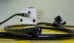
Endoscopic mucosal resection proves effective, durable for Barrett’s-associated neoplasia
Complete endoscopic mucosal resection is an effective, durable, and relatively safe treatment for Barrett’s esophagus with high-grade dysplasia, findings from a series of 107 patients suggest.
A particular benefit of endoscopic mucosal resection is that it provides large and intact tissue specimens that allow for accurate staging, according to a report by Dr. Vani J.A. Konda of the University of Chicago and her colleagues in the December issue of Clinical Gastroenterology and Hepatology (doi:10.1016/j.cgh.2014.04.010).
Based on an intention-to-treat analysis, the approach resulted in complete eradication of Barrett’s esophagus in 86 of the 107 patients (80%) who were referred to the Center for Endoscopic Research and Therapeutics at the University of Chicago between August 2003 and December 2012 for Barrett’s esophagus with suspected high-grade dysplasia or intramucosal carcinoma. Based on a per-protocol analysis, the approach resulted in complete eradication in 79 of 80 patients (98.8%), the investigators reported.
Endoscopic mucosal resection resulted in a change in the diagnosis in 27 cases (25.2%), which were upstaged based on assessment of the resection specimens. Four of the cases in which the diagnosis was changed were initially diagnosed as high-grade dysplasia on biopsy and were found on endoscopic mucosal resection to have evidence of submucosal invasion, they said.
Patients included in the series had a mean lesion length of 3.6 cm. All patients underwent complete endoscopic mucosal resection performed on an outpatient basis by a single endoscopist and were followed through January 2014 for a median of 33 months. Most (78.5%) were treated using the cap-assisted technique, while 8.4% were treated using band ligation, 2.8% were treated using a mixed band ligation and cap technique, and 11.2% were treated using an injection-assisted, free-hand technique. Two patients with suspected submucosal invasion underwent a combination of endoscopic submucosal dissection and endoscopic mucosal resection.
Esophageal strictures occurred in 44 patients (41.1%) and 40 were symptomatic; strictures required an average of 2.3 dilations. Symptomatic dysphagia developed in 37.3% of patients, and perforations occurred in two patients, suggesting a need for surgical back-up for patients undergoing endoscopic mucosal resection, the investigators said.High-grade dysplasia and intramucosal carcinoma each recurred in one patient, and both were treated successfully with endoscopic mucosal resection, they reported.
At the most recent analysis, 53 of 74 patients (71.6%) in a durability cohort were in complete remission from intestinal metaplasia, and all were in complete remission from high-grade dysplasia or intramucosal carcinoma, they noted.
Endoscopic therapy is now preferred over esophagectomy in most cases of Barrett’s esophagus with high-grade dysplasia or intramucosal carcinoma, and the current findings show that complete endoscopic mucosal resection is an effective and durable method to treat Barrett’s-associated neoplasia, the investigators concluded, adding that the results support the replacement of esophagectomy by endoscopic therapy for the condition.
“The most commonly encountered complication of esophageal stricture formation can be addressed with endoscopic dilation,” the investigators said.
However, long-term outcomes, particularly in those with advanced pathology who undergo endoscopic management, are still needed, as is an improved understanding of the behavior of the disease following treatment with specific modalities in order to optimize endoscopic therapy, they added.
This study was supported by the Francis L. Lederer Foundation. The authors reported having no disclosures.
Localized treatments aimed at the eradication of dysplasia, early cancer, and surrounding mucosa in Barrett’s esophagus have been a major advancement in gastroenterology. Although radiofrequency ablation (RFA) has become the procedure of choice, debate exists regarding the role of other mucosal extirpative procedures. The work by Dr. Konda and her colleagues gives life to another proposed procedure, complete endoscopic mucosal resection (EMR) of dysplastic and Barrett’s mucosa. In this study, complete EMR led to results comparable with previous reports on RFA (Gastroenterology 2013;145:96-104). EMR also has theoretical advantages over RFA, including avoidance of promoting protumorigenic mutations and clonal expansion in remaining mucosa (Am. J. Gastroenterol. 2013;108:1584-92).

|
| Dr. David A. Katzka |
EMR, like RFA, however, is not risk free; perforations occurred in three patients (one during dilation) and 38% developed symptomatic strictures. Furthermore, the endoscopic skill required to complete this protocol also necessitated expertise in cap technique, band ligation, injection-assisted free-hand technique, focal RFA, and endoscopic submucosal dissection. In other words, performing complete EMR is for a select group of endoscopic intensivists only. Unfortunately, endoscopic eradication of Barrett’s mucosa remains a procedure that is betwixt and between. It is between the mucosal and submucosal layer but is betwixt because of limitations on the depth of penetration of RFA (around 0.7 mm) and EMR (Dig. Dis. 2009;26:280-4) needed to eradicate the elusive Barrett’s stem cell, coupled with the increasing chance of esophageal stricture formation with these deeper levels of injury. Nevertheless, work such as this from Dr. Konda and her colleagues allows us to continue to claim at least a partial victory over Barrett’s dysplasia. It is hoped that with future techniques, we may come up with a “kinder” and easier method of removing all Barrett’s mucosa without the provocation of a substantial fibrotic reaction of the remaining esophageal wall.
Dr. David A. Katzka is in the department of gastroenterology and hepatology, Mayo Clinic, Rochester, Minn. He has no conflicts of interest.
Localized treatments aimed at the eradication of dysplasia, early cancer, and surrounding mucosa in Barrett’s esophagus have been a major advancement in gastroenterology. Although radiofrequency ablation (RFA) has become the procedure of choice, debate exists regarding the role of other mucosal extirpative procedures. The work by Dr. Konda and her colleagues gives life to another proposed procedure, complete endoscopic mucosal resection (EMR) of dysplastic and Barrett’s mucosa. In this study, complete EMR led to results comparable with previous reports on RFA (Gastroenterology 2013;145:96-104). EMR also has theoretical advantages over RFA, including avoidance of promoting protumorigenic mutations and clonal expansion in remaining mucosa (Am. J. Gastroenterol. 2013;108:1584-92).
|
|
| Dr. David A. Katzka |
EMR, like RFA, however, is not risk free; perforations occurred in three patients (one during dilation) and 38% developed symptomatic strictures. Furthermore, the endoscopic skill required to complete this protocol also necessitated expertise in cap technique, band ligation, injection-assisted free-hand technique, focal RFA, and endoscopic submucosal dissection. In other words, performing complete EMR is for a select group of endoscopic intensivists only. Unfortunately, endoscopic eradication of Barrett’s mucosa remains a procedure that is betwixt and between. It is between the mucosal and submucosal layer but is betwixt because of limitations on the depth of penetration of RFA (around 0.7 mm) and EMR (Dig. Dis. 2009;26:280-4) needed to eradicate the elusive Barrett’s stem cell, coupled with the increasing chance of esophageal stricture formation with these deeper levels of injury. Nevertheless, work such as this from Dr. Konda and her colleagues allows us to continue to claim at least a partial victory over Barrett’s dysplasia. It is hoped that with future techniques, we may come up with a “kinder” and easier method of removing all Barrett’s mucosa without the provocation of a substantial fibrotic reaction of the remaining esophageal wall.
Dr. David A. Katzka is in the department of gastroenterology and hepatology, Mayo Clinic, Rochester, Minn. He has no conflicts of interest.
Complete endoscopic mucosal resection is an effective, durable, and relatively safe treatment for Barrett’s esophagus with high-grade dysplasia, findings from a series of 107 patients suggest.
A particular benefit of endoscopic mucosal resection is that it provides large and intact tissue specimens that allow for accurate staging, according to a report by Dr. Vani J.A. Konda of the University of Chicago and her colleagues in the December issue of Clinical Gastroenterology and Hepatology (doi:10.1016/j.cgh.2014.04.010).
Based on an intention-to-treat analysis, the approach resulted in complete eradication of Barrett’s esophagus in 86 of the 107 patients (80%) who were referred to the Center for Endoscopic Research and Therapeutics at the University of Chicago between August 2003 and December 2012 for Barrett’s esophagus with suspected high-grade dysplasia or intramucosal carcinoma. Based on a per-protocol analysis, the approach resulted in complete eradication in 79 of 80 patients (98.8%), the investigators reported.
Endoscopic mucosal resection resulted in a change in the diagnosis in 27 cases (25.2%), which were upstaged based on assessment of the resection specimens. Four of the cases in which the diagnosis was changed were initially diagnosed as high-grade dysplasia on biopsy and were found on endoscopic mucosal resection to have evidence of submucosal invasion, they said.
Patients included in the series had a mean lesion length of 3.6 cm. All patients underwent complete endoscopic mucosal resection performed on an outpatient basis by a single endoscopist and were followed through January 2014 for a median of 33 months. Most (78.5%) were treated using the cap-assisted technique, while 8.4% were treated using band ligation, 2.8% were treated using a mixed band ligation and cap technique, and 11.2% were treated using an injection-assisted, free-hand technique. Two patients with suspected submucosal invasion underwent a combination of endoscopic submucosal dissection and endoscopic mucosal resection.
Esophageal strictures occurred in 44 patients (41.1%) and 40 were symptomatic; strictures required an average of 2.3 dilations. Symptomatic dysphagia developed in 37.3% of patients, and perforations occurred in two patients, suggesting a need for surgical back-up for patients undergoing endoscopic mucosal resection, the investigators said.High-grade dysplasia and intramucosal carcinoma each recurred in one patient, and both were treated successfully with endoscopic mucosal resection, they reported.
At the most recent analysis, 53 of 74 patients (71.6%) in a durability cohort were in complete remission from intestinal metaplasia, and all were in complete remission from high-grade dysplasia or intramucosal carcinoma, they noted.
Endoscopic therapy is now preferred over esophagectomy in most cases of Barrett’s esophagus with high-grade dysplasia or intramucosal carcinoma, and the current findings show that complete endoscopic mucosal resection is an effective and durable method to treat Barrett’s-associated neoplasia, the investigators concluded, adding that the results support the replacement of esophagectomy by endoscopic therapy for the condition.
“The most commonly encountered complication of esophageal stricture formation can be addressed with endoscopic dilation,” the investigators said.
However, long-term outcomes, particularly in those with advanced pathology who undergo endoscopic management, are still needed, as is an improved understanding of the behavior of the disease following treatment with specific modalities in order to optimize endoscopic therapy, they added.
This study was supported by the Francis L. Lederer Foundation. The authors reported having no disclosures.
Key clinical point: Endoscopic therapy should replace esophagectomy for Barrett’s-associated neoplasia.
Major finding: Barrett’s esophagus was completely eradicated in 86 of 107 patients (80%) on intention-to-treat analysis.
Data source: A series of 107 cases.
Disclosures: This study was supported by the Francis L. Lederer Foundation. The authors reported having no disclosures.
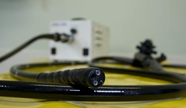
High failure rate seen with limited parathyroidectomy in patients with MEN-1
SAN FRANCISCO – Patients with hyperparathyroidism due to multiple endocrine neoplasia type 1 (MEN-1) have a 4 in 10 chance of persistent hyperparathyroidism if they undergo surgery that leaves at least one gland in place, according to a retrospective cohort study presented at the annual clinical congress of the American College of Surgeons.
“Limited initial parathyroidectomy in patients with MEN-1–associated primary hyperparathyroidism results in a high failure rate. Additional enlarged contralateral parathyroid glands are frequently missed by preoperative localizing studies,” commented lead investigator Dr. Naris Nilubol, a staff clinician with the endocrine oncology branch of the Center for Cancer Research, National Cancer Institute, Bethesda, Md.

“We conclude that limited parathyroidectomy in MEN-1 guided by preoperative localizing studies is associated with high failure rates and therefore should not be performed,” he maintained.
In an interview, session comoderator Dr. Marybeth S. Hughes, a staff clinician with the thoracic and gastrointestinal oncology branch, Center for Cancer Research, National Cancer Institute, commented, “In general, I would say that the data presented just reiterates the standard of care, that MEN-1 patients should have bilateral neck exploration with [removal of] three and half glands, or four glands with autotransplantation. So it just basically solidifies what is being done standardly. I don’t think there is a compelling argument to change the standard.”
Dr. Nilubol and colleagues reviewed the charts of 99 patients with MEN-1 who underwent at least one parathyroidectomy at the National Institutes of Health (NIH).
Of the 64 patients who had initial surgery at NIH and had preoperative localizing studies done, 32 had only a single enlarged gland identified by the tests, suggesting they would be good candidates for limited surgery, according to Dr. Nilubol. Bilateral neck dissection at the time of parathyroidectomy showed that in 22 (69%) of these 32 patients, the studies had correctly identified the largest gland; however, in 19 (87%) of those 22, it missed another enlarged gland on the contralateral side. Furthermore, in 5 (16%) of the 32, the largest gland was found on the contralateral side.
With a median follow-up of 23 months, the risk of persistent hyperparathyroidism was 41% for patients who had limited parathyroidectomy (three or fewer glands removed) at initial surgery, significantly and sharply higher than the 6% seen in patients who had subtotal parathyroidectomy or more extensive surgery (at least three and a half glands removed).

Looking at the cumulative number of glands removed during initial and subsequent surgeries, 57% of patients having two or fewer glands removed and 45% of those having two and a half to three glands removed had persistent hyperparathyroidism – both significantly higher than the 5% of patients having at least three and a half glands removed.
Regarding complications, 10% of the patients who had their initial surgery at NIH developed permanent hypoparathyroidism, reported Dr. Nilubol, who disclosed that he had no relevant conflicts of interest.
Session attendees asked about the use of parathyroid hormone levels intraoperatively to guide surgery and what strategy surgeons follow at his institution in this patient population.
Previous research has suggested that intraoperative parathyroid hormone levels do not add any information that would change the operative plan, Dr. Nilubol replied. “Everybody at NIH has preop localizing studies as part of the clinical investigation, but it doesn’t change the way we approach it. Everybody gets a bilateral neck exploration and three and a half–gland removal,” provided all glands can be found, he said.
Session attendee Dr. Michael J. Campbell, a surgeon at the University California, Davis, commented, “A 10% permanent hypoparathyroidism rate in these patients – and they have a tendency to be young, most of them in their late teens, early 20s – that’s a major complication. So could you take your data and make exactly the opposite argument, that maybe you should be doing less to these patients to limit that fairly life-altering complication?” Permanent hypothyroidism at that age is “a significant medical problem,” Dr. Nilubol agreed. However, “at the NIH, we don’t operate on everybody just because they have primary hyperparathyroidism. They have to fulfill metabolic complications before we choose to operate on them. We want to delay the surgeries and [time] between the surgeries because if they live long enough, it will recur, so we want to operate when we can make the most difference, meaning [addressing] kidney stone, bone loss, etc. The most common reason for young patients is they have kidney stones, which leads to surgery.”
SAN FRANCISCO – Patients with hyperparathyroidism due to multiple endocrine neoplasia type 1 (MEN-1) have a 4 in 10 chance of persistent hyperparathyroidism if they undergo surgery that leaves at least one gland in place, according to a retrospective cohort study presented at the annual clinical congress of the American College of Surgeons.
“Limited initial parathyroidectomy in patients with MEN-1–associated primary hyperparathyroidism results in a high failure rate. Additional enlarged contralateral parathyroid glands are frequently missed by preoperative localizing studies,” commented lead investigator Dr. Naris Nilubol, a staff clinician with the endocrine oncology branch of the Center for Cancer Research, National Cancer Institute, Bethesda, Md.
“We conclude that limited parathyroidectomy in MEN-1 guided by preoperative localizing studies is associated with high failure rates and therefore should not be performed,” he maintained.
In an interview, session comoderator Dr. Marybeth S. Hughes, a staff clinician with the thoracic and gastrointestinal oncology branch, Center for Cancer Research, National Cancer Institute, commented, “In general, I would say that the data presented just reiterates the standard of care, that MEN-1 patients should have bilateral neck exploration with [removal of] three and half glands, or four glands with autotransplantation. So it just basically solidifies what is being done standardly. I don’t think there is a compelling argument to change the standard.”
Dr. Nilubol and colleagues reviewed the charts of 99 patients with MEN-1 who underwent at least one parathyroidectomy at the National Institutes of Health (NIH).
Of the 64 patients who had initial surgery at NIH and had preoperative localizing studies done, 32 had only a single enlarged gland identified by the tests, suggesting they would be good candidates for limited surgery, according to Dr. Nilubol. Bilateral neck dissection at the time of parathyroidectomy showed that in 22 (69%) of these 32 patients, the studies had correctly identified the largest gland; however, in 19 (87%) of those 22, it missed another enlarged gland on the contralateral side. Furthermore, in 5 (16%) of the 32, the largest gland was found on the contralateral side.
With a median follow-up of 23 months, the risk of persistent hyperparathyroidism was 41% for patients who had limited parathyroidectomy (three or fewer glands removed) at initial surgery, significantly and sharply higher than the 6% seen in patients who had subtotal parathyroidectomy or more extensive surgery (at least three and a half glands removed).
Looking at the cumulative number of glands removed during initial and subsequent surgeries, 57% of patients having two or fewer glands removed and 45% of those having two and a half to three glands removed had persistent hyperparathyroidism – both significantly higher than the 5% of patients having at least three and a half glands removed.
Regarding complications, 10% of the patients who had their initial surgery at NIH developed permanent hypoparathyroidism, reported Dr. Nilubol, who disclosed that he had no relevant conflicts of interest.
Session attendees asked about the use of parathyroid hormone levels intraoperatively to guide surgery and what strategy surgeons follow at his institution in this patient population.
Previous research has suggested that intraoperative parathyroid hormone levels do not add any information that would change the operative plan, Dr. Nilubol replied. “Everybody at NIH has preop localizing studies as part of the clinical investigation, but it doesn’t change the way we approach it. Everybody gets a bilateral neck exploration and three and a half–gland removal,” provided all glands can be found, he said.
Session attendee Dr. Michael J. Campbell, a surgeon at the University California, Davis, commented, “A 10% permanent hypoparathyroidism rate in these patients – and they have a tendency to be young, most of them in their late teens, early 20s – that’s a major complication. So could you take your data and make exactly the opposite argument, that maybe you should be doing less to these patients to limit that fairly life-altering complication?” Permanent hypothyroidism at that age is “a significant medical problem,” Dr. Nilubol agreed. However, “at the NIH, we don’t operate on everybody just because they have primary hyperparathyroidism. They have to fulfill metabolic complications before we choose to operate on them. We want to delay the surgeries and [time] between the surgeries because if they live long enough, it will recur, so we want to operate when we can make the most difference, meaning [addressing] kidney stone, bone loss, etc. The most common reason for young patients is they have kidney stones, which leads to surgery.”
AT THE ACS CLINICAL CONGRESS
Key clinical point: Patients are more likely to have persistent hyperparathyroidism if a gland is left behind.
Major finding: The failure rate after initial parathyroidectomy was 41% with limited surgery versus 6% with subtotal or more extensive surgery.
Data source: A retrospective chart review of 99 patients with MEN-1–associated hyperparathyroidism.
Disclosures: Dr. Nilubol disclosed that he had no relevant conflicts of interest.

Expert panel endorses continued use of morcellation
VANCOUVER – Morcellation is an effective, lifesaving tool in gynecologic surgery when used appropriately and should not be abandoned despite recent concerns about the dissemination of occult cancers, according to an expert panel that weighed in on this issue at a meeting sponsored by AAGL.
Panelists presented new data to inform the intense debate over this procedure, which has culminated in the Food and Drug Administration (FDA) recommending against the use of power morcellators during fibroid removal by hysterectomy or myomectomy for most women.

Earlier this year, AAGL convened the Tissue Extraction Task Force to study this issue and respond to the controversy. The association presented a statement to the FDA on power morcellation and published the task force findings that morcellation can be done safely and effectively when performed by trained and experienced surgeons in informed, carefully screened premenopausal women (J. Minim. Invasive Gynecol. 2014;21:517-30).
Abandoning it may raise mortality
“The priority of this entire discussion needs to focus on the patient’s welfare,” contended panelist Dr. Jubilee Brown, an associate professor in the department of gynecologic oncology and reproductive medicine, University of Texas M.D. Anderson Cancer Center, Houston. “For every piece of data that we look at, we need to keep that in the back of our minds as we analyze this.”
In a new study, she and her colleagues retrospectively studied outcomes in 808 consecutive patients with planned laparoscopic supracervical hysterectomy with morcellation who had at least 5 years of follow-up. The leading indications for surgery were menorrhagia and leiomyomata.
Only a single woman had a leiomyosarcoma; she was converted to an open procedure without morcellation but nonetheless died from the disease. “What hasn’t shown up in much of the literature is the wisdom of the operating surgeon, who identified that this uterus looked abnormal and called our group in,” commented Dr. Brown, who is also AAGL’s designated spokesperson on tissue extraction. “Unfortunately, what’s also missed in much of the literature is that leiomyosarcoma is an aggressive and often deadly disease. … In her case, as in so many cases, the problem was not the surgery, the problem was the cancer.”
Among the 778 women who underwent the planned laparoscopic hysterectomy with morcellation, 16 were found to have endometrial hyperplasia, two had adenocarcinoma, and one had an endometrial stromal sarcoma – but reassuringly, none had evidence of disease at follow-up.
“I think that what this tells us is that we need to be absolutely meticulous in our preoperative evaluation of patients in whom we are considering morcellation,” Dr. Brown said. The findings “speak to our obligation to educate our membership and everybody performing preoperative sampling on these patients.”
A decision analysis study also reported at the meeting by first author Dr. R. Wendel Naumann, Carolinas Medical Center in Charlotte, N.C., showed that mortality from laparoscopic hysterectomy with power morcellation – even accounting for possible dissemination of undiagnosed leiomyosarcomas – was 0.077%, still less than the 0.085% mortality from abdominal hysterectomy. “Though it is a small difference, it is an absolute difference in favor of laparoscopic hysterectomy with power morcellation. In fact, if all women were converted to an open hysterectomy, 17 more women each year would die of open hysterectomy than of power morcellation,” Dr. Brown commented.
“Power morcellation is an important tool,” she concluded, reiterating AAGL’s position that its use should be improved, not abandoned.
Low risk of leiomyosarcomas
Panelist Dr. Marit Lieng, an associate professor and consultant in the gynecology department of Oslo University Hospital, and her coinvestigators retrospectively studied 4,765 women who underwent surgery at the hospital for uterine fibroids between 2000 and 2013.

There were 26 cases of leiomyosarcoma (the majority in postmenopausal women), for an incidence of 0.54%, or 1 in 183 women.
However, only a single patient with leiomyosarcoma had laparoscopic supracervical hysterectomy with morcellation, because the tumor was identified or suspected preoperatively or intraoperatively in the rest, reported Dr. Lieng, who is also with the Institute of Clinical Medicine at the University of Oslo.
Therefore, the risk of unintended morcellation of an undiagnosed leiomyosarcoma was just 1 in 4,765 women, or 0.02%.
“I think the findings of our study support the conclusions of the AAGL expert group. … You can do power morcellation in selected patients,” Dr. Lieng commented. “Given a thorough preoperative evaluation, including a cervical cytology, endometrial biopsy, and evaluation of the myometrium by ultrasound or MRI, the risk of unintended morcellation of a uterine leiomyosarcoma in premenopausal women appears to be very low.”
Leiomyosarcomas best removed en bloc
“When you are creating public health care policy, decision analysis must begin with scientifically valid evidence,” asserted panelist Dr. Elizabeth Pritts, medical director of the Wisconsin Fertility Institute, Middleton.

She and her colleagues undertook a comprehensive new meta-analysis assessing the prevalence of occult leiomyosarcomas at hysterectomy or myomectomy for presumed uterine fibroids, including 133 original articles describing 30,193 women having explicit pathology.
Analysis of all prospective data showed that the predicted prevalence rate of occult leiomyosarcoma was 0.12 per 1,000 operations for presumed benign fibroids.
The corresponding 1 in 8,300 operations needed to find a leiomyosarcoma in this new meta-analysis differs greatly from the 1 in 498 found in an FDA meta-analysis, mainly because of the differing evidence base, Dr. Pritts maintained. “It really has to do with initial search criteria,” she said, noting, for example, that the FDA’s search strategy missed studies in which no cancer was found and studies in languages other than English.
Dr. Pritts and her colleagues also conducted a new systematic review looking at outcomes after morcellation of an unsuspected leiomyosarcoma, which was recently published (J. Minim. Invasive Gynecol. 2014 Sept. 2 [doi: 10.1016/j.jmig.2014.08.781]).
Main analyses here were based on six papers that compared morcellation with en bloc removal of leiomyosarcomas, most of which found worse survival for women whose tumors were morcellated.
“Now this is not great evidence, but remember, in evidence-based medicine, you’ve got to look at the very best available evidence. This is it,” Dr. Pritts maintained. “En bloc removal confers benefit—don’t cut into these.”
On closer inspection, only 3 of the 81 cases of morcellation reported were confirmed to be power morcellation. Comparisons of outcome with power versus hand morcellation, albeit limited by small numbers, suggested no difference in survival or upstaging.
“There are no data to suggest that any type of morcellation is better or worse than another type, even when including simple tumor biopsies,” Dr. Pritts concluded.
Dr. Brown, Dr. Lieng, and Dr. Pritts disclosed that they had no relevant conflicts of interest.
VANCOUVER – Morcellation is an effective, lifesaving tool in gynecologic surgery when used appropriately and should not be abandoned despite recent concerns about the dissemination of occult cancers, according to an expert panel that weighed in on this issue at a meeting sponsored by AAGL.
Panelists presented new data to inform the intense debate over this procedure, which has culminated in the Food and Drug Administration (FDA) recommending against the use of power morcellators during fibroid removal by hysterectomy or myomectomy for most women.
Earlier this year, AAGL convened the Tissue Extraction Task Force to study this issue and respond to the controversy. The association presented a statement to the FDA on power morcellation and published the task force findings that morcellation can be done safely and effectively when performed by trained and experienced surgeons in informed, carefully screened premenopausal women (J. Minim. Invasive Gynecol. 2014;21:517-30).
Abandoning it may raise mortality
“The priority of this entire discussion needs to focus on the patient’s welfare,” contended panelist Dr. Jubilee Brown, an associate professor in the department of gynecologic oncology and reproductive medicine, University of Texas M.D. Anderson Cancer Center, Houston. “For every piece of data that we look at, we need to keep that in the back of our minds as we analyze this.”
In a new study, she and her colleagues retrospectively studied outcomes in 808 consecutive patients with planned laparoscopic supracervical hysterectomy with morcellation who had at least 5 years of follow-up. The leading indications for surgery were menorrhagia and leiomyomata.
Only a single woman had a leiomyosarcoma; she was converted to an open procedure without morcellation but nonetheless died from the disease. “What hasn’t shown up in much of the literature is the wisdom of the operating surgeon, who identified that this uterus looked abnormal and called our group in,” commented Dr. Brown, who is also AAGL’s designated spokesperson on tissue extraction. “Unfortunately, what’s also missed in much of the literature is that leiomyosarcoma is an aggressive and often deadly disease. … In her case, as in so many cases, the problem was not the surgery, the problem was the cancer.”
Among the 778 women who underwent the planned laparoscopic hysterectomy with morcellation, 16 were found to have endometrial hyperplasia, two had adenocarcinoma, and one had an endometrial stromal sarcoma – but reassuringly, none had evidence of disease at follow-up.
“I think that what this tells us is that we need to be absolutely meticulous in our preoperative evaluation of patients in whom we are considering morcellation,” Dr. Brown said. The findings “speak to our obligation to educate our membership and everybody performing preoperative sampling on these patients.”
A decision analysis study also reported at the meeting by first author Dr. R. Wendel Naumann, Carolinas Medical Center in Charlotte, N.C., showed that mortality from laparoscopic hysterectomy with power morcellation – even accounting for possible dissemination of undiagnosed leiomyosarcomas – was 0.077%, still less than the 0.085% mortality from abdominal hysterectomy. “Though it is a small difference, it is an absolute difference in favor of laparoscopic hysterectomy with power morcellation. In fact, if all women were converted to an open hysterectomy, 17 more women each year would die of open hysterectomy than of power morcellation,” Dr. Brown commented.
“Power morcellation is an important tool,” she concluded, reiterating AAGL’s position that its use should be improved, not abandoned.
Low risk of leiomyosarcomas
Panelist Dr. Marit Lieng, an associate professor and consultant in the gynecology department of Oslo University Hospital, and her coinvestigators retrospectively studied 4,765 women who underwent surgery at the hospital for uterine fibroids between 2000 and 2013.
There were 26 cases of leiomyosarcoma (the majority in postmenopausal women), for an incidence of 0.54%, or 1 in 183 women.
However, only a single patient with leiomyosarcoma had laparoscopic supracervical hysterectomy with morcellation, because the tumor was identified or suspected preoperatively or intraoperatively in the rest, reported Dr. Lieng, who is also with the Institute of Clinical Medicine at the University of Oslo.
Therefore, the risk of unintended morcellation of an undiagnosed leiomyosarcoma was just 1 in 4,765 women, or 0.02%.
“I think the findings of our study support the conclusions of the AAGL expert group. … You can do power morcellation in selected patients,” Dr. Lieng commented. “Given a thorough preoperative evaluation, including a cervical cytology, endometrial biopsy, and evaluation of the myometrium by ultrasound or MRI, the risk of unintended morcellation of a uterine leiomyosarcoma in premenopausal women appears to be very low.”
Leiomyosarcomas best removed en bloc
“When you are creating public health care policy, decision analysis must begin with scientifically valid evidence,” asserted panelist Dr. Elizabeth Pritts, medical director of the Wisconsin Fertility Institute, Middleton.
She and her colleagues undertook a comprehensive new meta-analysis assessing the prevalence of occult leiomyosarcomas at hysterectomy or myomectomy for presumed uterine fibroids, including 133 original articles describing 30,193 women having explicit pathology.
Analysis of all prospective data showed that the predicted prevalence rate of occult leiomyosarcoma was 0.12 per 1,000 operations for presumed benign fibroids.
The corresponding 1 in 8,300 operations needed to find a leiomyosarcoma in this new meta-analysis differs greatly from the 1 in 498 found in an FDA meta-analysis, mainly because of the differing evidence base, Dr. Pritts maintained. “It really has to do with initial search criteria,” she said, noting, for example, that the FDA’s search strategy missed studies in which no cancer was found and studies in languages other than English.
Dr. Pritts and her colleagues also conducted a new systematic review looking at outcomes after morcellation of an unsuspected leiomyosarcoma, which was recently published (J. Minim. Invasive Gynecol. 2014 Sept. 2 [doi: 10.1016/j.jmig.2014.08.781]).
Main analyses here were based on six papers that compared morcellation with en bloc removal of leiomyosarcomas, most of which found worse survival for women whose tumors were morcellated.
“Now this is not great evidence, but remember, in evidence-based medicine, you’ve got to look at the very best available evidence. This is it,” Dr. Pritts maintained. “En bloc removal confers benefit—don’t cut into these.”
On closer inspection, only 3 of the 81 cases of morcellation reported were confirmed to be power morcellation. Comparisons of outcome with power versus hand morcellation, albeit limited by small numbers, suggested no difference in survival or upstaging.
“There are no data to suggest that any type of morcellation is better or worse than another type, even when including simple tumor biopsies,” Dr. Pritts concluded.
Dr. Brown, Dr. Lieng, and Dr. Pritts disclosed that they had no relevant conflicts of interest.
AT THE AAGL GLOBAL CONGRESS

CT overutilized to diagnose appendicitis
SAN FRANCISCO – At least 25% of CT scans to diagnose appendicitis were unnecessary, potentially resulting in $1.8 million in costs at one institution and up to four new cancers from the radiation exposure, a retrospective study suggests.
The review of 1,054 patients who underwent appendectomy at the University of California, Davis, in 2005-2010 focused on costs for the patients who had high Alvarado scores, a clinical scoring system used to diagnose appendicitis, before they underwent appendectomy. CT scans to help diagnose appendicitis were performed on 77% of all patients.
Records showed that 26% of patients had an Alvarado score of 8-10, meaning that appendicitis was highly likely. CT was performed on 70% of patients with an Alvarado score of 8 and 77% of patients with a score of 9-10, comprising nearly 25% of all CT scans. That resulted in an estimated $1,813,399 in unnecessary costs for imaging, Dr. Adam Dougherty and his associates reported at the annual clinical congress of the American College of Surgeons.

This “overutilization” of CT scans delivered more than 4,009 mSv in unnecessary radiation exposure, averaging 19.75 mSV per scan, which is 20 times the annual limit suggested for safety, said Dr. Dougherty of the university. That excess radiation could be expected to produce up to four new cancers down the line, resulting in additional costs, he said.
The investigators also looked at the 9% of patients with low Alvarado scores, meaning that appendicitis was unlikely. CT scans were performed in 75% of patients with a score of 0-3 and 80% of patients with an Alvarado score of 4. In this subgroup, 24% showed normal/early pathology on appendectomy, which “argues against imaging and surgical treatment,” Dr. Dougherty said. The 44 CT scans in this subgroup resulted in an estimated $393,052 in unnecessary costs, he said.
That doesn’t include additional costs that could be expected from imaging, such as wait time, appendectomy and its sequelae, and potential workups of incidentalomas in the low-risk group, he added.
Previous studies have shown that a comprehensive clinical exam is as accurate as CT in diagnosing appendicitis, and that clinical assessment unaided by CT can reliably diagnose acute appendicitis, Dr. Dougherty said.
With a 72% increase in abdominal CT scans documented in other U.S. data from 2000 to 2005, he called for a “necessary, fundamental culture change” to restrain resource utilization “in order to maximize the value of the health care dollar while doing what is best for the patient.”
The investigators proposed a clinical pathway for the workup of suspected appendicitis that places greater emphasis on ultrasound imaging and conservative pathways, such as 23-hour admission for observation and next-day follow-up.
In the study, ultrasonography was underutilized across all subgroups as a viable alternative to CT scans, he said.
Dr. Dougherty reported having no financial disclosures.
On Twitter @sherryboschert
SAN FRANCISCO – At least 25% of CT scans to diagnose appendicitis were unnecessary, potentially resulting in $1.8 million in costs at one institution and up to four new cancers from the radiation exposure, a retrospective study suggests.
The review of 1,054 patients who underwent appendectomy at the University of California, Davis, in 2005-2010 focused on costs for the patients who had high Alvarado scores, a clinical scoring system used to diagnose appendicitis, before they underwent appendectomy. CT scans to help diagnose appendicitis were performed on 77% of all patients.
Records showed that 26% of patients had an Alvarado score of 8-10, meaning that appendicitis was highly likely. CT was performed on 70% of patients with an Alvarado score of 8 and 77% of patients with a score of 9-10, comprising nearly 25% of all CT scans. That resulted in an estimated $1,813,399 in unnecessary costs for imaging, Dr. Adam Dougherty and his associates reported at the annual clinical congress of the American College of Surgeons.
This “overutilization” of CT scans delivered more than 4,009 mSv in unnecessary radiation exposure, averaging 19.75 mSV per scan, which is 20 times the annual limit suggested for safety, said Dr. Dougherty of the university. That excess radiation could be expected to produce up to four new cancers down the line, resulting in additional costs, he said.
The investigators also looked at the 9% of patients with low Alvarado scores, meaning that appendicitis was unlikely. CT scans were performed in 75% of patients with a score of 0-3 and 80% of patients with an Alvarado score of 4. In this subgroup, 24% showed normal/early pathology on appendectomy, which “argues against imaging and surgical treatment,” Dr. Dougherty said. The 44 CT scans in this subgroup resulted in an estimated $393,052 in unnecessary costs, he said.
That doesn’t include additional costs that could be expected from imaging, such as wait time, appendectomy and its sequelae, and potential workups of incidentalomas in the low-risk group, he added.
Previous studies have shown that a comprehensive clinical exam is as accurate as CT in diagnosing appendicitis, and that clinical assessment unaided by CT can reliably diagnose acute appendicitis, Dr. Dougherty said.
With a 72% increase in abdominal CT scans documented in other U.S. data from 2000 to 2005, he called for a “necessary, fundamental culture change” to restrain resource utilization “in order to maximize the value of the health care dollar while doing what is best for the patient.”
The investigators proposed a clinical pathway for the workup of suspected appendicitis that places greater emphasis on ultrasound imaging and conservative pathways, such as 23-hour admission for observation and next-day follow-up.
In the study, ultrasonography was underutilized across all subgroups as a viable alternative to CT scans, he said.
Dr. Dougherty reported having no financial disclosures.
On Twitter @sherryboschert
AT THE ACS CLINICAL CONGRESS
Key clinical point: Records showed that 26% of patients who had CT scans for suspect appendicitis had an Alvarado score of 8-10.
Major finding: A quarter of CT scans were on patients with likely appendicitis by Alvarado score, producing $1.8 million in unnecessary costs.
Data source: A retrospective study of 1,054 patients undergoing appendectomy in 2005-2010 at one institution.
Disclosures: Dr. Dougherty reported having no financial disclosures.

Battle over morcellation lost ‘in the media’
VANCOUVER, B.C. – The media played a major role in determining the fate of uterine morcellation, suggested a study reported at a meeting sponsored by AAGL.
“How did we lose this battle of uterine morcellation? We lost it in the media,” asserted lead investigator Dr. Adrian C. Balica, director of the minimally invasive gynecologic surgery program at the Robert Wood Johnson Medical School in New Brunswick, N.J.

He and his colleagues used the Google Adwords Keyword Planner to collect Internet search data for a variety of relevant terms used in 2013 and in 2014 through August.
This interval spanned events that included the first report of the issue in the mainstream media (December 2013), the Food and Drug Administration’s initial statement discouraging use of power morcellation for uterine fibroids (April 2014), and the issuance of analyses and rebuttals by several medical professional associations (May 2104 and thereafter).
Subsequent to the AAGL meeting, the FDA issued a new warning on Nov. 24, 2014, not to use power morcellation in the majority of women undergoing hysterectomy or myomectomy for uterine fibroids because “there is no reliable method for predicting whether a woman with fibroids may have a uterine sarcoma” that morcellation could spread. The agency estimated that about 1 in 350 fibroid patients actually have an occult sarcoma.
Results showed that the average monthly number of Google searches for the term “morcellation” held steady throughout most of 2013 at about 250 per month, reported Dr. Balica. There was, however, a sharp uptick in December 2013 to more than 2,000 per month, and the number continued to rise to a peak of about 18,000 per month in July 2014. A similar pattern was seen for the terms “morcellator,” “fibroids in uterus,” and “morcellation of uterine fibroid.”
Additionally, there were virtually no searches for “power morcellation” in 2013, but there were about 1,900 per month at the peak in April 2014. Much the same pattern was seen for “power morcellator,” he said.
Dr. Balica noted that the rapid cascade of events set into motion has had a profound impact for the field, including banning of morcellation at many hospitals and Johnson & Johnson withdrawing its power morcellator, which accounted for an estimated 72% of the devices used, from the market.
“Medical and surgical practice is going to be changed by the media,” he predicted. Thus, studying how the morcellator controversy unfolded in this venue can help inform strategies for addressing public perceptions.
His own hospital has stopped using power morcellation, according to Dr. Balica. “This is just the battle. Hopefully, we aren’t going to lose the war,” he concluded.
Dr. Balica disclosed that he had no relevant conflicts of interest.
VANCOUVER, B.C. – The media played a major role in determining the fate of uterine morcellation, suggested a study reported at a meeting sponsored by AAGL.
“How did we lose this battle of uterine morcellation? We lost it in the media,” asserted lead investigator Dr. Adrian C. Balica, director of the minimally invasive gynecologic surgery program at the Robert Wood Johnson Medical School in New Brunswick, N.J.
He and his colleagues used the Google Adwords Keyword Planner to collect Internet search data for a variety of relevant terms used in 2013 and in 2014 through August.
This interval spanned events that included the first report of the issue in the mainstream media (December 2013), the Food and Drug Administration’s initial statement discouraging use of power morcellation for uterine fibroids (April 2014), and the issuance of analyses and rebuttals by several medical professional associations (May 2104 and thereafter).
Subsequent to the AAGL meeting, the FDA issued a new warning on Nov. 24, 2014, not to use power morcellation in the majority of women undergoing hysterectomy or myomectomy for uterine fibroids because “there is no reliable method for predicting whether a woman with fibroids may have a uterine sarcoma” that morcellation could spread. The agency estimated that about 1 in 350 fibroid patients actually have an occult sarcoma.
Results showed that the average monthly number of Google searches for the term “morcellation” held steady throughout most of 2013 at about 250 per month, reported Dr. Balica. There was, however, a sharp uptick in December 2013 to more than 2,000 per month, and the number continued to rise to a peak of about 18,000 per month in July 2014. A similar pattern was seen for the terms “morcellator,” “fibroids in uterus,” and “morcellation of uterine fibroid.”
Additionally, there were virtually no searches for “power morcellation” in 2013, but there were about 1,900 per month at the peak in April 2014. Much the same pattern was seen for “power morcellator,” he said.
Dr. Balica noted that the rapid cascade of events set into motion has had a profound impact for the field, including banning of morcellation at many hospitals and Johnson & Johnson withdrawing its power morcellator, which accounted for an estimated 72% of the devices used, from the market.
“Medical and surgical practice is going to be changed by the media,” he predicted. Thus, studying how the morcellator controversy unfolded in this venue can help inform strategies for addressing public perceptions.
His own hospital has stopped using power morcellation, according to Dr. Balica. “This is just the battle. Hopefully, we aren’t going to lose the war,” he concluded.
Dr. Balica disclosed that he had no relevant conflicts of interest.
AT THE AAGL GLOBAL CONGRESS
Key clinical point: Relevant Google searches rose sharply as the debate unfolded.
Major finding: The mean monthly number of searches for “morcellation” rose from about 250 in July 2013 to 18,000 in July 2014.
Data source: An analysis of Google searches for terms related to the power morcellator debate.
Disclosures: Dr. Balica disclosed that he had no relevant conflicts of interest.