User login
Official Newspaper of the American College of Surgeons
VIDEO: Clot aspiration equals retrieval for ischemic stroke
HOUSTON – Intracerebral clot aspiration was as safe and effective as stent retriever thrombectomy for restoring cerebral blood flow in a French multicenter, randomized trial with 381 acute ischemic stroke patients.
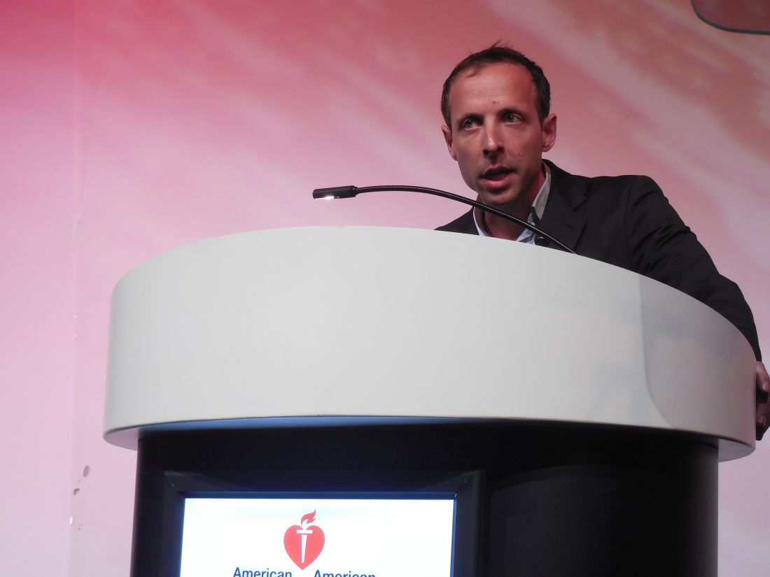
This study is the “first direct comparison of aspiration versus stent retrieval” as the initial strategy for clot removal in acute ischemic stroke, and it “opens the door to add a new tool” for clot removal, Bertrand Lapergue, MD, said at the International Stroke Conference sponsored by the American Heart Association.
The new results “are the first to show that aspiration first is as good as a stent retriever, but we need to also see the results from COMPASS,” a U.S. multicenter trial that is in the process of making the same comparison, commented Ricardo A. Hanel, MD, a vascular neurosurgeon at Baptist Health in Jacksonville, Fla. The COMPASS Trial: a Direct Aspiration First Pass Technique has now enrolled about two-thirds of its target patient number, and until the study is complete the role of direct aspiration for clot removal in stroke remains investigational for U.S. practice, said Dr. Hanel, a COMPASS investigator.
The aspiration catheter tested in ASTER is marketed by Penumbra and has already received Food and Drug Administration approval for revascularization of ischemic stroke patients. U.S. use of aspiration for treating acute ischemic stroke, however, has remained limited because there is no clear evidence of the method’s efficacy. Dr. Hanel said that he occasionally uses aspiration as an adjunct to clot removal with a stent retriever.

ASTER’s primary endpoint was the percentage of patients who achieved thrombolysis in cerebral infarction (TICI) 2b or 3 flow at the end of treatment, which occurred in 85% of patients treated with aspiration first and in 83% of those treated by clot removal first, a difference that was not statistically significant, Dr. Lapergue reported. The rate of patients who achieved either TICI 2b or 3 flow after the initial strategy only was 63% with aspiration and 68% with clot removal, also a nonsignificant difference. The two strategies also showed no significant difference for any measured safety parameter. The results showed a trend toward more vasospasm with clot removal – a 6% rate, versus 3% with clot aspiration – but this did not reach statistical significance.
Results from additional analyses of the clinical outcomes of patients in the trial and of cost efficacy will be reported later in 2017, Dr. Lapergue said.
ASTER received an unrestricted research grant from Penumbra, a company that markets clot removal aspiration catheters. Dr. Lapergue had no personal disclosures. Dr. Hanel has been a consultant to and received grant support from Medtronic. He has received research grants from MicroVention and has an ownership interest in InNeuroCo.
[email protected]
On Twitter @mitchelzoler
ASTER is an important trial. It shows for the first time that an aspiration device is probably as safe and reasonable for opening an acute occlusion in a large cerebral artery as is a stent retriever.
ASTER, however, was done entirely in a French population, making it uncertain whether the results are applicable to other populations. For example, U.S. acute ischemic stroke patients, especially African Americans and Hispanics, generally have more intracerebral atherostenotic disease than do patients from European countries, while French patients tend to have more embolic disease. Will aspiration be as effective in U.S. patients with atherostenotic blockages? I would love to see this study repeated in a U.S. population of ischemic stroke patients, and that is now happening in the COMPASS trial. It would be helpful to know if there are selected U.S. patients who might be better treated using either aspiration or a stent retriever first.
Although aspiration catheters have already received Food and Drug Administration approval for clot removal in acute ischemic stroke patients, many U.S. interventionalists have moved to deploying stent retrievers based on the very positive results reported with these devices about 2 years ago. For the moment, stent retrievers remain the most prominent devices to open large vessel occlusions.
Ralph L. Sacco, MD, is professor and chairman of neurology at the University of Miami. He had no relevant disclosures. He made these comments in a video interview and during a press conference.
The video associated with this article is no longer available on this site. Please view all of our videos on the MDedge YouTube channel
HOUSTON – Intracerebral clot aspiration was as safe and effective as stent retriever thrombectomy for restoring cerebral blood flow in a French multicenter, randomized trial with 381 acute ischemic stroke patients.
This study is the “first direct comparison of aspiration versus stent retrieval” as the initial strategy for clot removal in acute ischemic stroke, and it “opens the door to add a new tool” for clot removal, Bertrand Lapergue, MD, said at the International Stroke Conference sponsored by the American Heart Association.
The new results “are the first to show that aspiration first is as good as a stent retriever, but we need to also see the results from COMPASS,” a U.S. multicenter trial that is in the process of making the same comparison, commented Ricardo A. Hanel, MD, a vascular neurosurgeon at Baptist Health in Jacksonville, Fla. The COMPASS Trial: a Direct Aspiration First Pass Technique has now enrolled about two-thirds of its target patient number, and until the study is complete the role of direct aspiration for clot removal in stroke remains investigational for U.S. practice, said Dr. Hanel, a COMPASS investigator.
The aspiration catheter tested in ASTER is marketed by Penumbra and has already received Food and Drug Administration approval for revascularization of ischemic stroke patients. U.S. use of aspiration for treating acute ischemic stroke, however, has remained limited because there is no clear evidence of the method’s efficacy. Dr. Hanel said that he occasionally uses aspiration as an adjunct to clot removal with a stent retriever.
ASTER’s primary endpoint was the percentage of patients who achieved thrombolysis in cerebral infarction (TICI) 2b or 3 flow at the end of treatment, which occurred in 85% of patients treated with aspiration first and in 83% of those treated by clot removal first, a difference that was not statistically significant, Dr. Lapergue reported. The rate of patients who achieved either TICI 2b or 3 flow after the initial strategy only was 63% with aspiration and 68% with clot removal, also a nonsignificant difference. The two strategies also showed no significant difference for any measured safety parameter. The results showed a trend toward more vasospasm with clot removal – a 6% rate, versus 3% with clot aspiration – but this did not reach statistical significance.
Results from additional analyses of the clinical outcomes of patients in the trial and of cost efficacy will be reported later in 2017, Dr. Lapergue said.
ASTER received an unrestricted research grant from Penumbra, a company that markets clot removal aspiration catheters. Dr. Lapergue had no personal disclosures. Dr. Hanel has been a consultant to and received grant support from Medtronic. He has received research grants from MicroVention and has an ownership interest in InNeuroCo.
[email protected]
On Twitter @mitchelzoler
AT THE INTERNATIONAL STROKE CONFERENCE
Key clinical point:
Major finding: Recanalization occurred in 85% of patients treated with aspiration first and 83% treated with clot removal first.
Data source: ASTER, a multicenter, randomized French trial with 381 patients.
Disclosures: ASTER received an unrestricted research grant from Penumbra, a company that markets clot removal aspiration catheters. Dr. Lapergue had no personal disclosures. Dr. Hanel has been a consultant to and received grant support from Medtronic. He has received research grants from MicroVention and has an ownership interest in InNeuroCo.
CMS to alert docs of their MIPS status soon
ORLANDO – Want to know if you must participate in the new MIPS program? CMS is about to let you know.
Physicians who are right around the eligibility threshold for participation in the Quality Payment Program “want to know if they are eligible” for the Merit-based Incentive Payment System (MIPS), one of the QPP’s two tracks, Kate Goodrich, MD, said at the annual meeting of the Healthcare Information Management Systems Society. Within the next 6 weeks – about the first week of April – the Centers for Medicare & Medicaid Services will notify practices with less than $30,000 in Medicare payments or that serve less than 100 Medicare patients if they are exempt.

- Do the bare minimum and face no penalties.
- Submit 90 days worth of data and be eligible for a small bonus payment.
- Submit for the full year and be eligible for the full bonus that is to be determined.
Doing absolutely nothing will result in a 4% reduction in Medicare fee schedule payments in 2019.
“We have to expect that we will have some folks who do the minimum” in 2017, Dr. Goodrich, director of the Center for Clinical Standards and Quality and the chief medical officer for CMS, said. “They are just not ready to go beyond that. But even for folks who haven’t participated previously [in reporting programs], we are hearing they want to at least try to do more than just the bare minimum because they want to get ready for future years of the program.”
She said that CMS officials “are definitely hearing from some larger health systems, but even some medium and smaller practices that were really familiar with what we now call legacy programs, so meaningful use and PQRS [Physician Quality Reporting System] and so forth, that they’re ready.”
ORLANDO – Want to know if you must participate in the new MIPS program? CMS is about to let you know.
Physicians who are right around the eligibility threshold for participation in the Quality Payment Program “want to know if they are eligible” for the Merit-based Incentive Payment System (MIPS), one of the QPP’s two tracks, Kate Goodrich, MD, said at the annual meeting of the Healthcare Information Management Systems Society. Within the next 6 weeks – about the first week of April – the Centers for Medicare & Medicaid Services will notify practices with less than $30,000 in Medicare payments or that serve less than 100 Medicare patients if they are exempt.
- Do the bare minimum and face no penalties.
- Submit 90 days worth of data and be eligible for a small bonus payment.
- Submit for the full year and be eligible for the full bonus that is to be determined.
Doing absolutely nothing will result in a 4% reduction in Medicare fee schedule payments in 2019.
“We have to expect that we will have some folks who do the minimum” in 2017, Dr. Goodrich, director of the Center for Clinical Standards and Quality and the chief medical officer for CMS, said. “They are just not ready to go beyond that. But even for folks who haven’t participated previously [in reporting programs], we are hearing they want to at least try to do more than just the bare minimum because they want to get ready for future years of the program.”
She said that CMS officials “are definitely hearing from some larger health systems, but even some medium and smaller practices that were really familiar with what we now call legacy programs, so meaningful use and PQRS [Physician Quality Reporting System] and so forth, that they’re ready.”
AT HIMSS 2017
Most lung recipients gain 2-year survival benefit
Nearly three-quarters of lung transplant recipients are likely to gain at least 2 years of survival, according to new research.
In a study published in the February issue of the Annals of the American Thoracic Society, researchers used data from 13,040 adults listed for lung transplantation between May 2005 and September 2011 to develop a structural nested accelerated failure time model of the survival benefit of lung transplantation over time.
“A ‘structural nested model’ is [used to] compare the distribution of counterfactual residual survival if a patient were to receive a transplanted organ with the survival distribution if the patient did not receive that organ and never received one subsequently,” wrote David M. Vock, PhD, from the University of Minnesota, and coauthors.
Using this approach, they calculated that 73.8% of transplant recipients were predicted to achieve a 2-year survival benefit with transplantation. At 1 year posttransplantation, the relative survival benefit was 1.59, at 2 years it was 1.93, and at 3 years it was 2.23 (Ann Am Thorac Soc. 2017;14:172-81. doi: 10.1513/AnnalsATS.201606-507OC).
Patients’ lung allocation score at transplantation (LAS-T) – the score used to prioritize donated lungs for transplantation – had a significant impact on the survival benefit from transplantation. The relative survival benefit of transplantation increased by 59.4% as the lung allocation score increased from 30 to 35, and increased by 45.1% as the lung allocation score increased from 50 to 55.
However patients with a lung allocation score of 32.5 or less were more likely to die with a transplant than without, even over the long term, while patients with a score of 35 or more always gained a survival advantage from transplantation, even if their scores were as high as 50-100. The authors said this showed there should be no upper limit for the lung allocation score.
“It has been suggested that the LAS system may encourage patients who have clinically deteriorated to undergo transplantation even though it would be futile,” they wrote. “Our results reinforce the notion that lung transplantation should be considered an appropriate treatment option for patients with most advanced lung diseases and is expected to confer survival benefit in appropriately selected patients.”
Researchers also observed an interesting, borderline significant association between disease group and survival benefit, with individuals with obstructive lung disease showing the lowest relative survival gains and those with cystic fibrosis showing the highest. Head to head, the relative survival benefit of transplantation for those with cystic fibrosis was 54.4% greater than for those with obstructive lung disease.
Other factors such as transplant type, age, smoking, and center volume also influenced relative survival benefit. Bilateral transplants were associated with a 13.4% greater relative survival benefit, lungs from donors aged under 55 years showed a 17.9% relative survival benefit, and lungs from donors without a history of smoking showed a 10.5% increase in relative survival benefit.
However the researchers noted that their modeling focused on only the survival benefit of transplantation and did not take into account improvements in quality of life. This was likely to be particularly relevant in conditions such as chronic obstructive pulmonary disease where the quality of life benefits might justify transplantation even in the absence of a clear survival benefit.
“A comprehensive understanding of the survival benefit of lung transplantation and how that benefit varies by recipient characteristics is imperative to inform recipient selection, to justify the intensive health care resources allocated to this treatment, and to achieve an equitable allocation of donor lungs,” the researchers said.
The study was supported by the National Heart, Lung, and Blood Institute; the National Cancer Institute; and the National Institute of Allergy and Infectious Diseases. One author declared grants and personal fees from private industry for consultation on lung transplantation. No other conflicts of interest were declared.
Lung transplantation is the only option available for patients with treatment-resistant end-stage lung disease. However, the ability of this intervention to extend survival is still actively debated. The authors demonstrate that most adults undergoing lung transplantation experience a survival benefit that is mainly driven by the value of the lung allocation score at the time of transplantation and by the underlying lung disease.
It is reassuring to see that the two studies published so far that accounted for the course of patient disease after placement on a wait list reached essentially the same conclusions: Most of the patients experienced a survival benefit from lung transplantation.
Dr. Gabriel Thabut is from the service de pneumologie B and transplantation pulmonaire at the University of Paris. These comments are taken from an accompanying editorial (Ann Am Thorac Soc. 2017;14:163-4. doi: 10.1513/AnnalsATS.201611-853ED). No conflicts of interest were declared.
Nearly three-quarters of lung transplant recipients are likely to gain at least 2 years of survival, according to new research.
In a study published in the February issue of the Annals of the American Thoracic Society, researchers used data from 13,040 adults listed for lung transplantation between May 2005 and September 2011 to develop a structural nested accelerated failure time model of the survival benefit of lung transplantation over time.
“A ‘structural nested model’ is [used to] compare the distribution of counterfactual residual survival if a patient were to receive a transplanted organ with the survival distribution if the patient did not receive that organ and never received one subsequently,” wrote David M. Vock, PhD, from the University of Minnesota, and coauthors.
Using this approach, they calculated that 73.8% of transplant recipients were predicted to achieve a 2-year survival benefit with transplantation. At 1 year posttransplantation, the relative survival benefit was 1.59, at 2 years it was 1.93, and at 3 years it was 2.23 (Ann Am Thorac Soc. 2017;14:172-81. doi: 10.1513/AnnalsATS.201606-507OC).
Patients’ lung allocation score at transplantation (LAS-T) – the score used to prioritize donated lungs for transplantation – had a significant impact on the survival benefit from transplantation. The relative survival benefit of transplantation increased by 59.4% as the lung allocation score increased from 30 to 35, and increased by 45.1% as the lung allocation score increased from 50 to 55.
However patients with a lung allocation score of 32.5 or less were more likely to die with a transplant than without, even over the long term, while patients with a score of 35 or more always gained a survival advantage from transplantation, even if their scores were as high as 50-100. The authors said this showed there should be no upper limit for the lung allocation score.
“It has been suggested that the LAS system may encourage patients who have clinically deteriorated to undergo transplantation even though it would be futile,” they wrote. “Our results reinforce the notion that lung transplantation should be considered an appropriate treatment option for patients with most advanced lung diseases and is expected to confer survival benefit in appropriately selected patients.”
Researchers also observed an interesting, borderline significant association between disease group and survival benefit, with individuals with obstructive lung disease showing the lowest relative survival gains and those with cystic fibrosis showing the highest. Head to head, the relative survival benefit of transplantation for those with cystic fibrosis was 54.4% greater than for those with obstructive lung disease.
Other factors such as transplant type, age, smoking, and center volume also influenced relative survival benefit. Bilateral transplants were associated with a 13.4% greater relative survival benefit, lungs from donors aged under 55 years showed a 17.9% relative survival benefit, and lungs from donors without a history of smoking showed a 10.5% increase in relative survival benefit.
However the researchers noted that their modeling focused on only the survival benefit of transplantation and did not take into account improvements in quality of life. This was likely to be particularly relevant in conditions such as chronic obstructive pulmonary disease where the quality of life benefits might justify transplantation even in the absence of a clear survival benefit.
“A comprehensive understanding of the survival benefit of lung transplantation and how that benefit varies by recipient characteristics is imperative to inform recipient selection, to justify the intensive health care resources allocated to this treatment, and to achieve an equitable allocation of donor lungs,” the researchers said.
The study was supported by the National Heart, Lung, and Blood Institute; the National Cancer Institute; and the National Institute of Allergy and Infectious Diseases. One author declared grants and personal fees from private industry for consultation on lung transplantation. No other conflicts of interest were declared.
FROM ANNALS OF THE AMERICAN THORACIC SOCIETY
Key clinical point: Nearly three-quarters of lung transplant recipients are predicted to achieve a 2-year survival benefit with transplantation.
Major finding: Research suggests 73.8% of transplant recipients are likely to achieve a 2-year survival benefit with transplantation.
Data source: A structural nested accelerated failure time model of the survival benefit of lung transplantation using data from 13,040 adults listed for lung transplantation.
Disclosures: The study was supported by the National Heart, Lung, and Blood Institute; the National Cancer Institute; and the National Institute of Allergy and Infectious Diseases. One author declared grants and personal fees from private industry for consultation on lung transplantation. No other conflicts of interest were declared.
Three signs predict hypercalcemic crisis in hyperparathyroid patients
LAS VEGAS – A triad of signs – elevated serum calcium, elevated parathyroid hormone, and a history of kidney stones – can predict hypercalcemic crisis among patients with hyperparathyroidism, a study showed.
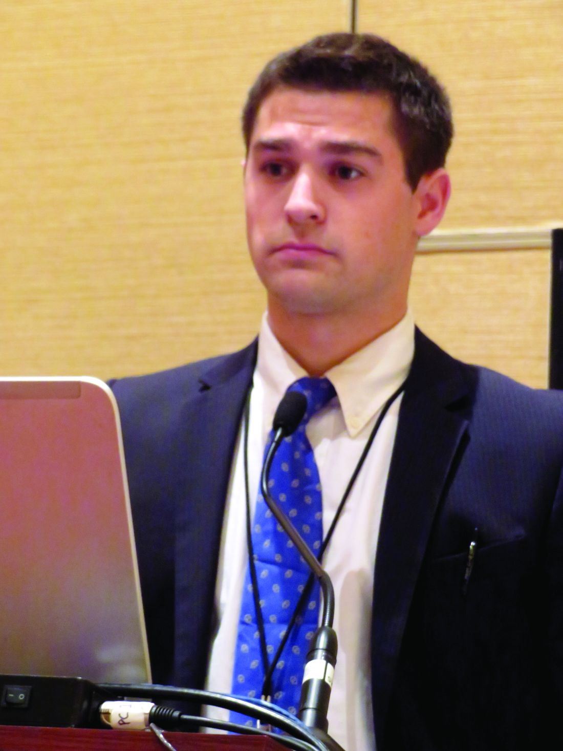
Patients who present with the trifecta should be considered for expedited parathyroidectomy, Andrew Lowell said at the Association for Academic Surgery/Society of University Surgeons Academic Surgical Congress.
The model was based on a retrospective analysis of 183 patients with hyperparathyroidism who were hospitalized and treated for hypercalcemia. These were divided into two groups: those who developed a hypercalcemic crisis (29) and those who did not (154).
There were no significant differences in age, sex, alcohol or tobacco use, body mass index, or Charlson comorbidity score. However, those who developed a crisis were significantly more likely to have had kidney stones (31% vs. 14%). Their preoperative serum calcium level was also significantly higher (median, 13.8 vs. 12.4 mg/dL), and they had significantly higher parathyroid hormone (PTH) levels (median, 318 vs. 160 pg/mL). Their preoperative vitamin D level was also significantly lower (median, 16 vs. 26 ng/mL).
Parathyroidectomy was equally effective in both groups, but twice as many patients with crisis needed a multigland resection (24% vs. 12%).
Mr. Lowell conducted a univariate, and then a multivariate, analysis to determine independent risk factors for hypercalcemic crisis. This revealed that a higher preoperative calcium level, an elevated PTH level, and a history of kidney stones were significantly associated with crisis.
Hypercalcemia developed in:
• 91% of those with a serum calcium higher than 13.25 mg/dL and 6% of those with a lower serum calcium level.
• 60% of those with a PTH of 394 pg/mL or higher and 19% of those with a PTH less than 394 pg/mL.
• 31% of those with a history of kidney stones and 14% of those without such a history.
The investigators created a decision tree that begins with a calcium level greater than 13.25 mg/dL, a PTH level higher than 394 pg/mL, and a Charlson comorbidity index of 4 or greater. The model carried an overall predictive accuracy of 90% and a positive predictive value of 76%, Mr. Lowell said.
Session moderator Benjamin Poulose, MD, of Vanderbilt University, Nashville, Tenn., said the model looks very good on paper, but might be challenging to implement when assessing emergent patients.
Mr. Lowell suggested that it would be better employed in an outpatient setting.
“I think this would be more useful in the situation of a physician who knows that patient’s comorbidities, in the context of counseling, to determine” the need for and timing of surgery, he said.
He had no relevant financial disclosures.
On Twitter @Alz_Gal
LAS VEGAS – A triad of signs – elevated serum calcium, elevated parathyroid hormone, and a history of kidney stones – can predict hypercalcemic crisis among patients with hyperparathyroidism, a study showed.
Patients who present with the trifecta should be considered for expedited parathyroidectomy, Andrew Lowell said at the Association for Academic Surgery/Society of University Surgeons Academic Surgical Congress.
The model was based on a retrospective analysis of 183 patients with hyperparathyroidism who were hospitalized and treated for hypercalcemia. These were divided into two groups: those who developed a hypercalcemic crisis (29) and those who did not (154).
There were no significant differences in age, sex, alcohol or tobacco use, body mass index, or Charlson comorbidity score. However, those who developed a crisis were significantly more likely to have had kidney stones (31% vs. 14%). Their preoperative serum calcium level was also significantly higher (median, 13.8 vs. 12.4 mg/dL), and they had significantly higher parathyroid hormone (PTH) levels (median, 318 vs. 160 pg/mL). Their preoperative vitamin D level was also significantly lower (median, 16 vs. 26 ng/mL).
Parathyroidectomy was equally effective in both groups, but twice as many patients with crisis needed a multigland resection (24% vs. 12%).
Mr. Lowell conducted a univariate, and then a multivariate, analysis to determine independent risk factors for hypercalcemic crisis. This revealed that a higher preoperative calcium level, an elevated PTH level, and a history of kidney stones were significantly associated with crisis.
Hypercalcemia developed in:
• 91% of those with a serum calcium higher than 13.25 mg/dL and 6% of those with a lower serum calcium level.
• 60% of those with a PTH of 394 pg/mL or higher and 19% of those with a PTH less than 394 pg/mL.
• 31% of those with a history of kidney stones and 14% of those without such a history.
The investigators created a decision tree that begins with a calcium level greater than 13.25 mg/dL, a PTH level higher than 394 pg/mL, and a Charlson comorbidity index of 4 or greater. The model carried an overall predictive accuracy of 90% and a positive predictive value of 76%, Mr. Lowell said.
Session moderator Benjamin Poulose, MD, of Vanderbilt University, Nashville, Tenn., said the model looks very good on paper, but might be challenging to implement when assessing emergent patients.
Mr. Lowell suggested that it would be better employed in an outpatient setting.
“I think this would be more useful in the situation of a physician who knows that patient’s comorbidities, in the context of counseling, to determine” the need for and timing of surgery, he said.
He had no relevant financial disclosures.
On Twitter @Alz_Gal
AT THE ACADEMIC SURGICAL CONGRESS
Key clinical point: .
Major finding: A model based on the biomarkers and comorbidity status had a predictive accuracy of 90% and a positive predictive value of 76%.
Data source: A study cohort consisting of 183 patients.
Disclosures: Mr. Lowell had no relevant financial disclosures.
Infections boost postop wound dehiscence risk
SAN DIEGO – Pre- and postsurgical infections top the list of factors in putting patients at risk of wound dehiscence after laparotomy, a database study has found.
Before surgery, a contaminated or dirty wound and sepsis doubled the risk of a post-laparotomy dehiscence, Anam Pal*, MD, said at the Association for Academic Surgery/Society of University Surgeons Academic Surgical Congress.
After surgery, a deep wound infection raised the risk by more than four times, and a superficial wound infection almost tripled the risk, said Dr. Pal, a second-year surgical resident at Hofstra Northwell School of Medicine at Staten Island University Hospital Program, New York.*

“Since infections are the strongest predictors, we need more aggressive efforts to prevent surgical site infections in these patients,” she said. Any patient who displays these risk factors should have retention sutures placed during closing as an extra measure of precaution against the potentially devastating complication.
Dr. Pal said the time is right for a new risk model of wound dehiscence after abdominal laparotomy. The existing predictive tool is almost 20 years old and was validated in the Veterans Affairs Surgical Quality Improvement Program database.
“This risk score was created using patient data gathered from 1996 to 1998 on the VA population. We know that this group is older and sicker than the general population,” she said. In fact, she ran that calculation on her own dataset and found that it “grossly overestimated” the risk of wound dehiscence in a general population. “This raises questions about the generalizability of that score.”
Among the 18,306 exploratory laparotomies in Dr. Pal’s dataset, there were 275 cases of wound dehiscence, for a rate of 1.5%.
There were striking baseline differences between the patient groups, she noted. Generally, patients with wound dehiscence were sicker and frailer than those without. “There was significantly more smoking, chronic obstructive pulmonary disease, diabetes, pneumonia and ventilator placement, obesity, and disseminated malignancy.”
She also noted significantly higher rates of wound infection and steroid use. Patients with dehiscence were significantly less likely to have lost weight during the 6 months before their laparotomy as well.
They were more likely to have sepsis or septic shock, to present emergently, and to have had a surgery within the 30 days prior. Functionally, they were significantly more likely to be rated as “totally dependent.”
A multivariate analysis identified six preoperative and four postoperative risk factors:
Preoperative
• Contaminated/dirty wound – odds ratio 2.00.
• Sepsis/septic shock – OR 1.85.
• Totally dependent status – OR 1.8.
• Male gender – OR 1.6.
• ASA class 3 or greater – OR 1.4.
• Smoking – OR 1.3.
• Weight loss protective – OR 0.44.
Postoperative
• Deep wound infection – OR 4.25.
• Superficial wound infection – OR 2.76.
• Reintubation – OR 2.38.
• Deep space infection – OR 1.67.
The investigators then split the data randomly into a 75% training cohort and 25% validation cohort. A receiver operator curve analysis determined that both cohorts had an AUC of around 0.70, meaning that the model was a moderate-good predictor of wound dehiscence.
“Our predictive model is just as good as the one that was developed 20 years ago,” and potentially, more appropriate for a general population, Dr. Pal concluded.
She had no financial disclosures.
[email protected]
On Twitter @Alz_Gal
*An earlier version of this article misstated Dr. Pal's name and affiliation.
SAN DIEGO – Pre- and postsurgical infections top the list of factors in putting patients at risk of wound dehiscence after laparotomy, a database study has found.
Before surgery, a contaminated or dirty wound and sepsis doubled the risk of a post-laparotomy dehiscence, Anam Pal*, MD, said at the Association for Academic Surgery/Society of University Surgeons Academic Surgical Congress.
After surgery, a deep wound infection raised the risk by more than four times, and a superficial wound infection almost tripled the risk, said Dr. Pal, a second-year surgical resident at Hofstra Northwell School of Medicine at Staten Island University Hospital Program, New York.*
“Since infections are the strongest predictors, we need more aggressive efforts to prevent surgical site infections in these patients,” she said. Any patient who displays these risk factors should have retention sutures placed during closing as an extra measure of precaution against the potentially devastating complication.
Dr. Pal said the time is right for a new risk model of wound dehiscence after abdominal laparotomy. The existing predictive tool is almost 20 years old and was validated in the Veterans Affairs Surgical Quality Improvement Program database.
“This risk score was created using patient data gathered from 1996 to 1998 on the VA population. We know that this group is older and sicker than the general population,” she said. In fact, she ran that calculation on her own dataset and found that it “grossly overestimated” the risk of wound dehiscence in a general population. “This raises questions about the generalizability of that score.”
Among the 18,306 exploratory laparotomies in Dr. Pal’s dataset, there were 275 cases of wound dehiscence, for a rate of 1.5%.
There were striking baseline differences between the patient groups, she noted. Generally, patients with wound dehiscence were sicker and frailer than those without. “There was significantly more smoking, chronic obstructive pulmonary disease, diabetes, pneumonia and ventilator placement, obesity, and disseminated malignancy.”
She also noted significantly higher rates of wound infection and steroid use. Patients with dehiscence were significantly less likely to have lost weight during the 6 months before their laparotomy as well.
They were more likely to have sepsis or septic shock, to present emergently, and to have had a surgery within the 30 days prior. Functionally, they were significantly more likely to be rated as “totally dependent.”
A multivariate analysis identified six preoperative and four postoperative risk factors:
Preoperative
• Contaminated/dirty wound – odds ratio 2.00.
• Sepsis/septic shock – OR 1.85.
• Totally dependent status – OR 1.8.
• Male gender – OR 1.6.
• ASA class 3 or greater – OR 1.4.
• Smoking – OR 1.3.
• Weight loss protective – OR 0.44.
Postoperative
• Deep wound infection – OR 4.25.
• Superficial wound infection – OR 2.76.
• Reintubation – OR 2.38.
• Deep space infection – OR 1.67.
The investigators then split the data randomly into a 75% training cohort and 25% validation cohort. A receiver operator curve analysis determined that both cohorts had an AUC of around 0.70, meaning that the model was a moderate-good predictor of wound dehiscence.
“Our predictive model is just as good as the one that was developed 20 years ago,” and potentially, more appropriate for a general population, Dr. Pal concluded.
She had no financial disclosures.
[email protected]
On Twitter @Alz_Gal
*An earlier version of this article misstated Dr. Pal's name and affiliation.
AT THE ACADEMIC SURGICAL CONGRESS
Key clinical point:
Major finding: Deep wound infection quadrupled the risk of wound dehiscence and superficial wound infection almost tripled it.
Data source: The ACS NSQIP review comprised more than 18,000 operations.
Disclosures: Dr. Pal had no financial disclosures.
Hot Threads in ACS Communities
Your colleagues already have a lot to say in 2017. Here are the top discussion threads in ACS Communities just prior to press time (communities in which the threads appear are listed in parentheses):
1. Music in the OR. (General Surgery)
2. Nephrologist to surgeon in 3 months! (General Surgery)
3. MACRA. (Advocacy)
4. Mini-fellowship – or how to “brush up” on trauma? (General Surgery)
5. Trauma/PEG for intubated polytrauma patient. (Trauma Surgery)
6. Students observing in OR. (General Surgery)
7. Pediatric appendectomy. (General Surgery)
8. Call-bladders. (General Surgery)
9. Physician rehabilitation. (General Surgery)
10. Letters to ACS Fellows, Members and Members of Congress. (Vascular Surgery)
To join communities, log in to ACS Communities at http://acscommunities.facs.org/home, go to “Browse All Communities” near the top of any page, and click the blue “Join” button next to the community you’d like to join. If you have any questions, please send them to [email protected].
Your colleagues already have a lot to say in 2017. Here are the top discussion threads in ACS Communities just prior to press time
FDA confirms complications from intragastric balloons
Complications from overinflation and acute pancreatitis can create problems for obesity patients treated with intragastric balloons, according to a statement from the Food and Drug Administration. In a letter to health care providers published on February 9, 2017, the FDA warned of the two specific issues that have been the subject of multiple adverse event reports.
“We recommend that you closely monitor patients with these devices for these adverse events, and to submit reports to help us better understand any complications from the use of these obesity treatment devices,” the letter said.
Most of the overinflation reports involved the Orbera Intragastric Balloon System (Apollo Endosurgery) that uses a single balloon, although some reports involved the ReShape Integrated Dual Balloon System (ReShape Medical) that uses two balloons. Neither product mentions overinflation risk in its labeling. “At this moment there is not enough information to determine what is causing the balloon to overinflate,” according to the FDA letter.
A separate set of adverse event reports noted the development of acute pancreatitis caused when the balloons compressed other gastrointestinal structures. Both the Orbera and ReShape products were associated with pancreatitis, although neither lists pancreatitis as a potential complication on their labels. Pancreatitis was reported as early as 3 days after implantation, and symptoms included severe back and abdominal pain.
The FDA letter recommends that health care providers consider overinflation and pancreatitis in their differential diagnoses of obesity patients with intragastric balloons who present with the symptoms described, and to report any type of serious adverse events associated with intragastric balloons to the FDA through the MedWatch program. For more information about reporting adverse events to the FDA, visit the MedWatch site.
Watch and wait often better than resecting in ground-glass opacities
Three years of follow-up is adequate for partially solid ground-glass opacity lesions that do not progress, while pure ground-glass opacity lesions that show no progression may require further follow-up care, a study suggests.
The results of the study strengthen the argument for taking a “watch and wait” approach, and raise the question of whether patient outcomes can be improved without more precise diagnostic criteria, said study author Shigei Sawada, MD, PhD, a researcher at the Shikoku Cancer Center in Matsuyama, Japan, and his colleagues. They drew these conclusions from performing a long-term outcome investigation of 226 patients with pure or mixed ground-glass opacity lesions shown by CT imaging to be 3 cm or less in diameter.
Once established that the disease has stabilized in a pure or mixed ground-glass opacity lesion, “the frequency of CT examinations could probably be reduced or ... discontinued,” the investigators wrote. The study is published online in Chest (2017;151[2]:308-15).
Because ground-glass opacities often can remain unchanged for years, reflexively choosing resection can result in a patient’s being overtreated. Meanwhile, the use of increasingly accurate imaging technology likely means detection rates of such lesions will continue to increase, leaving clinicians to wonder about optimal management protocols, particularly since several guidance documents include differing recommendations on the timing of surveillance CTs for patients with stable disease.
The study includes 10-15 years of follow-up data on the 226 patients, registered between 2000 and 2005. Across the study, there were nearly twice as many women as men, all with an average age of 61 years. About a quarter had multiple ground-glass opacities; about a quarter also had partially consolidated lesions. Of the 124 patients who’d had resections, all but one was stage IA. The most prominent histologic subtype was adenocarcinoma in situ in 63 patients, followed by 39 patients with minimally invasive adenocarcinomas, and 19 with lepidic predominant adenocarcinomas. Five patients had papillary-predominant adenocarcinomas.
Roughly one-quarter of the cohort did not receive follow-up examinations after 68 months, as their lesions either remained stable or were shown to have reduced in size. Another 45 continued to undergo follow-up examinations.
After initial detection of a pure ground-glass opacity, the CT examination schedule was every 3, 6, and 12 months, and then annually. After detection of a mixed ground-glass opacity, a CT examination was given every 3 months for the first year, then reduced to every 6 months thereafter. In patients with stable disease, the individual clinicians determined whether to obtain additional CT follow-up imaging.
A ground-glass lesion was determined to have progressed if the diameter increased, as it did in about a third of patients; or, if there was new or increased consolidation, as there was in about two-thirds of patients. The table of consolidation/tumor ratios (CTR) used included CTR zero, also referred to as a pure ground-glass lesion; CTR 1-25; CTR 26-50; and CTR equal to or greater than 51. When there were multiple lesions, the largest one detected was the target.
All cases of patients with a CTR of more than zero were identified within 3 years, while 13.6% of patients with a CTR of zero required more than 3 years to identify tumor growth. Aggressive cancer was detected in 4% of patients with a CTR of zero and in 70% of those with a CTR greater than 25% (P less than .001). Aggressive cancer was seen in 46% of those with consolidation/tumor ratios that increased during follow-up and in 8% of those whose tumors increased in diameter (P less than .007). After about 10 years of follow-up after resection, 1.6% of cancers recurred.
There were two deaths from lung cancer among the study’s patients. The first, a 54-year-old man, had an acinar-predominant adenocarcinoma, 5 mm in diameter with a consolidation/tumor ratio of 0.75 that increased during follow-up. The recurrence developed in the mediastinal lymph nodes 51 months after resection surgery. The second patient had a papillary-predominant adenocarcinoma appearing as a pure ground-glass opacity 27 mm in diameter. The consolidation/tumor ratio also increased during follow-up, with recurrences in the bone and mediastinal lymph nodes at 30 months post resectioning.
Neither patient was re-biopsied, and both were diagnosed according to CT imaging alone. There were 13 other patient deaths from non–lung cancer related causes.
Given the 3-year timespan necessary to detect tumor growth in all but the CTR zero group, and the study’s size and long-term nature, the investigators concluded that a follow-up period of 3 years for patients with part-solid lesions “should be adequate.”
By contrast, CHEST recommends CT scans be done for at least 3 years in patients with pure ground-glass lesions and between 3 and 5 years in the other CTR groups with nodules measuring 8 mm or less. The National Comprehensive Cancer Network guideline advises low-dose CT scanning until a patient is no longer eligible for definitive treatment.
Dr. Sawada and his colleagues did not use an exact criterion for tumor growth in their study, such as a precise ratio of increase in size or consolidation, in part because at the time of the study the most common form of CT evaluation was visual inspection; they reported that tumors exhibiting growth most commonly increased between 2 and 3 mm in either size or consolidation. “Evaluations based on visual inspections can be imprecise, and different physicians may arrive at different judgments,” the investigators wrote. “However, [the use of] computer-aided diagnosis systems are not yet commonly applied in clinical practice.”
Although imaging should have guided the decision to resect, according to Dr. Sawada and his coauthors, two-thirds of patients in the study were given the procedure even though their lesions were not shown by CT scans to have progressed. This was done either at the patient’s request, or per the clinical judgment of a physician.

Also becoming more specific about changing CTRs would be helpful in developing management protocols, according to Dr. Detterbeck. “In my opinion, we need to start factoring in the rate of change. A gradual 2 mm increase in size over a period of 5 years may not be an appropriate trigger for resection.”
Neither the investigators nor the editorial writer had any relevant disclosures.
[email protected]
On Twitter @whitneymcknight
Eric Gartman, MD, FCCP, comments: This study provides further support that the biology of ground-glass and part-solid nodules is different than fully solid nodules – and we should not be in a rush to resect these lesions. While the recommendations are likely to evolve over time as more information becomes available, this conservative approach toward nonsolid nodules is currently adopted in the Lung-RADS guidelines.
Eric Gartman, MD, FCCP, comments: This study provides further support that the biology of ground-glass and part-solid nodules is different than fully solid nodules – and we should not be in a rush to resect these lesions. While the recommendations are likely to evolve over time as more information becomes available, this conservative approach toward nonsolid nodules is currently adopted in the Lung-RADS guidelines.
Eric Gartman, MD, FCCP, comments: This study provides further support that the biology of ground-glass and part-solid nodules is different than fully solid nodules – and we should not be in a rush to resect these lesions. While the recommendations are likely to evolve over time as more information becomes available, this conservative approach toward nonsolid nodules is currently adopted in the Lung-RADS guidelines.
Three years of follow-up is adequate for partially solid ground-glass opacity lesions that do not progress, while pure ground-glass opacity lesions that show no progression may require further follow-up care, a study suggests.
The results of the study strengthen the argument for taking a “watch and wait” approach, and raise the question of whether patient outcomes can be improved without more precise diagnostic criteria, said study author Shigei Sawada, MD, PhD, a researcher at the Shikoku Cancer Center in Matsuyama, Japan, and his colleagues. They drew these conclusions from performing a long-term outcome investigation of 226 patients with pure or mixed ground-glass opacity lesions shown by CT imaging to be 3 cm or less in diameter.
Once established that the disease has stabilized in a pure or mixed ground-glass opacity lesion, “the frequency of CT examinations could probably be reduced or ... discontinued,” the investigators wrote. The study is published online in Chest (2017;151[2]:308-15).
Because ground-glass opacities often can remain unchanged for years, reflexively choosing resection can result in a patient’s being overtreated. Meanwhile, the use of increasingly accurate imaging technology likely means detection rates of such lesions will continue to increase, leaving clinicians to wonder about optimal management protocols, particularly since several guidance documents include differing recommendations on the timing of surveillance CTs for patients with stable disease.
The study includes 10-15 years of follow-up data on the 226 patients, registered between 2000 and 2005. Across the study, there were nearly twice as many women as men, all with an average age of 61 years. About a quarter had multiple ground-glass opacities; about a quarter also had partially consolidated lesions. Of the 124 patients who’d had resections, all but one was stage IA. The most prominent histologic subtype was adenocarcinoma in situ in 63 patients, followed by 39 patients with minimally invasive adenocarcinomas, and 19 with lepidic predominant adenocarcinomas. Five patients had papillary-predominant adenocarcinomas.
Roughly one-quarter of the cohort did not receive follow-up examinations after 68 months, as their lesions either remained stable or were shown to have reduced in size. Another 45 continued to undergo follow-up examinations.
After initial detection of a pure ground-glass opacity, the CT examination schedule was every 3, 6, and 12 months, and then annually. After detection of a mixed ground-glass opacity, a CT examination was given every 3 months for the first year, then reduced to every 6 months thereafter. In patients with stable disease, the individual clinicians determined whether to obtain additional CT follow-up imaging.
A ground-glass lesion was determined to have progressed if the diameter increased, as it did in about a third of patients; or, if there was new or increased consolidation, as there was in about two-thirds of patients. The table of consolidation/tumor ratios (CTR) used included CTR zero, also referred to as a pure ground-glass lesion; CTR 1-25; CTR 26-50; and CTR equal to or greater than 51. When there were multiple lesions, the largest one detected was the target.
All cases of patients with a CTR of more than zero were identified within 3 years, while 13.6% of patients with a CTR of zero required more than 3 years to identify tumor growth. Aggressive cancer was detected in 4% of patients with a CTR of zero and in 70% of those with a CTR greater than 25% (P less than .001). Aggressive cancer was seen in 46% of those with consolidation/tumor ratios that increased during follow-up and in 8% of those whose tumors increased in diameter (P less than .007). After about 10 years of follow-up after resection, 1.6% of cancers recurred.
There were two deaths from lung cancer among the study’s patients. The first, a 54-year-old man, had an acinar-predominant adenocarcinoma, 5 mm in diameter with a consolidation/tumor ratio of 0.75 that increased during follow-up. The recurrence developed in the mediastinal lymph nodes 51 months after resection surgery. The second patient had a papillary-predominant adenocarcinoma appearing as a pure ground-glass opacity 27 mm in diameter. The consolidation/tumor ratio also increased during follow-up, with recurrences in the bone and mediastinal lymph nodes at 30 months post resectioning.
Neither patient was re-biopsied, and both were diagnosed according to CT imaging alone. There were 13 other patient deaths from non–lung cancer related causes.
Given the 3-year timespan necessary to detect tumor growth in all but the CTR zero group, and the study’s size and long-term nature, the investigators concluded that a follow-up period of 3 years for patients with part-solid lesions “should be adequate.”
By contrast, CHEST recommends CT scans be done for at least 3 years in patients with pure ground-glass lesions and between 3 and 5 years in the other CTR groups with nodules measuring 8 mm or less. The National Comprehensive Cancer Network guideline advises low-dose CT scanning until a patient is no longer eligible for definitive treatment.
Dr. Sawada and his colleagues did not use an exact criterion for tumor growth in their study, such as a precise ratio of increase in size or consolidation, in part because at the time of the study the most common form of CT evaluation was visual inspection; they reported that tumors exhibiting growth most commonly increased between 2 and 3 mm in either size or consolidation. “Evaluations based on visual inspections can be imprecise, and different physicians may arrive at different judgments,” the investigators wrote. “However, [the use of] computer-aided diagnosis systems are not yet commonly applied in clinical practice.”
Although imaging should have guided the decision to resect, according to Dr. Sawada and his coauthors, two-thirds of patients in the study were given the procedure even though their lesions were not shown by CT scans to have progressed. This was done either at the patient’s request, or per the clinical judgment of a physician.
Also becoming more specific about changing CTRs would be helpful in developing management protocols, according to Dr. Detterbeck. “In my opinion, we need to start factoring in the rate of change. A gradual 2 mm increase in size over a period of 5 years may not be an appropriate trigger for resection.”
Neither the investigators nor the editorial writer had any relevant disclosures.
[email protected]
On Twitter @whitneymcknight
FROM CHEST
Key clinical point:
Major finding: Of 226 patients with ground-glass opacity lesions 3 cm or less in size, 124 had resection, 57 required no further follow-up, and 45 continue to receive follow-up.
Data source: Long-term study of 226 patients with pure or mixed ground-glass opacities of 3 cm or less given regular CT imaging between 2000 and 2005.
Disclosures: Neither the investigators nor the editorial writer had any relevant disclosures.

Want better patient engagement? Use social media
ORLANDO – Looking for a better way to reach your patients and guide them to being more engaged in their own health care? Then expand your social media presence.
That was the message Kevin Campbell, MD, a cardiologist at the University of North Carolina, Chapel Hill, told attendees at the annual meeting of the Healthcare Information and Management Systems Society.

“Social media is where our patients, customers, and consumers, as well as our colleagues are and where we need to be,” Dr. Campbell said.
He called out three specific social media tools – Twitter, Facebook Live, and blogging – as the most effective for physicians. The first two offer a key way to connect with patients and colleagues – the ability to use video.
“The most important thing out in the digital space in terms of engagement is video and the visual,” Dr. Campbell said. “You’ll get 6,000% more engagement with a video-type point of contact. That can be a link from Twitter or it can be a Facebook Live [session] or a YouTube video.”
Video messaging also helps doctors support their peers, Dr. Campbell said.
“We can provide timely information to our colleagues,” he noted. “We have Twitter chats with fellows-in-training … I can talk to fellows who are in Germany about a technique we’re using here and then they can learn more about that technique.”
Secure social media channels also can be used to share thoughts and ideas regarding a clinical query regarding a specific test or imaging result, he said.
“The patient benefits because there are more brains thinking about their problem,” he noted.
Blogging also can be effective for reaching out to patients and colleagues as it provides a way to disseminate information on topics such as new treatments or learning about disease states.
But if the blog is set up to be interactive, doctors can get insights into their patients’ health concerns, Dr. Campbell said.
“I let patients write on my blog and I actually learn a ton about the patient experience,” he said. “I don’t even think of X, Y, and Z situation. I will be more empathetic to this because of what [they] told me.”
The caveat, of course, is that specific, protected information cannot be shared or posted.
“The attorneys will really get upset,” he noted. “So you do not develop a legal duty to that patient. But you can talk about treatments and what’s out there and what’s available. ... There may be a new treatment for leukemia that’s experimental that’s coming out of a university in Philadelphia and I can talk about that treatment.”
Dr. Campbell also said social media can be effective for doctors to promote themselves, pointing to his own career where he serves as a regular commentator on both the national and local levels. Engaging with media outlets can help turn a doctor into a key opinion leader, he said.
“If you say something important, engage about important issues, patients, consumers, industry partners and other leaders in the field are going to engage with you,” he said.
Dr. Campbell reported no conflicts of interest.
ORLANDO – Looking for a better way to reach your patients and guide them to being more engaged in their own health care? Then expand your social media presence.
That was the message Kevin Campbell, MD, a cardiologist at the University of North Carolina, Chapel Hill, told attendees at the annual meeting of the Healthcare Information and Management Systems Society.
“Social media is where our patients, customers, and consumers, as well as our colleagues are and where we need to be,” Dr. Campbell said.
He called out three specific social media tools – Twitter, Facebook Live, and blogging – as the most effective for physicians. The first two offer a key way to connect with patients and colleagues – the ability to use video.
“The most important thing out in the digital space in terms of engagement is video and the visual,” Dr. Campbell said. “You’ll get 6,000% more engagement with a video-type point of contact. That can be a link from Twitter or it can be a Facebook Live [session] or a YouTube video.”
Video messaging also helps doctors support their peers, Dr. Campbell said.
“We can provide timely information to our colleagues,” he noted. “We have Twitter chats with fellows-in-training … I can talk to fellows who are in Germany about a technique we’re using here and then they can learn more about that technique.”
Secure social media channels also can be used to share thoughts and ideas regarding a clinical query regarding a specific test or imaging result, he said.
“The patient benefits because there are more brains thinking about their problem,” he noted.
Blogging also can be effective for reaching out to patients and colleagues as it provides a way to disseminate information on topics such as new treatments or learning about disease states.
But if the blog is set up to be interactive, doctors can get insights into their patients’ health concerns, Dr. Campbell said.
“I let patients write on my blog and I actually learn a ton about the patient experience,” he said. “I don’t even think of X, Y, and Z situation. I will be more empathetic to this because of what [they] told me.”
The caveat, of course, is that specific, protected information cannot be shared or posted.
“The attorneys will really get upset,” he noted. “So you do not develop a legal duty to that patient. But you can talk about treatments and what’s out there and what’s available. ... There may be a new treatment for leukemia that’s experimental that’s coming out of a university in Philadelphia and I can talk about that treatment.”
Dr. Campbell also said social media can be effective for doctors to promote themselves, pointing to his own career where he serves as a regular commentator on both the national and local levels. Engaging with media outlets can help turn a doctor into a key opinion leader, he said.
“If you say something important, engage about important issues, patients, consumers, industry partners and other leaders in the field are going to engage with you,” he said.
Dr. Campbell reported no conflicts of interest.
AT HIMSS17
Pheochromocytoma linked to higher risk of postop complications
LAS VEGAS – Patients with pheochromocytoma are likely to have preoperative comorbidities that predispose them to postoperative cardiopulmonary complications, leading to a longer length of stay and greater hospital charges.
A 5-year national database review found high rates of chronic lung disease and malignant hypertension among these patients, Punam P. Parikh, MD, said at the Association for Academic Surgery/Society of University Surgeons Academic Surgical Congress.
“They are also at an increased risk for vascular injury during surgery, perhaps because these tumors are so vascular in nature, and associated intraoperative blood transfusion,” said Dr. Parikh of the University of Miami. Postoperatively, patients with pheochromocytoma are twice as likely to experience respiratory complications and almost eight times as likely to experience cardiac complications as patients with other hormonally active adrenal tumors.
Dr. Parikh queried the National Inpatient Sample to find patients who underwent adrenalectomy for the rare adrenal tumor from 2006 to 2011. Of 27,312 patients who had adrenalectomy during the 5-year period, 22% had hormonally active adrenal tumors. Of these, just 1.4% (85) were pheochromocytoma. Other hormonally active adrenal tumors were Conn’s syndrome (65%) and Cushing’s syndrome (33%).
A number of comorbidities were significantly more common among pheochromocytoma patients than among those with Conn’s and Cushing’s syndromes, including congestive heart failure (12% vs. 4% in the other syndromes) and malignant hypertension (5% vs. 3% and 0.3%, respectively). A third of pheochromocytoma patients also had diabetes.
The rate of intraoperative complications was significantly higher in these patients (22%) than in those with Conn’s and Cushing’s (11% and 17%). Vascular injury occurred in 6% vs. 2% and 4%, respectively. Almost a quarter of pheochromocytoma patients (21%) needed an intraoperative transfusion, compared with 2% of Conn’s patients and 3% of Cushing’s patients.
There were also more postoperative complications among pheochromocytoma patients than Conn’s or Cushing’s patients, including cardiac (6% vs. 0.4% and 0.6%) and pulmonary complications (17% vs. 6% and 9%).
Not surprisingly, Dr. Parikh said, pheochromocytoma patients had longer hospital stays (5 days), compared with patients with the other tumors (3 days). Hospital charges were also higher for those with pheochromocytoma ($50,000) than those with Conn’s or Cushing’s ($35,500 and $46,334, respectively).
A multivariate analysis concluded that pheochromocytoma was an independent risk factor for intraoperative blood transfusion (odds ratio, 4.2), postoperative cardiac complications (OR, 7.6), and postoperative respiratory complications (OR, 1.9).
Dr. Parikh suggested that patients with pheochromocytoma could benefit from some preoperative preparation.
“Because of these issues, these high-risk patients should undergo appropriate preoperative medical optimization in preparation for their adrenalectomy,” she noted.
She had no financial disclosures.
[email protected]
On Twitter @Alz_Gal
AT THE ACADEMIC SURGICAL CONGRESS
Key clinical point: that predispose them to postoperative complications and prolonged hospital stays.
Major finding: Pheochromocytoma patients had more postoperative complications than Conn’s or Cushing’s patients, including cardiac (6% vs. 0.4% and 0.6%) and pulmonary complications (17% vs. 6% and 9%).
Data source: The database review comprised more than 27,000 patients with adrenal tumors.
Disclosures: Dr. Parikh had no financial disclosures.