User login
Vaginal dilation: When it’s indicated and tips on teaching it
Vaginal dilators are used to restore vaginal capacity, to expand the vagina in width and depth, to provide elasticity to the tissues, and to allow for comfortable sexual activity. Vaginal dilators are smooth plastic, rubber, or glass cylinder-shaped objects that come in a variety of graduated sizes and weights.
Several medical conditions may warrant the use of vaginal dilation, including superficial dyspareunia, high-tone pelvic floor dysfunction, vaginismus, provoked vestibulodynia, vaginal atrophy, vulvar dermatoses, vaginal agenesis, and postradiation adhesions. Dilation also can be used as deconditioning therapy for psychogenic dyspareunia.1-4 In addition, Masters and Johnson advocated the use of dilators for patients with female sexual dysfunction in order to interrupt the cycle of pain–fear–muscle spasm–more pain, and to build confidence “in the privacy of the marital bedroom.”5
Vaginal dilators often are sufficient to restore function, with dilator therapy considered successful if a woman is able to resume comfortable sexual intercourse or self-stimulation, as desired.1,6 Vaginal dilation also can be used as an adjunct to pelvic floor muscle physical therapy, psychotherapy, sex therapy, minimally absorbed local vaginal estrogen therapy, intravaginal muscle relaxants, lubricants, moisturizers, and vibrators.
Each patient in these case studies achieved success resuming sexual activity after several months of dilator therapy used in combination with other medical interventions.
CASE 1: Chronic vulvovaginal infection and pain
A 26-year-old G0P0 woman presented with a 2-year history of prohibitive penetrative dyspareunia. She had a history of chronic vulvovaginal candidiasis, treated by another clinician with multiple courses of intravaginal antifungal cream.
After extensive evaluation for sexual pain, a diagnosis of pelvic floor muscle spasm, sexual aversion, fear secondary to pain, and contact irritant dermatitis was reached. After vaginal fungal cultures indicated negative results, a size small dilator was introduced in the office using a hypoallergenic intravaginal moisturizer. After daily use of the vaginal dilator for 4 months, with progressed introduction of graduated sizes (small, medium, medium+, large), she was able to accommodate intravaginal intercourse with her partner.
CASE 2: Interstitial cystitis and fear of pain
A 58-year-old G3P3 postmenopausal woman presented with interstitial cystitis (IC), pelvic floor muscle hypertonus, vulvovaginal atrophy, and provoked vestibulodynia. Although her IC symptoms were well-controlled, she was fearful about reestablishing physical intimacy with her partner after 7 years of abstinence.
A program of intravaginal estrogen (Vagifem) 2 to 3 times per week, introital cutaneous lysate (Neogyn) vulvar soothing cream twice per day, and compounded muscle-relaxing intravaginal diazepam suppositories 2 to 3 times per week was initiated. After 2 months of treatment, she was taught in the office to use a size extra small vaginal dilator. She was delighted that use did not result in pain. Two months later, she was able to use a size small dilator, and 4 months later, a size medium dilator. At this point, the patient is confident that she can have sexual intercourse.
CASE 3: Lichen sclerosus
A 50-year-old G0P0 premenopausal woman had a history of IC and biopsy-proven lichen sclerosus. The white plaques surrounding her introitus had become so severe in the past year that she was no longer able to tolerate penile penetration without tearing. Nightly use of topical clobetasol cream and introital estrogen cream (Estrace) was recommended. After 30 days, the patient began twice-a-week maintenance with the creams and also began to use vaginal dilators. After success inserting a size extra large dilator following 5 months of dilator use, she was able to resume intercourse without tearing.
CASE 4: Vestibulodynia and vaginismus
A 25-year-old G0P0 woman underwent vestibulectomy for primary provoked vestibulodynia followed by pelvic floor muscle physical therapy for primary vaginismus. Her marriage of 6 years was unconsummated. Two weeks postoperatively, she began using a size small dilator daily and progressed to a size medium plus dilator after 6 weeks. She managed her chronic constipation and pelvic floor muscle hypertonus with daily fiber supplements, stool softeners, and self-transvaginal massage of the pelvic floor muscles. Seven weeks after surgery, she accomplished intercourse with her husband for the first time.
How to teach your patient to use vaginal dilation successfully
Before ordering vaginal dilation for your patient, 1) assess the levator ani muscle group for hypertonus or spasm and 2) choose the size dilator to start therapy that does not cause pain with insertion but enters with some resistance.
When beginning to teach your patient to use a vaginal dilator in the office, a mirror demonstration may be helpful. Be sure to instruct your patient regarding the following elements to help her achieve success with dilation therapy.
Relax and allow for privacy. About 10 to 15 minutes of privacy before vaginal dilation can help with the success of each individual therapy session. Relaxation can be facilitated with activities such as deep breathing, soaking in a warm bath, or using prescribed muscle relaxants 30 to 60 minutes prior to dilation.
Use proper positioning. Instruct the patient to lie on the bed with her knees bent and placed apart. Advise her to place the lubricated dilator in the vagina as far as it can go without causing any pain (FIGURE 1). It may be helpful for her to bear down when first inserting the dilator. An in-and-out motion is not necessary.
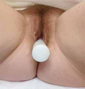
FIGURE 1 Inserting the dilator
Tell the patient to lie on the bed with her knees bent and to insert the lubricated dilator into her vagina as far as it will go without causing pain.Be sure to inform her to use a water-based lubricant—not lotion, petroleum jelly, or any non-water-based lubricant.
Dilate daily. The dilator should be used daily and left in place for 5 to 15 minutes. She may experience a small amount of spotting initially, but spotting should abate within 2 weeks of initiating dilator use. Each dilator size should be used for 3 to 4 weeks. When your patient is changing sizes, she should transition to the larger size over several days by dilating for the first few minutes with the smaller dilator, then changing to the larger dilator for the remainder of the time. If she experiences pain or heavy bleeding, she should cease dilation and follow-up with you.
Proper cleaning. Instruct her to wash the dilator with antibacterial soap and water and to dry it thoroughly between uses.
Follow-up. When undergoing vaginal dilation therapy, your patient should be following up with you at regular intervals, usually once a month, to facilitate compliance with the program.
You can purchase dilators for your medical practice and resell them in your office. Patients also can be directed to purchase dilators directly from a manufacturer (such as Syracuse Medical Devices or AmeriMed Direct) or through a number of Internet sites, including Middlesexmd.com6 or Vaginismus.com.7 Both of these Web sites also offer educational materials, including videos and books and a private support forum or blog.

FIGURE 2 Vaginal dilators come in 8 different circumferences
Vaginal dilators range in size from Extra Small at 1/2 in (13 mm) to Large Plus at 1 1/2 in (39 mm). Each is 6 in (15 cm) long, has one rounded end, and is constructed of sterilizable, medical-grade plastic.
Printed with permission of Syracuse Medical Devices, Inc., Syracuse, New York.Most dilators used in the United States are 6 in (15 cm) long and are made from sterilizable, medical-grade, latex-free, rigid plastic with a smooth surface. Some dilators are made of softer material such as silicone, and others have a vibrating inner wand.6 They are available to purchase as single dilators or in sets of 5 to 8 graduated sizes (FIGURE 2). Some sets come with a storage bag and universal handles that lock-on for insertion. Graduated circumference sizes are fairly universal in the United States (TABLE).
Average dilator sizes and circumferences*
| Size | Circumference |
|---|---|
| Extra small | 1/2 in; 13 mm |
| Extra small plus | 11/16 in; 18 mm |
| Small | 7/8 in; 22 mm |
| Small plus | 1 in; 25 mm |
| Medium | 1 1/8 in; 29 mm |
| Medium plus | 1 1/4 in; 32 mm |
| Large | 1 3/8 in; 35 mm |
| Large plus | 1 5/8 in; 38 mm |
| *Based on Syracuse Medical Devices, Inc. product information. | |
Restoring her sexual health: Our goal
Sexual health is a vitally important quality-of-life issue; restoring that health should be our priority. We need to educate our patients on nonprescription methods to promote their vaginal and sexual health, as vaginal dilation therapy can result in the reduction or elimination of dyspareunia.
We want to hear from you! Tell us what you think.
Your age-based guide to comprehensive well-woman care
Robert L. Barbieri, MD (October 2012)
Sexual dysfunction
Barbara S. Levy, MD (Update, September 2012)
New study: ObGyns aren’t fully addressing their patients’ sexual function
(Web News, April 2012)
How to prepare your patient for the many nuances of postpartum sexuality
Roya Rezaee, MD, and Sheryl Kingsberg, PhD (January 2012)

Susan Kellogg Spadt, PhD, CRNP
Dr. Kellogg Spadt is Director of Sexual Medicine.

Jennifer Iorio, MSN, CRNP
Ms. Iorio is a Nurse Practitioner, Female Sexual Medicine.

Jennifer Yonaitis Fariello, MSN, CRNP
Ms. Fariello is a Nurse Practitioner, Male and Female Sexual Medicine.

Kristene E. Whitmore, MD
Dr. Whitmore is Medical Director and Chief of Surgery.
All are colleagues at the Pelvic & Sexual Health Institute, Philadelphia, Pennsylvania.
The authors report no financial relationships relevant to this article.
Susan Kellogg Spadt, PhD, CRNP
Dr. Kellogg Spadt is Director of Sexual Medicine.
Jennifer Iorio, MSN, CRNP
Ms. Iorio is a Nurse Practitioner, Female Sexual Medicine.
Jennifer Yonaitis Fariello, MSN, CRNP
Ms. Fariello is a Nurse Practitioner, Male and Female Sexual Medicine.
Kristene E. Whitmore, MD
Dr. Whitmore is Medical Director and Chief of Surgery.
All are colleagues at the Pelvic & Sexual Health Institute, Philadelphia, Pennsylvania.
The authors report no financial relationships relevant to this article.
Vaginal dilators are used to restore vaginal capacity, to expand the vagina in width and depth, to provide elasticity to the tissues, and to allow for comfortable sexual activity. Vaginal dilators are smooth plastic, rubber, or glass cylinder-shaped objects that come in a variety of graduated sizes and weights.
Several medical conditions may warrant the use of vaginal dilation, including superficial dyspareunia, high-tone pelvic floor dysfunction, vaginismus, provoked vestibulodynia, vaginal atrophy, vulvar dermatoses, vaginal agenesis, and postradiation adhesions. Dilation also can be used as deconditioning therapy for psychogenic dyspareunia.1-4 In addition, Masters and Johnson advocated the use of dilators for patients with female sexual dysfunction in order to interrupt the cycle of pain–fear–muscle spasm–more pain, and to build confidence “in the privacy of the marital bedroom.”5
Vaginal dilators often are sufficient to restore function, with dilator therapy considered successful if a woman is able to resume comfortable sexual intercourse or self-stimulation, as desired.1,6 Vaginal dilation also can be used as an adjunct to pelvic floor muscle physical therapy, psychotherapy, sex therapy, minimally absorbed local vaginal estrogen therapy, intravaginal muscle relaxants, lubricants, moisturizers, and vibrators.
Each patient in these case studies achieved success resuming sexual activity after several months of dilator therapy used in combination with other medical interventions.
CASE 1: Chronic vulvovaginal infection and pain
A 26-year-old G0P0 woman presented with a 2-year history of prohibitive penetrative dyspareunia. She had a history of chronic vulvovaginal candidiasis, treated by another clinician with multiple courses of intravaginal antifungal cream.
After extensive evaluation for sexual pain, a diagnosis of pelvic floor muscle spasm, sexual aversion, fear secondary to pain, and contact irritant dermatitis was reached. After vaginal fungal cultures indicated negative results, a size small dilator was introduced in the office using a hypoallergenic intravaginal moisturizer. After daily use of the vaginal dilator for 4 months, with progressed introduction of graduated sizes (small, medium, medium+, large), she was able to accommodate intravaginal intercourse with her partner.
CASE 2: Interstitial cystitis and fear of pain
A 58-year-old G3P3 postmenopausal woman presented with interstitial cystitis (IC), pelvic floor muscle hypertonus, vulvovaginal atrophy, and provoked vestibulodynia. Although her IC symptoms were well-controlled, she was fearful about reestablishing physical intimacy with her partner after 7 years of abstinence.
A program of intravaginal estrogen (Vagifem) 2 to 3 times per week, introital cutaneous lysate (Neogyn) vulvar soothing cream twice per day, and compounded muscle-relaxing intravaginal diazepam suppositories 2 to 3 times per week was initiated. After 2 months of treatment, she was taught in the office to use a size extra small vaginal dilator. She was delighted that use did not result in pain. Two months later, she was able to use a size small dilator, and 4 months later, a size medium dilator. At this point, the patient is confident that she can have sexual intercourse.
CASE 3: Lichen sclerosus
A 50-year-old G0P0 premenopausal woman had a history of IC and biopsy-proven lichen sclerosus. The white plaques surrounding her introitus had become so severe in the past year that she was no longer able to tolerate penile penetration without tearing. Nightly use of topical clobetasol cream and introital estrogen cream (Estrace) was recommended. After 30 days, the patient began twice-a-week maintenance with the creams and also began to use vaginal dilators. After success inserting a size extra large dilator following 5 months of dilator use, she was able to resume intercourse without tearing.
CASE 4: Vestibulodynia and vaginismus
A 25-year-old G0P0 woman underwent vestibulectomy for primary provoked vestibulodynia followed by pelvic floor muscle physical therapy for primary vaginismus. Her marriage of 6 years was unconsummated. Two weeks postoperatively, she began using a size small dilator daily and progressed to a size medium plus dilator after 6 weeks. She managed her chronic constipation and pelvic floor muscle hypertonus with daily fiber supplements, stool softeners, and self-transvaginal massage of the pelvic floor muscles. Seven weeks after surgery, she accomplished intercourse with her husband for the first time.
How to teach your patient to use vaginal dilation successfully
Before ordering vaginal dilation for your patient, 1) assess the levator ani muscle group for hypertonus or spasm and 2) choose the size dilator to start therapy that does not cause pain with insertion but enters with some resistance.
When beginning to teach your patient to use a vaginal dilator in the office, a mirror demonstration may be helpful. Be sure to instruct your patient regarding the following elements to help her achieve success with dilation therapy.
Relax and allow for privacy. About 10 to 15 minutes of privacy before vaginal dilation can help with the success of each individual therapy session. Relaxation can be facilitated with activities such as deep breathing, soaking in a warm bath, or using prescribed muscle relaxants 30 to 60 minutes prior to dilation.
Use proper positioning. Instruct the patient to lie on the bed with her knees bent and placed apart. Advise her to place the lubricated dilator in the vagina as far as it can go without causing any pain (FIGURE 1). It may be helpful for her to bear down when first inserting the dilator. An in-and-out motion is not necessary.
FIGURE 1 Inserting the dilator
Tell the patient to lie on the bed with her knees bent and to insert the lubricated dilator into her vagina as far as it will go without causing pain.Be sure to inform her to use a water-based lubricant—not lotion, petroleum jelly, or any non-water-based lubricant.
Dilate daily. The dilator should be used daily and left in place for 5 to 15 minutes. She may experience a small amount of spotting initially, but spotting should abate within 2 weeks of initiating dilator use. Each dilator size should be used for 3 to 4 weeks. When your patient is changing sizes, she should transition to the larger size over several days by dilating for the first few minutes with the smaller dilator, then changing to the larger dilator for the remainder of the time. If she experiences pain or heavy bleeding, she should cease dilation and follow-up with you.
Proper cleaning. Instruct her to wash the dilator with antibacterial soap and water and to dry it thoroughly between uses.
Follow-up. When undergoing vaginal dilation therapy, your patient should be following up with you at regular intervals, usually once a month, to facilitate compliance with the program.
You can purchase dilators for your medical practice and resell them in your office. Patients also can be directed to purchase dilators directly from a manufacturer (such as Syracuse Medical Devices or AmeriMed Direct) or through a number of Internet sites, including Middlesexmd.com6 or Vaginismus.com.7 Both of these Web sites also offer educational materials, including videos and books and a private support forum or blog.
FIGURE 2 Vaginal dilators come in 8 different circumferences
Vaginal dilators range in size from Extra Small at 1/2 in (13 mm) to Large Plus at 1 1/2 in (39 mm). Each is 6 in (15 cm) long, has one rounded end, and is constructed of sterilizable, medical-grade plastic.
Printed with permission of Syracuse Medical Devices, Inc., Syracuse, New York.Most dilators used in the United States are 6 in (15 cm) long and are made from sterilizable, medical-grade, latex-free, rigid plastic with a smooth surface. Some dilators are made of softer material such as silicone, and others have a vibrating inner wand.6 They are available to purchase as single dilators or in sets of 5 to 8 graduated sizes (FIGURE 2). Some sets come with a storage bag and universal handles that lock-on for insertion. Graduated circumference sizes are fairly universal in the United States (TABLE).
Average dilator sizes and circumferences*
| Size | Circumference |
|---|---|
| Extra small | 1/2 in; 13 mm |
| Extra small plus | 11/16 in; 18 mm |
| Small | 7/8 in; 22 mm |
| Small plus | 1 in; 25 mm |
| Medium | 1 1/8 in; 29 mm |
| Medium plus | 1 1/4 in; 32 mm |
| Large | 1 3/8 in; 35 mm |
| Large plus | 1 5/8 in; 38 mm |
| *Based on Syracuse Medical Devices, Inc. product information. | |
Restoring her sexual health: Our goal
Sexual health is a vitally important quality-of-life issue; restoring that health should be our priority. We need to educate our patients on nonprescription methods to promote their vaginal and sexual health, as vaginal dilation therapy can result in the reduction or elimination of dyspareunia.
We want to hear from you! Tell us what you think.
Your age-based guide to comprehensive well-woman care
Robert L. Barbieri, MD (October 2012)
Sexual dysfunction
Barbara S. Levy, MD (Update, September 2012)
New study: ObGyns aren’t fully addressing their patients’ sexual function
(Web News, April 2012)
How to prepare your patient for the many nuances of postpartum sexuality
Roya Rezaee, MD, and Sheryl Kingsberg, PhD (January 2012)
Odanacatib Adds Bone in Alendronate-Pretreated Osteoporosis
MINNEAPOLIS – Odanacatib added bone density in postmenopausal women with osteoporosis previously treated with alendronate, according to results from a study presented at the annual meeting of the American Society for Bone and Mineral Research.
In a phase II trial involving 243 patients, the investigational oral cathepsin K inhibitor began to distinguish itself from placebo at 12 months. At 2 years, the change from baseline in femoral neck bone mineral density (BMD) was significantly increased by 1.73%, compared with a loss of 0.94% for placebo (P less than .001).

BMD changes in the treatment group at 2 years were also significantly different from placebo at the trochanter (1.83% vs. –1.35%) and for the total hip (0.83% vs. –1.87%, both P less than .001).
All of the women studied had received alendronate (Fosamax) for at least 3 years. Patients could have been off the bisphosphonate for up to 3 months prior to enrollment, but many switched directly over to odanacatib, said study coauthor Dr. Albert Leung, executive director of clinical research at Merck Research Laboratories.
"This drug has the potential to give additional benefits when [patients] have been treated with alendronate for a number of years and the treatment effect has reached a plateau and they may need a different treatment," he said in an interview.
In July 2012, a pivotal 16,731-patient, phase III trial of odanacatib was stopped early after an interim analysis showed a "favorable benefit-risk profile" for fracture risk reduction in postmenopausal women with previously untreated osteoporosis.
"It could be quite a population that may benefit from this drug," Dr. Leung remarked.
The study’s data monitoring committee, however, flagged safety concerns in "certain selected areas" for further follow-up. Those safety risks have not been identified as the trial is still closing, but will be monitored along with efficacy in a double-blind, placebo-controlled, extension trial going out to 5 years of treatment, he said.
Merck, which plans to submit regulatory applications for odanacatib in the United States and Europe in the first half of 2013, has high hopes for odanacatib despite competition from generic drugs because of its novel mechanism of action.
Odanacatib inhibits cathepsin K, the primary protease in osteoclasts that breaks down bone collagen during bone resorption. Unlike traditional antiresorptive drugs like bisphosphonates, however, odanacatib does not interfere with the function of the entire osteoclast or reduce the number of osteoclasts. This characteristic is important, as osteoclasts secrete signaling factors to stimulate osteoblasts, the cells responsible for bone formation. As a result, there is greater bone formation with odanacatib, Dr. Leung explained.
The phase II trial enrolled women at least 60 years of age (mean 71 years) with a BMD T score of –2.5 to more than –3.5 at any hip site without a prior fragility fracture or those with a history of fragility fracture (except hip fracture), and a BMD T score of –1.5 and more than –3.5 at any hip site. The women were randomly assigned to odanacatib 50 mg once weekly or placebo for 24 months, as well as 5,600 IU of vitamin D3 per week and calcium at dosages up to 1,200 mg/day. The study was not powered to assess fractures.
At 24 months, the change in BMD at the lumbar spine was significant at 2.28% for odanacatib vs. a loss of 0.30% with placebo (P less than .001).
BMD change was not significant at the distal forearm, with losses of 0.92% vs. 1.14%, respectively.
As expected, urinary collagen type I cross-linked N-telopeptide, a biomarker of bone resorption, increased with placebo, compared with a significant 47% decrease with odanacatib.
The bone formation marker, serum type I procollagen, rose inexplicably with placebo, but this increase was surpassed by a significant gain of 31.2% with odanacatib.
Most unexpected, however, was an increase in the resorption marker collagen type I cross-linked C-telopeptide (sCTx) with once-weekly odanacatib. Dr. Leung said the finding appears to correlate with the bone density changes because sCTx levels remained relatively stable during the first 12 months of treatment before rising in the second year of the study.
Finally, adverse events were similar in both groups. The most common adverse events in the odanacatib and placebo arms were urinary tract infection (11.5% vs. 16.5%, respectively), back pain (11.5% vs. 9.9%), arthralgia (9% vs. 9.9%), and fractures (4.9% vs. 13.2%). Treatment discontinuation rates due to adverse events were 9% vs. 3.3%, he said.
Dr. Leung and several of his coauthors are employees of Merck, the trial sponsor.
MINNEAPOLIS – Odanacatib added bone density in postmenopausal women with osteoporosis previously treated with alendronate, according to results from a study presented at the annual meeting of the American Society for Bone and Mineral Research.
In a phase II trial involving 243 patients, the investigational oral cathepsin K inhibitor began to distinguish itself from placebo at 12 months. At 2 years, the change from baseline in femoral neck bone mineral density (BMD) was significantly increased by 1.73%, compared with a loss of 0.94% for placebo (P less than .001).
BMD changes in the treatment group at 2 years were also significantly different from placebo at the trochanter (1.83% vs. –1.35%) and for the total hip (0.83% vs. –1.87%, both P less than .001).
All of the women studied had received alendronate (Fosamax) for at least 3 years. Patients could have been off the bisphosphonate for up to 3 months prior to enrollment, but many switched directly over to odanacatib, said study coauthor Dr. Albert Leung, executive director of clinical research at Merck Research Laboratories.
"This drug has the potential to give additional benefits when [patients] have been treated with alendronate for a number of years and the treatment effect has reached a plateau and they may need a different treatment," he said in an interview.
In July 2012, a pivotal 16,731-patient, phase III trial of odanacatib was stopped early after an interim analysis showed a "favorable benefit-risk profile" for fracture risk reduction in postmenopausal women with previously untreated osteoporosis.
"It could be quite a population that may benefit from this drug," Dr. Leung remarked.
The study’s data monitoring committee, however, flagged safety concerns in "certain selected areas" for further follow-up. Those safety risks have not been identified as the trial is still closing, but will be monitored along with efficacy in a double-blind, placebo-controlled, extension trial going out to 5 years of treatment, he said.
Merck, which plans to submit regulatory applications for odanacatib in the United States and Europe in the first half of 2013, has high hopes for odanacatib despite competition from generic drugs because of its novel mechanism of action.
Odanacatib inhibits cathepsin K, the primary protease in osteoclasts that breaks down bone collagen during bone resorption. Unlike traditional antiresorptive drugs like bisphosphonates, however, odanacatib does not interfere with the function of the entire osteoclast or reduce the number of osteoclasts. This characteristic is important, as osteoclasts secrete signaling factors to stimulate osteoblasts, the cells responsible for bone formation. As a result, there is greater bone formation with odanacatib, Dr. Leung explained.
The phase II trial enrolled women at least 60 years of age (mean 71 years) with a BMD T score of –2.5 to more than –3.5 at any hip site without a prior fragility fracture or those with a history of fragility fracture (except hip fracture), and a BMD T score of –1.5 and more than –3.5 at any hip site. The women were randomly assigned to odanacatib 50 mg once weekly or placebo for 24 months, as well as 5,600 IU of vitamin D3 per week and calcium at dosages up to 1,200 mg/day. The study was not powered to assess fractures.
At 24 months, the change in BMD at the lumbar spine was significant at 2.28% for odanacatib vs. a loss of 0.30% with placebo (P less than .001).
BMD change was not significant at the distal forearm, with losses of 0.92% vs. 1.14%, respectively.
As expected, urinary collagen type I cross-linked N-telopeptide, a biomarker of bone resorption, increased with placebo, compared with a significant 47% decrease with odanacatib.
The bone formation marker, serum type I procollagen, rose inexplicably with placebo, but this increase was surpassed by a significant gain of 31.2% with odanacatib.
Most unexpected, however, was an increase in the resorption marker collagen type I cross-linked C-telopeptide (sCTx) with once-weekly odanacatib. Dr. Leung said the finding appears to correlate with the bone density changes because sCTx levels remained relatively stable during the first 12 months of treatment before rising in the second year of the study.
Finally, adverse events were similar in both groups. The most common adverse events in the odanacatib and placebo arms were urinary tract infection (11.5% vs. 16.5%, respectively), back pain (11.5% vs. 9.9%), arthralgia (9% vs. 9.9%), and fractures (4.9% vs. 13.2%). Treatment discontinuation rates due to adverse events were 9% vs. 3.3%, he said.
Dr. Leung and several of his coauthors are employees of Merck, the trial sponsor.
AT THE ANNUAL MEETING OF THE AMERICAN SOCIETY FOR BONE AND MINERAL RESEARCH
Major Finding: The change from baseline in femoral neck bone mineral density at 2 years was +1.73% with odanacatib vs. –0.94% for placebo (P less than .001).
Data Source: This study was a phase II trial in 243 women with postmenopausal osteoporosis previously treated with alendronate.
Disclosures: Dr. Leung and several of his coauthors are employees of Merck, the trial sponsor.

Limit HT Use to Menopause Symptoms, Task Force Reaffirms
Combined estrogen and progestin should not be used for the prevention of osteoporosis or other chronic conditions in postmenopausal women, according to recommendations issued by the U.S. Preventive Services Task Force.
Hormone therapy currently has Food and Drug Administration approval for use in the prevention of osteoporosis in postmenopausal women.
The task force, an independent body of volunteer experts that advises the Department of Health and Human Services, issued the recommendations Oct. 22 as an update of its 2005 statement on hormone therapy for prevention of disease in postmenopausal women.

Using the most recent scientific evidence available, including long-term follow-up data from the Women's Health Initiative (WHI) studies of hormone therapy use in postmenopausal women, the task force reached the same conclusions as it had in 2005, advising against combined estrogen and progestin for prevention of chronic conditions, and also against the use of estrogen alone for the prevention of chronic conditions in postmenopausal women who have had a hysterectomy.
The task force emphasized that hormone therapy was still indicated for the management of menopausal symptoms, such as hot flashes or vaginal dryness. It additionally made clear that its recommendation against hormone therapy for disease prevention does not apply to women younger than 50 years of age who have undergone surgical menopause.
Prior to the WHI studies, a series of government-funded trials that began in the 1990s, with follow-up ending in 2010, hormones had been widely used for the prevention of bone disease in postmenopausal women. Both estrogen and combined estrogen and progestin are known to reduce fracture risk.
However, both forms of hormone therapy were shown during the WHI studies to also increase the risk of serious adverse events, to the point where the trials were stopped early. In one randomized, placebo-controlled trial, estrogen alone was associated with a significantly higher risk of stroke, deep vein thrombosis, and gallbladder disease, while combined therapy was associated with an increased risk of stroke, invasive breast cancer, dementia, gallbladder disease, deep vein thrombosis, and pulmonary embolism.
Reproductive endocrinologist Jan L. Shifren of the department of obstetrics and gynecology and reproductive biology at Harvard Medical School and director of the menopause program at Massachusetts General Hospital, both in Boston, said in an interview that the task force’s updated position largely reflected the current consensus of the ob.gyn. community, "which is that HT should not be used to prevent the diseases of aging."
The task force was "very careful to point out that they are not saying HT should not be used for the treatment of vasomotor symptoms or vaginal atrophy. It’s not that hormones aren’t indicated; they’re just not indicated for prevention. They remain an appropriate treatment for otherwise healthy, very symptomatic women at the menopause transition," said Dr. Shifren, who is not a task force member.
FDA-approved indications for hormone therapy in postmenopausal women include treatment of menopausal symptoms and prevention of osteoporosis. A black box warning indicates that estrogen with or without progestin should be prescribed at the lowest effective dose and for the shortest time possible. The task force’s findings were based on the dosages and formulations used in the WHI trials: oral conjugated equine estrogen (0.625 mg/day plus medroxyprogesterone acetate, 2.5 mg/day) or estrogen 0.625 mg/day alone.
Dr. Shifren said that there are some practitioners "who believe that hormone therapy could still be appropriate for the prevention of osteoporosis in people who absolutely cannot tolerate any other therapy. But what I would argue is that it is incredibly rare that there is a patient who can’t tolerate one of the very many other FDA-approved treatments for the prevention of osteoporosis."
The task force members declared no relevant financial conflicts of interest.
Combined estrogen and progestin should not be used for the prevention of osteoporosis or other chronic conditions in postmenopausal women, according to recommendations issued by the U.S. Preventive Services Task Force.
Hormone therapy currently has Food and Drug Administration approval for use in the prevention of osteoporosis in postmenopausal women.
The task force, an independent body of volunteer experts that advises the Department of Health and Human Services, issued the recommendations Oct. 22 as an update of its 2005 statement on hormone therapy for prevention of disease in postmenopausal women.
Using the most recent scientific evidence available, including long-term follow-up data from the Women's Health Initiative (WHI) studies of hormone therapy use in postmenopausal women, the task force reached the same conclusions as it had in 2005, advising against combined estrogen and progestin for prevention of chronic conditions, and also against the use of estrogen alone for the prevention of chronic conditions in postmenopausal women who have had a hysterectomy.
The task force emphasized that hormone therapy was still indicated for the management of menopausal symptoms, such as hot flashes or vaginal dryness. It additionally made clear that its recommendation against hormone therapy for disease prevention does not apply to women younger than 50 years of age who have undergone surgical menopause.
Prior to the WHI studies, a series of government-funded trials that began in the 1990s, with follow-up ending in 2010, hormones had been widely used for the prevention of bone disease in postmenopausal women. Both estrogen and combined estrogen and progestin are known to reduce fracture risk.
However, both forms of hormone therapy were shown during the WHI studies to also increase the risk of serious adverse events, to the point where the trials were stopped early. In one randomized, placebo-controlled trial, estrogen alone was associated with a significantly higher risk of stroke, deep vein thrombosis, and gallbladder disease, while combined therapy was associated with an increased risk of stroke, invasive breast cancer, dementia, gallbladder disease, deep vein thrombosis, and pulmonary embolism.
Reproductive endocrinologist Jan L. Shifren of the department of obstetrics and gynecology and reproductive biology at Harvard Medical School and director of the menopause program at Massachusetts General Hospital, both in Boston, said in an interview that the task force’s updated position largely reflected the current consensus of the ob.gyn. community, "which is that HT should not be used to prevent the diseases of aging."
The task force was "very careful to point out that they are not saying HT should not be used for the treatment of vasomotor symptoms or vaginal atrophy. It’s not that hormones aren’t indicated; they’re just not indicated for prevention. They remain an appropriate treatment for otherwise healthy, very symptomatic women at the menopause transition," said Dr. Shifren, who is not a task force member.
FDA-approved indications for hormone therapy in postmenopausal women include treatment of menopausal symptoms and prevention of osteoporosis. A black box warning indicates that estrogen with or without progestin should be prescribed at the lowest effective dose and for the shortest time possible. The task force’s findings were based on the dosages and formulations used in the WHI trials: oral conjugated equine estrogen (0.625 mg/day plus medroxyprogesterone acetate, 2.5 mg/day) or estrogen 0.625 mg/day alone.
Dr. Shifren said that there are some practitioners "who believe that hormone therapy could still be appropriate for the prevention of osteoporosis in people who absolutely cannot tolerate any other therapy. But what I would argue is that it is incredibly rare that there is a patient who can’t tolerate one of the very many other FDA-approved treatments for the prevention of osteoporosis."
The task force members declared no relevant financial conflicts of interest.

Denosumab/Teriparatide Combo Bests Single-Agent Bone Therapy
MINNEAPOLIS – Combining the antiresorptive denosumab with the anabolic agent teriparatide increased bone mineral density more than either drug alone in postmenopausal women at high fracture risk in the ongoing DATA study.
At 12 months, the combination of denosumab (Prolia) and teriparatide (Forteo) significantly increased bone mineral density (BMD) by 8.9% at the spine, 4.5% at the femoral neck, and 4.9% at the total hip.

The increases in BMD observed in the combination group are larger than those seen in prior combination anabolic and antiresorptive trials, Dr. Benjamin Z. Leder reported at the annual meeting of the American Society for Bone and Mineral Research.
The DATA (Denosumab, Teriparatide or Both for the Treatment of Postmenopausal Osteoporosis) trial is the first to study denosumab in combination with an anabolic agent. Prior trials combining teriparatide and bisphosphonates have shown inconsistent effects on BMD or, in some cases, a blunting effect of the anabolic agent.
The mechanisms underlying the additive effects of denosumab and teriparatide are unclear, but they may be related to the ability of denosumab to fully block teriparatide’s pro-resorptive effects while still allowing for continued modeling-based bone formation and, perhaps, an expansion of the anabolic window, said Dr. Leder, an endocrinologist with Massachusetts General Hospital in Boston.
"If these results persist in the second year of therapy and are confirmed in larger studies, the combination of these two agents may eventually prove to be a beneficial treatment in patients who are at particularly high risk of fracture," he said.
The trial randomized 100 women aged 45 years or older who were at least 3 years post menopause to daily teriparatide 20 mcg subcutaneous or denosumab 60 mg subcutaneous every 6 months or both. All patients received calcium 1,200 mg and vitamin D 400 IU.
Enrollment criteria were a BMD T-score of –2.5 or less at any anatomic site or a T-score of –2 or less with one risk factor (fracture or parental hip fracture after age 50, prior hyperthyroidism, inability to rise from a chair with arms elevated, or current smoker) or a T-score of –1 or less with a history of fragility fracture.
Patients were excluded if they had received oral bisphosphonates in the past 6 months; glucocorticoids for more than 14 days in the past 6 months; and any prior use of teriparatide, strontium, or parenteral bisphosphonates.
Patients were stratified by age and spine BMD. The 94 evaluable patients had an average age of 66 years.
At 12 months, the average increase in total hip BMD was 0.7% with teriparatide, 2.5% with denosumab, and 4.9% with combination therapy. Femoral neck BMD increased 0.8%, 2.1%, and 4.5% and spine BMD increased 6.2%, 5.5%, and 8.9%, respectively.
At the distal one-third of the radius, there was a decrease in BMD of 1.8% with teriparatide, an increase of 1.7% with denosumab, and a gain of 2.5% with the combination, Dr. Leder said. The difference in BMD was significant between the combination and teriparatide groups (P less than .001) but not between the combination and denosumab groups.
Changes in bone density were not significantly different between bisphosphonate-naive patients and those with prior bisphosphonate exposure.
Bone formation biomarker analysis showed significant suppression of osteocalcium with denosumab monotherapy at 3 months that continued through the 12-month study, while there was no change at 3 months and a more modest suppression thereafter in the combination group, he said.
Denosumab monotherapy significantly inhibited procollagen type I N-terminal propeptide at 3 and 6 months, but both groups were similar at 12 months.
The data on bone turnover marker C-telopeptide of type I collagen were distinct, with suppression identical in the denosumab alone and combination groups, Dr. Leder observed.
During a discussion of the study, Dr. Leder said bone biopsies were not available but that data at the distal radius and tibia that have not yet been analyzed "may provide some additional idea of what is going on, specifically in the trabecular and cortical compartments."
Session comoderator Dr. Aliya Khan, director of the calcium disorders clinic at St. Joseph’s Healthcare, McMaster University in Hamilton, Ont., said in an interview that the results shouldn’t be universally applied, but "if someone has a fracture, we can certainly consider this approach."
She went on to say that "combination therapy may be a way to improve bone strength, and it may actually enable us to avoid conditions such as atypical femoral fractures, which appear to be associated with oversuppression of bone remodeling."
Eli Lilly and Amgen sponsored the trial. Dr. Leder reported consulting for Amgen and Merck. Dr. Khan reported no disclosures.
MINNEAPOLIS – Combining the antiresorptive denosumab with the anabolic agent teriparatide increased bone mineral density more than either drug alone in postmenopausal women at high fracture risk in the ongoing DATA study.
At 12 months, the combination of denosumab (Prolia) and teriparatide (Forteo) significantly increased bone mineral density (BMD) by 8.9% at the spine, 4.5% at the femoral neck, and 4.9% at the total hip.
The increases in BMD observed in the combination group are larger than those seen in prior combination anabolic and antiresorptive trials, Dr. Benjamin Z. Leder reported at the annual meeting of the American Society for Bone and Mineral Research.
The DATA (Denosumab, Teriparatide or Both for the Treatment of Postmenopausal Osteoporosis) trial is the first to study denosumab in combination with an anabolic agent. Prior trials combining teriparatide and bisphosphonates have shown inconsistent effects on BMD or, in some cases, a blunting effect of the anabolic agent.
The mechanisms underlying the additive effects of denosumab and teriparatide are unclear, but they may be related to the ability of denosumab to fully block teriparatide’s pro-resorptive effects while still allowing for continued modeling-based bone formation and, perhaps, an expansion of the anabolic window, said Dr. Leder, an endocrinologist with Massachusetts General Hospital in Boston.
"If these results persist in the second year of therapy and are confirmed in larger studies, the combination of these two agents may eventually prove to be a beneficial treatment in patients who are at particularly high risk of fracture," he said.
The trial randomized 100 women aged 45 years or older who were at least 3 years post menopause to daily teriparatide 20 mcg subcutaneous or denosumab 60 mg subcutaneous every 6 months or both. All patients received calcium 1,200 mg and vitamin D 400 IU.
Enrollment criteria were a BMD T-score of –2.5 or less at any anatomic site or a T-score of –2 or less with one risk factor (fracture or parental hip fracture after age 50, prior hyperthyroidism, inability to rise from a chair with arms elevated, or current smoker) or a T-score of –1 or less with a history of fragility fracture.
Patients were excluded if they had received oral bisphosphonates in the past 6 months; glucocorticoids for more than 14 days in the past 6 months; and any prior use of teriparatide, strontium, or parenteral bisphosphonates.
Patients were stratified by age and spine BMD. The 94 evaluable patients had an average age of 66 years.
At 12 months, the average increase in total hip BMD was 0.7% with teriparatide, 2.5% with denosumab, and 4.9% with combination therapy. Femoral neck BMD increased 0.8%, 2.1%, and 4.5% and spine BMD increased 6.2%, 5.5%, and 8.9%, respectively.
At the distal one-third of the radius, there was a decrease in BMD of 1.8% with teriparatide, an increase of 1.7% with denosumab, and a gain of 2.5% with the combination, Dr. Leder said. The difference in BMD was significant between the combination and teriparatide groups (P less than .001) but not between the combination and denosumab groups.
Changes in bone density were not significantly different between bisphosphonate-naive patients and those with prior bisphosphonate exposure.
Bone formation biomarker analysis showed significant suppression of osteocalcium with denosumab monotherapy at 3 months that continued through the 12-month study, while there was no change at 3 months and a more modest suppression thereafter in the combination group, he said.
Denosumab monotherapy significantly inhibited procollagen type I N-terminal propeptide at 3 and 6 months, but both groups were similar at 12 months.
The data on bone turnover marker C-telopeptide of type I collagen were distinct, with suppression identical in the denosumab alone and combination groups, Dr. Leder observed.
During a discussion of the study, Dr. Leder said bone biopsies were not available but that data at the distal radius and tibia that have not yet been analyzed "may provide some additional idea of what is going on, specifically in the trabecular and cortical compartments."
Session comoderator Dr. Aliya Khan, director of the calcium disorders clinic at St. Joseph’s Healthcare, McMaster University in Hamilton, Ont., said in an interview that the results shouldn’t be universally applied, but "if someone has a fracture, we can certainly consider this approach."
She went on to say that "combination therapy may be a way to improve bone strength, and it may actually enable us to avoid conditions such as atypical femoral fractures, which appear to be associated with oversuppression of bone remodeling."
Eli Lilly and Amgen sponsored the trial. Dr. Leder reported consulting for Amgen and Merck. Dr. Khan reported no disclosures.
AT THE ANNUAL MEETING OF THE AMERICAN SOCIETY FOR BONE AND MINERAL RESEARCH
Major Finding: The combination of denosumab and teriparatide significantly increased bone mineral density by 8.9% at the spine, 4.5% at the femoral neck, and 4.9% at the total hip at the end of 12 months of therapy.
Data Source: These findings come from an open-label, randomized controlled trial in 94 postmenopausal women at high fracture risk.
Disclosures: Amgen and Eli Lilly sponsored the study. Dr. Leder reported consulting for Amgen and Merck. Dr. Khan reported no disclosures.

Low-Fat Diet a Dud for Women's Heart Disease
ESTES PARK, COLO. – Perhaps the least-known finding of the landmark Women’s Health Initiative was the complete failure of a structured low-fat diet intervention to lower the risks of coronary heart disease, stroke, or colon cancer.
"This has gotten very little press. But the results made me very happy because it gave me one less thing to worry about, which is eating a low-fat diet. It doesn’t seem to have the same magnitude of effect in women as it does in men," Dr. Nanette Santoro said at a conference on internal medicine sponsored by the University of Colorado.

The Women’s Health Initiative Randomized Controlled Dietary Modification Trial involved 48,835 postmenopausal women aged 50-79 at 40 U.S. centers who were randomized 40/60 to a low-fat diet intervention or a control group.
During a mean follow-up of 8.1 years, the diet intervention and control groups didn’t show any significant differences in rates of coronary heart disease (hazard ratio, 0.97); stroke (1.02); or cardiovascular disease (0.98) (JAMA 2006;295:655-66).
Similarly, the event-rate curves for cardiovascular outcomes as well as for colon cancer in the intervention and control arms were virtually identical the entire time, with no hint of either early or late benefit for the low-fat diet (JAMA 2006;295:643-54).
There was a nonsignificant trend for less invasive breast cancer in the low-fat diet group, where the annualized incidence rate was 0.42%, a 9% relative risk reduction compared with the 0.45% rate in controls (JAMA 2006;295:629-42).
"So if there’s any possible benefit to a low-fat diet, there might be some for breast cancer," commented Dr. Santoro, professor and chair of the department of ob.gyn. at the university.
The diet intervention entailed an intensive behavioral modification program with 18 group sessions during year 1 and quarterly maintenance sessions thereafter, with supplemental individualized contact. The goal was to reduce dietary fat intake by boosting consumption of fruits and vegetables to at least five servings daily, along with at least six servings of grains daily. Weight loss goals weren’t part of the study, which was designed in the 1990s before the obesity epidemic was apparent.
The intervention was effective in terms of accomplishing lasting dietary change. At baseline, fat accounted for about 38% of total daily energy intake. After 1 year, this figure dropped to 24% in the diet intervention arm. At year 6, fat accounted for 29% of daily energy intake in the diet group compared with 37% in controls, a difference Dr. Santoro called "huge" in light of the enormous number of participants and the women’s diverse ethnicities and backgrounds.
The intervention group averaged 3.6 servings per day of fruits and vegetables at baseline and 4.9 by year 6, compared with 3.8 in controls. Efforts to increase consumption of grains were unsuccessful, however. The intervention group averaged 4.7 servings per day at baseline and 4.3 at year 6, compared with 3.8 in controls.
The Women’s Health Initiative Randomized Controlled Dietary Modification Trial was funded by the National Heart, Lung, and Blood Institute. Dr. Santoro reported that she has a research grant from Bayer.
ESTES PARK, COLO. – Perhaps the least-known finding of the landmark Women’s Health Initiative was the complete failure of a structured low-fat diet intervention to lower the risks of coronary heart disease, stroke, or colon cancer.
"This has gotten very little press. But the results made me very happy because it gave me one less thing to worry about, which is eating a low-fat diet. It doesn’t seem to have the same magnitude of effect in women as it does in men," Dr. Nanette Santoro said at a conference on internal medicine sponsored by the University of Colorado.
The Women’s Health Initiative Randomized Controlled Dietary Modification Trial involved 48,835 postmenopausal women aged 50-79 at 40 U.S. centers who were randomized 40/60 to a low-fat diet intervention or a control group.
During a mean follow-up of 8.1 years, the diet intervention and control groups didn’t show any significant differences in rates of coronary heart disease (hazard ratio, 0.97); stroke (1.02); or cardiovascular disease (0.98) (JAMA 2006;295:655-66).
Similarly, the event-rate curves for cardiovascular outcomes as well as for colon cancer in the intervention and control arms were virtually identical the entire time, with no hint of either early or late benefit for the low-fat diet (JAMA 2006;295:643-54).
There was a nonsignificant trend for less invasive breast cancer in the low-fat diet group, where the annualized incidence rate was 0.42%, a 9% relative risk reduction compared with the 0.45% rate in controls (JAMA 2006;295:629-42).
"So if there’s any possible benefit to a low-fat diet, there might be some for breast cancer," commented Dr. Santoro, professor and chair of the department of ob.gyn. at the university.
The diet intervention entailed an intensive behavioral modification program with 18 group sessions during year 1 and quarterly maintenance sessions thereafter, with supplemental individualized contact. The goal was to reduce dietary fat intake by boosting consumption of fruits and vegetables to at least five servings daily, along with at least six servings of grains daily. Weight loss goals weren’t part of the study, which was designed in the 1990s before the obesity epidemic was apparent.
The intervention was effective in terms of accomplishing lasting dietary change. At baseline, fat accounted for about 38% of total daily energy intake. After 1 year, this figure dropped to 24% in the diet intervention arm. At year 6, fat accounted for 29% of daily energy intake in the diet group compared with 37% in controls, a difference Dr. Santoro called "huge" in light of the enormous number of participants and the women’s diverse ethnicities and backgrounds.
The intervention group averaged 3.6 servings per day of fruits and vegetables at baseline and 4.9 by year 6, compared with 3.8 in controls. Efforts to increase consumption of grains were unsuccessful, however. The intervention group averaged 4.7 servings per day at baseline and 4.3 at year 6, compared with 3.8 in controls.
The Women’s Health Initiative Randomized Controlled Dietary Modification Trial was funded by the National Heart, Lung, and Blood Institute. Dr. Santoro reported that she has a research grant from Bayer.
EXPERT ANALYSIS FROM A CONFERENCE ON INTERNAL MEDICINE SPONSORED BY THE UNIVERSITY OF COLORADO

How to ask about, and manage, the undertreated problem of sexual dysfunction
Novel Estrogen Combination Shows Favorable Coagulation Profile
HOUSTON – The novel investigational tissue selective estrogen complex comprising bazedoxifene plus conjugated estrogens displayed an overall favorable coagulation profile in a pooled analysis of three randomized, placebo-controlled, double-blind, phase III clinical trials.
Moreover, the risk of venous thromboembolism or cerebrovascular events was low, at 0.2% or less during up to 2 years of treatment, a rate not significantly different from that of control patients, Dr. Sven O. Skouby of Herlev (Denmark) Hospital said at the annual meeting of the Endocrine Society.
He presented an analysis of serial coagulation parameters obtained in 1,978 postmenopausal women with intact uteri who were randomized to bazedoxifene at 20 mg per day plus either 0.45 or 0.625 mg of conjugated estrogens or to placebo in the first, fourth, and fifth SMART (Selective Estrogens, Menopause, and Response to Therapy) trials. SMART-1 assessed changes in coagulation variables at 12 and 24 months, while SMART-4 and -5 did so at 12 months.
Changes from baseline in protein C activity, d-dimer level, and prothrombin and partial thromboplastin times were similar in the two bazedoxifene/conjugated estrogens study arms and in placebo-treated controls.
The two active treatment arms showed modest but not clinically meaningful decreases in plasminogen activator-1, antithrombin III, and fibrinogen, compared with placebo. Similarly, plasminogen activity in the two active treatment groups increased by a mean of 0.1 L/L over the course of 24 months, a statistically greater increase than the 0.06 L/L seen in the control group, but not a clinically significant difference, Dr. Skouby observed.
SMART-1, -4, and -5 included 3,549 randomized patients with follow-up data on hard clinical thromboembolic events. Three patients on bazedoxifene/conjugated estrogens experienced deep vein thrombosis during 2 years of therapy, as did one on placebo. There were no cases of pulmonary embolism. Two of the treated patients and no controls had a cerebrovascular event.
In a separate presentation, Dr. Sebastian Mirkin reported on a pooled analysis of lipid changes in 4,409 postmenopausal women in SMART-1 through -5.
At 24 months, LDL cholesterol fell by a mean 7.4% from baseline in the bazedoxifene 20 mg/conjugated estrogens 0.45 mg group, and by 8.4% in those on bazedoxifene 20 mg/conjugated estrogens 0.625 mg, compared with a 2% increase in controls.
HDL levels rose by 7.3% in the lower-dose and 7.9% in the higher-dose groups, a significantly greater benefit than the 2.8% increase with placebo. Apolipoprotein B declined by a mean of 0.4% in the lower- and 0.5% in the higher-dose bazedoxifene/conjugated estrogens group, compared with a 4.3% increase in controls.
The one negative was that triglyceride levels increased by 20.3% in the lower- and 18.7% in the higher-dose groups, significantly greater than the 7.1% rise with placebo, reported Dr. Mirkin.
Bazedoxifene is not approved in the United States. Pfizer submitted a new drug application to the Food and Drug Administration in 2006, and in 2007 got an "approvable letter" that asked for more information. There is no public record of Pfizer’s response. Pfizer has announced that it will submit an application to the FDA for the bazedoxifene/conjugated estrogen combination this year.
The SMART trials were sponsored by Pfizer. Dr. Skouby serves as a consultant to the company. Dr. Mirkin is a Pfizer employee.
AT THE ANNUAL MEETING OF THE ENDOCRINE SOCIETY
Major Finding: An overall favorable coagulation profile was documented in women on an investigational tissue-selective estrogen complex for up to 2 years.
Data Source: This was a pooled analysis of several thousand postmenopausal women with intact uteri who were randomized to bazedoxifene 20 mg/conjugated estrogens 0.45 mg, bazedoxifene 20 mg/conjugated estrogens 0.625 mg, or placebo for up to 2 years in three phase III clinical trials.
Disclosures: The SMART trials were sponsored by Pfizer. Dr. Skouby serves as a consultant to the company. Dr. Mirkin is a Pfizer employee.
Denosumab Shows Favorable Results in FREEDOM Extension
HOUSTON – Denosumab continued to achieve reduced fractures rates, progressively rising bone mineral density, and sustained reduction in bone turnover biomarkers through 6 years of continuous use in the ongoing 7-year extension of the landmark FREEDOM trial.
FREEDOM (Fracture Reduction Evaluation of Denosumab in Osteoporosis Every 6 Months) was the pivotal phase III, double-blind, placebo-controlled, 3-year randomized trial that led to marketing approval of denosumab (Prolia) at 60 mg subcutaneously every 6 months for treatment of postmenopausal osteoporosis (N. Engl. J. Med. 2009;361:756-65).
The FREEDOM extension study is evaluating the long-term safety and efficacy of denosumab for an additional 7 years. Thus, participants who were in the active treatment arm of the original study will receive denosumab for a total of 10 years, whereas patients who crossed over to denosumab after 3 years on placebo will be on the novel RANKL (receptor-activated nuclear factor–kappaB ligand) inhibitor for 7 years.
At the conference, Dr. Henry G. Bone presented updated study results through 6 years. In all, 4,550 postmenopausal women from the original randomized trial were enrolled in the extension and have been followed for an additional 3 years.
In the original FREEDOM trial, 3 years of denosumab resulted in a 10.1% increase in BMD in the lumbar spine, compared with baseline. With an additional 3 years of open-label therapy, this figure has increased to 15.1%. The gain in the crossover group paralleled that seen with active therapy in the original trial; that is, after 6 years – the last 3 on open-label denosumab – those patients had a 9.4% increase over baseline in lumbar spine BMD, reported Dr. Bone, chief of the endocrinology department at St. John Hospital and Medical Center, Detroit, and director of the Michigan Bone and Mineral Clinic.
Total hip BMD rose from a 5.7% increase over baseline after 3 years of denosumab to a 7.5% increase after 6 years. The crossover group had a 4.8% increase at this site after 3 years of active therapy.
During the first 3 years of the FREEDOM trial, new vertebral fractures occurred in 2.3% of the denosumab group, compared with 7.2% of placebo-treated controls. During the subsequent 3 years of open-label therapy, the original denosumab cohort had a 3.5% incidence of new vertebral fractures. Because it would have been unethical to have a placebo arm during those second 3 years, statistical modeling using a method known as "virtual twins" was used to project outcomes had the original control group remained on placebo. Their expected rate of new vertebral fractures during the first 3 years of the extension study was 6.3%.
The incidence of nonvertebral fractures during the 3-year, double-blind phase of FREEDOM was 8.0% in the placebo arm and significantly lower, at 6.5%, in the denosumab group. During the second 3-year period of denosumab therapy, the incidence was 3.8%.
"It’s interesting that the nonvertebral fracture rate in the second 3 years is actually quite a bit lower than in the first 3 years, suggesting there may be an additional benefit of long treatment," Dr. Bone said.
The projected rate of new nonvertebral fractures during years 4-6 in the "virtual twins" was 7.5%.
Turning to safety issues, the endocrinologist said that there were two adjudicated cases of osteonecrosis of the jaw during 6 years of denosumab therapy, and two more in the control group that occurred after crossover to active treatment. There has been one documented case of atypical femoral fracture during 6 consecutive years on denosumab. There has been one serious skin infection in the control group after they were crossed to 3 years of denosumab, and several more during years 4-6 in the long-term extension group.
The FREEDOM study extension is sponsored by Amgen. The presenter is a consultant to and on the speakers bureau for the company.
Fracture Reduction Evaluation of Denosumab in Osteoporosis Every 6 Months, Prolia, postmenopausal osteoporosis, RANKL, receptor-activated nuclear factor–kappaB ligand inhibitor, Dr. Henry G. Bone, postmenopausal women,
HOUSTON – Denosumab continued to achieve reduced fractures rates, progressively rising bone mineral density, and sustained reduction in bone turnover biomarkers through 6 years of continuous use in the ongoing 7-year extension of the landmark FREEDOM trial.
FREEDOM (Fracture Reduction Evaluation of Denosumab in Osteoporosis Every 6 Months) was the pivotal phase III, double-blind, placebo-controlled, 3-year randomized trial that led to marketing approval of denosumab (Prolia) at 60 mg subcutaneously every 6 months for treatment of postmenopausal osteoporosis (N. Engl. J. Med. 2009;361:756-65).
The FREEDOM extension study is evaluating the long-term safety and efficacy of denosumab for an additional 7 years. Thus, participants who were in the active treatment arm of the original study will receive denosumab for a total of 10 years, whereas patients who crossed over to denosumab after 3 years on placebo will be on the novel RANKL (receptor-activated nuclear factor–kappaB ligand) inhibitor for 7 years.
At the conference, Dr. Henry G. Bone presented updated study results through 6 years. In all, 4,550 postmenopausal women from the original randomized trial were enrolled in the extension and have been followed for an additional 3 years.
In the original FREEDOM trial, 3 years of denosumab resulted in a 10.1% increase in BMD in the lumbar spine, compared with baseline. With an additional 3 years of open-label therapy, this figure has increased to 15.1%. The gain in the crossover group paralleled that seen with active therapy in the original trial; that is, after 6 years – the last 3 on open-label denosumab – those patients had a 9.4% increase over baseline in lumbar spine BMD, reported Dr. Bone, chief of the endocrinology department at St. John Hospital and Medical Center, Detroit, and director of the Michigan Bone and Mineral Clinic.
Total hip BMD rose from a 5.7% increase over baseline after 3 years of denosumab to a 7.5% increase after 6 years. The crossover group had a 4.8% increase at this site after 3 years of active therapy.
During the first 3 years of the FREEDOM trial, new vertebral fractures occurred in 2.3% of the denosumab group, compared with 7.2% of placebo-treated controls. During the subsequent 3 years of open-label therapy, the original denosumab cohort had a 3.5% incidence of new vertebral fractures. Because it would have been unethical to have a placebo arm during those second 3 years, statistical modeling using a method known as "virtual twins" was used to project outcomes had the original control group remained on placebo. Their expected rate of new vertebral fractures during the first 3 years of the extension study was 6.3%.
The incidence of nonvertebral fractures during the 3-year, double-blind phase of FREEDOM was 8.0% in the placebo arm and significantly lower, at 6.5%, in the denosumab group. During the second 3-year period of denosumab therapy, the incidence was 3.8%.
"It’s interesting that the nonvertebral fracture rate in the second 3 years is actually quite a bit lower than in the first 3 years, suggesting there may be an additional benefit of long treatment," Dr. Bone said.
The projected rate of new nonvertebral fractures during years 4-6 in the "virtual twins" was 7.5%.
Turning to safety issues, the endocrinologist said that there were two adjudicated cases of osteonecrosis of the jaw during 6 years of denosumab therapy, and two more in the control group that occurred after crossover to active treatment. There has been one documented case of atypical femoral fracture during 6 consecutive years on denosumab. There has been one serious skin infection in the control group after they were crossed to 3 years of denosumab, and several more during years 4-6 in the long-term extension group.
The FREEDOM study extension is sponsored by Amgen. The presenter is a consultant to and on the speakers bureau for the company.
Fracture Reduction Evaluation of Denosumab in Osteoporosis Every 6 Months, Prolia, postmenopausal osteoporosis, RANKL, receptor-activated nuclear factor–kappaB ligand inhibitor, Dr. Henry G. Bone, postmenopausal women,
Fracture Reduction Evaluation of Denosumab in Osteoporosis Every 6 Months, Prolia, postmenopausal osteoporosis, RANKL, receptor-activated nuclear factor–kappaB ligand inhibitor, Dr. Henry G. Bone, postmenopausal women,
AT THE ANNUAL MEETING OF THE ENDOCRINE SOCIETY
Major Finding: The incidence of nonvertebral fractures in a large group of postmenopausal women with osteoporosis was 6.5% during their first 3 years on denosumab and significantly lower at 3.8% during their next 3 years on the drug, which suggests additional benefit with longer-term therapy.
Data Source: These data come from an ongoing, 7-year, open-label extension of the original 7,868-patient, phase-III FREEDOM study.
Disclosures: The FREEDOM study extension is sponsored by Amgen. The presenter is a consultant to and on the speakers bureau for the company.
PRODUCT UPDATE
AIRSTRIP OB: MOBILE ACCESS TO FETAL HEART-RATE MONITORS
AirStrip OB, the first app to receive FDA medical device clearance, enables ObGyns to remotely view up to 4 hours of data from fetal heart-rate monitors. With access to fetal heartbeats, maternal contraction patterns, vital signs, and nursing notes, physicians can communicate with L&D staff to respond quickly when clinical attention is needed. AirStrip OB from AirStrip Technologies is available for iPhone, Android, BlackBerry, and several Windows Mobile devices, and is HIPAA compliant.
FOR MORE INFORMATION, VISIT www.airstriptech.com
MENOPAUSAL SYMPTOMS RELIEVED WITH METAGENICS’ ESTROVERA™
ERr 731, the key ingredient in the supplement Estrovera, is an extract of Siberian rhubarb root that Metagenics claims provides effective relief for menopausal hot flashes comparable to conventional hormone therapies, but without the associated potential adverse side effects of estrogen. In published clinical studies, ERr 731 has been shown to significantly reduce the number and severity of hot flashes, night sweats, and other menopausal symptoms. Estrovera is not FDA approved.
FOR MORE INFORMATION, VISIT www.metagenics.com
EATING DISORDERS—AN ONLINE TRAINING MODULE FROM THE AMA
The American Medical Association offers an online training module to help physicians better screen for and manage eating disorders. Early intervention and communication has proven highly effective in combating these diseases. This CME resource will help physicians improve the process of identification, treatment, and management of these mental illnesses.
FOR MORE INFORMATION, VISIT www.ama-cmeonline.com/eating_disorders
RENESSA® IN-OFFICE PROCEDURE FOR SUI: A NONSURGICAL APPROACH
Renessa, developed by Novasys Medical, treats the most common type of incontinence, stress urinary incontinence (SUI), in a single, in-office, 20-minute procedure. This FDA approved, nonsurgical option uses radiofrequency energy to gently heat the tissue of the urethra near the bladder and activate a natural collagen regeneration process that firms the tissue. Renessa has been found to reduce or eliminate urine leakage during routine activities such as laughing, coughing, or sneezing without the use of mesh, implants, or foreign materials.
FOR MORE INFORMATION, VISIT www.renessa.com
HEALTHTAP: A MOBILE HEALTH PLATFORM THAT CONNECTS PATIENTS TO PHYSICIANS
HealthTap is a new comprehensive, consumer-facing mobile health platform. Using a free app, patients can connect with a Medical Expert Network of 12,000 US-licensed physicians via phone or messaging. Also available are doctor-to-doctor ratings and up-to-date health information. Premium features (for payment) include secure “Private Health Conversations” and storage of health records. Available for iPhone, iPad, and Android.
FOR MORE INFORMATION, VISIT www.healthtap.com
VITERA HEALTHCARE SOLUTIONS FOR EHR AND PRACTICE MANAGEMENT
Vitera Healthcare Solutions is a provider of electronic health records (EHR) and practice management software and services. The Intergy v8.00 contains a refreshed user interface for EHR, a new claim management module, a physician portal, a scheduling tool, online management of referrals and reports for radiology and imaging centers, patient charts with one-click access, and additional state-of-the-art features.
FOR MORE INFORMATION, VISIT www.viterahealthcare.com
What is the optimal interval for osteoporosis screening in postmenopausal women before fracture occurrence and osteoporosis treatment initiation?
Update on Osteoporosis
Steven R. Goldstein, MD (November 2011)
The optimal screening interval for bone density assessment in menopausal women is an extremely complicated but important issue because osteoporosis and fragility fracture are a major health concern. There are nearly 2 million osteoporotic fractures each year, accounting for 432,000 hospital admissions, 25 million office visits, and an increased risk of disability and death, all at a cost of up to $18 billion.1
There is no question that determination of bone mass (achieved through bone mineral density [BMD] testing by dual energy x-ray absorptiometry [DXA] and reported as T scores) will diagnose osteopenia and osteoporosis, correlate with fracture risk (the lower the bone mass, the higher the incidence of fracture), and monitor changes in bone mass over time.
Medicare allows for BMD testing every 23 months, and that has become standard for many clinicians.
Details of the trial
Gourlay and colleagues studied nearly 5,000 basically healthy women, the youngest of whom was 67 years of age. Women who had osteoporosis and who were taking medication for fracture reduction were excluded, as were women who had a history of pre-existing fracture.
The researchers concluded that the better the initial bone-density score at age 67, the longer it would take for a woman to develop osteoporosis. For instance, if a woman older than 67 years had a T score of –1.00 or better, it would take her 16.8 years (95% confidence interval [CI], 11.5 to 24.6) to reach osteoporosis. In contrast, a woman with a T score of –2.00 would reach osteoporosis in only 1.1 years. Current estrogen use was found to be significantly associated with higher BMD and a longer testing interval, although the authors did not recommend modifying the screening interval on the basis of estrogen use.
These findings certainly call into question the notion that all patients should be screened for osteoporosis every 23 months. Perhaps it is better to think of screening as a way to initially triage patients for decisions relative to follow-up.
Limitations and considerations
Some extremely important observations must be made:
- The article by Gourlay and colleagues created tremendous media attention, most of which implied that there is too much screening with DXA scans. Nothing can be further from the truth. Only 13% of women older than age 65 are actually getting a baseline DXA scan.2
- The data in this report apply only to white women older than 67 years who have no pre-existing fracture and are not taking any medications for osteoporosis. Extrapolation to younger women or other groups is not valid.
- We should not be interested in the development of an arbitrary T score for bone mass but rather the determination of whether a particular patient has a level of fracture risk that warrants pharmacologic intervention.
These observations support use of a model like FRAX (see “What is FRAX?”), which can be used annually even without an updated DXA of the hip. FRAX is much more appropriate than DXA testing every 23 months and should become the clinical standard of care.
The Fracture Risk Assessment (FRAX®) Tool1 has been developed by the World Health Organization (WHO). It is based on individual patient models that integrate the risks associated with clinical factors as well as bone mineral density at the femoral neck. FRAX models have been developed from studying population-based cohorts from Europe, North America, Asia, and Australia.
FRAX algorithms give the 10-year probability of hip fracture and of a major osteoporotic fracture (clinical spine, forearm, hip, or shoulder fracture).
WHO offers sophisticated computer-driven models or simplified, printable versions of FRAX for office use—available at http://www.who-frax.org/.
Reference
1. Welcome to FRAX. FRAX: WHO Fracture Risk Assessment Tool Web site. http://www.shef.ac.uk/FRAX/. Accessed July 7, 2012.
Remember, there are more fragility fractures in nonosteoporotic women than in osteoporotic women. The risk (incidence per 10,000 women) is greater in osteoporotic women, but the absolute number in the population is greater in women who have not yet reached that threshold.
In older healthy women, BMD follow-up arbitrarily at 23 months makes little sense. The interval before reassessment can substantially lengthen for some women with excellent initial T scores, while more frequent assessments should be performed for women with worse initial T scores. Furthermore, strict reliance solely on T scores is not the best method for predicting fracture risk or when to start pharmacologic intervention. Yearly assessment using a tool like FRAX should become the standard.
STEVEN R. GOLDSTEIN, MD
We want to hear from you! Tell us what you think.
1. Lewiecki EM, Laster AJ, Miller PF, Bilezikian JP. More bone density testing is needed, not less. J Bone Miner Res. 2012;27(4):739-742.
2. American Society for Bone and Mineral Research. The American Society for Bone and Mineral Research response to media coverage of New England Journal of Medicine study: “Bone Density Testing Interval and Transition to osteoporosis in older women.” http://www.asbmr.org/about/pressreleases/detail.aspx?cid=3801baff-0df3-47c0-874f-08a185d67001. Published February 1, 2012. Accessed April 9, 2012.
Update on Osteoporosis
Steven R. Goldstein, MD (November 2011)
The optimal screening interval for bone density assessment in menopausal women is an extremely complicated but important issue because osteoporosis and fragility fracture are a major health concern. There are nearly 2 million osteoporotic fractures each year, accounting for 432,000 hospital admissions, 25 million office visits, and an increased risk of disability and death, all at a cost of up to $18 billion.1
There is no question that determination of bone mass (achieved through bone mineral density [BMD] testing by dual energy x-ray absorptiometry [DXA] and reported as T scores) will diagnose osteopenia and osteoporosis, correlate with fracture risk (the lower the bone mass, the higher the incidence of fracture), and monitor changes in bone mass over time.
Medicare allows for BMD testing every 23 months, and that has become standard for many clinicians.
Details of the trial
Gourlay and colleagues studied nearly 5,000 basically healthy women, the youngest of whom was 67 years of age. Women who had osteoporosis and who were taking medication for fracture reduction were excluded, as were women who had a history of pre-existing fracture.
The researchers concluded that the better the initial bone-density score at age 67, the longer it would take for a woman to develop osteoporosis. For instance, if a woman older than 67 years had a T score of –1.00 or better, it would take her 16.8 years (95% confidence interval [CI], 11.5 to 24.6) to reach osteoporosis. In contrast, a woman with a T score of –2.00 would reach osteoporosis in only 1.1 years. Current estrogen use was found to be significantly associated with higher BMD and a longer testing interval, although the authors did not recommend modifying the screening interval on the basis of estrogen use.
These findings certainly call into question the notion that all patients should be screened for osteoporosis every 23 months. Perhaps it is better to think of screening as a way to initially triage patients for decisions relative to follow-up.
Limitations and considerations
Some extremely important observations must be made:
- The article by Gourlay and colleagues created tremendous media attention, most of which implied that there is too much screening with DXA scans. Nothing can be further from the truth. Only 13% of women older than age 65 are actually getting a baseline DXA scan.2
- The data in this report apply only to white women older than 67 years who have no pre-existing fracture and are not taking any medications for osteoporosis. Extrapolation to younger women or other groups is not valid.
- We should not be interested in the development of an arbitrary T score for bone mass but rather the determination of whether a particular patient has a level of fracture risk that warrants pharmacologic intervention.
These observations support use of a model like FRAX (see “What is FRAX?”), which can be used annually even without an updated DXA of the hip. FRAX is much more appropriate than DXA testing every 23 months and should become the clinical standard of care.
The Fracture Risk Assessment (FRAX®) Tool1 has been developed by the World Health Organization (WHO). It is based on individual patient models that integrate the risks associated with clinical factors as well as bone mineral density at the femoral neck. FRAX models have been developed from studying population-based cohorts from Europe, North America, Asia, and Australia.
FRAX algorithms give the 10-year probability of hip fracture and of a major osteoporotic fracture (clinical spine, forearm, hip, or shoulder fracture).
WHO offers sophisticated computer-driven models or simplified, printable versions of FRAX for office use—available at http://www.who-frax.org/.
Reference
1. Welcome to FRAX. FRAX: WHO Fracture Risk Assessment Tool Web site. http://www.shef.ac.uk/FRAX/. Accessed July 7, 2012.
Remember, there are more fragility fractures in nonosteoporotic women than in osteoporotic women. The risk (incidence per 10,000 women) is greater in osteoporotic women, but the absolute number in the population is greater in women who have not yet reached that threshold.
In older healthy women, BMD follow-up arbitrarily at 23 months makes little sense. The interval before reassessment can substantially lengthen for some women with excellent initial T scores, while more frequent assessments should be performed for women with worse initial T scores. Furthermore, strict reliance solely on T scores is not the best method for predicting fracture risk or when to start pharmacologic intervention. Yearly assessment using a tool like FRAX should become the standard.
STEVEN R. GOLDSTEIN, MD
We want to hear from you! Tell us what you think.
1. Lewiecki EM, Laster AJ, Miller PF, Bilezikian JP. More bone density testing is needed, not less. J Bone Miner Res. 2012;27(4):739-742.
2. American Society for Bone and Mineral Research. The American Society for Bone and Mineral Research response to media coverage of New England Journal of Medicine study: “Bone Density Testing Interval and Transition to osteoporosis in older women.” http://www.asbmr.org/about/pressreleases/detail.aspx?cid=3801baff-0df3-47c0-874f-08a185d67001. Published February 1, 2012. Accessed April 9, 2012.